Article

Profiling of Potential Antibacterial Compounds of Lactic Acid Bacteria against Extremely Drug Resistant (XDR) Acinetobacter baumannii

1
Tropical Infectious Diseases Research and Education Center (TIDREC), Higher Institution Center of Excellence (HiCOE), Universiti Malaya, Kuala Lumpur 50603, Malaysia
2
Department of Medical Microbiology, Faculty of Medicine, Universiti Malaya, Kuala Lumpur 50603, Malaysia
3
Department of Chemistry, Center of Theoretical and Computational Physics (CTCP), Faculty of Science, Universiti Malaya, Kuala Lumpur 50603, Malaysia
4
School of Science, Monash University Malaysia, Jalan Lagoon Selatan, Bandar Sunway 47500, Malaysia
*
Author to whom correspondence should be addressed.
Molecules 2021, 26(6), 1727; https://doi.org/10.3390/molecules26061727
Submission received: 29 January 2021 / Revised: 11 March 2021 / Accepted: 17 March 2021 / Published: 19 March 2021
(This article belongs to the Section Natural Products Chemistry)

Abstract

:
A total of 20 of isolates of lactic acid bacteria (LAB) were selected and screened for antagonistic activity against clinical strains of 30 clinical isolates of extremely drug-resistant (XDR) Acinetobacter baumannii using the well diffusion assay method. Results showed that 50% of the highly LAB strains possessed inhibitory activity against (up to 66%) of the XDR A. baumannii strains tested. The supernatant of the twenty LAB strains was subjected to gas chromatography mass spectrometry (GCMS) revealed that the common compound found in the active isolates against XDR A. baumannii was 3-Isobutyl-2,3,6,7,8,8a-hexahydropyrrolo[1,2-a]pyrazine-1,4-dione, a known potential diketopiperazine group. The molecular docking study against potential antibacterial targets with selected ligands was performed to predict the binding mode of interactions, which is responsible for antibacterial activity. The docking analysis of the potent compounds supported the potential antibacterial activity exhibiting high inhibition constant and binding affinity in silico.

1. Introduction

Lactic acid bacteria (LAB) have been widely studied for their various applications as probiotics and food products. Culture supernatants from LAB were previously characterized for inhibition of bacteria and fungi and further attributed to the production of antifungal peptides in apple [1]. The mechanisms of action of antibacterial activity against other pathogens such as Shigella sonnei was due to the production of organic acids and macromolecules involved in inhibition [2]. Studies have also shown that lactic acid bacteria have a vast amount of bioactive compounds that are beneficial as antifungal [3], antibiotic-resistant uropathogens [3], and other human pathogenic bacteria [4], in addition to other potential uses such as anti-cancer, anti-hypertensive, anti-thrombotic, lowering of cholesterol [5], anti-oxidant, and immunomodulation from food proteins [6] in the receptors of the gut epithelium [7]. Further assessment and classification of these bacteria as potential probiotic typically requires in vitro and in vivo study. This study will need to characterize the lactic acid bacteria to survive GI tract conditions, production of antimicrobial substances, tolerance to gastric acid and bile, and ability to adhere or co-aggregate to the intestinal epithelial cells as well as evaluated for the possible transferable antibiotic resistance prior to human clinical trials [8].
The considerable interest in using LAB for the various applications was due to its generally regarded as safe (GRAS) status and as a natural antimicrobial substance. It is also considered as an appropriate alternative for antibiotic treatment and a better pharmaceutical approach [9]. Multidrug resistance (MDR) is a global concern, particularly with extreme and pan drug resistance being reported at an increasing rate in large, highly specialized hospitals [10]. Outbreaks of antimicrobial resistances include Staphylococcus aureus, Escherichia coli, Klebsiella pneumoniae, Acinetobacter baumannii, Pseudomonas aeruginosa, or Enterococcus faecalis in various hospital settings. A. baumannii is the most common and critical nosocomial pathogen of human infection, especially in intensive care units (ICUs) worldwide [11,12,13,14,15]. A. baumannii infection is often associated with ventilator-associated pneumonia (VAP), septicemia, meningitis, endocarditis, urinary tract infection (UTI), keratitis, and ophthalmitis, with high morbidity and mortality rates up to 60% [11,13,14]. The antibiotic resistance of A. baumannii has gradually developed from multidrug resistance (MDR) to extremely-drug resistance (XDR) [16] and has resulted in several antibiotics such as ureidopenicillin, aminopenicillin, cephalosporin, cefoxitin, chloramphenicol, etc. [12] or all authorized antibiotics except tigecycline and polymyxins [11], showing low efficacy in A. baumannii infection treatment. The development of antibiotic resistance is caused by the inactivation of enzymes or the production of enzymes [13], and the resistance genes transfer through plasmids, mutations of targeted genes [14], membrane pore proteins alterations, and active efflux mechanisms, etc. [13,16]. Due to their strong environmental adaptability for a prolonged period [11,16], especially on inanimate objects, this adaptation contributes to their persistence in the medical environment including hand sanitizers, medical personnel belongings, and medical equipment [12]. One of the critical sources of infection is from the biofilm-related contaminated respiratory support devices or suction devices [14] and transmitted via aerosolized A. baumannii from infected patients [15]. Although there are still antibiotics available against XDR A. baumannii including carbapenems and fluoroquinolones, the minimum inhibitory concentrations (MICs) has gradually increased [12], and some carbapenems-resistant A. baumannii has been reported [15,16]. The limited antibiotics against A. baumannii show the importance of alternative treatment needed for A. baumannii infection. Various strategies are being employed to address MDR including antibiotic stewardship and even redeveloping old antibiotics. Acinetobacter baumannii infections have been an ongoing challenge as carbapenems are still the preferred antimicrobial treatment for Acinetobacter infections. However, limited study has explored the potential use of lactic acid bacteria against extremely resistant bacteria.
Therefore, the purpose of this study was to screen and identify the potential antagonistic properties of LAB against XDR Acinetobacter baumannii.

2. Results

2.1. Antimicrobial Assay

Twenty active LAB isolates were selected based on the profile of hydrogen peroxide production and tested against A. baumannii ATCC 19606, A. haemolyticus ATCC 19002, A. iwoffii ATCC 15309, and XDR A. baumannii. In this antimicrobial assay, findings of the antagonistic activity from twenty of the cell-free supernatants (CFS) from LAB isolates (L001 to L020) were tabulated as Table S1. The isolates were considered active for exhibiting inhibition zones of 10 mm to 12 mm (results not shown). Highly active isolates were found to belong to Lactobacillus plantarum and Pediococcus acidilactici, for instance, L001 had the highest antagonistic activity up to 66.6% of the XDR A. baumannii. The range observed was from 0% of strains susceptible to LAB CFS to as high as 66.6% of the isolates being susceptible. LAB have been widely studied and explored for their potential antimicrobial properties against antibiotic-resistant uropathogens [17] and Helicobacter pylori [18]. According to Manzoor, Ul-Haq [17], L. plantarum has a remarkable inhibition against the tested multiresistant uropathogens including Enterococcus faecalis and Escherichia coli up to 29 mm of the inhibition zone, suggesting the potential antibacterial metabolites produced from L. plantarum against uropathogens.

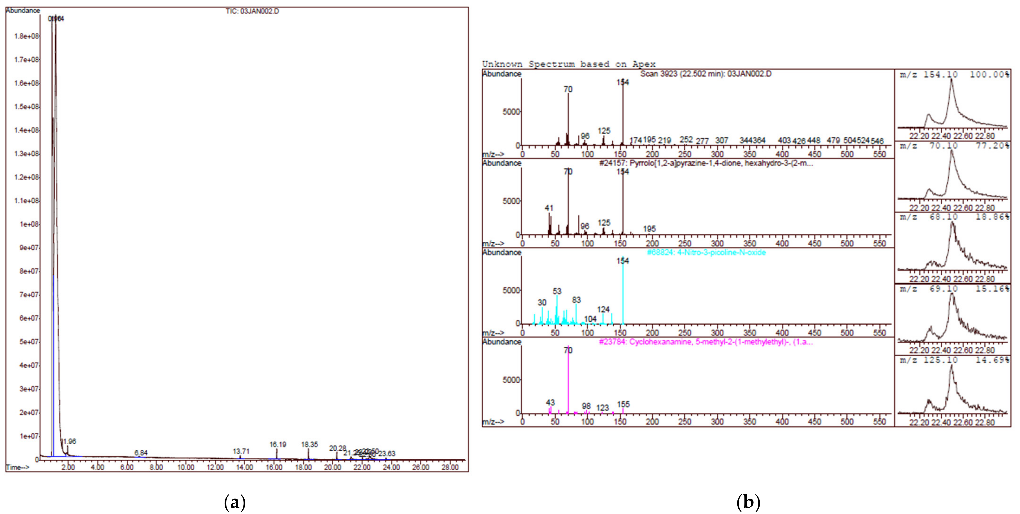
2.2. Gas Chromatography Mass Spectrometry (GCMS) Analysis

GCMS analysis of the twenty strains showed that a total of 16 different compounds were detected from the LAB strains (Table 1). Two strains of Pediococcus spp. (L010 and L013) did not show any antagonistic activity against all XDR A. baumannii, marked as a “0” value, while the highest activity was found in L001 (Figure 1), which was active against 20 out of 30 XDR isolates. A heat map of the compounds and their antagonistic activity value are shown in Figure 2. Three of the most common compounds produced by LAB strains were pyrrolo(2,1-F)pyrazine-1,4-dione,2,3,6,7,8,8A-hexahydro-3-(phenylmethyl)-, 2,4-di-tert-butylphenol, and 3-Isobutyl-2,3,6,7,8,8a-hexahydropyrrolo[1,2-a]pyrazine-1,4-dione. Based on the current finding, the common compound that was found in the strains with antagonistic activity was 3-Isobutyl-2,3,6,7,8,8a-hexahydropyrrolo[1,2-a]pyrazine-1,4-dione and pyrrolo(2,1-F)pyrazine-1,4-dione,2,3,6,7,8,8A-hexahydro-3-(phenylmethyl), which are both cyclodipeptides (CDPs) with proline. When comparing between the three active compounds, it was shown that LAB strains with 0% antagonistic activities did not produce 3-Isobutyl-2,3,6,7,8,8a-hexahydropyrrolo[1,2-apyrazine-1,4-dione while pyrrolo(2,1-F)pyrazine-1,4-dione,2,3,6,7,8,8A-hexahydro-3-(phenylmethyl) showed an antagonistic effect in 16 out of the 18 compound producing LAB strains, suggesting the potential role of CDPs with proline against XDR A. baumannii strains.

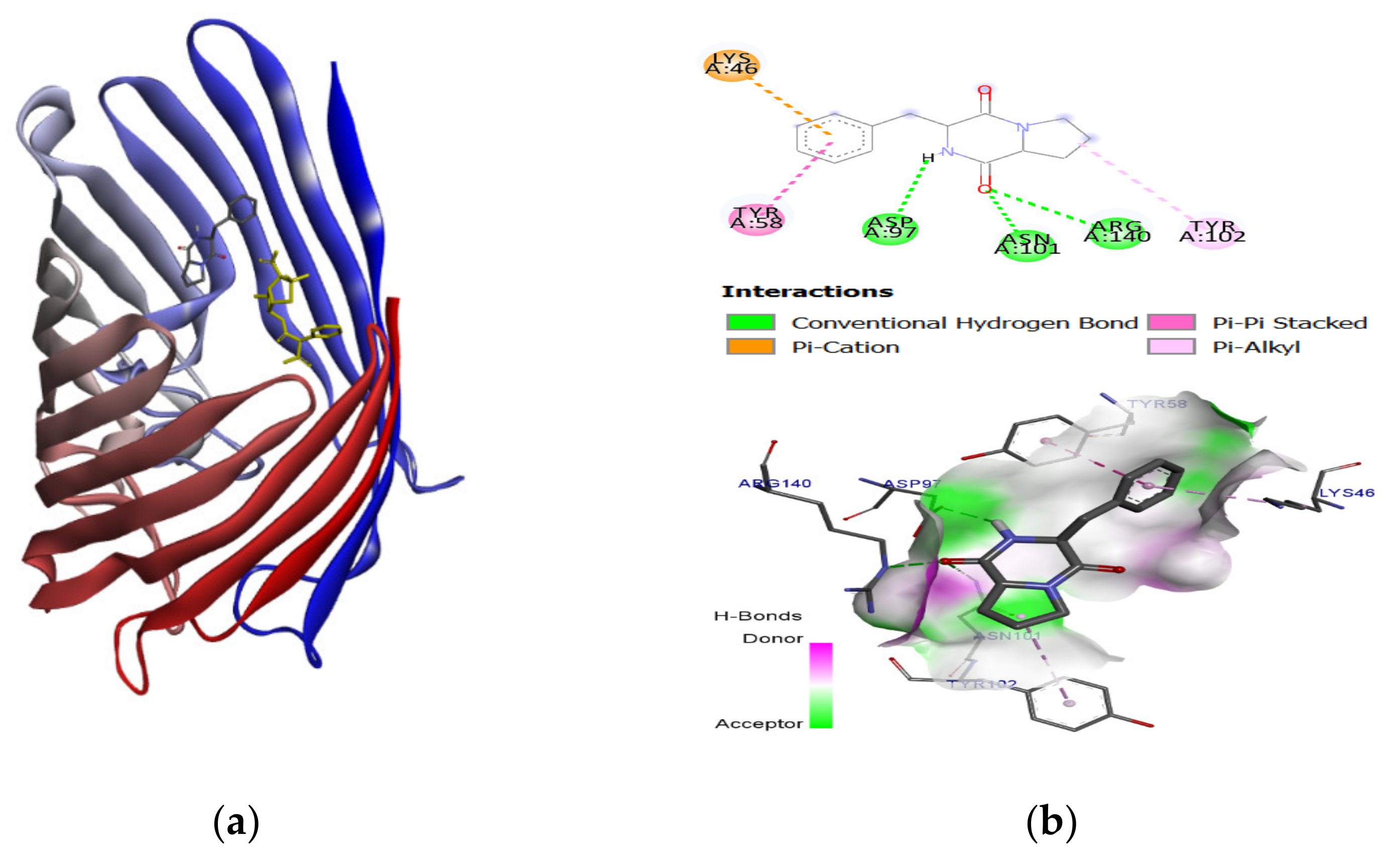
2.3. Molecular Docking and Binding Energy Evaluation

The small ribosomal subunit (30S) fragment and protein structures were selected for interaction with an isolated bioactive compound. One important mechanism of bacterial resistance is the decrease in the permeability of the bacterial membrane. In our investigation for the potential mechanism in bacterial biosynthesis and outer membrane protein, the OmpF proteins complexed with inhibitors, ampicillin (PDB ID: 4GCP), carbenicillin (PDB ID: 4GCQ), and ertapenem (PDB ID: 4GCS) were used to determine the binding interaction with selected compounds. The inhibitors were removed before the docking.
In general, to evaluate the prediction of the accuracy of the binding affinity between ligands and target protein from molecular docking, the lower values of binding energy (ΔG) indicate the binding strength of the ligands. The binding energy of ligand-protein docked complexes were analyzed based on minimum binding energy values and the ligand interaction (hydrogen/hydrophobic) pattern. Docking results justified that among the selected candidates, compounds (Pyrrolo(1,2-F)pyrazine-1,4-dione,2,3,6,7,8,8A-hexahydro-3-(phenylmethyl), 4-Nitro-4′-Chlorodiphenylsulphoxide, Dodecamethylcyclohexasiloxane) possessed good binding energy values (−7.1 to −6.0 kcal/mol) compared to the bioactive compound (−8.6 to −6.1 kcal/mol), as mentioned in Table 2. It has been observed that since the structural skeleton of compounds was comparable to the binding pocket; therefore, the binding energy values were also in a similar pattern. In all docking affinity values, the predicted energy values were not higher than 2.5 kcal/mol, indicating that the selected ligands fitted well in the active pocket of the targeted protein. Therefore, the in vitro results were focused on checking their binding profile. The best-ranked pose of the selected compound (Pyrrolo (1,2-F) pyrazine-1,4-dione,2,3,6,7,8,8A-hexahydro-3-(phenylmethyl) was extracted from each binding site and visualized for 2D/3D binding interaction in Figure 3 and Figure 4.
The docking study explored the actual binding pattern within the active site of the target protein. The ligand, pyrrolo(1,2-F)pyrazine-1,4-dione,2,3,6,7,8,8A-hexahydro-3-(phenylmethyl), confined within the same active site with the drug Paromomycin of the target DNA and having a strong binding interaction (−7.0 kcal/mol) with two hydrogens bonding and π π interaction with the target DNA, is as shown as Figure 3. There is a possibility of this ligand, pyrrolo(1,2-F)pyrazine-1,4-dione,2,3,6,7,8,8A-hexahydro-3-(phenylmethyl) to also have good binding (−6.8 kcal/mol) with the OmpF porin protein (Figure 4). The binding pocket analysis showed that pyrrolo(1,2-F)pyrazine-1,4-dione,2,3,6,7,8,8A-hexahydro-3-(phenylmethyl) binds with several amino acids, Lys46, Tyr58, Asp97, Asn101, Tyr102, and Arg140, in the binding region of the target protein. It has been observed that three hydrogen bonds were observed at Asp97, Asn101, and Arg140 with 14705-60-3 and π π ,   π c a t i o n ,   and   π π -alkyl interaction were found. Our results justified that pyrrolo(1,2-F)pyrazine-1,4-dione,2,3,6,7,8,8A-hexahydro-3-(phenylmethyl) firmly binds to both bacterial membranes with active binding residues of the OmpF porin protein and 16S rRNA, which could functionally participate in the inhibition of DNA synthesis.

3. Discussion

The clinical strains of A. baumannii used in this study are extremely drug-resistant, especially to β-lactam (AP, SAM, AMC, CXM, CRO, CTX, IPM, MEM, FEP, and TZP) and fluoroquinolone (CIP) antibiotics, which are active against all strains. The β-lactam antibiotic is known to interfere with the synthesis of bacterial cell wall through irreversible transpeptidase enzyme inhibition whereas fluoroquinolone inhibits DNA replication through DNA gyrase interaction [19]. Porin channel and outer membrane protein (OMP) embedded in the microbial membrane allow for the exchange of molecules including antibiotics between intracellular and outer space. Downregulation of OMP gene expression increased antibiotic-resistant of A. baumannii as the access of the antibiotic to intracellular was reduced, which plays an important role in resistance mechanisms. The presence of β-lactam in periplasmic space triggers A. baumannii to possess an enzymatic reaction of β-lactamases such as Acinetobacter-derived cephalosporinases (ADCs) through AmpC gene expression, which is involved in the hydrolysis of β-lactam such as IPM, MEM, and FEP, or efflux pump to eliminate antibiotics from cell cytoplasm [20,21]. Production of CDPs by LAB has been shown to potentiate the inhibition of XDR A. baumannii. The majority of CDPs are produced as secondary metabolites by bacteria and are considered as the smallest possible CDPs among cyclic organic compounds in diketopiperazines (DKPs), which consist of two nitrogen atoms of a piperazine 6-membered ring forming an amide bridge [22]. The stable and rigid structure, chiral nature, protease resistance, and functionalized structures of CDPs make them possible to bind to the various receptors with high affinity as reported in 3-Isobutyl-2,3,6,7,8,8a-hexahydropyrrolo[1,2-a]pyrazine-1,4-dione (−5.5 kcal/mol) and Pyrrolo(2,1-F)pyrazine-1,4-dione,2,3,6,7,8,8A-hexahydro-3-(phenylmethyl) (−6.8 kcal/mol). The high binding of CDPs to microbial membrane receptors such as OMP exerts a radical scavenging effect and an overall antagonistic effect against various biological cells including pathogenic bacteria, viruses, fungi, and tumors. These properties predominate as an attractive model for drug design [22,23,24]. The inhibition mechanism of CDPs could be due to their hydrophobic nature, which could interfere with the bacterial cell membrane, influencing their function and loss of cell integrity, causing cell death [25]. Interestingly, the high binding of CDPs with 16S rRNA was observed in this study (up to −7.0 kcal/mol), suggesting the possibility of transportation of CDPs into intracellular of XDR A. baumannii and further binding to 16S rRNA and interrupting DNA synthesis. Findings in the current study may be supported by the fact that CDPs are biosynthesized by LAB, which is believed to involve protease enzymes that cleave terminal ends of amino acids, creating dipeptides and cyclize naturally involving enzymes such as non-ribosomal peptide synthetases (NRPSs) and CDPs synthases [22]. The compound, 3-Isobutyl-2,3,6,7,8,8a-hexahydropyrrolo[1,2-a]pyrazine-1,4-dione is also known as cyclo (d-Pro-l-Leu) or cyclo (l-Pro-d-Leu). Antimicrobial activity of 3-Isobutyl-2,3,6,7,8,8a-hexahydropyrrolo[1,2-a]pyrazine-1,4-dione has been widely reported against Candida albicans with minimum inhibitory concentration (MIC) of 16 μg/mL and minimum fungicidal concentration (MFC) of 32 μg/mL [24]; Aspergillus flavus and Aspergillus niger with MIC of 8 μg/mL and 32 μg/mL, respectively, which are 500-fold and 156-fold lower compared to the control [26]; Mycobacterium tuberculosis with MIC of 8 μg/mL and minimum bactericidal concentration (MBC) of 16 μg/mL [23]; Vibro anguillarum with MIC of 0.13 μg/mL, which was about four times lower than oxytetracycline (0.5 μg/mL) [27]. Pyrrolo(2,1-F)pyrazine-1,4-d-one,2,3,6,7,8,8A-hexahydro-3-(phenylmethyl)- is also known as cyclo (d-Phe-l-Pro). Cyclo (l-Pro-d-Phe) obtained from Streptomyces sp. DA18 shown moderate antimicrobial activity against Escherichia coli, Bacillus subtilis, Pseudomonas fluorescens, and Candida albican [28]. Carvalho and Abraham (2012) reported the potential of cyclo (d-Phe-l-Pro) in exerting moderate inhibitory activity against V. anguillarum with a MIC of 0.13 μg/mL compared to its isomer, cyclo (d-Phe-d-Pro) with a MIC of 0.03 μg/mL [29]. Despite their isomerization, Liu et al. (2017) reported that the proline-based CDPs had significant antagonistic activity in bacteria [30]. For example, cyclo (Leu-Pro)a and cyclo (Phe-Pro)b produced by Lactobacillus plantarum LBP-K10 have antibacterial activity against Gram-positive such as B. subtilis (MIC: 13.55 μg/mLa and 45.88 μg/mLb) and MDR S. aureus 11471 (MIC: 17.28 μg/mLa and 46.22 μg/mLb) and Gram-negative such as E. coli (MIC: 10.41 μg/mLa and 35.68 μg/mLb) and MDR Salmonella typhimurium 12219 (MIC: 18.19 μg/mLa and 43.70 μg/mLb). Confirmation of CDP’s role in the inhibition of XDR strains still require further investigation; however, the preliminary results showed that inhibition was apparent in this study.
Another exciting compound identified in this study was 2,4-di-tert-butylphenol(2,4 DTBP), which was detected in 11 out of 20 LAB strains. All strains showed antagonistic activity against at least 10% up to 70% of the XDR strains, except that L010 showed no effect. This could be due to the concentration of this compound produced did not reach the threshold to exert the effect, which also applied to L013 or due to the presence of some other compounds that interfere with 2,4 DTBP activity. Lertcanawa et al. (2016) reported that the combination of vancomycin and 2,4 DTBP led to inactivation of antagonistic activity against methicillin-resistant Staphylococcus aureus (MRSA) due to enzyme catalyzation that contributes to the formation of other compounds that may have no effect opposing MRSA, and eventually reduce the concentration of 2,4 DTBP [31]. On the other hand, 2,4 DTBP obtained from Streptomyces species has reported exerting a similar effect as vancomycin in MRSA by cell deformation, with a MIC of 31.25 μg/mL and 1.56 μg/mL, which are much lower compared to oxacillin (>100.00 μg/mL) [32]. The mechanism action of 2,4 DTBP against MRSA is found through their penetration into the cell membrane and interfere with the cell integrity and permeability by interaction with the hydrophobic tails of the phospholipid bilayer, causing cytolysis and the release of genetic material, hence cell death [31]. According to Aissaoui et al. (2018), thermophilic bacteria isolates were found to produce ~15% of 2,4 DTBP, which was identified as the essential compound to inhibit MDR bacteria including Pseudomonas aeruginosa and Staphylococcus aureus [33]. The compound 2,4 DTBP was also shown to have antibiofilm formation of pathogenic bacteria, Streptococcus pyogenes up to 79% inhibition at a 48 μg/mL concentration [34].
The molecular docking studies buoyed that selected compounds can work as a lead inhibitor of bacterial DNA synthesis and membrane permeation due to conformational fitting in the active site of the targeted protein, especially compound pyrrolo (2,1-F) pyrazine-1,4-dione,2,3,6,7,8,8A-hexahydro-3-(phenylmethyl)-(14705-60-3), which has a greater binding affinity (−6.4 kcal/mol) compared to antibiotic Ertapenem (−6.1 kcal/mol). While there are many in silico models and detection of compounds against MDR and XDR tuberculosis [35,36], limited studies have reported active compounds and in silico docking against XDR A. baumannii. Therefore, to the best of our knowledge, this is the first report on the identification of DKP against XDR A. baumannii.

4. Materials and Methods

4.1. Approval by Institutional Biosafety and Biosecurity Council and Ethics Committee

Approval for this project was obtained from Institutional Biosafety and Biosecurity Council University Malaysia (UMIBBC/NOI/R/FOM/TIDREC-003/2017-rev01) for the work with potentially infectious materials. Collection of clinical bacterial strains and secondary data such as patients’ demographic and clinical data were conducted under the approval of Universiti Malaya Medical Center (UMMC) Medical Research Ethics Committee (MREC) (IRB Reference number: 1073.21). The UMMC MREC did not require the written informed consent from participants because the isolates were obtained from the hospital’s diagnostic lab, and patients were not directly involved in this study.

4.2. Sampling and Isolation of Lactic Acid Bacteria

A total number of 150 samples from food sources and the environment was collected for the sampling process using the modified method from Monika, Savitri [37]. In brief, 10 g of samples were diluted in 90 mL of MRS broth for LAB. Samples were then homogenized and incubated for 24–48 h using the anaerobic condition at 37 °C. Samples were then streaked onto MRS agar and further incubated up to 48 h to obtain colonies. Presumptive colonies were isolated and purified for identification using the molecular method.

4.3. Molecular Identification and Hydrogen Peroxide Production

Pure colonies were obtained from streaking and resuspended in 500 µL of distilled water. DNA extraction was performed using the boil cell method [38]; in brief, the suspension was incubated in the dry block at 100 °C for 20 min and immediately transferred to freezing at −20 °C for 10 min. Tubes were then centrifuged under 12,000 rpm for 2 min, and the supernatant was used as a DNA template.
LAB isolated from food samples were identified using 16S rRNA primers 27F (5′-AGAGTTTGATCCTGGCTCAG-3′) and 1492R2 (5′-TACGGYTACCTTGTTACGACTT-3′) described in [39]. The polymerase chain reaction mix using GoTaq® Green Polymerase reaction consisted of 1x colorless buffer, 3 mM of MgCl2, 2.5 mM of dNTP mix, 300 pmol of primers, 1.5 U Taq Polymerase, and 3 uL of template DNA. Gel electrophoresis was performed at 100 V for 40 min in 1% agarose and further proceeded for sequencing services.
Isolates were also screened for hydrogen peroxide production using the Prussian-blue agar method described in Saito, Seki [40], and were characterized based on light blue changes in the agar.

4.4. Well-Diffusion Antimicrobial Assay

Evaluation of antimicrobial activity was conducted using the well diffusion method described in Hoover and Harlander [41]. Each LAB colony was purified to obtain a single colony, and glycerol stock was prepared. Glycerol stock was used to inoculate MRS broth for Lactobacillus bacteria. The estimation of cell count was based on the McFarland standard and OD600 reading to obtain 1010 CFU/mL using an appropriate blank control.
LAB cultures were then centrifuged at 12,000× g 5 min, and the supernatant was treated to exclude the inhibitory effect of other compounds including organic acids by adjusting to pH 6.0–6.5 with 5 N NaOH and hydrogen peroxide by adding catalase to a final concentration of 1 mg/mL. The cell-free supernatant (CFS) was subjected to filter sterilization using a 0.2 µm syringe filter and transferred into the new sterile tube for analysis. A total of 395 CFS were screened for antibacterial activity against Acinetobacter baumannii ATCC 19606, Acinetobacter haemolyticus ATCC 19002, and Acinetobacter iwoffii ATCC 15309. Control strains were grown on Mueller Hinton broth and adjusted to 108 CFU/mL and seeded onto Mueller Hinton Agar. Agar wells were punctured to create a 5 mm diameter well. The wells were loaded with 10 µL of the filter-sterilized CFS from lactic acid bacteria. Agar plates were then incubated at 37 °C for 24 h, and results were interpreted as ‘positive’ or ‘negative’. Any zone of inhibition above 1 mm was considered as ‘positive’ and selected to further evaluation.

4.5. Screening Against Clinical Isolates of Extremely Drug Resistance (XDR) Acinetobacter baumannii

The twenty most active strains of lactic acid bacteria with low hydrogen peroxide production were selected to reduce the possibility of inhibition resulting from hydrogen peroxide and tested against 30 strains of XDR A. baumannii. Clinical isolates of XDR A. baumannii were obtained from the University Malaya Medical Center (UMMC). XDR strains were grown in TSB and adjusted to a concentration of 106 CFU/mL for well diffusion disc agar antimicrobial assay, as described above. Agar plates were then incubated at 37 °C for 24 h, and results were interpreted as ‘positive’ or ‘negative’. Antibiotic resistance profile of XDR A. baumannii was tabulated as Table S2.

4.6. GCMS

The selected twenty strains of LAB (L001–L020) were cultured in 100 mL of MRS broth (Himedia, India) in a CO2 incubator and adjusted to 1010 CFU/mL using a spectrophotometer at 600 nm. Cultures were centrifuged at 5000× g for 10 min, and the cell-free supernatant was collected. The supernatant was partially characterized as described above, and freeze-dried, followed by the addition of an equal volume of methanol and allowed to stand for 48 h and subjected to the rotary evaporator for methanol extraction at 50 °C for 1 h. The compounds were collected for GCMS analysis using a HP–5 column, 0.32 mm × 30 mm × 0.25 µm nominal with helium as the carrier gas at a flow rate at 7.7 mL/min. The interpretation on the mass spectrum from GCMS was performed using the database of the National Institute Standard and Technology (NIST) 98. The mass spectrum of the compounds detected from the methanolic extract was compared with the spectrum of known compounds stored in the NIST library to determine the name, molecular weight, and structure of the compound. The determination of the identity of the compound was set to be at above 80% for the quality of signal and identity.

4.7. In Silico Investigation toward Antimicrobial Multi-Protein Targets

To elucidate the potential mechanism, selected target proteins in bacterial biosynthesis and outer-membrane protein F (OmpF) porin were accessed from the Protein Data Bank (PDB) (www.rcsb.org (accessed on 18 July 2020)) as follows:
  • 4GCP: Crystal structure of E. coli OmpF porin in complex with ampicillin
  • 4GCQ: Crystal structure of E. coli OmpF porin in complex with carbenicillin
  • 4GCS: Crystal structure of E. coli OmpF porin in complex with ertapenem
  • 1YRJ: Crystal structure of apramycin bound to a ribosomal RNA
  • 1MWL: Crystal structure of geneticin bound to the eubacterial 16S rRNA
  • 1J7T: Crystal structure of paromomycin and the 16S rRNA
  • 1LC4: Crystal structure of tobramycin bound to the eubacterial 16S rRNA
The selected target proteins were minimized with the Amber force field by employing a conjugate gradient algorithm in UCSF Chimera 1.10.1 [42]. All protein structures were prepared for docking using the protein preparation in Chimera software with the default protocol for PDB2PQR and Dock Prep. Protonation state was assigned using PROPKA at pH 7.0, and gasteiger charges were assigned for protein. Selected ligands were downloaded from PubChem and optimized using the AM1 level using the Gaussian software package. Molecular docking was performed with a local search algorithm using Autodock Vina in PyRx virtual screening software (http://pyrx.scripps.edu/ (accessed on 18 July 2020) considered the target conformation as a rigid unit while the ligands were allowed to be flexible and adaptable to the target. The amino acid binding sites were selected at the ligand center from the X-ray structure. The grid box was set at 20 × 20 × 20 Å3 with the default grid spacing of 0.375. The 2D and 3D interactions were visualized by BIOVIA Discovery Studio software [43] for different conformations for each ligand and the lowest binding affinity conformations were predicted.

5. Conclusions

LAB are prospective antibiotic replacements due to their ability to produce CDPs as metabolites, namely 3-Isobutyl-2,3,6,7,8,8a-hexahydropyrrolo[1,2-a]pyrazine-1,4-dione and pyrrolo (2,1-F)pyrazine-1,4-dione,2,3,6,7,8,8A-hexahydro-3-(phenylmethyl). These compounds potentiate the inactivation of XDR A. baumanii clinical strains through their highly binding affinity to A. baumannii membrane receptors and intracellular 16S rRNA (−5.4 to −7.1 kcal/mol). However, the mechanisms of DKPs and CDPs in microbial inactivation should be further investigated and their potential in antibiotic stewardship by the reduction of antibiotic utilization, hence combating antibiotic-resistance.

Supplementary Materials

The following are available online, Table S1: Identification of lactic acid bacteria strains selected for further screening. Table S2: Antibiotic resistance profile of XDR A. baumannii strains tested in this study.

Author Contributions

Conceptualization, C.S.J.T., A.S.A., and H.Y.L.; Formal analysis, P.-C.Y., N.A., J.J.W., C.S.J.T., A.S.A., V.S.L. and H.Y.L.; Funding acquisition, H.Y.L.; Methodology, P.-C.Y., N.A., J.J.W., C.S.J.T., A.S.A., and V.S.L.; Validation, P.-C.Y., J.J.W. and C.S.J.T., Writing—original draft, P.-C.Y., N.A., V.S.L., H.Y.L., and S.A.; Writing—review & editing, C.S.J.T., A.S.A., V.S.L., S.A., and H.Y.L. All authors have read and agreed to the published version of the manuscript.

Funding

This research was funded under BKP056-2016 with a partial contribution by FP049-2018A- Fundamental Research Grant Scheme Ministry of Higher Education Malaysia, Project MO002-2019 by Higher Institution Center of Excellence (HICoE) program and grant TIDREC RU0008-2018 and UM.00000091/ADA.AK.PI.

Institutional Review Board Statement

The collection of clinical bacterial strains and secondary data such as patients’ demographic and clinical data were conducted under the approval of Universiti Malaya Medical Centre (UMMC) Medical Research Ethics Committee (MREC) (IRB Reference number: 1073.21.

Informed Consent Statement

The UMMC MREC did not require the written informed consent from participants because the isolates were obtained from the hospital’s diagnostic lab and patients were not directly involved in this study.

Data Availability Statement

All data generated and analyzed during this study are included in this manuscript.

Acknowledgments

We acknowledge the funding from the Ministry of Education, Malaysia for a niche area research under the Higher Institution Center of Excellence (HICoE) program (Project MO002-2019). N.A. and P.-C.Y. received the Graduate Research Assistantship Scheme from Universiti Malaya during their studies.

Conflicts of Interest

The authors declare no conflict of interest. The funders had no role in the design of the study; in the collection, analyses, or interpretation of data; in the writing of the manuscript, or in the decision to publish the results.
Sample Availability: Sample of the compound L001 is available from the authors.

References

  1. Rouse, S.; Harnett, D.; Vaughan, A.; Van Sinderen, D. Lactic acid bacteria with potential to eliminate fungal spoilage in foods. J. Appl. Microbiol. 2007, 104, 915–923. [Google Scholar] [CrossRef]
  2. Zhang, Y.-C.; Zhang, L.-W.; Ma, W.; Yi, H.-X.; Yang, X.; Du, M.; Shan, Y.-J.; Han, X.; Zhang, L.-L. Screening of probiotic lactobacilli for inhibition of Shigella sonnei and the macromolecules involved in inhibition. Anaerobe 2012, 18, 498–503. [Google Scholar] [CrossRef] [PubMed]
  3. Bazukyan, I.; Matevosyan, L.; Toplaghaltsyan, A.; Trchounian, A. Antifungal activity of lactobacilli isolated from Armenian dairy products: An effective strain and its probable nature. AMB Express 2018, 8, 1–8. [Google Scholar] [CrossRef]
  4. Halder, D.; Mandal, M.; Chatterjee, S.S.; Pal, N.K.; Mandal, S. Indigenous probiotic Lactobacillus isolates presenting antibiotic like activity against human pathogenic bacteria. Biomedicines 2017, 5, 31. [Google Scholar] [CrossRef] [PubMed] [Green Version]
  5. Damodharan, K.; Lee, Y.S.; Palaniyandi, S.A.; Yang, S.H.; Suh, J.-W. Preliminary probiotic and technological characterization of Pediococcus pentosaceus strain KID7 and in vivo assessment of its cholesterol-lowering activity. Front. Microbiol. 2015, 6, 768. [Google Scholar] [CrossRef] [PubMed] [Green Version]
  6. Pessione, E. Lactic acid bacteria contribution to gut microbiota complexity: Lights and shadows. Front. Cell. Infect. Microbiol. 2012, 2, 86. [Google Scholar] [CrossRef] [PubMed] [Green Version]
  7. Hayes, M.; Stanton, C.; Fitzgerald, G.F.; Ross, R.P. Putting microbes to work: Dairy fermentation, cell factories and bioactive peptides. Part II: Bioactive peptide functions. Biotechnol. J. 2007, 2, 435–449. [Google Scholar] [CrossRef]
  8. FAO/WHO. Joint FAO/WHO Working Group Report on Drafting Guidelines for the Evalution of Probiotics in Food; FAO/WHO: London, ON, Canada, 2002. [Google Scholar]
  9. Ahmadi, S.; Soltani, M.; Shamsaie, M.; Islami, H.R.; Peyghan, R. Comparative effect of Pediococcus acidilactici as probiotic and vitamin C on survival, growth performance and enzyme activities of white leg shrimp (Litopenaeus vannamei). J. Anim. Vet. Adv. 2014, 13, 877–885. [Google Scholar] [CrossRef]
  10. Bassetti, M.; De Waele, J.J.; Eggimann, P.; Garnacho-Montero, J.; Kahlmeter, G.; Menichetti, F.; Nicolau, D.P.; Paiva, J.A.; Tumbarello, M.; Welte, T.; et al. Preventive and therapeutic strategies in critically ill patients with highly resistant bacteria. Intensiv. Care Med. 2015, 41, 776–795. [Google Scholar] [CrossRef] [PubMed]
  11. Almaghrabi, M.K.; Joseph, M.R.P.; Assiry, M.M.; Hamid, M.E. Multidrug-Resistant Acinetobacter baumannii: An emerging health threat in Aseer Region, Kingdom of Saudi Arabia. Can. J. Infect. Dis. Med. Microbiol. 2018, 2018, 1–4. [Google Scholar] [CrossRef] [PubMed] [Green Version]
  12. Asif, M.; Alvi, I.A.; Rehman, S.U. Insight into Acinetobacter baumannii: Pathogenesis, global resistance, mechanisms of resistance, treatment options, and alternative modalities. Infect. Drug Resist. 2018, 11, 1249–1260. [Google Scholar] [CrossRef] [Green Version]
  13. Pourhajibagher, M.; Hashemi, F.B.; Pourakbari, B.; Aziemzadeh, M.; Bahador, A. Antimicrobial resistance of Acinetobacter baumannii to imipenem in Iran: A systematic review and meta-analysis. Open Microbiol. J. 2016, 10, 32–42. [Google Scholar] [CrossRef] [PubMed] [Green Version]
  14. Qi, L.; Li, H.; Zhang, C.; Liang, B.; Li, J.; Wang, L.; Du, X.; Liu, X.; Qiu, S.; Song, H. Relationship between antibiotic resistance, biofilm formation, and biofilm-specific resistance in Acinetobacter baumannii. Front. Microbiol. 2016, 7, 483. [Google Scholar] [CrossRef] [PubMed] [Green Version]
  15. Wong, D.; Nielsen, T.B.; Bonomo, R.A.; Pantapalangkoor, P.; Luna, B.; Spellberg, B. Clinical and pathophysiological overview of Acinetobacter infections: A century of challenges. Clin. Microbiol. Rev. 2016, 30, 409–447. [Google Scholar] [CrossRef] [Green Version]
  16. Qin, L.-J.; Wang, X. A review on Acinetobacter baumannii. J. Acute Dis. 2019, 8, 16. [Google Scholar] [CrossRef]
  17. Manzoor, A.; Ul-Haq, I.; Baig, S.; Qazi, J.I.; Niazi, R.K. Efficacy of locally isolated lactic acid bacteria against antibiotic-resistant uropathogens. Jundishapur J. Microbiol. 2015, 8, 18952. [Google Scholar] [CrossRef]
  18. Zheng, P.-X.; Fang, H.-Y.; Yang, H.-B.; Tien, N.-Y.; Wang, M.-C.; Wu, J.-J. Lactobacillus pentosus strain LPS16 produces lactic acid, inhibiting multidrug-resistant Helicobacter pylori. J. Microbiol. Immunol. Infect. 2016, 49, 168–174. [Google Scholar] [CrossRef] [PubMed] [Green Version]
  19. Buyana, B.; Alven, S.; Nqoro, X.; Aderibigbe, B. Antibiotics encapsulated scaffolds as potential wound dressings. In Antibiotic Materials in Healthcare; Kokkarachedu, V., Kanikireddy, V., Sadiku, R., Eds.; Academic Press: Cambridge, MA, USA, 2020; pp. 111–128. [Google Scholar]
  20. Lee, C.-R.; Lee, J.H.; Park, M.; Park, K.S.; Bae, I.K.; Kim, Y.B.; Cha, C.-J.; Jeong, B.C.; Lee, S.H. Biology of Acinetobacter baumannii: Pathogenesis, antibiotic resistance mechanisms, and prospective treatment options. Front. Cell. Infect. Microbiol. 2017, 7, 55. [Google Scholar] [CrossRef] [PubMed] [Green Version]
  21. Manchanda, V.; Sinha, S.; Singh, N.P. Multidrug resistant Acinetobacter. J. Glob. Infect. Dis. 2010, 2, 291–304. [Google Scholar] [CrossRef] [PubMed]
  22. Mishra, A.K.; Choi, J.; Choi, S.-J.; Baek, K.-H. Cyclodipeptides: An overview of their biosynthesis and biological activity. Molecules 2017, 22, 1796. [Google Scholar] [CrossRef] [Green Version]
  23. Kumar, S.N.; Mohandas, C. Antimycobacterial activity of cyclic dipeptides isolated from Bacillus sp. N strain associated with entomopathogenic nematode. Pharm. Biol. 2013, 52, 91–96. [Google Scholar] [CrossRef] [PubMed] [Green Version]
  24. Kumar, S.N.; Mohandas, C.; Nambisan, B. Purification of an antifungal compound, cyclo(l-Pro-d-Leu) for cereals produced by Bacillus cereus subsp. thuringiensis associated with entomopathogenic nematode. Microbiol. Res. 2013, 168, 278–288. [Google Scholar] [CrossRef]
  25. Kumar, S.N.; Mohandas, C.; Siji, J.; Rajasekharan, K.; Nambisan, B. Identification of antimicrobial compound, diketopiperazines, from a Bacillus sp. N strain associated with a rhabditid entomopathogenic nematode against major plant pathogenic fungi. J. Appl. Microbiol. 2012, 113, 914–924. [Google Scholar] [CrossRef]
  26. Kumar, S.N.; Nambisan, B.; Mohandas, C.; Sundaresan, A. In vitro synergistic activity of diketopiperazines alone and in combination with amphotericin B or clotrimazole against Candida albicans. Folia Microbiol. 2013, 58, 475–482. [Google Scholar] [CrossRef] [PubMed]
  27. Fdhila, F.; Vázquez, V.; Sánchez, J.L.; Riguera, R. dd-Diketopiperazines: Antibiotics active against Vibrio anguillarum isolated from marine bacteria associated with cultures of Pecten maximus. J. Nat. Prod. 2003, 66, 1299–1301. [Google Scholar] [CrossRef] [PubMed]
  28. Gao, Y.; Yu, L.; Peng, C.; Li, Z.; Guo, Y. Diketopiperazines from two strains of South China Sea sponge-associated microorganisms. Biochem. Syst. Ecol. 2010, 38, 931–934. [Google Scholar] [CrossRef]
  29. De Carvalho, M.P. Antimicrobial and biofilm inhibiting Diketopiperazines. Curr. Med. Chem. 2012, 19, 3564–3577. [Google Scholar] [CrossRef] [PubMed]
  30. Liu, R.; Kim, A.H.; Kwak, M.-K.; Kang, S.-O. Proline-based cyclic dipeptides from korean fermented vegetable kimchi and from Leuconostoc mesenteroides LBP-K06 have activities against multidrug-resistant bacteria. Front. Microbiol. 2017, 8, 761. [Google Scholar] [CrossRef] [Green Version]
  31. Lertcanawa, M.; Chawawisit, K.; Bhoopong, P.; Phupong, W. Combination effect between 2, 4-Di-tert-butylphenol produced by Streptomyces sp. KB1 TISTR 2304 and vancomycin against methicillin-resistant Staphylococcus aureus (MRSA). Int. J. Pharmacol. 2016, 12, 838–844. [Google Scholar] [CrossRef]
  32. Chawawisit, K.; Bhoopong, P.; Phupong, W.; Lertcanawa, M. Anti-MRSA activity, mode of action and cytotoxicity of 2, 4-Di-tert-butylphenol produced by Streptomyces sp. KB1. Int. J. Pharm. Sci. Rev. Res. 2015, 35, 114–119. [Google Scholar]
  33. Aissaoui, N.; Mahjoubi, M.; Nas, F.; Mghirbi, O.; Arab, M.; Souissi, Y.; Hoceini, A.; Masmoudi, A.S.; Mosbah, A.; Cherif, A.; et al. Antibacterial potential of 2,4-Di-tert-Butylphenol and calixarene-based prodrugs from thermophilic Bacillus licheniformis isolated in Algerian Hot Spring. Geomicrobiol. J. 2018, 36, 53–62. [Google Scholar] [CrossRef]
  34. Viszwapriya, D.; Prithika, U.; Deebika, S.; Balamurugan, K.; Pandian, S.K. In vitro and in vivo antibiofilm potential of 2,4-Di- tert -butylphenol from seaweed surface associated bacterium Bacillus subtilis against group A Streptococcus. Microbiol. Res. 2016, 191, 19–31. [Google Scholar] [CrossRef]
  35. Modi, P.; Patel, S.; Chhabria, M.T. Identification of some novel pyrazolo[1,5-a]pyrimidine derivatives as InhA inhibitors through pharmacophore-based virtual screening and molecular docking. J. Biomol. Struct. Dyn. 2018, 37, 1736–1749. [Google Scholar] [CrossRef] [PubMed]
  36. Hameed, H.; Islam, M.M.; Chhotaray, C.; Wang, C.; Liu, Y.; Tan, Y.; Li, X.; Tan, S.; Delorme, V.; Yew, W.W. Molecular targets related drug resistance mechanisms in MDR-, XDR-, and TDR-Mycobacterium tuberculosis strains. Front. Cell. Infect. Microbiol. 2018, 8, 114. [Google Scholar] [CrossRef]
  37. Monika, S.; Kumar, V.; Kumari, A.; Angmo, K.; Bhalla, T.C. Isolation and characterization of lactic acid bacteria from traditional pickles of Himachal Pradesh, India. J. Food Sci. Technol. 2017, 54, 1945–1952. [Google Scholar] [CrossRef]
  38. Lee, H.-Y.; Chai, L.-C.; Tang, S.-Y.; Jinap, S.; Ghazali, F.M.; Nakaguchi, Y.; Nishibuchi, M.; Son, R. Application of MPN-PCR in biosafety of Bacillus cereus s.l. for ready-to-eat cereals. Food Control. 2009, 20, 1068–1071. [Google Scholar] [CrossRef]
  39. Garcia, E.F.; Luciano, W.A.; Xavier, D.E.; Da Costa, W.C.A.; Oliveira, K.D.S.; Franco, O.L.; Júnior, M.A.D.M.; Lucena, B.T.L.; Picão, R.C.; Magnani, M.; et al. Identification of lactic acid bacteria in fruit pulp processing byproducts and potential probiotic properties of selected Lactobacillus Strains. Front. Microbiol. 2016, 7, 1371. [Google Scholar] [CrossRef] [PubMed] [Green Version]
  40. Saito, M.; Seki, M.; Iida, K.-I.; Nakayama, H.; Yoshida, S.-I. A novel agar medium to detect hydrogen peroxide-producing bacteria based on the prussian blue-forming reaction. Microbiol. Immunol. 2007, 51, 889–892. [Google Scholar] [CrossRef] [PubMed]
  41. Hoover, D.G.; Harlander, S.K. CHAPTER 2—Screening methods for detecting bacteriocin activity. In Bacteriocins of Lactic Acid Bacteria; Hoover, D.G., Steenson, L.R., Eds.; Academic Press: Cambridge, MA, USA, 1993; pp. 23–39. [Google Scholar] [CrossRef]
  42. Pettersen, E.F.; Goddard, T.D.; Huang, C.C.; Couch, G.S.; Greenblatt, D.M.; Meng, E.C.; Ferrin, T.E. UCSF Chimera—A visualization system for exploratory research and analysis. J. Comput. Chem. 2004, 25, 1605–1612. [Google Scholar] [CrossRef] [PubMed] [Green Version]
  43. Biovia, D.S. Discovery Studio Modeling Environment; Dassault Systèmes: San Diego, CA, USA, 2018. [Google Scholar]
Figure 1. (a). GCMS chromatogram of methanolic extract of L001 and (b) spectrum analysis of compounds in L001 against pyrolo(1,2-a)pyrazine-1,4-dione.
Molecules 26 01727 g001
Figure 2. Heat map showing compounds from the lactic acid bacteria strains and antagonistic activities in some XDR Acinetobacter baumannii strains (in percentage, n = 30).
Molecules 26 01727 g002
Figure 3. (a) In silico interaction of selected ligand pyrrolo (1,2-F) pyrazine-1,4-dione,2,3,6,7,8,8A-hexahydro-3-(phenylmethyl) in stick mode, carbenicillin in yellow and target protein, OmpF porin (4GCQ) (b) 2D (top) and 3D level interaction (bottom) of the best-docked complexes illustrate the mode of interaction of the amino acid in the binding pocket. Non-polar hydrogens are not shown.
Molecules 26 01727 g003
Figure 4. In silico interaction between selected ligand pyrrolo(1,2-F)pyrazine-1,4-d-one,2,3,6,7,8,8A-hexahydro-3-(phenylmethyl) and target RNA fragment (1YRJ). Poses of docked complexes are in the stick model where the x-ray ligand (Paromomycin) in yellow color indicates a prominent active site where the ligand interacted with hydrogen bonding in green and π π interaction in pink. Non-polar hydrogens are not shown.
Molecules 26 01727 g004
Table 1. List of compounds identified from lactic acid bacteria strains and their antagonistic activity against 30 extremely drug resistance (XDR) Acinetobacter baumannii isolates.
Sample IDAntagonistic Activity in 30 XDR A. Baumannii Strains (%)CAS NO.Compound ID
L001
(Lactobacillus plantarum)
20/30 (66.0)000541-01-5Hexadecamethylheptasiloxane
005654-86-43-Isobutyl-2,3,6,7,8,8a-hexahydropyrrolo[1,2-a]pyrazine-1,4-dione
024535-53-34-Nitro-4′-chlorodiphenylsulfoxide
000107-52-8Tetradecamethylhexasiloxane
L002
(Lactobacillus plantarum)
19/30 (63.3)000084-66-2Diethyl phthalate
000096-76-42,4-Di-tert-Butylphenol
000540-97-6Dodecamethylcyclohexasiloxane
005654-86-43-Isobutyl-2,3,6,7,8,8a-hexahydropyrrolo[1,2-a]pyrazine-1,4-dione
014705-60-3Pyrrolo(2,1-F)pyrazine-1,4-dione,2,3,6,7,8,8A-hexahydro-3-(phenylmethyl)-
000107-50-6Tetradecamethyl Cycloheptasiloxane
L003
(Lactobacillus plantarum)
18/30 (60.0)000096-76-42,4-Di-tert-Butylphenol
000112-88-91-Octadecene
000111-82-0Methyl laurate
014705-60-3Pyrrolo(2,1-F)pyrazine-1,4-dione,2,3,6,7,8,8A-hexahydro-3-(phenylmethyl)-
000107-50-6Tetradecamethyl Cycloheptasiloxane
L004
(Pediococcus pentosaceus)
5/30 (16.7)000096-76-42,4-Di-tert-Butylphenol
000540-97-6Dodecamethylcyclohexasiloxane
000541-01-5Hexadecamethylheptasiloxane
005654-86-43-Isobutyl-2,3,6,7,8,8a-hexahydropyrrolo[1,2-a]pyrazine-1,4-dione
000544-76-3Hexadecane
014705-60-3Pyrrolo(2,1-F)pyrazine-1,4-dione,2,3,6,7,8,8A-hexahydro-3-(phenylmethyl)-
000107-50-6Tetradecamethyl Cycloheptasiloxane
L005
(Pediococcus pentosaceus)
4/30 (13.3)000096-76-42,4-Di-tert-Butylphenol
005654-86-43-Isobutyl-2,3,6,7,8,8a-hexahydropyrrolo[1,2-a]pyrazine-1,4-dione
077899-03-71-Heneicosyl formate
014705-60-3Pyrrolo (2,1-F) pyrazine-1,4-dione,2,3,6,7,8,8A-hexahydro-3-(phenylmethyl)-
L006
(Pediococcus pentosaceus)
3/30 (10.0)000096-76-42,4-Di-tert-Butylphenol
000112-88-91-Octadecene
005654-86-43-Isobutyl-2,3,6,7,8,8a-hexahydropyrrolo[1,2-a]pyrazine-1,4-dione
024535-53-34-Nitro-4′-chlorodiphenylsulphoxide
014705-60-3Pyrrolo(2,1-F)pyrazine-1,4-dione,2,3,6,7,8,8A-hexahydro-3-(phenylmethyl)
L007
(Pediococcus pentosaceus)
1/30 (3.3)000096-76-42,4-Di-tert-Butylphenol
005654-86-43-Isobutyl-2,3,6,7,8,8a-hexahydropyrrolo[1,2-a]pyrazine-1,4-dione
014705-60-3Pyrrolo (2,1-F) pyrazine-1,4-dione,2,3,6,7,8,8A-hexahydro-3-(phenylmethyl)
L008
(Pediococcus pentosaceus)
1/30 (3.3)000096-76-42,4-Di-tert-Butylphenol
000540-97-6Dodecamethylcyclohexasiloxane
005654-86-43-Isobutyl-2,3,6,7,8,8a-hexahydropyrrolo [1,2-a] pyrazine-1,4-dione
014705-60-3Pyrrolo(2,1-F)pyrazine-1,4-dione,2,3,6,7,8,8A-hexahydro-3-(phenylmethyl)
L009
(Pediococcus acidilactici)
1/30 (3.3)000096-76-42,4-Di-tert-Butylphenol
005654-86-43-Isobutyl-2,3,6,7,8,8a-hexahydropyrrolo[1,2-a]pyrazine-1,4-dione
014705-60-3Pyrrolo(2,1-F)pyrazine-1,4-dione,2,3,6,7,8,8A-hexahydro-3-(phenylmethyl)-
L010
(Pediococcus spp.)
0/30 (0)000096-76-42,4-Di-tert-Butylphenol
014705-60-3Pyrrolo(2,1-F)pyrazine-1,4-dione,2,3,6,7,8,8A-hexahydro-3-(phenylmethyl)-
L011
(Pediococcus acidilactici)
1/30 (3.3)000096-76-42,4-Di-tert-Butylphenol
000060-12-8Phenethyl alcohol
014705-60-3Pyrrolo(2,1-F)pyrazine-1,4-dione,2,3,6,7,8,8A-hexahydro-3-(phenylmethyl)
L012
(Pediococcus pentosaceus)
1/30 (3.3)005654-86-43-Isobutyl-2,3,6,7,8,8a-hexahydropyrrolo[1,2-a]pyrazine-1,4-dione
005875-45-62,5-di-tert-butylphenol
014705-60-3Pyrrolo(2,1-F)pyrazine-1,4-dione,2,3,6,7,8,8A-hexahydro-3-(phenylmethyl)
000107-50-6Tetradecamethyl Cycloheptasiloxane
L013
(Pediococcus pentosaceus)
0/30 (0)014705-60-3Pyrrolo(2,1-F)pyrazine-1,4-dione,2,3,6,7,8,8A-hexahydro-3-(phenylmethyl)
L014
(Pediococcus pentosaceus)
5/30 (16.7)005654-86-43-Isobutyl-2,3,6,7,8,8a-hexahydropyrrolo[1,2-a]pyrazine-1,4-dione
014705-60-3Pyrrolo(2,1-F)pyrazine-1,4-dione,2,3,6,7,8,8A-hexahydro-3-(phenylmethyl)-
L015
(Pediococcus pentosaceus)
12/30 (40.0)014705-60-3Pyrrolo(2,1-F)pyrazine-1,4-dione,2,3,6,7,8,8A-hexahydro-3-(phenylmethyl)-
L016
(Enterococcus spp.)
2/30 (6.7)005654-86-43-Isobutyl-2,3,6,7,8,8a-hexahydropyrrolo[1,2-a]pyrazine-1,4-dione
014705-60-3Pyrrolo(2,1-F)pyrazine-1,4-dione,2,3,6,7,8,8A-hexahydro-3-(phenylmethyl)
L017
(Pediococcus acidilactici)
6/30 (20.0)005654-86-43-Isobutyl-2,3,6,7,8,8a-hexahydropyrrolo[1,2-a]pyrazine-1,4-dione
014705-60-3Pyrrolo (2,1-F) pyrazine-1,4-dione,2,3,6,7,8,8A-hexahydro-3-(phenylmethyl)
L018
(Pediococcus pentosaceus)
13/20 (43.3)000096-76-42,4-Di-tert-Butylphenol
005654-86-43-Isobutyl-2,3,6,7,8,8a-hexahydropyrrolo [1,2-a] pyrazine-1,4-dione
014705-60-3Pyrrolo (2,1-F) pyrazine-1,4-dione,2,3,6,7,8,8A-hexahydro-3-(phenylmethyl)
L019
(Lactobacillus paraplantarum)
1/30 (3.3)005654-86-43-Isobutyl-2,3,6,7,8,8a-hexahydropyrrolo [1,2-a] pyrazine-1,4-dione
014705-60-3Pyrrolo (2,1-F) pyrazine-1,4-dione,2,3,6,7,8,8A-hexahydro-3-(phenylmethyl)
L020
(Enterococcus spp.)
1/30 (3.3)005654-86-43-Isobutyl-2,3,6,7,8,8a-hexahydropyrrolo [1,2-a] pyrazine-1,4-dione
Table 2. Docking energy values (ΔG in kcal/mol) of the selected compound. The highlighted compounds were bound well over the selected investigation with the targets.
Compound ID (CAS NO.)Autodock Vina Binding Affinity (kcal/mol)
OmpF Porin Protein16S rRNA
4GCP4GCQ4GCS1YRJ1MWL1J7T1LC4
Tetradecamethylhexasiloxane (107-52-8 Si)−4.8−4.6−4.5−6.3−5.6−5.9−5.3
Dodecamethylcyclohexasiloxane (540-97-6 Si)−5.8−6.4−5.6−6.0−6.2−6.1−6.6
Hexadecamethylheptasiloxane (541-01-5 Si)−5.0-4.9−4.2−6.1−5.3−5.8−6.2
3-Isobutyl-2,3,6,7,8,8a-hexahydropyrrolo[1,2-A] Pyrazine-1,4-Dione (5654-86-4)−5.4−5.4−5.5−6.1−5.7−6.0−5.9
4-Nitro-4′-Chlorodiphenylsulphoxide (24535-53-3)−6.1−6.7−6.1−6.4−6.6−6.5−6.3
Diethyl Phthalate (84-66-2)−5.3−5.6−5.1−5.3−5.7−5.8−5.3
2,4-Di-Tert-Butylphenol (96-76-4)−5.9−6.0−5.9−5.3−5.1−5.7−5.2
Pyrrolo (1,2-F) pyrazine-1,4-dione,2,3,6,7,8,8A-hexahydro-3-(phenylmethyl) (14705-60-3)−6.4−6.8−6.4−7.1−6.5−7.0−6.5
1-Octadecene (112-88-9)−4.2−4.9−4.1−3.6−3.2−4.0−3.4
Hexadecane (544-76-3)−4.1−4.6−4.7−3.5−3.2−3.5−3.0
PhenethylAlcohol (60-12-8)−4.3−4.9−4.3−4.0−4.3−4.4−4.2
1-Heneicosylformate (77899-03-7)−4.5−4.2−4.2−4.0−3.9−4.4−3.9
Methyl laurate (111-82-0)−4.5−4.7−4.7−4.1−3.7−4.2−3.7
2,5-di-tert-butylphenol (5875-45-6)−5.8−6.0−5.8−5.7−5.3−5.6−5.6
Xray LigandAmpicillinCarbenicillinErtapenemApramycinGeneticinParomomycinTobramycin
−7.1−7.1−6.1−8.2−7.2−8.6−7.6
Publisher’s Note: MDPI stays neutral with regard to jurisdictional claims in published maps and institutional affiliations.

