
The Cyclobutanocucurbit[5–8]uril Family: Electronegative Cavities in Contrast to Classical Cucurbituril while the Electropositive Outer Surface Acts as a Crystal Packing Driver


Abstract
:
1. Introduction
2. Results and Discussion
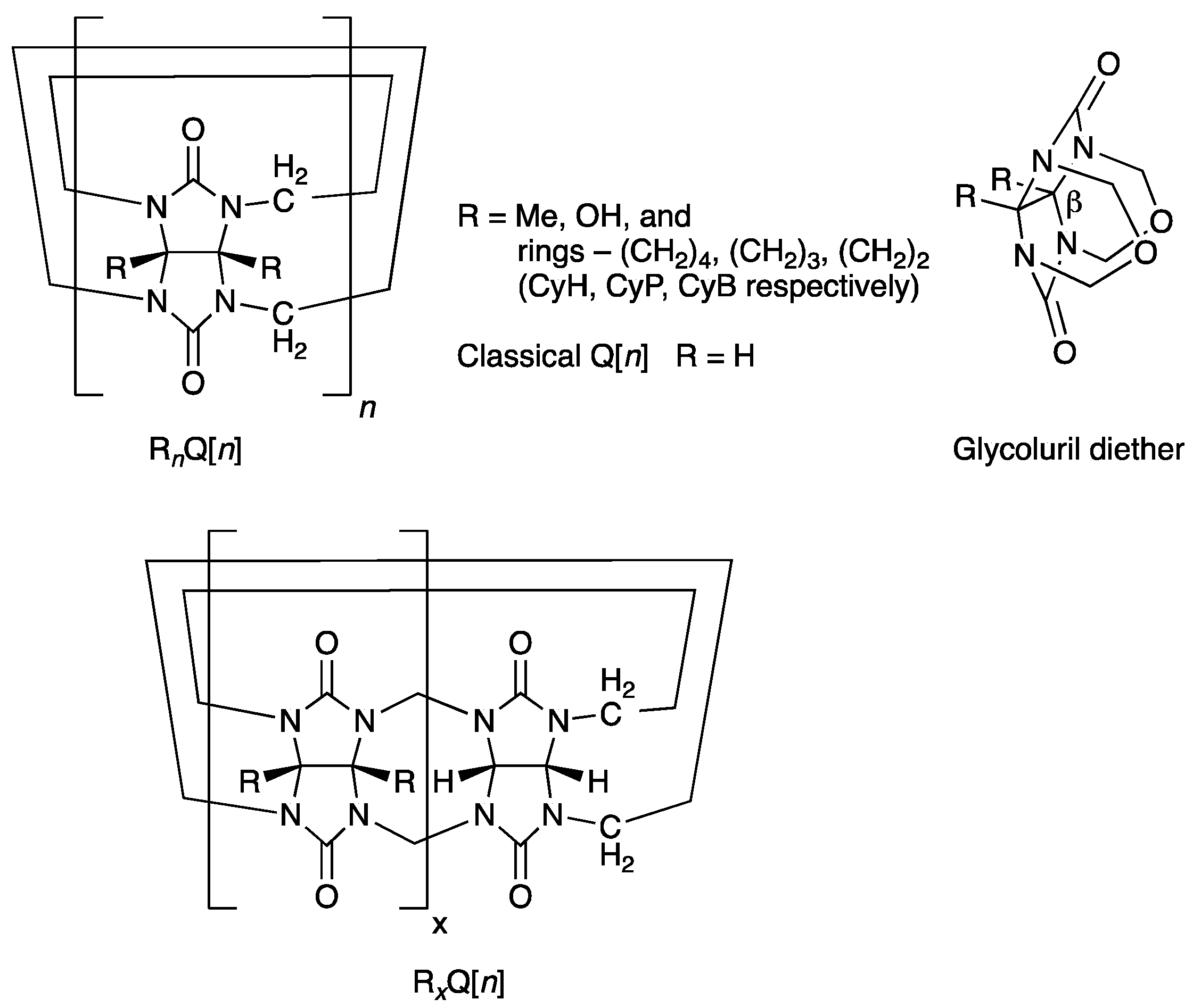
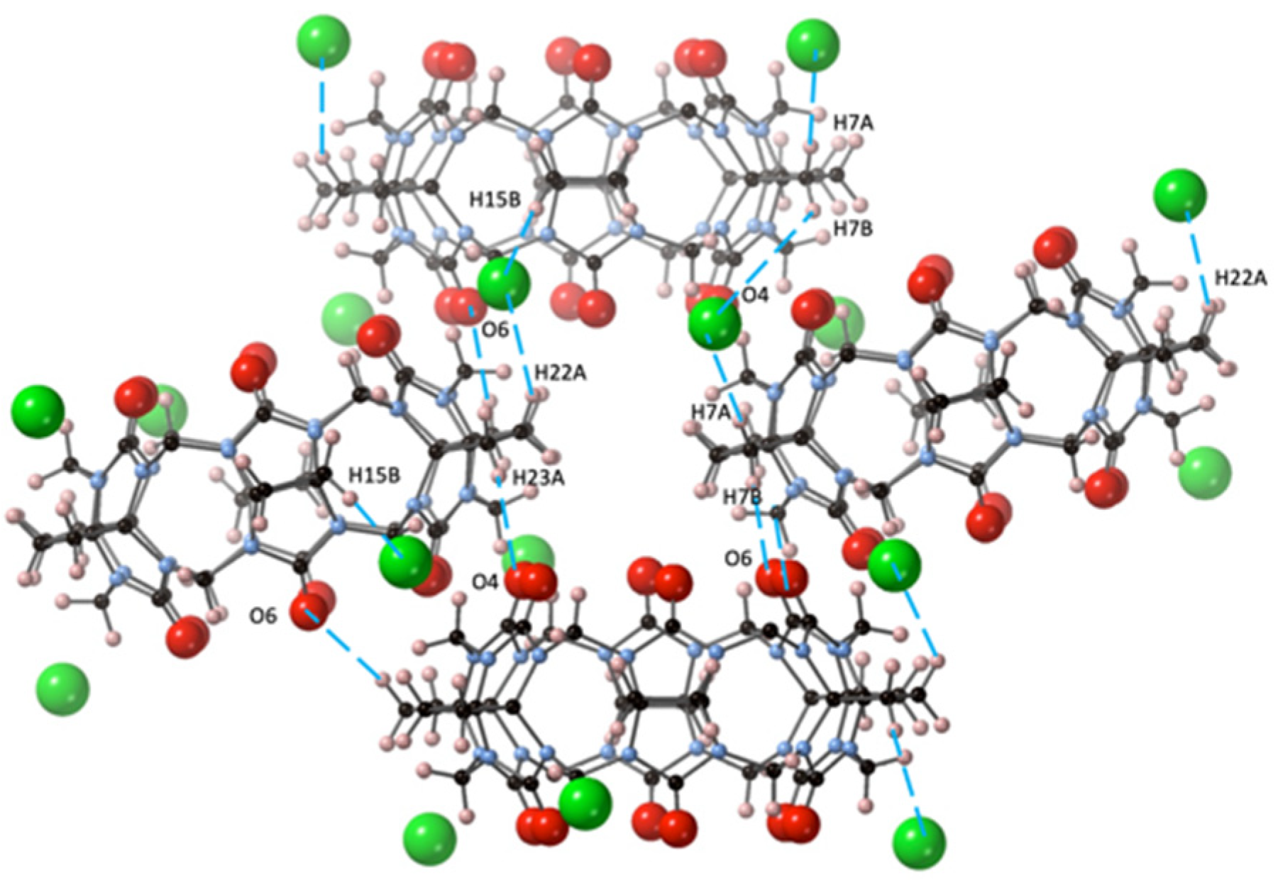
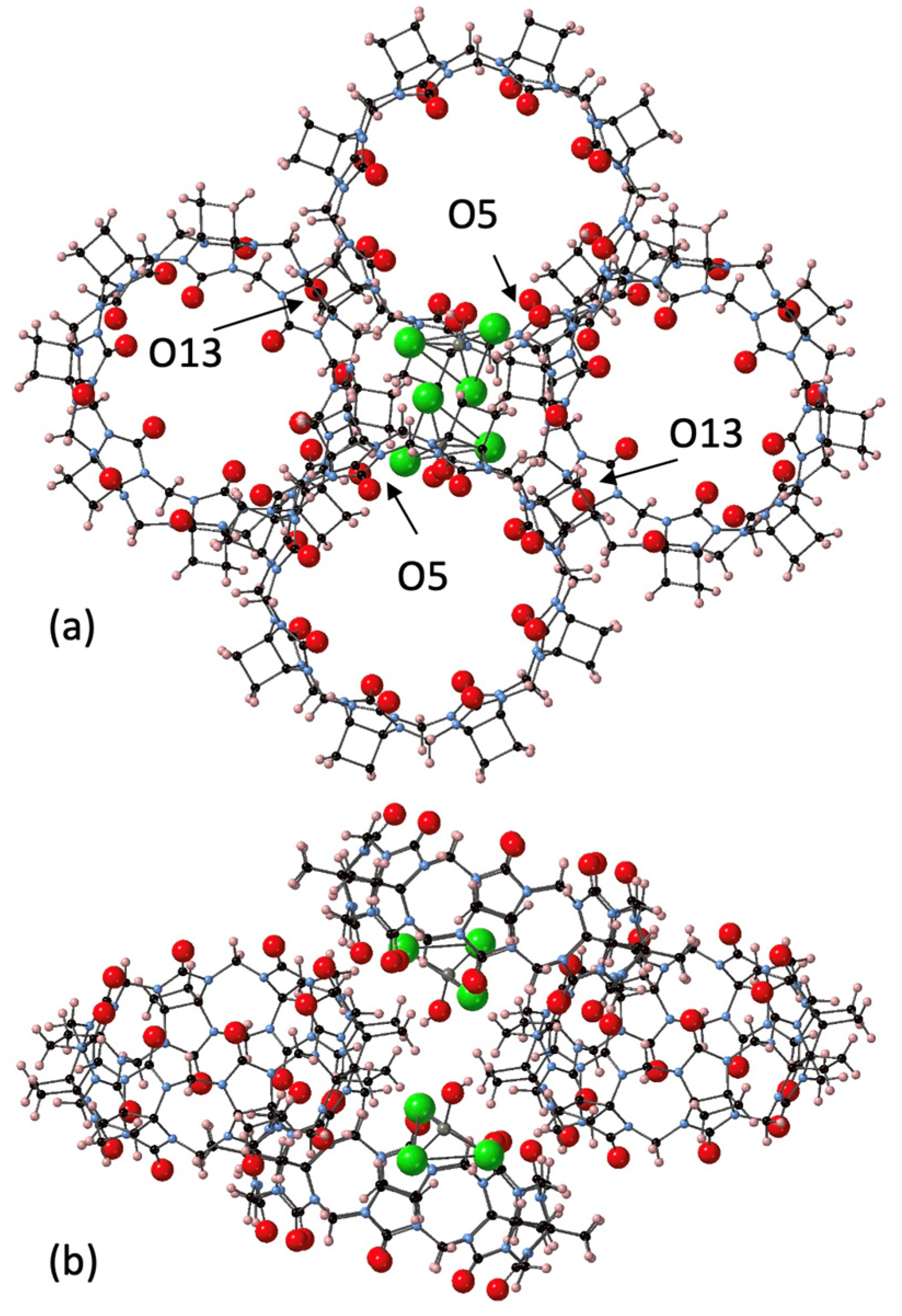
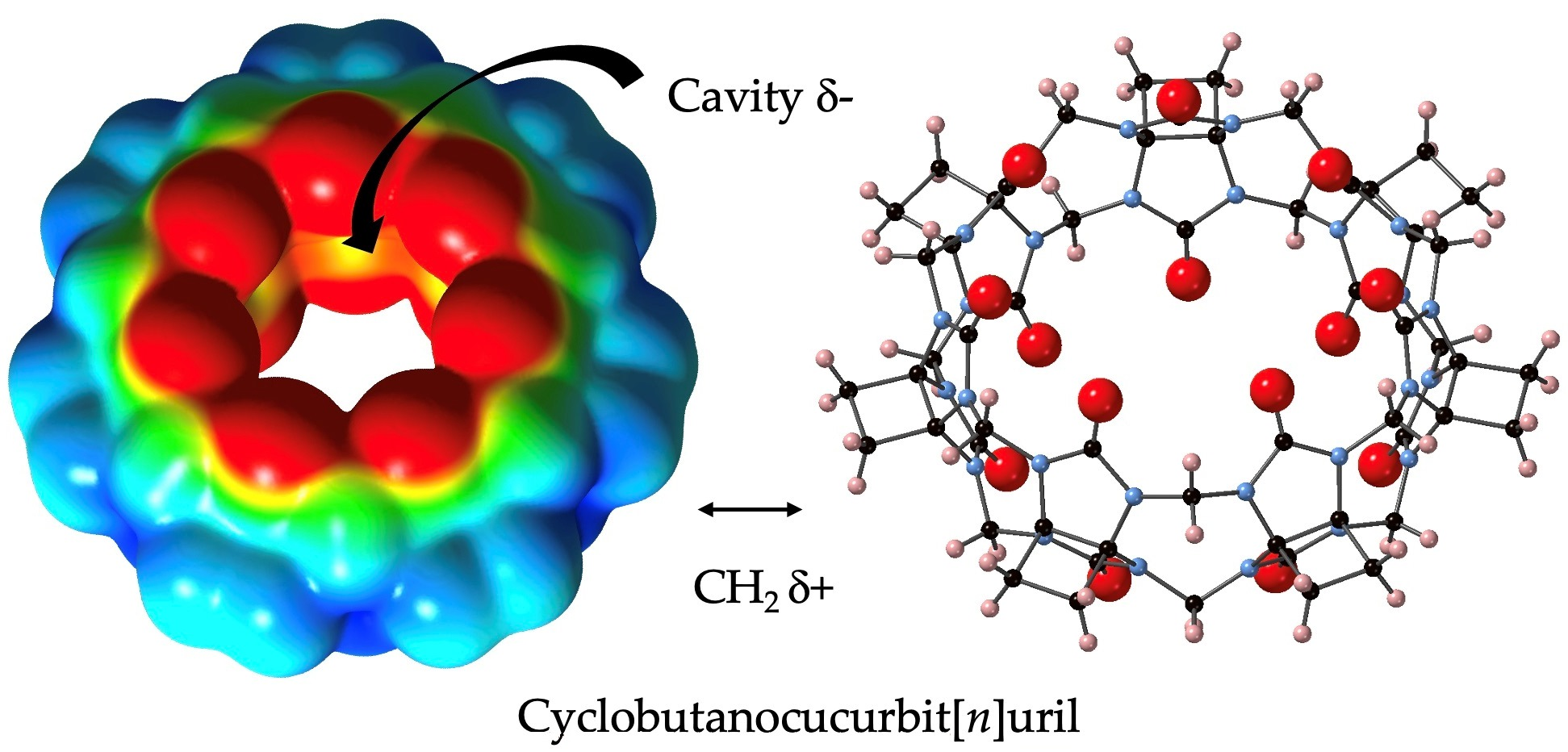
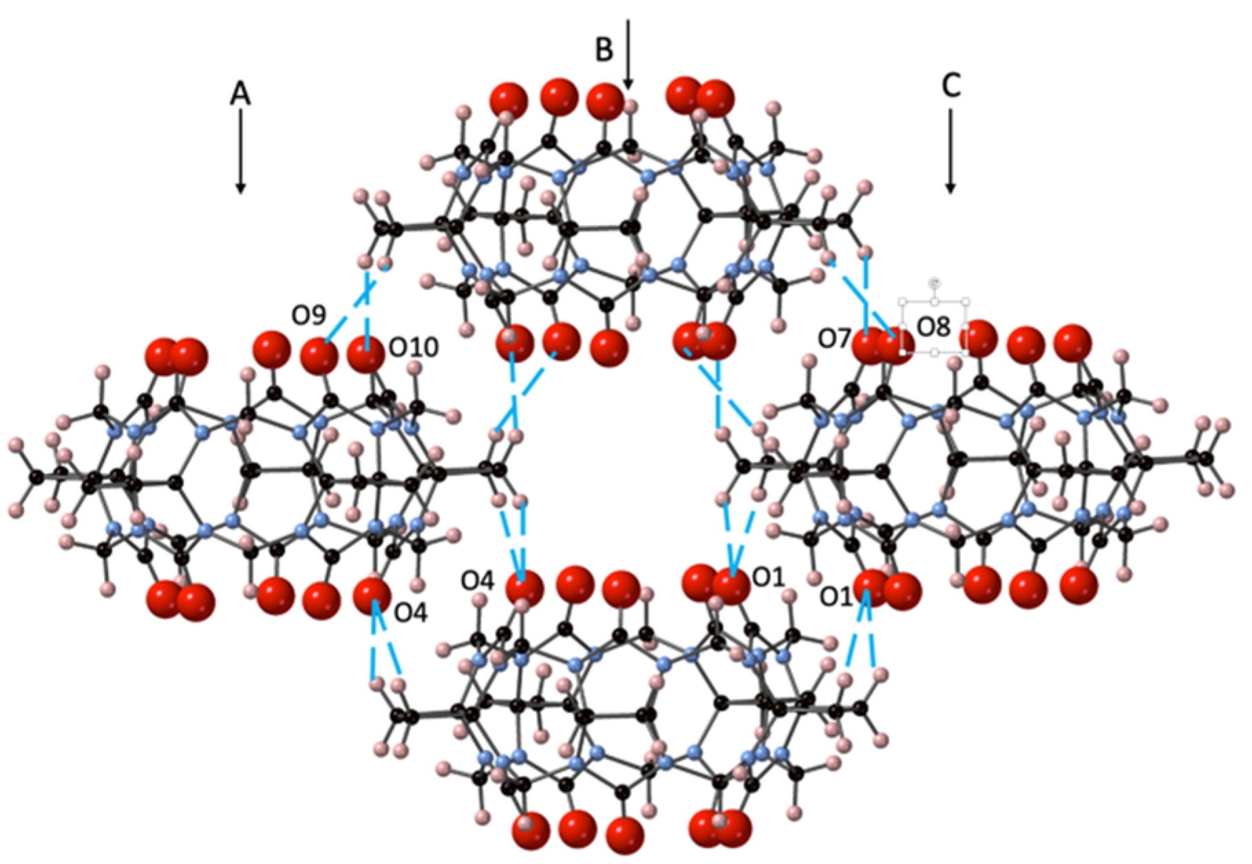
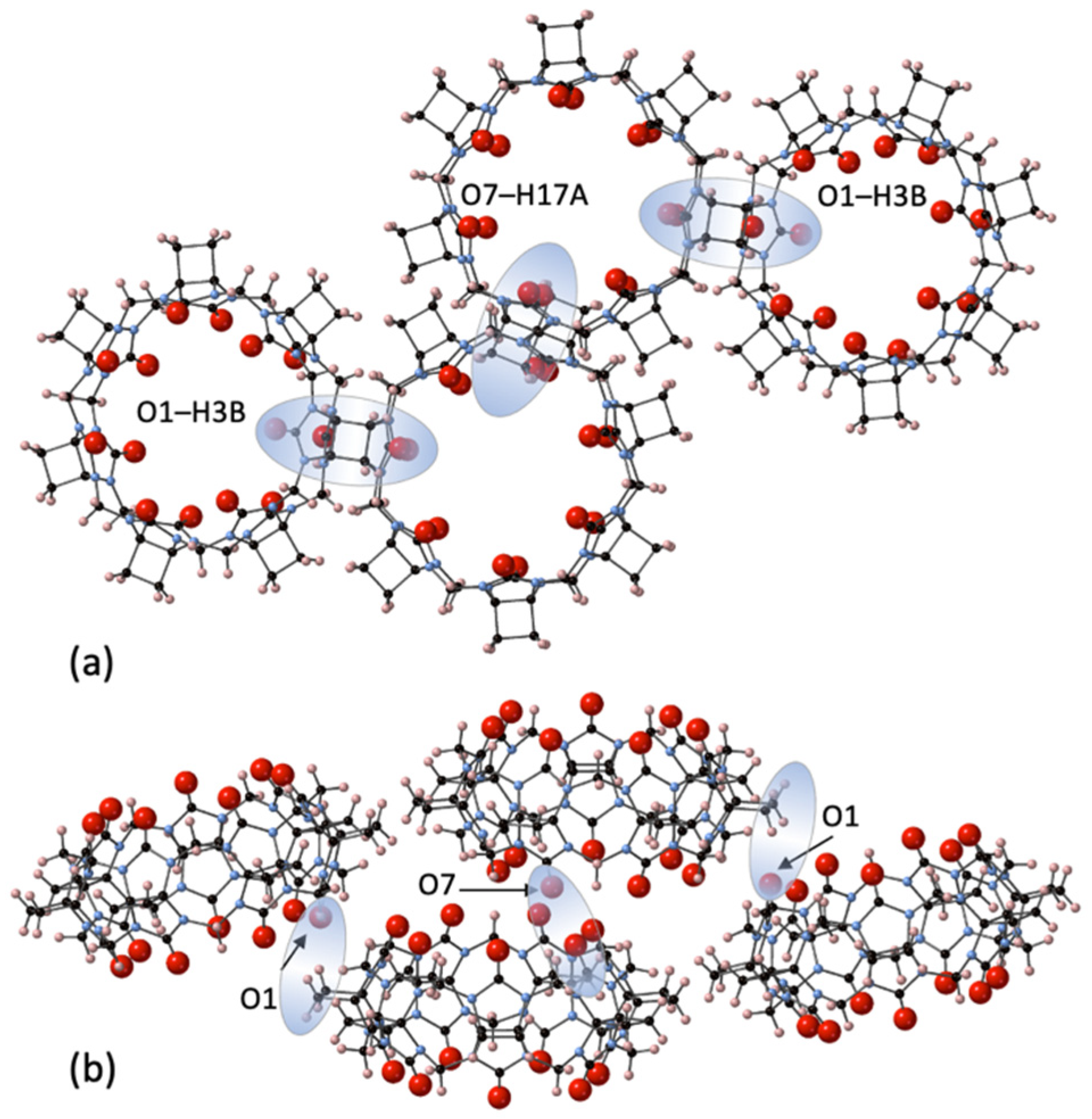
Highlighted Structural Features for 1, 2, 3, and 4 and Their Outer Surface Interactions
3. Materials and Methods
3.1. Purification of CyB5Q[5]·7H2O (1) and CyB6Q[6]·2Cl−·2(H3O+)·14H2O (2)
3.2. Purification of CyB7Q[7]·12H2O (3) and CyB8Q[8]·(ZnCl3·H2O)−·H3O+·10H2O (4)
3.3. X-ray Crystallography
3.4. Theoretical Method
4. Conclusions
Supplementary Materials
Author Contributions
Funding
Data Availability Statement
Conflicts of Interest
Sample Availability
References
- Wu, X.; Chen, Y.; Yu, Q.; Li, F.Q.; Lui, Y. A cucurbituril/polysaccharide/carbazole ternary supramolecular assembly for targeted cell imaging. Chem. Commun. 2019, 55, 4343–4346. [Google Scholar] [CrossRef]
- Koo, B.; Jun, E.; Liu, H.; Kim, E.J.; Park, Y.Y.; Lim, S.B.; Kim, S.C.; Shin, Y. A biocomposite-based rapid sampling assay for circulating cell-free DNA in liquid biopsy samples from human cancers. Sci. Rep. 2020, 10, 14932. [Google Scholar] [CrossRef] [PubMed]
- Huang, Y.; Gao, R.H.; Liu, M.; Chen, L.X.; Ni, X.L.; Xiao, X.; Cong, H.; Zhu, Q.J.; Chen, K.; Tao, Z. Cucurbit[n]uril-based supramolecular frameworks assembled through outer-surface interactions. Angew. Chem. Int. Ed. 2021, 60, 15166–151192. [Google Scholar] [CrossRef] [PubMed]
- Jin, Y.; Huang, T.; Zhao, W.; Yang, X.; Meng, Y.; Ma, P. A study on the self-assembly mode and supramolecular framework of complexes of cucurbit[6]urils and 1-(4-methoxyphenyl)piperazine. RSC Adv. 2020, 10, 37369–37373. [Google Scholar] [CrossRef]
- Yang, M.-X.; Tang, Q.; Yang, M.; Wang, Q.; Tao, Z.; Xiao, X.; Huang, Y. pH-stimulus response dye-cucurbituril sensor for amino acids in aqueous solution. Spectrochim. Acta A Mol. Biomol. Spectrosc. 2020, 230, 118076. [Google Scholar] [CrossRef] [PubMed]
- Olson, J.E.; Braegelman, A.S.; Zou, L.; Webber, M.J.; Camden, J.P. Capture of phenylalanine and Phenylalanine-Terminated Peptides Using a Supramolecular Macrocycle for Surface-Enhanced Raman Scattering Detection. Appl. Spectrosc. 2020, 74, 1374–1383. [Google Scholar] [CrossRef] [PubMed]
- Liu, M.; Kan, J.; Yao, Y.; Zhang, Y.; Bian, B.; Tao, Z.; Zhu, Q.; Xiao, X. Facile preparation and application of luminescent cucurbit[10]uril-based porous supramolecular frameworks. Sens. Actuators B Chem. 2019, 283, 290–297. [Google Scholar] [CrossRef]
- Li, Q.; Sun, J.; Zhou, J.; Hua, B.; Shao, L.; Huang, F. Barium cation-responsive supra-amphiphile constructed by a new twisted cucurbit[15]uril/paraquat recognition motif in water. Org. Chem. Front. 2018, 5, 1940–1944. [Google Scholar] [CrossRef]
- Cheng, Q.; Cao, Z.; Hao, A.; Zhao, Y.; Xing, P. Fluorescent imprintable hydrogels via organic/inorganic supramolecular coassembly. ACS Appl. Mater. Interfaces 2020, 12, 15491–15499. [Google Scholar] [CrossRef]
- Shan, P.-H.; Zhao, J.; Deng, X.-Y.; Lin, R.-L.; Bian, B.; Tao, Z.; Xiao, X.; Liu, J.-X. Selective recognition and determination of phenylalanine by a fluorescent probe based on cucurbit[8]uril and palmatine. Anal. Chim. Acta 2020, 1104, 164–171. [Google Scholar] [CrossRef] [PubMed]
- Zhang, X.; Xie, J.; Xu, Z.; Tao, Z.; Zhang, Q. The interaction between cucurbit[8]uril and baicalein and the effect on baicalein properties. Beilstein J. Org. Chem. 2020, 16, 71–77. [Google Scholar] [CrossRef]
- Zhang, X.; Shi, X.; Su, S.; Zhou, G.; Ni, X.-L. Tunable fluorescent pseudorotaxane from axle-length-dependent cucurbit[7]uril complexation. Dyes Pig. 2020, 172, 107785. [Google Scholar] [CrossRef]
- Ouari, O.; Bardelang, D. Nitroxide radicals with cucurbit[n]urils and other cavitands. Isr. J. Chem. 2018, 58, 343–356. [Google Scholar] [CrossRef]
- Tian, X.; Chen, L.X.; Yao, Y.Q.; Chen, K.; Chen, M.-D.; Zeng, X.; Tao, Z. 4-Sulfocalix[4]arene/Cucurbit[7]uril-Based supramolecular assemblies through the outer surface interactions of cucurbit[n]uril. ACS Omega 2018, 3, 6665–6672. [Google Scholar] [CrossRef] [PubMed] [Green Version]
- Zhang, Z.-Y.; Chen, Y.; Liu, Y. Efficient Room-Temperature Phosphorescence of a Solid-State Supramolecule Enhanced by Cucurbit[6]uril. Angew. Chem. Int. Ed. 2019, 58, 6028–6032. [Google Scholar] [CrossRef]
- Wang, J.; Huang, Z.; Ma, X.; Tian, H. Visible-Light-Excited Room-Temperature Phosphorescence in Water by Cucurbit[8]uril-Mediated Supramolecular Assembly. Angew. Chem. Int. Ed. 2020, 59, 9928–9933. [Google Scholar] [CrossRef] [PubMed]
- Lian, Z.; Jiang, M.; Qiao, F.; Chen, M.-N.; Wang, R.-Z.; Zhuo, S.; Xing, L.-B. Artificial light-harvesting supramolecular assemblies with different morphology formed by cucurbit[n]urils-based host-guest complexation. J. Photochem. Photobiol. A 2020, 386, 112135. [Google Scholar] [CrossRef]
- Li, F.; Wang, M.; Guan, S.; Huang, Z.; Liu, S.; Li, X.; Jiang, X.; Luo, Q.; Xu, J.; Liu, J. Cucurbit[8]uril-based supramolecular polymer nanocapsules as an effective siRNA delivery platform for gene therapy. Polym. Chem. 2019, 10, 5659–5664. [Google Scholar] [CrossRef]
- Tang, M.-J.; Liu, M.-L.; Wang, D.-A.; Shao, D.-D.; Wang, H.-J.; Cui, Z.; Cao, X.-L.; Sun, S.-P. Precisely patterned nanostrand surface of cucurbituril[n]-based nanofiltration membranes for effective alcohol−water condensation. Nano Lett. 2020, 20, 2717–2723. [Google Scholar] [CrossRef] [PubMed]
- Wu, W.-J.; Wu, F.; Day, A.I. Molecular snuggle and stretch of a tetraammonium chain in the construction of a hetero-[4]pseudorotaxane with cyclopentanoQ[6] and classical Q[7]. J. Org. Chem. 2017, 82, 5507–5515. [Google Scholar] [CrossRef]
- Li, Z.-F.; Wu, F.; Zhou, F.-G.; Ni, X.-L.; Feng, X.; Xiao, X.; Zhang, Y.-Q.; Xue, S.-F.; Zhu, Q.-J.; Lindoy, L.F.; et al. Approach to 10-unit ‘‘bracelet’’ frameworks based on coordination of alkyl-substituted cucurbit[5]urils and potassium ions. Cryst. Growth Des. 2010, 10, 5113–5116. [Google Scholar] [CrossRef]
- Lin, R.-L.; Li, R.; Shi, H.; Zhang, K.; Meng, D.; Sun, W.-Q.; Chen, K.; Liu, J.-X. Symmetrical-tetramethyl-cucurbit[6]uril-driven movement of cucurbit[7]uril gives rise to heterowheel[4]pseudorotaxanes. J. Org. Chem. 2020, 85, 3568–3575. [Google Scholar] [CrossRef]
- Wagner, A.; Ly, K.H.; Heidary, N.; Szabó, I.; Földes, T.; Assaf, K.I.; Barrow, S.J.; Sokołowski, K.; Al-Hada, M.; Kornienko, N.; et al. Host−guest chemistry meets electrocatalysis: Cucurbit[6]uril on a Au surface as a hybrid system in CO2 reduction. ACS Catal. 2020, 10, 751–761. [Google Scholar] [CrossRef] [PubMed] [Green Version]
- Lin, R.-L.; Dong, Y.-P.; Tang, M.; Liu, Z.; Tao, Z.; Liu, J.-X. Selective recovery and detection of gold with cucurbit[n]urils (n = 5−7). Inorg. Chem. 2020, 59, 3850–3855. [Google Scholar] [CrossRef]
- Flinn, A.; Hough, G.C.; Stoddart, F.J.; Williams, D.J. Decamethylcucurbit[5]uril. Angew. Chem. Int. Ed. 1992, 31, 1475–1477. [Google Scholar] [CrossRef]
- Sasmal, S.; Sinha, M.K.; Keinan, E. Facile purification of rare cucurbiturils by affinity chromatography. Org. Lett. 2004, 6, 1225–1228. [Google Scholar] [CrossRef] [PubMed]
- Zhao, J.; Kim, H.-J.; Oh, J.; Kim, S.-Y.; Lee, J.W.; Sakamoto, S.; Yamaguchi, K.; Kim, K. Cucurbit[n]uril derivatives soluble in water and organic solvents. Angew. Chem. Int. Ed. 2001, 40, 4233–4235. [Google Scholar] [CrossRef]
- Jon, S.Y.; Selvapalam, N.; Oh, D.H.; Kang, J.-K.; Kim, S.-Y.; Jeon, Y.J.; Lee, J.W.; Kim, K. Facile synthesis of cucurbit[n]uril derivatives via direct functionalization: Expanding utilization of cucurbit[n]uril. J. Am. Chem. Soc. 2003, 125, 10186–10187. [Google Scholar] [CrossRef]
- Wu, F.; Wu, L.-H.; Xiao, X.; Zhang, Y.-Q.; Xue, S.-F.; Tao, Z.; Day, A.I. Locating the cyclopentano cousins of the cucurbit[n]uril family. J. Org. Chem. 2012, 77, 606–611. [Google Scholar] [CrossRef]
- Zhao, Y.; Mandadapu, V.; Iranmanesh, H.; Beves, J.E.; Day, A.I. The inheritance angle: A determinant for the number of members in the substituted cucurbit[n]uril family. Org. Lett. 2017, 19, 4034–4037. [Google Scholar] [CrossRef]
- Vinciguerra, B.; Zavalij, P.Y.; Isaacs, L. Synthesis and recognition properties of cucurbit[8]uril derivatives. Org. Lett. 2015, 17, 5068–5071. [Google Scholar] [CrossRef] [PubMed]
- Huber, G.; Berthault, P.; Nguyen, A.L.; Pruvost, A.; Barruet, E.; Rivollier, J.; Heck, M.-P.; Prieur, B. Cucurbit[5]uril derivatives as oxygen carriers. Supramol. Chem. 2019, 31, 668–675. [Google Scholar] [CrossRef] [Green Version]
- Zhang, J.; Ren, H.; Liu, L. Trifluoromethanesulfonate anion-linked supramolecular frameworks of cucurbit[5]uril and cucurbit[7]uril. Chem. Lett. 2010, 39, 1016–1017. [Google Scholar] [CrossRef]
- Jeon, Y.-M.; Kim, J.; Whang, D.; Kim, K. Molecular container assembly capable of controlling binding and release of its guest molecules: Reversible encapsulation of organic molecules in sodium ion complexed cucurbituril. J. Am. Chem. Soc. 1996, 118, 9790–9791. [Google Scholar] [CrossRef]
- Chen, C.; Ge, W.; Chen, K.; Tao, Z.; Zhang, Y.; Zhu, Q.J. Preparation and adsorption properties of a facile solid cucurbit[8]uril-based porous supramolecular assembly. J. Chem. Res. 2019, 43, 412–418. [Google Scholar] [CrossRef]
- Xiao, X.; Zhang, Y.-Q.; Xue, S.-F.; Zhu, Q.-J.; Tao, Z. Crystal Structure of the Decamethylcucurbit[5]uril Complexes with Some Metal Ions. Chin. J. Inorg. Chem. 2009, 25, 596. [Google Scholar]
- Cao, L.; Zhang, Y.-Q.; Lin, R.-L.; Liu, J.-X.; Tao, Z. Controllable synthesis of dodecamethylcucurbit[6]uril and its application in separating phenylenediamine isomers. Cryst. Growth Des. 2021, 21, 2993–2999. [Google Scholar] [CrossRef]


| Q[n] | Portal O–O Average Diam Å | Cavity C–C Average Diam Å | Depth Å |
|---|---|---|---|
| Q[5] CyB5Q[5] | 2.11 2.15 | 5.22 5.22 | 9.13 [33] 9.13 |
| Q[6]Na CyB6Q[6] | 3.80 3.96 | 6.80 6.77 | 9.08 [34] 9.15 |
| Q[7] CyB7Q[7] | 5.17 5.21 | 8.20 8.22 | 9.09 [33] 9.13 |
| Q[8] CyB8Q[8] | 6.91 6.99 | 9.79 9.76 | 9.13 [35] 9.16 |
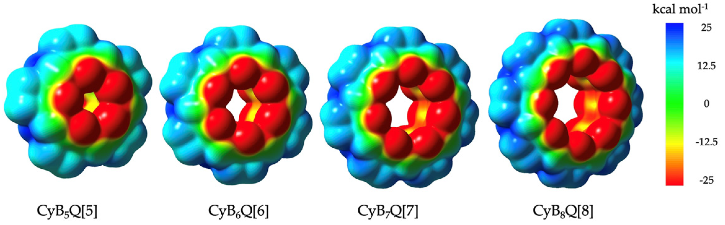
| Cavity C–C | Equatorial CH/CH2 | Portal C=O | |
|---|---|---|---|
| Q[n] | 0 ± 5 | +25 | −25 [4] |
| CyBnQ[n] | −12.5 | +12.5 | −25 |
| Average β° | (CyBnQ[n]–Q[n]) | (CyBnQ[n]–Me2nQ[n]) ∆° | ||
|---|---|---|---|---|
| N–C–N | ∆° | |||
| Me10Q[5] | 112.17 | [36] [33] | ||
| Q[5] | 115.14 | |||
| CyB5Q[5] | 115.98 | 0.84 | 3.81 | |
| Me12Q[6] | 112.84 | [37] [34] | ||
| Q[6] | 114.63 | |||
| CyB6Q[6] | 115.83 | 1.2 | 2.99 | |
| Q[7] | 114.65 | [33] | ||
| CyB7Q[7] | 115.2 | 0.55 | - 1 | |
| Q[8] | 114.51 | [35] | ||
| CyB8Q[8] | 115.21 | 0.7 | - 1 |
| Substituent R = | Calculated β° | Measured β° [29,30] |
|---|---|---|
| H | 111.9 | - |
| CyB | 112.8 | 111.77 |
| CyP | 110.5 | 110.08 |
| CyH | 109.8 | 109.31 |
| Me | 110.2 | 108.88 |
| 1 | 2 | 3 | 4 | |
|---|---|---|---|---|
| Formula | C40H54N20O17 | C48H82Cl2N24O28 | C56H80N28O26 | C64H89Cl3N32-O28Zn |
| FW | 1087.03 | 1514.27 | 1561.48 | 1926.39 |
| T/K | 100 | 293 | 293 | 110 |
| Crystal system | triclinic | monoclinic | orthorhombic | triclinic |
| Space group | P-1 | P1 21/c 1 | P nma | P-1 |
| a [Å] | 12.3691(7) | 11.3747(6) | 13.4181(8) | 15.8972(12) |
| b [Å] | 14.8265(17) | 17.0087(7) | 23.0795(18) | 18.3412(14) |
| c [Å] | 17.6974(10) | 17.4352(7) | 28.3746(19) | 20.3221(14) |
| α [°] | 75.361(7) | 90 | 90 | 78.560(6) |
| β [°] | 89.902(5) | 93.656(4) | 90 | 75.665(6) |
| γ [°] | 74.370(7) | 90 | 90 | 70.009(7) |
| V [Å3] | 3016.4(4) | 3366.3(3) | 8787.1(10) | 5352.8(7) |
| Z | 2 | 2 | 4 | 2 |
| R [I > 2σ(I)] 1 | 0.1181 | 0.0877 | 0.0954 | 0.0912 |
| wR [I > 2σ(I)] 2 | 0.3249 | 0.2500 | 0.2575 | 0.2256 |
Publisher’s Note: MDPI stays neutral with regard to jurisdictional claims in published maps and institutional affiliations. |
© 2021 by the authors. Licensee MDPI, Basel, Switzerland. This article is an open access article distributed under the terms and conditions of the Creative Commons Attribution (CC BY) license (https://creativecommons.org/licenses/by/4.0/).
Share and Cite
Chen, M.; Lv, N.; Zhao, W.; Day, A.I. The Cyclobutanocucurbit[5–8]uril Family: Electronegative Cavities in Contrast to Classical Cucurbituril while the Electropositive Outer Surface Acts as a Crystal Packing Driver. Molecules 2021, 26, 7343. https://doi.org/10.3390/molecules26237343
Chen M, Lv N, Zhao W, Day AI. The Cyclobutanocucurbit[5–8]uril Family: Electronegative Cavities in Contrast to Classical Cucurbituril while the Electropositive Outer Surface Acts as a Crystal Packing Driver. Molecules. 2021; 26(23):7343. https://doi.org/10.3390/molecules26237343
Chicago/Turabian StyleChen, Minghua, Naixia Lv, Weiwei Zhao, and Anthony I. Day. 2021. "The Cyclobutanocucurbit[5–8]uril Family: Electronegative Cavities in Contrast to Classical Cucurbituril while the Electropositive Outer Surface Acts as a Crystal Packing Driver" Molecules 26, no. 23: 7343. https://doi.org/10.3390/molecules26237343
APA StyleChen, M., Lv, N., Zhao, W., & Day, A. I. (2021). The Cyclobutanocucurbit[5–8]uril Family: Electronegative Cavities in Contrast to Classical Cucurbituril while the Electropositive Outer Surface Acts as a Crystal Packing Driver. Molecules, 26(23), 7343. https://doi.org/10.3390/molecules26237343