
Luminescent Complexes of Europium (III) with 2-(Phenylethynyl)-1,10-phenanthroline: The Role of the Counterions

 ,
,  ,
,  and
and 
Abstract
:1. Introduction
2. Results and Discussion
2.1. Characterization of Polycrystalline Samples
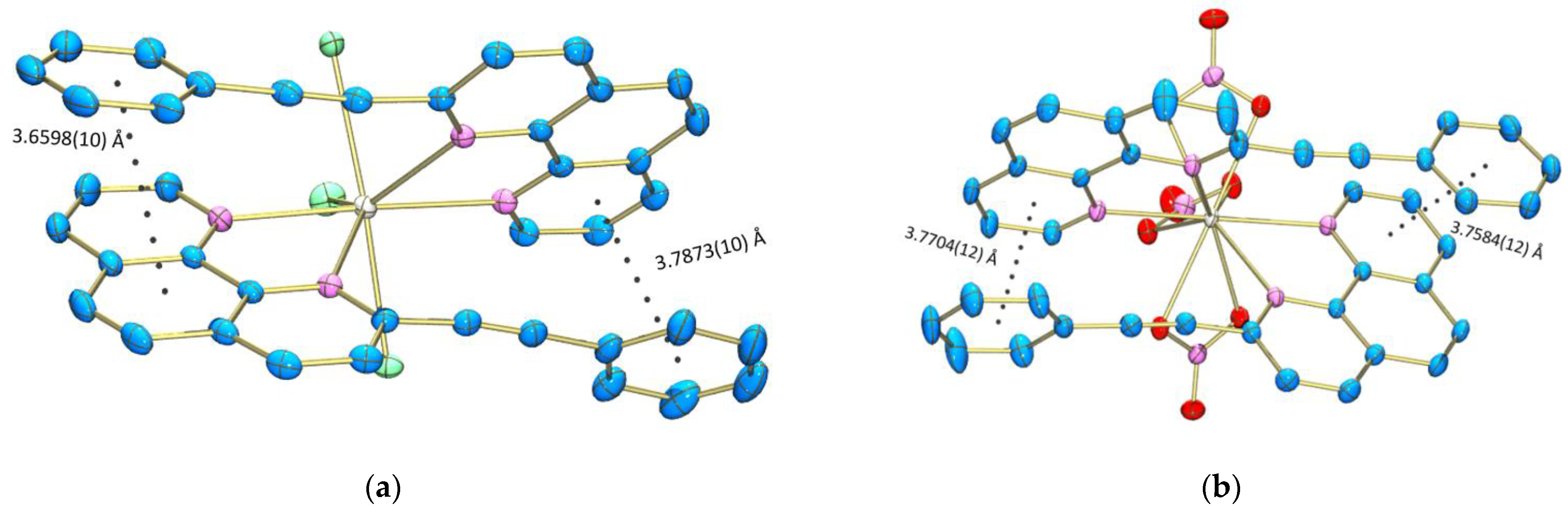
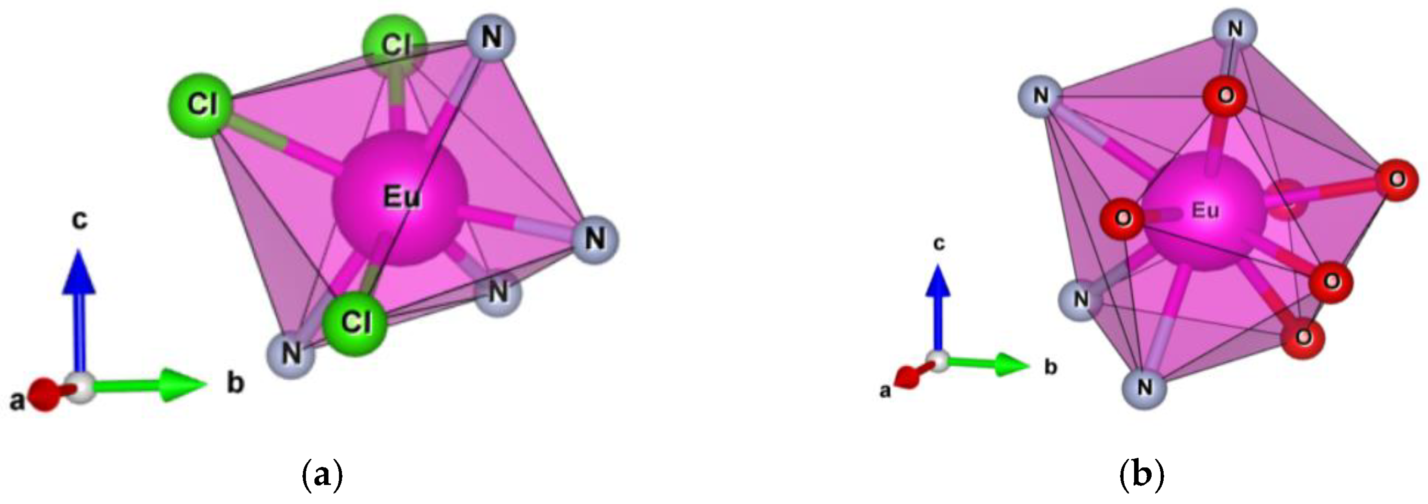
2.2. Crystal Structure
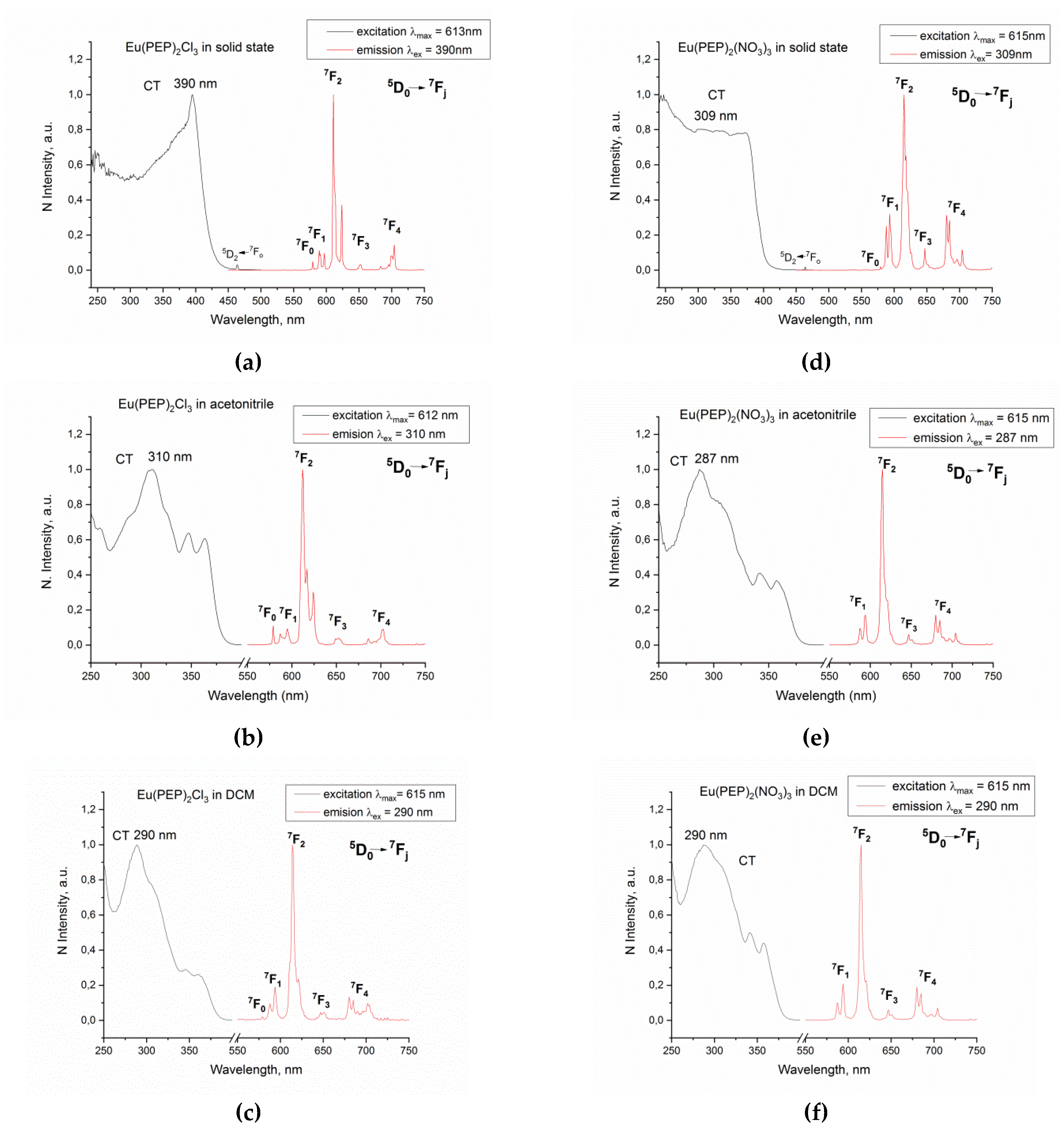
2.3. Optical Properties
3. Conclusions
4. Materials and Methods
4.1. Materials Used
4.2. Preparation of the Ligand and the Complexes
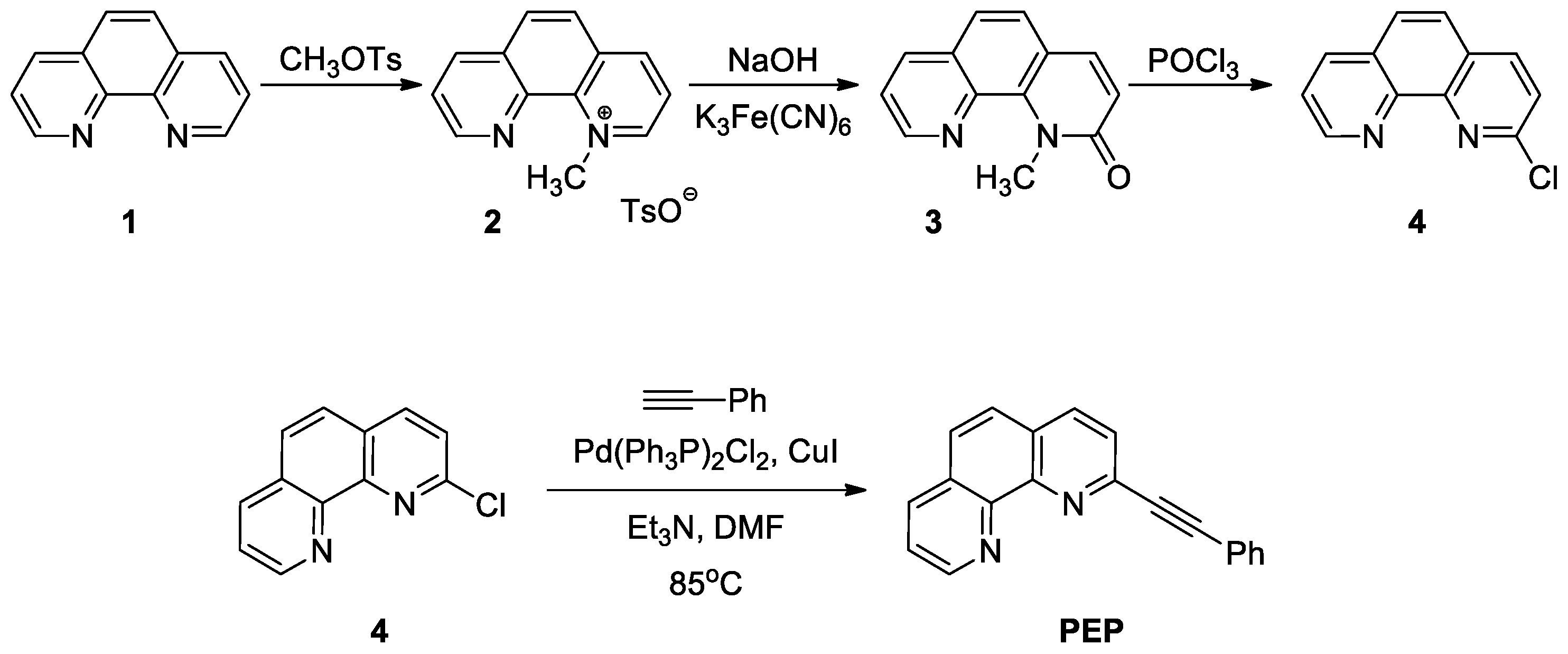
4.2.1. Synthesis of the 2-(Phenylethynyl)-1,10-phenanthroline Ligand
4.2.2. Synthesis of the Eu (III) Complexes with PEP
4.3. Characterization
4.4. Computational Protocol
Supplementary Materials
Author Contributions
Funding
Institutional Review Board Statement
Informed Consent Statement
Data Availability Statement
Acknowledgments
Conflicts of Interest
Sample Availability
References
- Zhang, Q.; Wang, J.; Kirillov, A.M.; Dou, W.; Xu, C.; Xu, C.; Yang, L.; Fang, R.; Liu, W. Multifunctional Ln-MOF Luminescent Probe for Efficient Sensing of Fe3+, Ce3+, and Acetone. ACS Appl. Mater. Interfaces 2018, 10, 23976–23986. [Google Scholar] [CrossRef] [PubMed]
- Gao, Y.; Yu, G.; Liu, K.; Wang, B. Luminescent mixed-crystal Ln-MOF thin film for the recognition and detection of pharmaceuticals. Sens. Actuators B Chem. 2018, 257, 931–935. [Google Scholar] [CrossRef]
- Yu, H.; Fan, M.; Liu, Q.; Su, Z.; Li, X.; Pan, Q.; Hu, X. Two Highly Water-Stable Imidazole-Based Ln-MOFs for Sensing Fe3+,Cr2O72-/CrO42- in a Water Environment. Inorg. Chem. 2020, 59, 2005–2010. [Google Scholar] [CrossRef]
- Bünzli, J.C.G. Lanthanide luminescent bioprobes (LLBs). Chem. Lett. 2009, 38, 104–109. [Google Scholar] [CrossRef]
- Hamon, N.; Roux, A.; Beyler, M.; Mulatier, J.-C.; Andraud, C.; Nguyen, C.; Maynadier, M.; Bettache, N.; Duperray, A.; Grichine, A.; et al. Pyclen-Based Ln(III) Complexes as Highly Luminescent Bioprobes for In Vitro and In Vivo One- and Two-Photon Bioimaging Applications. J. Am. Chem. Soc. 2020, 142, 10184–10197. [Google Scholar] [CrossRef]
- Gallardo, H.; Braga, H.C.; Tuzimoto, P.; Bortoluzzi, A.; Salla, C.A.M.; Bechtold, I.H.; Martins, J.S.; Legnani, C.; Quirino, W.G. Synthesis, structure and OLED application of a new europium(III) complex: {tris-(thenoyltrifluoroacetonate)[1,2,5]selenadiazolo[3,4-f][1,10]phenanthroline}europium(III). Inorg. Chim. Acta 2018, 473, 75–82. [Google Scholar] [CrossRef]
- Utochnikova, V.V. Chapter 318-Lanthanide complexes as OLED emitters. In Handbook on the Physics ans Chemistry of Rare Earths; Bünzli, J.-C.G., Pecharsky, V.K., Eds.; Elsevier: Moscow, Russia, 2021; Volume 59, pp. 1–91. [Google Scholar]
- Gahlaut, N.; Miller, L.W. Time-resolved microscopy for imaging lanthanide luminescence in living cells. Cytom. Part. A 2010, 77A, 1113–1125. [Google Scholar] [CrossRef] [Green Version]
- Deng, Q.; Zhu, Z.; Shu, X. Auto-Phase-Locked Time-Resolved Luminescence Detection: Principles, Applications, and Prospects. Front. Chem. 2020, 8, 562. [Google Scholar] [CrossRef]
- Bünzli, J.-C.G. Piguet Taking advantage of luminescent lanthanide ions. Chem. Soc. Rev. 2005, 34, 1048–1077. [Google Scholar] [CrossRef]
- Kostova, I.; Trendafilova, N.; Momekov, G. Theoretical, spectral characterization and antineoplastic activity of new lanthanide complexes. J. Trace Elem. Med. Biol. 2008, 22, 100–111. [Google Scholar] [CrossRef]
- Chundawat, N.S.; Jadoun, S.; Zarrintaj, P.; Chauhan, N.P.S. Lanthanide complexes as anticancer agents: A review. Polyhedron 2021, 207, 115387. [Google Scholar] [CrossRef]
- Bao, G. Lanthanide complexes for drug delivery and therapeutics. J. Lumin. 2020, 228, 117622. [Google Scholar] [CrossRef]
- Chan, W.-L.; Xie, C.; Lo, W.-S.; Bünzli, J.-C.G.; Wong, W.-K.; Wong, K.-L. Lanthanide–tetrapyrrole complexes: Synthesis, redox chemistry, photophysical properties, and photonic applications. Chem. Soc. Rev. 2021, 50, 12189–12257. [Google Scholar] [CrossRef]
- Bünzli, J.-C.G. On the design of highly luminescent lanthanide complexes. Coord. Chem. Rev. 2014, 293, 19–47. [Google Scholar] [CrossRef]
- Weissman, S.I. Intramolecular Energy Transfer The Fluorescence of Complexes of Europium. J. Chem. Phys. 1942, 10, 214–217. [Google Scholar] [CrossRef]
- Petoud, S.; Cohen, S.M.; Bünzli, J.C.G.; Raymond, K.N. Stable Lanthanide Luminescence Agents Highly Emissive in Aqueous Solution: Multidentate 2-Hydroxyisophthalamide Complexes of Sm3+, Eu3+, Tb3+, Dy3+. J. Am. Chem. Soc. 2003, 125, 13324–13325. [Google Scholar] [CrossRef]
- Eliseeva, S.V.; Pleshkov, D.N.; Lyssenko, K.A.; Lepnev, L.S.; Bünzli, J.-C.G.; Kuzmina, N.P. Deciphering three beneficial effects of 2,2′-bipyridine-N,N′-dioxide on the luminescence sensitization of lanthanide(III) hexafluoroacetylacetonate ternary complexes. Inorg. Chem. 2011, 50, 5137–5144. [Google Scholar] [CrossRef] [Green Version]
- Shavaleev, N.M.; Eliseeva, S.V.; Scopelliti, R.; Bünzli, J.C.G. Influence of Symmetry on the Luminescence and Radiative Lifetime of Nine-Coordinate Europium Complexes. Inorg. Chem. 2015, 54, 9166–9173. [Google Scholar] [CrossRef]
- Danchova, N.; Gutzov, S. Functionalization of Sol-Gel Zirconia Composites with Europium Complexes. Z. Naturforsch. B 2014, 69, 224–230. [Google Scholar] [CrossRef]
- Lenaerts, P. Covalent coupling of luminescent lanthanide complexes on hybrid materials and solid polymer supports. Kathol. Univ. Leuven 2005. Available online: https://www.researchgate.net/publication/28359751_Covalent_coupling_of_luminescent_lanthanide_complexes_on_hybrid_materials_and_solid_polymer_supports (accessed on 10 November 2021).
- Lunstroot, K.; Nockemann, P.; Van Hecke, K.; Van Meervelt, L.; Go, C.; Binnemans, K.; Driesen, K. Visible and Near-Infrared Emission by Samarium ( III ) -Containing Ionic Liquid Mixtures. Inorg. Chem. 2009, 48, 3018–3026. [Google Scholar] [CrossRef]
- Binnemans, K.; Görller-Walrand, C. Lanthanide-containing liquid crystals and surfactants. Chem. Rev. 2002, 102, 2303–2345. [Google Scholar] [CrossRef] [PubMed]
- Cui, Y.; Zhang, J.; He, H.; Qian, G. Photonic functional metal-organic frameworks. Chem. Soc. Rev. 2018, 47, 5740–5785. [Google Scholar] [CrossRef]
- Parreira, R.L.T.; Nassar, E.J.; da Silva, E.H.; Rocha, L.A.; Pedro, P.A.; Ferreira, C.M.A.; Kar, T.; Fonseca, D.E.P.; Coimbra, D.F.; Caramori, G.F. Electronic properties and metal-ligand bonding situation in Eu (III) complexes containing tris(pyrazolyl)borate and phenantroline ligands. J. Lumin. 2017, 182, 137–145. [Google Scholar] [CrossRef]
- Binnemans, K. Lanthanide-based luminescent hybrid materials. Chem. Rev. 2009, 109, 4283–4374. [Google Scholar] [CrossRef] [Green Version]
- Miller, M.T.; Gantzel, P.K.; Karpishin, T.B. Structures of the Copper(I) and Copper(II) Complexes of 2,9-Diphenyl-1,10-phenanthroline: Implications for Excited-State Structural Distortion. Inorg. Chem. 1998, 37, 2285–2290. [Google Scholar] [CrossRef]
- Alvariño, C.; Simond, D.; Lorente, P.M.; Besnard, C.; Williams, A.F. Chains, Necklaces and Weaving Chain-link Grids from Self-Assembly Reactions. Chem. Eur. J. 2015, 21, 8851–8858. [Google Scholar] [CrossRef]
- Maouche, R.; Belaid, S.; Benmerad, B.; Bouacida, S.; Freslon, S.; Daiguebonne, C.; Suffren, Y.; Calvez, G.; Bernot, K.; Roiland, C.; et al. Luminescence properties of lanthanide complexes-based molecular alloys. Inorg. Chim. Acta 2020, 501, 119309. [Google Scholar] [CrossRef]
- Akerboom, S.; Van Den Elshout, J.J.M.H.; Mutikainen, I.; Siegler, M.A.; Fu, W.T.; Bouwman, E. Substituted phenanthrolines as antennae in luminescent EuIII complexes. Eur. J. Inorg. Chem. 2013, 6137–6146. [Google Scholar] [CrossRef]
- Georgieva, I.; Trendafilova, N.; Zahariev, T.; Danchova, N.; Gutzov, S. Theoretical insight in highly luminescent properties of Eu (III) complex with phenanthroline. J. Lumin. 2018, 202, 192–205. [Google Scholar] [CrossRef]
- Zahariev, T.; Shandurkov, D.; Gutzov, S.; Trendafilova, N.; Enseling, D.; Jüstel, T.; Georgieva, I. Phenanthroline chromophore as efficient antenna for Tb3+ green luminescence: A theoretical study. Dye. Pigment. 2021, 185, 108890. [Google Scholar] [CrossRef]
- Werner, F.; Tada, K.; Ishii, A.; Takatab, M.; Hasegawa, M. The key role of accurate lattice parameters in revealing subtle structural differences-A case study in the system [Ln(phen/phen-d8) 2(NO3)3]. CrystEngComm 2009, 11, 1197–1200. [Google Scholar] [CrossRef]
- Accorsi, G.; Listorti, A.; Yoosaf, K.; Armaroli, N. 1,10-Phenanthrolines: Versatile building blocks for luminescent molecules, materials and metal complexes. Chem. Soc. Rev. 2009, 38, 1690–1700. [Google Scholar] [CrossRef]
- Cabral Campello, M.P.; Palma, E.; Correia, I.; Paulo, P.M.R.; Matos, A.; Rino, J.; Coimbra, J.; Pessoa, J.C.; Gambino, D.; Paulo, A.; et al. Lanthanide complexes with phenanthroline-based ligands: Insights into cell death mechanisms obtained by microscopy techniques. Dalt. Trans. 2019, 48, 4611–4624. [Google Scholar] [CrossRef]
- Sahoo, J.; Jaiswar, S.; Jena, H.S.; Subramanian, P.S. Sensing of Phosphate and ATP by Lanthanide Complexes in Aqueous Medium and Its Application on Living Cells. ChemistrySelect 2020, 5, 12878–12884. [Google Scholar] [CrossRef]
- Simonnet, M.; Suzuki, S.; Miyazaki, Y.; Kobayashi, T.; Yokoyama, K.; Yaita, T. Lanthanide Intra-series Separation by a 1,10-Phenanthroline Derivative: Counterion Effect. Solvent Extr. Ion. Exch. 2020, 38, 430–440. [Google Scholar] [CrossRef]
- Simonnet, M.; Kobayashi, T.; Shimojo, K.; Yokoyama, K.; Yaita, T. Study on Phenanthroline Carboxamide for Lanthanide Separation: Influence of Amide Substituents. Inorg. Chem. 2021, 60, 13409–13418. [Google Scholar] [CrossRef]
- Yang, X.F.; Ren, P.; Yang, Q.; Geng, J.S.; Zhang, J.Y.; Yuan, L.Y.; Tang, H.B.; Chai, Z.F.; Shi, W.Q. Strong Periodic Tendency of Trivalent Lanthanides Coordinated with a Phenanthroline-Based Ligand: Cascade Countercurrent Extraction, Spectroscopy, and Crystallography. Inorg. Chem. 2021, 60, 9745–9756. [Google Scholar] [CrossRef]
- Healy, M.R.; Ivanov, A.S.; Karslyan, Y.; Bryantsev, V.S.; Moyer, B.A.; Jansone-Popova, S. Efficient Separation of Light Lanthanides(III) by Using Bis-Lactam Phenanthroline Ligands. Chem. Eur. J. 2019, 25, 6326–6331. [Google Scholar] [CrossRef]
- Mirochnik, A.G.; Bukvetskii, B.V.; Zhikhareva, P.A.; Karasev, V.E. Crystal Structure and Luminescence of the [Eu(Phen)2(NO3)3] Complex. The Role of the Ion-Coactivator. Russ. J. Coord. Chem. 2001, 27, 443–448. [Google Scholar] [CrossRef]
- Zheng, Y.-Q.Q.; Zhou, L.-X.X.; Lin, J.-L.L.; Zhang, S.W. Syntheses and Crystal Structures of Ln(phen)2(NO3)3 with Ln = Pr, Nd, Sm, Eu, Dy, and phen = 1,10-phenanthroline. Z. Anorg. Allg. Chem. 2001, 627, 1643–1646. [Google Scholar] [CrossRef]
- Hart, F.A.; Laming, F.P. Complexes of 1,10-phenanthroline with lanthanide chlorides and thiocyanates. J. Inorg. Nucl. Chem. 1964, 26, 579–585. [Google Scholar] [CrossRef]
- Shurygin, A.V.; Vovna, V.I.; Korochentsev, V.V.; Mirochnik, A.G.; Kalinovskaya, I.V.; Sergienko, V.I. Electronic structure and optical properties of Ln(III) nitrate adducts with 1,10-phenanthroline. Spectrochim. Acta Part A Mol. Biomol. Spectrosc. 2019, 213, 176–183. [Google Scholar] [CrossRef]
- Sadikov, G.G.; Antsyshkina, A.S.; Kuznetsova, I.A.; Rodnikova, M.N. Synthesis and structure of two crystalline modifications of bis(1,10-Phenanthroline)trinitratoeuropium(III) Eu(NO3) 3(Phen)2. Crystallogr. Rep. 2006, 51, 47–52. [Google Scholar] [CrossRef]
- Tsaryuk, V.I.; Zhuravlev, K.P.; Vologzhanina, A.V.; Kudryashova, V.A.; Zolin, V.F. Structural regularities and luminescence properties of dimeric europium and terbium carboxylates with 1,10-phenanthroline (CN=9). J. Photochem. Photobiol. A Chem. 2010, 211, 7–19. [Google Scholar] [CrossRef]
- Pan, Z.; Jia, G.; Duan, C.K.; Wong, W.Y.; Wong, W.T.; Tanner, P.A. Crystal structure, spectroscopy and crystal field analysis of substituted 1,10-phenanthroline-europium complexes. Eur. J. Inorg. Chem. 2011, 637–646. [Google Scholar] [CrossRef]
- Binnemans, K. Interpretation of europium(III) spectra. Coord. Chem. Rev. 2015, 295, 1–45. [Google Scholar] [CrossRef] [Green Version]
- Krapcho, A.P.; Lanza, J.B. Improved Synthesis of 2,9-Dichloro-1,10- phenanthroline. Org. Prep. Proced. Int. 2007, 39, 603–620. [Google Scholar] [CrossRef]
- Sjogren, M.; Hansson, S.; Norrby, P.-O.; Akermark, B.; Cucciolito, M.E.; Vitagliano, A. Selective Stabilization of the Anti Isomer of (η3-Allyl)palladium and -platinum Complexes. Organometallics 1992, 11, 3954–3964. [Google Scholar] [CrossRef]
- Sheldrick, G.M. A short history of SHELX. Acta Crystallogr. A 2008, 64, 112–122. [Google Scholar] [CrossRef] [Green Version]
- Sheldrick, G.M. Crystal structure refinement with SHELXL. Acta Crystallogr. Sect. C 2015, 71, 3–8. [Google Scholar] [CrossRef]
- Hübschle, C.B.; Sheldrick, G.M.; Dittrich, B. ShelXle: A Qt graphical user interface for SHELXL. J. Appl. Crystallogr. 2011, 44, 1281–1284. [Google Scholar] [CrossRef] [Green Version]
- Zhang, Y.; Qi, D.; Cai, X.; Jiang, J. Vibrational spectra of mixed (phthalocyaninato)(porphyrinato) yttrium(III) double-decker complexes: Density functional theory calculations. Vib. Spectrosc. 2009, 51, 184–192. [Google Scholar] [CrossRef]
- Tarakanova, E.N.; Tarakanov, P.A.; Simakov, A.O.; Furuyama, T.; Kobayashi, N.; Konev, D.V.; Goncharova, O.A.; Trashin, S.A.; De Wael, K.; Sulimenkov, I.V.; et al. Synthesis and characterization of heteroleptic rare earth double-decker complexes involving tetradiazepinoporphyrazine and phthalocyanine macrocycles. Dalt. Trans. 2021, 50, 6245–6255. [Google Scholar] [CrossRef]
- Qi, D.; Zhang, L.; Wan, L.; Zhang, Y.; Bian, Y.; Jiang, J. Conformational effects, molecular orbitals, and reaction activities of bis(phthalocyaninato) lanthanum double-deckers: Density functional theory calculations. Phys. Chem. Chem. Phys. 2011, 13, 13277–13286. [Google Scholar] [CrossRef]
- Chai, J.D.; Head-Gordon, M. Long-range corrected hybrid density functionals with damped atom-atom dispersion corrections. Phys. Chem. Chem. Phys. 2008, 10, 6615–6620. [Google Scholar] [CrossRef] [Green Version]
- Zhang, Y.; Thor, W.; Wong, K.-L.; Tanner, P.A. Determination of Triplet State Energy and the Absorption Spectrum for a Lanthanide Complex. J. Phys. Chem. C 2021, 125, 7022–7033. [Google Scholar] [CrossRef]
- Dolg, M.; Stoll, H.; Savin, A.; Preuss, H. Energy-adjusted pseudopotentials for the rare earth elements. Theor. Chim. Acta 1989, 75, 173–194. [Google Scholar] [CrossRef]
- Runge, E.; Gross, E.K.U. Density-Functional Theory for Time-Dependent Systems. Phys. Rev. Lett. 1984, 52, 997–1000. [Google Scholar] [CrossRef]
- Miertuš, S.; Scrocco, E.; Tomasi, J. Electrostatic interaction of a solute with a continuum. A direct utilizaion of AB initio molecular potentials for the prevision of solvent effects. Chem. Phys. 1981, 55, 117–129. [Google Scholar] [CrossRef]
- Frisch, M.J.; Trucks, G.W.; Schlegel, H.B.; Scuseria, G.E.; Robb, M.A.; Cheeseman, J.R.; Scalmani, G.; Barone, V.; Mennucci, B.; Petersson, G.A.; et al. Gaussian 09W, A.02; Gaussian, Inc.: Wallingford, CT, USA, 2009. [Google Scholar]


| Eu(PEP)2Cl3 | Eu(PEP)2(NO3)3 | |||
|---|---|---|---|---|
| R [Å] | Eu–N | 2.5320(13) | Eu–N | 2.6006(15) |
| Eu–N≡ | 2.5822(14) | Eu–N≡ | 2.6253(15) | |
| Eu–N | 2.5400(14) | Eu–N | 2.5958(15) | |
| Eu–N≡ | 2.5806(12) | Eu–N≡ | 2.6542(15) | |
| Eu1–Cl1 | 2.6930(5) | Eu–O1 | 2.5247(14) | |
| Eu-Cltop | 2.6893(7) | Eu–O2 | 2.4813(13) | |
| Eu–Cl2 | 2.6744(5) | Eu–Otop | 2.5273(14) | |
| Eu–Otop | 2.5164(14) | |||
| Eu–O7 | 2.5371(14) | |||
| Eu–O8 | 2.5022(13) | |||
| Θ [o] | Cl1–Eu–Cl2 | 165.30(3) | O1–Eu–O2 | 51.07(4) |
| Cl2–Eu–Cltop | 82.71(2) | Otop–Eu–Otop | 50.60(5) | |
| Cl1–Eu-Cltop | 82.59(2) | O7–Eu–O8 | 50.74(4) | |
| N–Eu–N | 64.30(4) | N–Eu–N | 63.42(5) | |
| N≡–Eu–N | 64.45(4) | N–Eu–N≡ | 63.31(5) | |
| Eu(PEP)2Cl3 | Eu(PEP)2(NO3)3 | ||||
|---|---|---|---|---|---|
| (d) | (a) | (d) | (a) | ||
| Eu–N | 2.651 | 2.647 | Eu–N | 2.627 | 2.628 |
| Eu–N≡ | 2.664 | 2.654 | Eu–N≡ | 2.685 | 2.675 |
| Eu–N | 2.651 | 2.648 | Eu–N | 2.627 | 2.628 |
| Eu–N≡ | 2.664 | 2.653 | Eu–N≡ | 2.685 | 2.675 |
| Eu–Cl≡ | 2.778 | 2.791 | Eu–O | 2.548 | 2.554 |
| Eu–Cltop | 2.744 | 2.759 | Eu–O | 2.539 | 2.542 |
| Eu–Cl | 2.778 | 2.791 | Eu–Otop | 2.511 | 2.519 |
| Eu–Otop | 2.511 | 2.519 | |||
| Eu–O | 2.548 | 2.554 | |||
| Eu–O | 2.539 | 2.542 | |||
| Sample | Life Time in µs | QY% |
|---|---|---|
| Eu(PEP)2Cl3 solid state | 460 ± 11 | - |
| Eu(PEP)2(NO3)3 solid state | 1337 ± 2 | - |
| Eu(PEP)2Cl3 in solution/acetonitrile | 703 ± 1 | 8.8 ± 0.5 |
| Eu(PEP)2(NO3)3 in solution/acetonitrile | 1393 ± 25 | 9.6 ± 0.5 |
| Eu(PEP)2Cl3 in solution/DCM | 572 ± 4 | 1.4 ± 0.2 |
| Eu(PEP)2(NO3)3 in solution/DCM | 1665 ± 35 | 28.1 ± 0.5 |
| Eu(PEP)2Cl3 | Eu(PEP)2(NO3)3 | PEP | ||||
|---|---|---|---|---|---|---|
| (d) | (a) | (d) | (a) | (d) | (a) | |
| Ev (S0→S1) | 4.10 | 4.11 | 4.23 | 4.23 | 4.17 | 4.18 |
| f (S0→S1) | 0.1313 | 0.1193 | 0.0577 | 0.0528 | 1.1996 | 1.1412 |
| Ev (S0→T1) | 3.10 | 3.10 | 3.16 | 3.16 | 3.02 | 3.02 |
| Ev (T1→S0) | 2.14 | 2.13 | 2.35 | 2.35 | 2.29 | 2.29 |
| E0–0 (T1→S0) | 2.45 | 2.45 | 2.62 | 2.62 | 2.67 | 2.67 |
Publisher’s Note: MDPI stays neutral with regard to jurisdictional claims in published maps and institutional affiliations. |
© 2021 by the authors. Licensee MDPI, Basel, Switzerland. This article is an open access article distributed under the terms and conditions of the Creative Commons Attribution (CC BY) license (https://creativecommons.org/licenses/by/4.0/).
Share and Cite
Elenkova, D.; Lyapchev, R.; Romanova, J.; Morgenstern, B.; Dimitrova, Y.; Dimov, D.; Tsvetkov, M.; Zaharieva, J. Luminescent Complexes of Europium (III) with 2-(Phenylethynyl)-1,10-phenanthroline: The Role of the Counterions. Molecules 2021, 26, 7272. https://doi.org/10.3390/molecules26237272
Elenkova D, Lyapchev R, Romanova J, Morgenstern B, Dimitrova Y, Dimov D, Tsvetkov M, Zaharieva J. Luminescent Complexes of Europium (III) with 2-(Phenylethynyl)-1,10-phenanthroline: The Role of the Counterions. Molecules. 2021; 26(23):7272. https://doi.org/10.3390/molecules26237272
Chicago/Turabian StyleElenkova, Denitsa, Rumen Lyapchev, Julia Romanova, Bernd Morgenstern, Yana Dimitrova, Deyan Dimov, Martin Tsvetkov, and Joana Zaharieva. 2021. "Luminescent Complexes of Europium (III) with 2-(Phenylethynyl)-1,10-phenanthroline: The Role of the Counterions" Molecules 26, no. 23: 7272. https://doi.org/10.3390/molecules26237272
APA StyleElenkova, D., Lyapchev, R., Romanova, J., Morgenstern, B., Dimitrova, Y., Dimov, D., Tsvetkov, M., & Zaharieva, J. (2021). Luminescent Complexes of Europium (III) with 2-(Phenylethynyl)-1,10-phenanthroline: The Role of the Counterions. Molecules, 26(23), 7272. https://doi.org/10.3390/molecules26237272