Article

Synthesis and Evaluation of the Tetracyclic Ring-System of Isocryptolepine and Regioisomers for Antimalarial, Antiproliferative and Antimicrobial Activities

1
Department of Chemistry, Bioscience and Environmental Engineering, University of Stavanger, NO-4036 Stavanger, Norway
2
Discovery Biology, Griffith Institute for Drug Discovery, Griffith University, Don Young Road, Nathan, QLD 4111, Australia
3
The Norwegian College of Fishery Science, Marbio, UiT—The Arctic University of Norway, Breivika, NO-9037 Tromsø, Norway
4
CRC for Cancer Therapeutics, Griffith University, Nathan, QLD 4111, Australia
5
School of Environment & Science, Nathan, Griffith University, Nathan, QLD 4111, Australia
*
Author to whom correspondence should be addressed.
Molecules 2021, 26(11), 3268; https://doi.org/10.3390/molecules26113268
Submission received: 28 April 2021 / Revised: 18 May 2021 / Accepted: 26 May 2021 / Published: 30 May 2021
(This article belongs to the Special Issue Total Synthesis of Natural Products)

Abstract

:
A series of novel quinoline-based tetracyclic ring-systems were synthesized and evaluated in vitro for their antiplasmodial, antiproliferative and antimicrobial activities. The novel hydroiodide salts 10 and 21 showed the most promising antiplasmodial inhibition, with compound 10 displaying higher selectivity than the employed standards. The antiproliferative assay revealed novel pyridophenanthridine 4b to be significantly more active against human prostate cancer (IC50 = 24 nM) than Puromycin (IC50 = 270 nM) and Doxorubicin (IC50 = 830 nM), which are used for clinical treatment. Pyridocarbazoles 9 was also moderately effective against all the employed cancer cell lines and moreover showed excellent biofilm inhibition (9a: MBIC = 100 µM; 9b: MBIC = 100 µM).

1. Introduction

Malaria and cancer are two major health issues affecting millions of lives annually. Malaria is a parasitic blood disease caused by protozoans of the Plasmodium genus. Although five Plasmodium strains are known to infect humans, namely P. falciparum, P. vivax, P. ovale, P. knowlesi and P. malariae, infections by P. falciparum are responsible for the majority of malaria-related deaths [1,2]. The World Health Organization (WHO) estimated the number of malaria cases to be 229 million in 2019, claiming approximately 409,000 lives [1], despite considerable global efforts to combat the disease. A major obstacle in the battle against malaria has been the rapid appearance and spread of resistant strains across endemic areas [3]. An excess of 90% of all malaria incidents occur in sub-Saharan Africa [1], a region sorely dependent on the availability of affordable treatments. Originally, malaria-endemic regions were primarily limited to the immediate areas surrounding the tropics. The increasing surface air temperatures as a consequence of global warming is predicted to change this, leaving also temperate climates susceptible to the disease, and with it, a larger part of the human population [4]. Following the widespread appearance of chloroquine (CQ)-resistant strains of P. falciparum, artemisinin-based therapies have been the gold standard of malaria treatment [5]. However, in 2008, the first reports of artemisinin-based resistance were observed in Cambodia [6] and ten years later, over 30 independent cases had been documented throughout southeast Asia [7]. Therefore, the development of novel and affordable treatments remains of paramount importance.
Contrary to malaria, which is an infectious disease, cancer is a noncommunicable disease, ranking as the second leading cause of death globally, responsible for approximately 1 in 6 deaths. Estimates from WHO put the number of cancer cases in 2018 at 18.1 million, accompanied by 9.6 million fatalities [8]. The five most diagnosed cancers are lung, breast, colorectal, prostate and stomach. A variety of anti-cancer therapies are currently available, however, those treated suffer from the unwanted side effect of being highly immunosuppressed. Patients suffering from a compromised immune system following cancer treatments are therefore more likely to contract nosocomial infections [9], such as infection caused by drug-resistant Staphylococcus aureus, increasing the overall burden of nosocomial infectious diseases [10]. This is further complicated by the increased likelihood of formations of multidrug-resistant biofilms, which are notoriously hard to treat [11,12]. Bacterial infections are also known to be a cause of cancer on their own, and according to the WHO, roughly 13% of all cancers globally occur as a result of chronic infections [8]. Additionally, research in recent years has started to uncover a direct link between the formation of microbial biofilms in the body and the growth of certain cancers [13,14,15]. The availability of anticancer drugs with the dual capability of inhibiting biofilm growth is severely limited, making the development of such drugs greatly needed.
Natural products have proven to be an invaluable source of lead compounds for medicinal research in the past and present due to their wide array of structural diversity [16,17,18]. As of 2020, roughly 40% of all Food and Drugs Administration (FDA) approved drugs have natural origins [19], further demonstrating the importance of natural products in drug discovery. Accordingly, discovery and characterization of natural products and their semi-synthetic derivatives remain pivotal in the search for novel drug candidates [20]. The quinoline core represents a versatile structural motif, possessing applications in the fields of material science, the dye industry and moreover constitute an important building block in the design of pharmaceutical compounds [21]. In particular, due to the presence of the quinoline skeleton in numerous natural products displaying a vast array of biological activities, quinoline-based natural products and their derivatives are attractive medicinal targets [22,23,24,25].
Almost exclusively found in the West African climbing shrub Cryptolepis sanguinolenta [26,27], the indoloquinoline natural products cryptolepine (1), neocryptolepine (2), and isocryptolepine (3a) (Figure 1) represent a unique class of bioactive compounds. These alkaloids are characterized by a fused quinoline and indole moiety [28] and long before the constituents of C. sanguinolenta were identified, the extracts were used in herbal remedies to treat malarial fevers among other ailments [29]. The major bioactive component of the shrub was eventually determined to be cryptolepine (1), which has subsequently received the most attention in the literature of the three regioisomeric indoloquinolines 1, 2, and 3a. A host of biological properties have been observed in cryptolepine (1) assays, such as antiplasmodial, antimalarial [30,31,32,33,34,35], anti-inflammatory [33], antifungal [36,37,38], antimicrobial [39,40,41,42], antiproliferative [43,44,45,46] and antiviral [40]. The linearly arranged planar structure of cryptolepine (1) is believed to be related to its high level of undesired cytotoxicity, resulting in its ability to non-specifically intercalate into DNA, inhibiting topoisomerase II [44,47,48,49]. Neocryptolepine (2) and isocryptolepine (3a) have also been demonstrated to possess similar biological profiles, although inferior to cryptolepine (1) [28,50]. Despite the lower potency, both neocryptolepine (2) and isocryptolepine (3a) were revealed to be significantly less cytotoxic than cryptolepine (1), allowing for the possibility of their derivatives to be developed into new lead compounds [49,51].
The biological activities for the core structures of C. sanguinolenta have been extensively studied while their regioisomers have been largely undescribed. In particular, the novel pyridophenanthridine scaffold 4a (Figure 2) unveiled in our previous study [52], represents an interesting target for biological evaluation. The pyridophenanthridine skeleton may be regarded as a regioisomer to the pyridoacridines (the core structure of which is illustrated in compound 5 in Figure 2), a well-studied class of marine alkaloids most notably known for exhibiting potent antiproliferative qualities [53,54,55,56,57]. Similarly, to cryptolepine (1), nearly all naturally occurring pyridoacridines have been shown to act as DNA intercalating agents, resulting in cytotoxic effects in cultured tumor cells [54,55,58]. They also possess the ability to inhibit topoisomerase II [53,58] and further contain biological profiles such as antibacterial, antifungal, antiviral, antiparasitic and insecticidal [53,56,57,59,60,61]. Consequently, it is postulated that the pyridoacridines and their synthetic derivatives are pivotal for the future generation of medicinal compounds [58].
Recently, we described the preparation of several isocryptolepine regioisomers and certain chemoisomers [52]. In this paper, we present modifications to our previous synthetic strategies which allowed for the realization of novel tetracyclic ring-systems (compounds 4b, 8, and 9) along with the N-alkylation of several compounds to furnish new analogues (compounds 3b, 3c, 10, and 21). Moreover, the newly synthesized compounds, along with our existing library of natural products and analogues, were evaluated for their in vitro antiplasmodial activity against Plasmodium falciparum 3D7 parasites; cytotoxicity against normal mammalian cell line (HEK293), and three cancer cell lines (HCT116, MDA-MB-231 and PC-3). The compounds were also evaluated as antimicrobial agents against common pathogenic bacteria as well as their ability to inhibit biofilm growth.

2. Results and Discussion

2.1. Chemistry

We recently reported a concise synthesis of isocryptolepine (3a) and some regioisomers in which the two key synthetic steps were a Suzuki-Miyaura cross-coupling reaction followed by a palladium-catalyzed intramolecular cyclization [52,62]. The most unexpected result of our previous endeavor was the formation of a pyridophenanthridine scaffold 4a, when biaryl 7a was treated with palladium under our intramolecular cyclization conditions (Path A, Scheme 1). Shortly after our report, Kumar and co-workers reported the formation of compound 4a by a palladium-catalyzed arylation technique utilizing diaryliodinium salts [63]. To the best of our knowledge, these two preparations of pyridophenanthridine 4a remain the only descriptions in the literature. However, Beauchard and coworkers describe the accidental synthesis of the functionalized pyridophenanthridine 4aa in 2006 (Figure 2) [64]. This was the result of attempting to synthesize isocryptolepine analogues by a microwave-induced thermal decomposition of a benzotriazole-coupled quinoline.
Intrigued by these results, we decided to investigate further and wondered if the regioselectivity would be the same utilizing a different synthetic strategy. Drawing inspiration from Timári et al. [65] in the synthesis of isocryptolepine and further expanded on by Hostyn et al. [66] for the synthesis of isoneocryptolepine, a Suzuki-Miyaura cross-coupling reaction and nitrene insertion approach was undertaken. Standard azidation of biaryl 7a via installation of a diazonium salt yielded the aryl azide 7I, which upon thermal decomposition in refluxing 1,2-dichlorobenzene interestingly gave pyridocarbazole 9a as the only product without any traces of its regioisomer 4a (Scheme 1, Path B). Thereby, it was concluded that 4a and 9a can be achieved from a common starting material by following reaction pathway A and B, respectively, in Scheme 1. A fluoro-substituted analogue of compound 9a, namely 9b, was further possible to construct starting from boronic acid 6b. To conclude the synthetic pathways, compounds 4 and 9a were finally regioselectively N-methylated using excess iodomethane in refluxing acetonitrile [49] to realize tetracycles 8 and 10.
In Timári et al.’s original synthesis of isocryptolepine (3a) by means of a thermally induced nitrene insertion, only one regioisomeric product was observed, namely isocryptolepine precursor 14 [65]. Following the same conditions in our laboratories, the approach primarily resulted in the construction of indoloquinoline 14 but its regioisomer 13 was also formed in minor quantities (Scheme 2). Applying the nitrene insertion approach to biaryls 15, 17, and 19, we were able to significantly improve the yields of tetracycles 18 and 20 compared to our previous endeavors (Scheme 3, previous yields in brackets) [52]. Following a literature procedure, neocryptolepine (2) was obtained in good yield starting from its precursor 13 (Scheme 4) [67].
Modification of our previously reported conditions for the N-methylation of tetracycle 14 to furnish isocryptolepine (3a) [62], allowed the formation of two novel isocryptolepine analogues 3b and 3c, albeit in lower yields than the parent alkaloid (Scheme 4). Of the remaining tetracycles, namely compounds 16, 18, and 20, only compound 16 was successfully N-methylated using the same conditions as reported in our previous work [62]. Efforts to explain the failure of tetracycles 18 and 20 to undergo N-alkylation at the most reactive ring-nitrogen, presumably the quinoline moiety, is currently under way in our laboratories.

2.2. Antiplasmodial Assay

The prepared natural products and their derivatives were evaluated for their in vitro antiplasmodial activities against the Plasmodium falciparum 3D7 strain. The compounds were further tested for their in vitro cytotoxicity against HEK293 cells (human embryonic kidney cells) for the determination of their selectivity indices. Furthermore, to serve as positive controls for our analyses, chloroquine (CQ), dihydroartemisinin (DHA) and puromycin were employed. Results from these studies are summarized in Table 1.
The tested compounds were found to possess diverse activities against the Pf3D7 cell line. Albeit being well documented to have antiplasmodial activity in the literature, the parent alkaloid neocryptolepine (2) has thus far not been evaluated for in vitro antimalarial activity against Pf3D7 (IC50 = 7249 nM), showing a lower potency compared to isocryptolepine (3a) (IC50 = 1211 nM). Out of the two novel isocryptolepine derivatives, allyl variant 3c showed a marginal improvement compared to the natural product (IC50 = 1198 nM), while ethyl variant 3b showed a lower activity (IC50 = 1318 nM). Both derivatives were revealed to be notably more cytotoxic than the parent alkaloid 3a.
The neocryptolepine precursor 13 was revealed to display no antiplasmodial inhibition, which is in accordance with a previous study conducted by Jonckers et al., where they highlighted the importance of the N-5 methyl group for activity in certain halogen-substituted indolo[3,2-b]quinolines [68]. The regioisomer 15 was also shown to be inactive against Pf3D7. Contrary to this, the isocryptolepine precursor 14 displayed more potent antimalarial activities (IC50 = 977 nM) compared to the parent alkaloid 3a. For the isocryptolepine precursor 14, it has been shown through previous work that by introduction of certain basic side chains at C-9, the in vitro antimalarial activity against the K-1 strain of P. falciparum was dramatically increased compared to isocryptolepine (3a). The authors argued that these observations could be attributed to the basic properties allowing the compound to experience a lower degree of hydrophobicity [69], a quality also observed for CQ [28].
Pyridophenanthridines 4 (a: IC50 = 548 nM; b: IC50 = 866 nM) outperformed both neocryptolepine (2) and isocryptolepine (3a) in terms of activity and selectivity; however, it displayed an unfavorable increase in cytotoxicity. Keeping in mind the effects observed by Jonckers et al. [68] for the functionalization of the isocryptolepine precursor 14, addition of appropriate substituents to pyridophenanthridine 4a could potentially result in increased antiplasmodial activity. Evidently, the presence of the methoxy substituent in compound 4b negatively impacted both the antiplasmodial activity and cytotoxicity compared to the naked pyridophenanthridine 4a. Interestingly, the addition of an N-methyl group to pyridophenanthridines 4 to furnish compounds 8 (a: IC50 = 1698 nM; b: IC50 = 1546 nM) negatively impacts the antiplasmodial activity. For the indoloquinoline natural products, the N-methyl group is considered an instrumental aspect for their parasitic inhibition [28], this is evidently not the case for the pyridophenanthridines, possibly suggesting the presence of a novel mode of action against the parasitic life cycle. As this represents the first case in the literature of the antiplasmodial evaluation of a pyridophenanthridine, other functionalizations of the core scaffold should nonetheless be further researched.
The two most prominent results of our studies were the novel hydroiodide salts 10 (IC50 = 128 nM) and 21 (IC50 = 380 nM), where the latter showed improved selectivity compared to the standards. Their precursors 9a and 15 showed little to no activity, highlighting the importance of the N-methyl functionality. These results are possibly aided by the fact that the salt structure likely promotes increased solubility in aqueous media, further increasing the biological availability of the compounds, a fact which should be carefully considered when exploring new lead compounds.

2.3. Antiproliferative Assay

All prepared samples were evaluated in vitro against a panel of three cancer cell lines, including HCT116 (human colon cancer), MDA-MB-231 (human breast adenocarcinoma) and PC-3 (human prostate cancer) using a resazurin assay. Puromycin and Doxorubicin were employed as positive controls for the obtained IC50 results, which are summarized in Table 2.
Both parent alkaloids neocryptolepine (2) and isocryptolepine (3a) performed best against the HCT116 cell line (2: 6218 nM; 3a: 667 nM) (Table 2). It is evident that isocryptolepine (3a) had an overall better performance against the tested cancer cell lines than neocryptolepine (2). The same was observed for the isocryptolepine derivatives 3b and 3c; however, the potency was less than for the parent isocryptolepine (3a). Derivatives 3b and 3c were revealed to become less potent with increasing alkyl chain length for the human colon cancer (3b: IC50 = 742 nM; 3c: 1243 nM) and human breast adenocarcinoma (3b: IC50 = 998 nM; 3c: 3064 nM) cell lines. Interestingly, for the human prostate cancer cell line, a different trend was observed (3b: IC50 = 2440 nM; 3c: 1296 nM). The N-allyl group outperformed both the methyl and ethyl groups in terms of activity, suggesting that the alkene functionality is somehow important to the mechanism of cell growth inhibition. It is believed that the indoloquinolines inhibit cell growth by direct interactions with DNA, although the exact mechanism(s) remain uncertain [28,44,47,50,70].
Several of the tested compounds were found to display no activity against the panel of cancer cell lines, including novel compounds 10 and 21. Another compound which was observed to be inactive was neocryptolepine precursor 13, being inactive against all three cell lines. The isocryptolepine precursor 14 showed poor activity against all cancer cell lines and further highlights the necessity of the N-methyl group for cell growth inhibition.
The importance of incorporating an N-methyl is further demonstrated in compounds 4a and its corresponding N-methylated product 8a, showing an increase in activity against the HCT116 and MDA-MB-231 cell lines, favoring the inclusion of an N-methyl group. In the PC-3 cell lines, the pyridophenanthridines 4 showed a decrease in activity with the addition of an N-methyl substituent to give the corresponding compound 8. However, the assay revealed the methoxy pyridophenanthridine 4b to contain potent anticarcinogenic properties (IC50 = 24 nM) against the PC-3 cell line. Compound 4b showed a 10-fold and 35-fold increase in activity compared to the positive controls Puromycin (IC50 = 270 nM) and Doxorubicin (IC50 = 830 nM), respectively. The positioning of the methoxy substituent at C-6 of the pyridophenanthridine scaffold appears to be key to the observed increase in activity, as the naked pyridophenanthridine 4a showed only modest activity against the PC-3 cell line (IC50 = 1630 nM). A previous study by Lu and coworkers demonstrated the potential of the strategic installation of appropriate ring-substituents to obtain increased antiproliferative activity in various indolo[3,2-b]quinolines [71]. Similar to the observations made in this work, Lu et al. noted the potency of C-9 ester substituted indoloquinolines in their screening of several cancer cell lines [71], despite the parent neocryptolepine (2) displaying only minor inhibition of cell growth. The N-methylated pyridophenanthridine 8a evaluated in this work was further shown to be more potent against the MDA-MB-231 (IC50 = 360 nM) cell line than Doxorubicin (IC50 = 590 nM). Being novel compounds, the mode of action of the pyridophenanthridines against proliferative cancer is naturally unknown. Thus, proceeding studies have the potential to unveil a new mode of action. The discovery of new modes of action is regarded as highly important in the field of drug discovery [72], further illustrating the potential for the novel pyridophenanthridine scaffold as a lead for subsequent development into a new anticancer therapy.

2.4. Antimicrobial and Biofilm Iinhibition Assay

The prepared samples were tested for in vitro antimicrobial activity against E. faecalis, E. coli, P. aeruginosa, S. aureus, Streptococcus agalactiae and S. epidermis using gentamycin as a reference compound. The compounds were tested at 100, 75, 50, 25, 12.5, 10, 6.3, 3.1 and 1.6 µM and the obtained minimal inhibitory concentrations (MIC) and minimal bacterial inhibition concentrations (MBIC) can be seen in Table 3. Several of the screened compounds contained no antibacterial properties against the tested panel of bacteria, including tetracycles 8a, 10, 13-14, 16, 18, and 20-21, while compounds 3b, 3c, and 21 were not tested.
Neocryptolepine (2) showed only modest activity against Streptococcus agalactiae (MIC = 100 µM), while its precursor 13 was inactive against all bacterial strains. It has been shown previously that neocryptolepine (2) only possesses bacteriostatic properties against Gram-positive bacteria and displays no activity whatsoever against Gram-negative bacteria [40,72,73,74], which fits well with our observations. With the exception of P. aeruginosa, isocryptolepine (3a) contained modest activity against all the tested strains and excellent inhibition of biofilm growth.
The novel pyridophenanthridines 4a and 8a were both effective against the Gram-positive bacteria E. faecalis (4a: MIC = 100 µM; 8a: MIC = 75 µM) and S. aureus (4a: MIC = 100 µM; 8a: MIC = 75 µM) but were inactive against the rest. These results are comparable to previous observations for the indolo[2,3-b]quinolines (i.e., neocryptolepines), showing that the presence of an N-methyl substituent is essential for antimicrobial inhibition [73]. Methoxy substituted pyridophenanthridine 4b was proven to be the most successful in the evaluated series, being moderately effective against E. coli (MIC = 50 µM) and S. aureus (MIC = 75 µM). Interestingly, addition of the N-methyl functionality to produce pyridophenanthridine 8b, resulted in a complete loss of activity. Representing unknown scaffolds, the mode of action of the pyridophenanthridines are naturally not known; however, these data indicate that the methoxy substituted 4b and 8b could differ from their non-functionalized counterparts 4a and 8a.
Novel pyridocarbazoles 9 showed excellent biofilm formation inhibition (9a: MBIC = 100 µM; 9b: MIC = 100 µM) and variant 9a was also active against Streptococcus agalactiae (MBIC = 100 µM). The incorporation of a fluorine into a molecule is usually associated with a significant increase in biological activity [75], which is not the case for compound 9, having the non-fluorinated 9a performing better overall. In general, pyridocarbazoles have been primarily studied for their antiproliferative qualities in the past, with natural products such as the ellipticines containing potent anticancer properties [76]. The ellipticines are currently employed clinically as antiproliferative agents, though little is known about the inherent antimicrobial potential of such motifs. Although the antimicrobial activities observed for compound 9 were not particularly significant, this structural motif should be explored in greater detail in future research to uncover its full potential as a dual antimicrobial and antiproliferative agent.

3. Materials and Methods

3.1. Chemistry

3.1.1. General

Nuclear magnetic resonance (NMR) spectra were recorded on a Bruker AscendTM 400 series (Billerica, MA, USA), operating at 400.13 MHz for 1H, 376.49 MHz for 19F and 100.61 MHz for 13C, respectively. Chemical shifts (δ) are expressed in ppm relative to residual chloroform-d (1H, 7.26 ppm; 13C, 77.16 ppm), DMSO-d6 (1H, 2.50 ppm; 13C, 39.52 ppm), methanol-d4 (1H, 3.31 ppm; 13C, 49.00 ppm), acetone-d6 (1H, 2.09 ppm; 13C, 30.60 ppm), dichloromethane-d2 (1H, 5.32 ppm; 13C, 53.84 ppm) or α,α,α-trifluorotoluene (19F, −62.61 ppm) [77] as an external reference. The assignment of signals in various NMR spectra were often assisted by conducting correlation spectroscopy (COSY), heteronuclear single-quantum correlation spectroscopy (HSQC), heteronuclear multiple bond correlation spectroscopy (HMBC) and nuclear Overhauser effect spectroscopy (NOESY).
Reactions were monitored by thin-layer chromatography (TLC) carried out on 0.25 mm silica gel F254 coated aluminum sheets using UV light as a visualizing agent. Silica gel 60 (particle size 40–63 μm) was used for flash chromatography.
In addition to TLC, low resolution mass spectrometry (LRMS) was routinely used to monitor and identify the various components of reaction mixtures. The LRMS spectra were obtained using an Advion expressions CMS mass spectrometer operating at 3.5 kV in electrospray ionization (ESI) mode.
Infrared spectroscopy (IR) was performed on a Agilent Technologies Cary 360 FTIR spectrophotometer (Santa Clara, CA, USA). Solids were dissolved in CHCl3 or CH2Cl2 and adsorbed on a NaCl plate, or by placing the sample directly onto the crystal of an attenuated total reflectance (ATR) module. Melting points were measured using a Stuart Scientific SMP3 melting point apparatus and are uncorrected. High resolution mass spectrometry (HRMS) were conducted externally at the University of Bergen (UiB) or the University of Tromsø, using ESI mode. The microwave-assisted experiments were performed in a CEM Focused MicrowaveTM Synthesis System (Charlotte, NC, USA), model type Discover, operating at 0–300 W, a pressure of 0–290 psi, at a temperature of 118 °C, using reactor vial volumes of either 10 or 35 mL. Commercially available chemicals were used as delivered from the supplier unless otherwise noted.
Detailed experimental procedures and full characterizations for compounds 3a, 4, 5b, 7, 15, 17, and 19 are available through our previous works [52,62].

3.1.2. 4-Fluoro-2-(4,4,5,5-tetramethyl-1,3,2-dioxaborolan-2-yl)aniline (6b)

To a mixture of 2-bromo-4-fluoroaniline (1000.0 mg, 5.26 mmol), anhydrous Et3N (2.93 m, 21.04 mmol), PdCl2(PPh3)2 (369.2 mg, 0.53 mmol, 10 mol%) in 20 mL anhydrous dioxane, was added 4,4,5,5-tetramethyl-1,3,2-dioxaborolane (2.30 mL, 15.79 mmol) dropwise. The resulting mixture was refluxed for 22 h and then allowed to cool to rt before being quenched by addition of suitable amounts of sat. aq. NH4Cl. The crude was subsequently extracted using CH2Cl2 (3 × 20 mL) and the combined organic phases were washed with water (1 × 20 mL), brine (1 × 20 mL), dried (MgSO4), filtered and concentrated in vacuo. The concentrate was then evaporated onto celite and purification by silica gel column chromatography (pet. ether/EtOAc, 9:1 v/v) and concentration of the relevant fractions [Rf = 0.33 (pet. ether/EtOAc, 9:1 v/v)] gave the target compound 6b as a red solid (975.7 mg, 78%), mp 49–50 °C (lit. [78] 50–52 °C); IR (ATR): νmax 3481, 3388, 2978, 2931, 1621, 1431, 1137, 854 cm−1; 1H NMR (400 MHz, CDCl3): δ 7.28 (dd, J = 9.1 Hz, 3.1 Hz, 1H), 6.92 (ddd, J = 8.6 Hz, 8.3 Hz, 3.1 Hz, 1H), 6.53 (dd, J = 8.8 Hz, 4.3 Hz, 1H), 4.55 (bs, 2H), 1.34 (s, 12H); 13C NMR (100 MHz, CDCl3): δ 155.3 (d, JCF = 235.0 Hz), 149.9, 121.6 (d, JCF = 20.3 Hz), 119.8 (d, JCF = 23.0 Hz), 116.1 (d, JCF = 6.9 Hz), 83.9, 25.0 (one carbon was obscured or overlapping); 19F NMR (376 MHz, CDCl3): δ −129.0. The spectroscopic data are in accordance with previously reported data [78].

3.1.3. 4-Fluoro-2-(quinolin-5-yl)aniline (7c)

To a solution of 5-bromoquinoline (5a) (512.3 mg, 2.46 mmol) in 25 mL 1,2-dimethoxyethane (DME) under an argon atmosphere was added 4-fluoro-2-(4,4,5,5-tetramethyl-1,3,2-dioxaborolan-2-yl)aniline (6b) (875.7 mg, 3.69 mmol), an aqueous solution of Cs2CO3 (2805.3 mg, 8.61 mmol in 5 mL H2O) and Pd(PPh3)4 (142.1 mg, 0.12 mmol). The resulting mixture was stirred at 80 °C for 17 h before being allowed to cool to rt. The volatiles were then removed under reduced pressure and the concentrate was evaporated onto celite. Purification by silica gel column chromatography (pet. ether/EtOAc, 1:1 v/v) and concentration of the relevant fractions [Rf = 0.10 (pet. ether/EtOAc, 1:1 v/v)] gave the target compound 7c as an orange solid (419.5 mg, 72%), mp 196–197 °C; IR (ATR): νmax 3041, 2921, 2852, 1635, 1490, 1192, 900, 792 cm−1; 1H NMR (400 MHz, CD2Cl2): δ 8.90 (dd, J = 4.1 Hz, 1.7 Hz, 1H), 8.14–8.12 (m, 1H), 7.96 (ddd, J = 8.5 Hz, 1.6 Hz, 0.8 Hz, 1H), 7.79 (dd, J = 8.5 Hz, 7.1 Hz, 1H), 7.51 (dd, J = 7.0 Hz, 1.1 Hz, 1H), 7.36 (dd, J = 8.5 Hz, 4.2 Hz, 1H), 7.00 (td, J = 8.6 Hz, 3.0 Hz, 1H), 6.88 (dd, J = 9.0 Hz, 3.0 Hz, 1H), 6.79 (dd, J = 8.8 Hz, 4.8 Hz, 1H), 3.42 (bs, 2H) (Figure S3.1); 13C NMR (100 MHz, CD2Cl2): δ 156.4 (d, JCF = 235.8 Hz), 151.0, 149.1, 141.4, 136.8, 134.5, 130.1, 129.6, 128.3, 127.0, 125.7 (d, JCF = 7.2 Hz), 121.8, 117.7 (d, JCF = 22.1 Hz), 116.6 (d, JCF = 8.0 Hz), 115.9 (d, JCF = 22.1 Hz) (Figure S3.2); 19F NMR (376 MHz, CD2Cl2): δ -128.0 (Figure S3.3); HRMS (ESI): calcd. for C15H11FN2 [M + H+] 239.0979, found 239.0988.

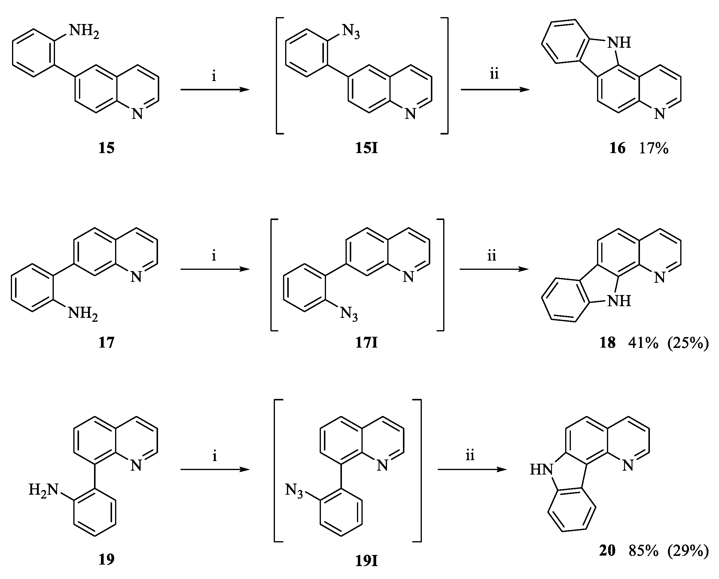
3.1.4. Intramolecular Cyclization to Form Tetracycles 9, 13, 14, 18 and 20

General Procedures

Method 1—palladium-catalyzed intramolecular C-H activation/C-N bond formation: The appropriate biaryl (1 equiv.) in a suitable amount of glacial acetic acid was added to a premixed solution of PdCl2(dppf) (10 mol%), 1,3-bis(2,4,6-trimethylphenyl)imidazolium (IMes) (5 mol%), H2O2 (35 wt%, 29 mol%) and a suitable amount of glacial acetic acid. The reaction mixture was then placed in a sealed reactor tube and immersed into the cavity of a microwave oven and heated at 118 °C until completion as indicated by TLC analysis. The reaction mixture was then transferred to a round-bottom flask with the aid of EtOAc/CHCl3 and the volatiles were removed under reduced pressure. The reaction mixture was finally evaporated onto celite and purified by column chromatography with the eluents as indicated in order to give the target compounds.
Method 2—diazotization-azidation-nitrene insertion: The appropriate biaryl (1 equiv.) was dissolved in a suitable amount of aq. HCl (37%) and the mixture was cooled to 0 °C using an ice bath. Next, to ice-cooled aq. NaNO2 (0.4 M) was added the solution dropwise and the resulting mixture was stirred at 0 °C for 1.5 h. An ice-cooled aq. solution of NaN3/NaOAc (2.1 equiv./14 equiv. in an appropriate amount of H2O) was added dropwise and the mixture stirred for 1 h while keeping the temperature at 0 °C. The reaction mixture was quenched by addition of appropriate amounts of sat. aq. K2CO3 and subsequently extracted with CH2Cl2 (3 × 20 mL). The combined organic phases were washed with water (1 × 20 mL), brine (1 × 20 mL), dried (MgSO4), filtered and concentrated in vacuo. The obtained residue was dissolved in a suitable amount of 1,2-dichlorobenzene and flushed with argon. The resulting mixture was stirred at 180 °C for 3 h under an argon atmosphere before being cooled to rt. The solvent was removed under reduced pressure and the concentrate was evaporated onto celite and purified by column chromatography using the eluents as indicated in order to give the target compounds.

7H-Pyrido[2,3-c]carbazole (9a)

Method 2: Following the general procedure, the title compound was prepared from 2-(quinolin-5-yl)aniline (7a) (100.0 mg, 0.45 mmol), HCl (37%, 3 mL), NaNO2 (82.9 mg, 1.20 mmol in 3 mL H2O), NaN3 (61.4 mg, 0.94 mmol) and NaOAc (516.8 mg, 6.30 mmol in 5 mL H2O). After formation of the azide was confirmed by IR, the cyclization was carried out using 3 mL of 1,2-dichlorobenzene. The crude was purified by silica gel column chromatography (CH2Cl2/EtOAc, 95:5 → 9:1 v/v) and concentration of the relevant fractions [Rf = 0.22 (CH2Cl2/EtOAc, 95:5 v/v)] gave the target compound 9a as a light brown solid (78.8 mg, 80%), mp 204–205 °C; IR (ATR): νmax 3045, 2919, 2842, 1523, 1274, 956, 804, 728 cm−1; 1H NMR (400 MHz, DMSO-d6): δ 11.92 (bs, 1H), 9.17 (dd, J = 8.4 Hz, 1.4 Hz, 1H), 8.84 (dd, J = 4.1 Hz, 1.4 Hz, 1H), 8.60 (d, J = 8.0 Hz, 1H), 8.03–7.98 (m, 2H), 7.69–7.65 (m, 2H), 7.47–7.44 (m, 1H), 7.34.7.31 (m, 1H) (Figure S6.1, S6.3, and S6.4); 13C NMR (100 MHz, DMSO-d6): δ 146.4, 144.3, 139.0, 136.9, 130.8, 127.7, 124.5, 124.3, 122.9, 121.6, 121.5, 119.8, 116.8, 113.6, 111.9 (Figure S6.2, S6.5, and S6.6); HRMS (ESI): calcd. for C15H10N2 [M + H+] 219.0917, found 219.0927.

10-Fluoro-7H-pyrido[2,3-c]carbazole (9b)

Method 2: Following the general procedure, the title compound was prepared from 4-fluoro-2-(quinolin-5-yl)aniline (7c) (419.5 mg, 1.76 mmol), HCl (37%, 8 mL), NaNO2 (137.9 mg, 2.00 mmol), NaN3 (240.5 mg, 3.70 mmol) and NaOAc (2021.2 mg, 24.64 mmol in 15 mL H2O). The crude was essentially pure by 1H NMR and 100.0 mg of the azide was dissolved in 2 mL 1,2-dichlorobenzene and reacted without any further purification. The cyclization yielded a reaction crude which was also pure by NMR and the target compound 9b was obtained as a dark green solid (87.3 mg, 97%), mp 256–257 °C; IR (ATR): νmax 3137, 2974, 2746, 1460, 1149, 789 cm−1; 1H NMR (400 MHz, CD3OD): δ 9.07 (ddd, J = 8.4 Hz, 1.6 Hz, 0.8 Hz, 1H), 8.74 (dd, J = 4.4 Hz, 1.6 Hz, 1H), 8.18–8.15 (m, 1H), 7.98 (dd, J = 9.1 Hz, 0.7 Hz, 1H), 7.91 (d, J = 9.1 Hz, 1H), 7.67 (dd, J = 8.4 HZ, 4.4 HZ, 1H), 7.58 (ddd, J = 8.8 Hz, 4.5 Hz, 0.5 Hz, 1H), 7.25-7.20 (m, 1H) (Figure S7.1, S7.4, and S7.5); 13C NMR (100 MHz, CD3OD): δ 159.2 (d, JCF = 234.5 Hz), 146.9, 145.1, 139.8, 137.3, 132.9, 127.9, 126.4, 124.7 (d, JCF = 9.5 Hz), 122.7, 118.3, 115.1 (d, JCF = 5.3 Hz), 113.7 (d, JCF = 24.0 Hz), 113.5 (d, JCF = 7.2 Hz), 107.6 (d, JCF = 24.8 Hz) (Figure S7.2, S7.6, and S7.7); 19F NMR (376 MHz, CD3OD): δ -123.6 (Figure S7.3); HRMS (ESI): calcd. for C15H9FN2 [M + H+] 237.0823, found 237.0830.

6H-Indolo[2,3-b]quinoline (13) and 11H-indolo[3,2-c]quinoline (14)

Method 2: Following the general procedure, the title compounds were prepared starting from 2-(quinolin-3-yl)aniline (12) (100.0 mg, 0.45 mmol), HCl (37%, 3 mL), NaNO2 (82.8 mg, 1.20 mmol in 3 mL H2O), NaN3 (61.4 mg, 0.94 mmol) and NaOAc (516.8 mg, 6.30 mmol in 5 mL H2O). After formation of the azide was confirmed by IR, the cyclization was carried out using 3 mL 1,2-dichlorobenzene. The crude was purified by silica gel column chromatography (CH2Cl2/EtOAc, 8:2 → 0:1 v/v) and concentration of the relevant fractions [Rf = 0.56 (CH2Cl2/EtOAc, 2:8 v/v)] gave compound 13 as off-white crystals (4.2 mg, 4%) along with compound 14 as an off-white solid (86.4 mg, 88%).

Characterization of Compound 13

mp 341–342 °C (lit. [79] 342–346 °C); IR (ATR): νmax 3139, 2923, 2849, 1402, 725 cm−1; 1H NMR (400 MHz, DMSO-d6): δ 11.72 (bs, 1H), 9.06 (s, 1H), 8.26 (d, J = 7.7 Hz, 1H), 8.11 (dd, J = 8.1 Hz, 1.3 Hz, 1H), 7.99–7.97 (m, 1H), 7.75–7.70 (m, 1H), 7.55–7.46 (m, 3H), 7.29–7.25 (m, 1H); 13C NMR (100 MHz, DMSO-d6): δ 152.7, 146.1, 141.4, 128.7, 128.6, 128.2, 127.7, 126.8, 123.6, 122.8, 121.8, 120.3, 119.7, 118.0, 110.9. The spectroscopic data are in accordance with previously reported data [79].

Characterization of Compound 14

mp 333-335 °C (lit. [62] 340–341 °C); IR (NaCl): νmax 3060, 2958, 2854, 1682, 1582, 1515, 1493 cm−1; 1H NMR (400 MHz, DMSO-d6): δ 12.74 (bs, 1H), 9.60 (s, 1H), 8.52 (dd, J = 7.8 Hz, 1.1 Hz, 1H), 8.33–8.31 (m, 1H), 8.14 (dd, J = 8.4 Hz, 1.1 Hz, 1H), 7.77–7.68 (m, 3H), 7.52–7.48 (m, 1H), 7.37–7.33 (m, 1H); 13C NMR (100 MHz, DMSO-d6): δ 145.3, 144.7, 139.8, 138.8, 129.4, 128.1, 125.7, 125.6, 122.1, 121.9, 120.6, 120.1, 117.1, 114.3, 111.9. The spectroscopic data are in accordance with previously reported data [62].

11H-Pyrido[2,3-a]carbazole (18)

Method 2: Following the general procedure, the title compound was prepared from 2-(quinolin-7-yl)aniline (17) (100.0 mg, 0.45 mmol), HCl (37%, 3 mL), NaNO2 (82.8 mg, 1.20 mmol in 3 mL H2O), NaN3 (61.4 mg, 0.94 mmol) and NaOAc (516.8 mg, 6.30 mmol in 3 mL H2O). After formation of the azide was confirmed by IR, the cyclization was carried out using 3 mL 1,2-dichlorobenzene. The crude was purified by silica gel column chromatography (CH2Cl2/EtOAc, 9:1 v/v) and concentration of the relevant fractions [Rf = 0.36 (CH2Cl2/EtOAc, 9:1 v/v)] gave the target compound 18 as off-white crystals (40.0 mg, 41%), mp 164 °C (lit. [52] 165–167 °C); IR (ATR): νmax 3263, 3043, 2923, 2854, 1523, 1369, 820, 734 cm−1; 1H NMR (400 MHz, CDCl3): δ 10.20 (bs, 1H), 8.92 (dd, J = 4.4 Hz, 1.5 Hz, 1H), 8.35 (dd, J = 8.3 Hz, 1.5 Hz, 1H), 8.24 (d, J = 8.5 Hz, 1H), 8.19–8.17 (m, 1H), 7.62–7.60 (m, 2H), 7.51–7.47 (m, 2H), 7.35–7.31 (m, 1H); 13C NMR (100 MHz, CDCl3): δ 147.8, 139.2, 137.4, 136.8, 134.9, 127.3, 125.9, 123.8, 121.7, 120.8, 120.5, 120.4, 120.2, 118.8, 111.8. The spectroscopic data are in accordance with previously reported data [52].

7H-Pyrido[3,2-c]carbazole (20)

Method 2: Following the general procedure, the title compound was prepared from 2-(quinolin-8-yl)aniline (19) (450.0 mg, 2.04 mmol), HCl (37%, 10 mL), NaNO2 (137.9 mg, 2.00 mmol in 5 mL H2O), NaN3 (278.5 mg, 4.28 mmol) and NaOAc (2342.8 mg, 28.56 mmol in 10 mL H2O). After formation of the azide was confirmed by IR, the cyclization was carried out using 5 mL 1,2-dichlorobenzene. The crude was purified by silica gel column chromatography (pet. ether/EtOAc, 1:1 v/v) and concentration of the relevant fractions [Rf = 0.85 (pet. ether/EtOAc, 1:1 v/v)] gave the target compound 20 as a dark red oil (195.9 mg, 44%). IR (ATR): νmax 3207, 2976, 2919, 2850, 2740, 2605, 2499 cm−1; 1H NMR (400 MHz, DMSO-d6): δ 11.92 (bs, 1H), 9.02 (dd, J = 4.4 Hz, 1.8 Hz, 1H), 8.90–8.88 (m, 1H), 8.46 (dd, J = 8.1 Hz, 1.4 Hz, 1H), 7.92 (d, J = 8.8 Hz, 1H), 7.84 (d, J = 8.8 Hz, 1H), 7.66–7.64 (m, 1H), 7.49 (dd, J = 8.0 Hz, 4.3 Hz, 1H), 7.46–7.42 (m, 1H), 7.33-7.29 (m, 1H); 13C NMR (100 MHz, DMSO-d6): δ 149.8, 145.3, 139.6, 138.5, 136.5, 126.0, 124.5, 123.1, 122.9, 122.8, 119.7, 118.3, 115.3, 114.2, 111.4. The spectroscopic data are in accordance with previously reported data [52].

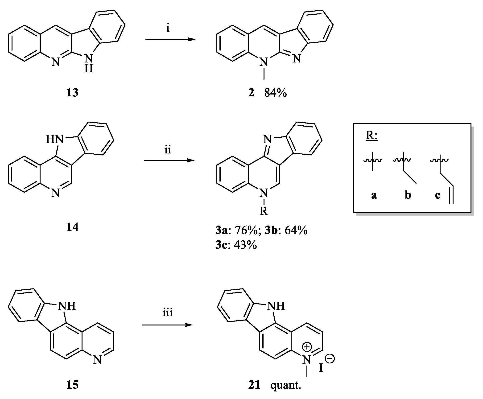
3.1.5. Neocryptolepine (2)

To a solution of 6H-indolo[2,3-b]quinoline (13) (23.0 mg, 0.10 mmol) in 2 mL THF, iodomethane (0.66 mL, 10.0 mmol) was added and the resulting mixture refluxed for 24 h. The volatiles were then removed under reduced pressure and the concentrate was evaporated onto celite. Purification by silica gel column chromatography (CH2Cl2/MeOH, 95:5 v/v) and concentration of the relevant fractions [Rf = 0.18 (CH2Cl2/MeOH, 95:5 v/v)] gave the hydroiodide salt of neocryptolepine. To obtain the free base, the hydroiodide salt was dissolved in a 20 mL 1:1 mixture of CH2Cl2 and NH3(aq) (20%) and stirred at rt for 30 min. The organic layer was then separated and the aqueous layers were extracted with CH2Cl2 (3 × 10 mL) and the combined organic layers were washed with water (1 × 10 mL), brine (1 × 10 mL), dried (MgSO4), filtered and concentrated in vacuo to give neocryptolepine (2) as a dark yellow solid (19.5 mg, 84%), mp 85–86 °C (lit. [67] 104–105 °C); IR (ATR): νmax 3051, 2961, 2923, 2852, 1494, 1012, 741 cm−1; 1H NMR (400 MHz, CD3OD): δ 8.67 (s, 1H), 8.05-8.02 (m, 2H), 7.90 (d, J = 8.6 Hz, 1H), 7.83–7.78 (m, 1H), 7.59–7.57 (m, 1H), 7.50–7.45 (m, 2H), 7.19 (td, J = 7.5 Hz, 1.0 Hz, 1H), 4.23 (s, 3H); 13C NMR (100 MHz, CD3OD): δ 156.9, 155.4, 138.1, 132.0, 131.2, 130.4, 130.1, 128.4, 124.9, 123.6, 122.4, 122.2, 121.2, 117.7, 115.7, 33.7. The spectroscopic data are in accordance with previously reported data [67].

3.1.6. 5-Ethyl-5H-indolo[3,2-c]quinoline (3b)

To a solution of 11H-indolo[3,2-c]quinoline (14) (15.0 mg, 0.068 mmol) in 3 mL toluene, ethyl iodide (1.1 mL, 13.68 mmol) was added and the resulting mixture was refluxed for 3 h. The volatiles were then removed under reduced pressure and the concentrate was evaporated onto celite. Purification by silica gel column chromatography (CHCl3/MeOH, 9:1 v/v) and concentration of the relevant fractions [Rf = 0.21 (CHCl3/MeOH, 9:1 v/v)] gave the hydroiodide salt of compound 3b. To obtain the free base, the hydroiodide salt was dissolved in a 40 mL 1:1 mixture of CH2Cl2 and NH3 (aq) (20%) and stirred at rt for 5 min. The organic layer was then separated and the aqueous layers were extracted with CH2Cl2 (2 × 10 mL) and the combined organic layers were washed with brine (1 × 10 mL), dried (MgSO4), filtered and concentrated in vacuo to give the target compound 3b as a yellow solid (11.0 mg, 64%), mp 198 °C. IR (NaCl): νmax 3371, 3049, 2960, 2927, 2856, 1731, 1640, 1598, 1455, 1392, 1353 cm−1; 1H NMR (400 MHz, DMSO-d6): δ 9.59 (s, 1H), 8.84 (dd, J = 8.1 Hz, 1.3 Hz, 1H), 8.23 (d, J = 8.7 Hz, 1H), 8.20–8.18 (m, 1H), 7.93–7.89 (m, 1H), 7.81 (d, J = 8.1 Hz, 1H), 7.79–7.75 (m, 1H), 7.51–7.47 (m, 1H), 7.34–7.30 (m, 1H), 4.81 (q, J = 7.1 Hz, 2H), 1.56 (t, J = 7.1 Hz, 3H) (Figure S1.1, S1.3, and S1.4); 13C NMR (100 MHz, DMSO-d6): δ 151.2, 150.5, 138.6, 134.3, 130.1, 126.1, 125.8, 124.7, 124.4, 120.5, 120.2, 119.9, 117.8, 117.2, 115.9, 49.7, 15.0 (Figure S1.2, S1.5, and S1.6); HRMS (ESI): calcd. for C17H14N2 [M + H+] 247.1235, found 247.1238.

3.1.7. 5-Allyl-5H-indolo[3,2-c]quinoline (3c)

To a solution of 11H-indolo[3,2-c]quinoline (14) (30.0 mg, 0.14 mmol) in 5 mL toluene, allyl bromide (1.14 mL, 13.76 mmol) was added and the resulting mixture was refluxed for 22 h. The volatiles were then removed under reduced pressure and the concentrate was evaporated onto celite. Purification by silica gel column chromatography (CHCl3/MeOH, 95:5 → 9:1 v/v) and concentration of the relevant fractions [Rf = 0.18 (CHCl3/MeOH, 9:1 v/v)] gave the hydroiodide salt of compound 3c. To obtain the free base, the hydroiodide salt was dissolved in a 20 mL 1:1 mixture of CH2Cl2 and NH3 (aq) (20%) and stirred at rt for 45 min. The organic layer was then separated and the aqueous layers were extracted with CH2Cl2 (3 × 10 mL) and the combined organic layers were washed with water (1 × 10 mL), brine (1 × 10 mL), dried (MgSO4), filtered and concentrated in vacuo to give the target compound 3c as a yellow viscous oil (15.5 mg, 43%). IR (ATR): νmax 2924, 2723, 1596, 1349, 1204, 927, 743 cm−1; 1H NMR (400 MHz, DMSO-d6): δ 9.67 (s, 1H), 8.84 (dd, J = 8.0 Hz, 1.3 Hz, 1H), 8.22–8.20 (m, 1H), 8.15 (d, J = 8.7 Hz, 1H), 7.92–7.88 (m, 1H), 7.84–7.77 (m, 2H), 7.55–7.51 (m, 1H), 7.37–7.34 (m, 1H), 6.26–6.17 (m, 1H), 5.46–5.44 (m, 2H), 5.31–5.28 (m, 1H), 5.15–5.11 (m, 1H) (Figure S2.1, S2.3, and S2.4); 13C NMR (100 MHz, DMSO-d6): δ 150.0, 149.9, 139.6, 134.9, 132.8, 130.3, 126.5, 126.1, 124.4, 124.3, 121.0, 120.1, 119.7, 118.5, 118.2, 116.8, 115.7, 56.5 (Figure S2.2 and S2.5); HRMS (ESI): calcd. for C18H14N2 [M + H+] 259.1230, found 259.1232.

3.1.8. 4-Methyl-4H-pyrido[4,3,2-gh]phenanthridine (8a)

To a solution of 7H-pyrido[4,3,2-gh]phenanthridine (4a) (70.0 mg, 0.32 mmol) in 2 mL acetonitrile, iodomethane (2.0 mL, 32.0 mmol) was added and the resulting mixture was refluxed for 2 h. The volatiles were then removed under reduced pressure and the concentrate was evaporated onto celite. Purification by silica gel column chromatography (CHCl3/MeOH, 95:5 + 0.3% NH3 (aq) v/v) and concentration of the relevant fractions [Rf = 0.33 (CHCl3/MeOH, 95:5 + 0.3% NH3(aq) v/v)] gave the hydroiodide salt of compound 8a. To obtain the free base, the hydroiodide salt was dissolved in a 20 mL 1:1 mixture of CH2Cl2 and NH3 (aq) (20%) and stirred at rt for 20 min. The organic layer was separated and the aqueous layers were extracted with Et2O (2 × 20 mL) and the combined organic layers were washed with water (1 × 10 mL), brine (1 × 10 mL), dried (MgSO4), filtered and concentrated in vacuo to give the target compound 8a as dark yellow crystals (52.8 mg, 71%), mp 182–183 °C; IR (ATR): νmax 3485, 3051, 2922, 2851, 2574, 1601, 1327, 820, 748 cm−1; 1H NMR (400 MHz, DMSO-d6): δ 8.29 (dd, J = 8.1 Hz, 1.1 Hz, 1H), 7.97 (d, J = 7.9 Hz, 1H), 7.71 (t, J = 8.1 Hz, 1H), 7.56 (dd, J = 8.2 Hz, 1.2 Hz, 1H), 7.51–7.47 (m, 2H), 7.30–7.26 (m, 1H), 7.05 (d, J = 8.1 Hz, 1H), 6.18 (d, J = 7.6 Hz, 1H), 3.45 (s, 3H) (Figure S4.1, S4.3, and S4.4); 13C NMR (100 MHz, DMSO-d6): "δ" 152.9, 145.9, 141.0, 140.9, 133.8, 131.6, 129.2, 127.0, 123.2, 122.9, 121.3, 119.6, 112.2, 108.8, 106.2, 39.6 (Figure S4.2, S4.5, and S4.6); HRMS (ESI): calcd. for C16H12N2 [M + H+] 233.1073, found 233.1073.

3.1.9. 6-Methoxy-4-methyl-4H-pyrido[4,3,2-gh]phenanthridine (8b)

To a solution of 6-methoxy-7H-pyrido[4,3,2-gh]phenanthridine (4b) (90.0 mg, 0.36 mmol) in 10 mL acetonitrile, iodomethane (2.25 mL, 36.3 mmol) was added and the resulting mixture refluxed for 2 h. The volatiles were then removed under reduced pressure and the concentrate was evaporated onto celite. Purification by silica gel column chromatography (EtOH + 0.1 → 5% NH3 (aq) v/v) and concentration of the relevant fractions [Rf = 0.23 (EtOH)] gave the hydroiodide salt of compound 8b. To obtain the free base, the hydroiodide salt was dissolved in a 20 mL 1:1 mixture of CH2Cl2 and NH3 (aq) (20%) and stirred at rt for 20 min. The organic layer was separated and the aqueous layers were extracted with CH2Cl2 (4 × 20 mL) and the combined organic layers were washed with water (1 × 20 mL), brine (1 × 20 mL), dried (MgSO4), filtered and concentrated in vacuo to give the target compound 8b as a dark yellow gel (55.1 mg, 58%). IR (ATR): νmax 2918, 2850, 1600, 1255, 1059, 745 cm−1; 1H NMR (400 MHz, CD2Cl2): δ 8.19 (dd, J = 8.2 Hz, 1.3 Hz, 1H), 7.79 (dd, J = 8.2 Hz, 1.0 Hz, 1H), 7.72 (d, J = 7.9 Hz, 1H), 7.57–7.51 (m, 2H), 7.34–7.30 (m, 1H), 6.74–6.72 (m, 2H), 3.85 (s, 3H), 3.34 (s, 3H) (Figure S5.1, S5.3 and S5.4); 13C NMR (100 MHz, CD2Cl2): δ 148.1, 146.3, 140.8, 139.9, 134.8, 130.9, 129.4, 128.6, 124.1, 122.7, 122.5, 119.9, 110.6, 107.7, 57.1, 40.4 (one carbon was obscured or overlapping) (Figure S5.2, S5.5 and S5.6); HRMS (ESI): calcd. for C17H14N2O 263.1179, found 263.1188.

3.1.10. 4-Methyl-7H-pyrido[2,3-c]carbazolium Iodide (10)

To a solution of 7H-pyrido[2,3-c]carbazole (9a) (40.7 mg, 0.19 mmol) in 5 mL acetonitrile, iodomethane (1.20 mL, 19.6 mmol) was added and the resulting mixture refluxed for 20 h. The volatiles were then removed under reduced pressure and the concentrate was evaporated onto celite. Purification by alumina gel column chromatography (CHCl3/MeOH, 9:1 v/v + 1% NH3 (aq)) and concentration of the relevant fractions [Rf = 0.12 (CHCl3/MeOH, 9:1 v/v + 2% NH3 (aq))] gave the target compound 10 as a bright yellow solid (20.9 mg, 47%), mp 284–286; IR (ATR): νmax 3353, 3043, 3006, 2960, 2921, 2853, 1556, 1370, 1326, 741 cm−1; 1H NMR (400 MHz, DMSO-d6): δ 12.84 (bs, 1H), 9.99 (d, J = 8.4 Hz, 1H), 9.39 (d, J = 5.6 Hz, 1H), 8.76 (d, J = 8.1 Hz, 1H), 8.50 (d, J = 9.3 Hz, 1H), 8.43 (d, J = 9.4 Hz, 1H), 8.22 (dd, J = 8.5 Hz, 5.7 Hz, 1H), 7.82 (d, J = 8.2 Hz, 1H), 7.64-7.60 (m, 1H), 7.48–7.44 (m, 1H), 4.74 (s, 3H) (Figure S8.1, S8.3, and S8.4); 13C NMR (100 MHz, DMSO-d6): δ 145.0, 140.8, 139.8, 137.5, 134.5, 126.6, 125.8, 122.4, 122.1, 121.9, 121.6, 121.1, 116.0, 114.3, 112.8, 46.3 (Figure S8.2, S8.5 and S8.6); HRMS (ESI): calcd. for C16H13N2I [M – I-] 233.1073, found 233.1073.

3.1.11. 4-Methyl-11H-pyrido[3,2-a]carbazolium Iodide (21)

To a solution of 11H-pyrido[3,2-a]carbazole (15) (32.1 mg, 0.15 mmol) in 5 mL acetonitrile, iodomethane (0.92 mL, 14.72 mmol) was added and the resulting mixture stirred at reflux for 20 h. The volatiles were then removed under reduced pressure and the obtained yellow crystals were thoroughly washed with n-hexanes and dried in vacuo to give the target compound 21 as a dark orange crystalline solid (47.6 mg, quant.), mp 279–280 °C; IR (ATR): νmax 3416, 3165, 3077, 2997, 2905, 1599, 1452, 1371, 740 cm−1; 1H NMR (400 MHz, CD3OD): δ 9.64 (d, J = 8.6 Hz, 1H), 9.28 (d, J = 5.7 Hz, 1H), 8.99 (d, J = 9.1 Hz, 1H), 8.35–8.33 (m, 1H), 8.15–8.10 (m, 2H), 7.79–7.77 (m, 1H), 7.65–7.61 (m, 1H), 7.46–7.42 (m, 1H), 4.76 (s, 3H) (Figure S9.1, S9.3, and S9.4); 13C NMR (100 MHz, CD3OD): δ 148.2, 141.7, 141.6, 139.1, 136.1, 131.1, 128.4, 123.6, 122.4, 122.3, 121.6, 121.1, 119.9, 113.1, 109.0, 46.9 (Figure S9.2, S9.5 and S9.6); HRMS (ESI): calcd. for C16H13N2I [M – I-] 233.1073, found 233.1075.

3.2. Biological Testing Assay

3.2.1. General

All compounds for antimicrobial testing were diluted to a final assay concentration of 40 µL, 0.4% DMSO, and tested in full dose-response using three concentrations per log dose (16 points with a concentration range of 0.33 nM–40 µM, for reference compounds: 21 points with a concentration of 0.01 nM–40 µM).
All compounds for antiplasmodial testing were diluted to a final assay concentration of 40 µL, 0.4% DMSO, and tested in full dose-response using three concentrations per log dose (16 points with a concentration range of 0.4 nM–40 µM, for reference compounds: 16 points with a concentration rage of 0.4 nM–40 µM for chloroquine and puromycin: 0.001 nM–0.1 µM for artemisinin). Compounds tested in the antiproliferative assays were tested in 11 dilution points (0.02 µM–40 µM or 0.04 µM–80 µM).

Antiplasmodial Imaging Assay

Antiplasmodial activity was determined as previously described by Duffy and Avery [80]. Briefly, compounds were incubated with 2% parasitemia in 0.3% hematocrit, in an assay volume of 50 µL for 72 h at 37 °C and 5% CO2 in CellCarrier Ultra 384-well PDL-imaging plates. After incubation, plates were stained with 4’,6-diamidino-2-phenylindole (DAPI) in a permeabilization buffer for 5 h at rt in the dark. Plates were imaged on the Opera confocal microplate image reader (PerkinElmer). Parasite inhibition was calculated using the minimum (0.4% DMSO) and maximum (5 µM puromycin) controls, and IC50 values determined using GraphPad Prism software.

Cytotoxicity Assay

The cytotoxicity of compounds was determined using a resazurin-based viability assay in HEK293 (ATCC®, CRL-1573), as described by Fletcher and Avery [81]. Compounds were added to TC-treated 384-well plates (Greiner, Kremsmünster, AT) containing 2500 HEK293 cells per well, total assay volume of 50 µL and incubated for 72 h at 37 °C, 5% CO2. After incubation, media was removed, replaced with 44 µM resazurin and plates incubated 6 h under the same experimental conditions. Fluorescence was measured using an EnSight plate reader (PerkinElmer, Waltham, MA, USA). Cell viability was calculated using positive (45 µM puromycin) and negative (0.4% DMSO) controls, and the IC50 values determined using GraphPad Prism software.

Antiproliferative Assay

Antiproliferative activity was assessed in HCT116 (ATCC® CCL-247; 1000 cells/well), MDA-MB-231 (ATCC® HTB-26; 2000 cells/well) and PC-3 (ATCC® CRL-1435; 1000 cells/well) cells. HCT116 cells were maintained in McCoy’s 5A media (Life Technologies, CA, USA), MDA-MB-231 cells were cultured in DMEM media (Life Technologies) with 10 mM HEPES (Life Technologies), whilst PC-3 cells were maintained in RPMI media (Life Technologies). All media were supplemented with 10% heat-inactivated fetal bovine serum (Australian source; Corning, CA, USA) and all cells were incubated at 37 °C in a humidified incubator with 5% CO2.
Cells were seeded in 384-well plates (Greiner Bio-One, NC, USA) using the respective complete media. After 24 h cell seeding, compounds were added and antiproliferative activity was determined using the resazurin assay after 72 h compound incubation. Briefly, cells were incubated with 60 µM resazurin (Cayman, MI, USA) for 6 h at 37 °C and fluorescence signals were measured using a microplate reader (EnSight, PerkinElmer, Waltham, MA, USA). Fluorescence signals were normalized to 0.4% DMSO and 50 µM puromycin and IC50 values were calculated from non-linear dose-response curves using GraphPad Prism 7 software (La Jolla, CA, USA).

3.2.2. Growth Inhibition Assay

To determine and quantify antimicrobial activity, a bacteria growth inhibition assay in liquid media was executed. Compounds 23, 4, 810, 1314, 16, 18, and 20 were tested against Staphylococcus aureus (ATCC 25923), Escherichia coli (ATCC 259233), Enterococcus faecialis (ATCC 29122), Pseudomonas aeruginosa (ATCC 27853) and Streptococcus agalactiae (ATCC 12386); all strains from LGC Standards (Teddington, UK). S. aureus, E. coli, and P. aeruginosa were grown in Muller Hinton broth (275730, Becton, Franklin Lakes, NJ, USA). E. faecalis and S. agalactiae were cultured in brain hearth infusion broth (53286, Sigma, St Louise, MO, USA). Fresh bacterial colonies were transferred in the respective medium and incubated at 37 °C overnight. The bacterial cultures were diluted to a culture density representing the log phase and µL/well were pipetted into a 96-well microtiter plate (734–2097, NunclonTM, Thermo Scientific, Waltham, MA, USA). The final cell density was 1500–15.000 colony forming units/well. The compound was diluted in 2% (v/v) DMSO in ddH2O, providing a final assay concentration of 50% of the prepared sample, since 50 µL of sample in DMSO/water were added to 50 µL bacterial culture. After adding the samples to the plates, they were incubated overnight at 37 °C and the growth was determined by measuring the optical density at λ = 600 nm (OD600) with a 1420 Multilabel Counter VICTOR3TM (Perkin Elmer, Waltham, MA, USA). A water sample was used as a reference control, growth medium without bacteria was used as a negative control and dilution series of gentamycin (A2712, Merck, Darmstadt, DE) from 32 to 0.01 µg/mL was used as positive control and visually inspected for bacterial growth. The positive control was used as a system suitability test and the results of the antimicrobial assay were only considered valid when positive control was passed. The final concentration of DMSO in the assays was ≤2% (v/v) and was known to have no effect in the tested bacteria. The data was processed using GraphPad Prism 8.

3.2.3. Biofilm Formation Inhibition Assay

For testing the inhibition of biofilm formation, the biofilm-producing Staphylococcus epidermidis (ATCC 35984) was grown in Tryptic Soy Broth (TSB, 105459, Merck, Kenilworth, NJ, USA) overnight at 37 °C. The overnight culture was diluted in fresh medium with 1% glucose (D9434, Sigma) before being transferred to a 96-well microtiter plate; 50 µL/well were incubated overnight with 50 µL of the test compound dissolved in 2% (v/v) DMSO aq. added in duplicates. The bacterial culture was removed from the plate and the plate was washed with tap water. The biofilm was fixed at 65 °C for 1 h before 70 µL 0.1% crystal violet (115940, Millipore, Burlington, MA, USA) was added to the wells for 10 min of incubation and 70 µL of 70% ethanol was then added to each well and the plate incubated on a shaker for 5–10 min. Biofilm formation inhibition was assessed by the presence of violet color and measured at 60 nm absorbance using a 1420 Multilabel Counter VICTOR3TM; 50 µL of a non-biofilm forming Staphylococcus haemolyticus (clinical isolate 8-7A, University Hospital of North Norway Tromsø, Norway) mixed in 50 µL autoclaved Milli-Q water was used as a control; 50 µL S. epidermidis mixed in 50 µL autoclaved Milli-Q water was used as the control for biofilm formation; and 50 µL TSB with 50 µL autoclaved Milli-Q water was used as a medium blank control.

4. Conclusions

In conclusion, a series of quinoline-based tetracyclic ring-systems were prepared and evaluated for their in vitro antiplasmodial, antiproliferative and antimicrobial activities against selected strains. Through these studies, it was determined that the ionic pyridocarbazoles 10 and 21 showed the best antiplasmodial activity against the Plasmodium falciparum 3D7 strains (10: IC50 = 128 nM; 21: IC50 = 380 nM) of the evaluated compounds. The antiproliferative assay revealed that the novel pyridophenanthridine scaffold 4 was the most active. In particular, compound 4b showed excellent potency against the PC-3 cell line (IC50 = 24 nM), significantly outperforming Puromycin (IC50 = 270 nM) and Doxorubicin (IC50 = 830 nM). The pyridophenanthridines 4 were also active against certain strains of Gram-positive and Gram-negative bacteria, with compound 4b being moderately active against E. coli (MIC = 50 µM) and Streptococcus agalactiae (MIC = 75 µM). The antimicrobial studies further demonstrated pyridocarbazoles 9 to be highly potent against biofilm growth (9a: MBIC = 100 µM; 9b: MBIC = 100 µM). Overall, this study has highlighted the potential for the novel pyridophenanthridine motif 4 and the studied pyridocarbazoles 9 to be developed into future drug candidates, with emphasis on the formulation of a dual antimicrobial and antiproliferative agent.

Supplementary Materials

Figure S1.1: 1H NMR of 5-ethyl-5H-indolo[3,2-c]quinoline (3b), Figure S1.2: 13C NMR of 5-ethyl-5H-indolo[3,2-c]quinoline (3b), Figure S1.3: COSY of 5-ethyl-5H-indolo[3,2-c]quinoline (3b), Figure S1.4: NOESY of 5-ethyl-5H-indolo[3,2-c]quinoline (3b), Figure S1.5: HSQC of 5-ethyl-5H-indolo[3,2-c]quinoline (3b), Figure S1.6: HMBC of 5-ethyl-5H-indolo[3,2-c]quinoline (3b), Figure S2.1: 1H NMR of 5-allyl-5H-indolo[3,2-c]quinoline (3c), Figure S2.2: 13C NMR of 5-allyl-5H-indolo[3,2-c]quinoline (3c), Figure S2.3: COSY of 5-allyl-5H-indolo[3,2-c]quinoline (3c), Figure S2.4: NOESY of 5-allyl-5H-indolo[3,2-c]quinoline (3c), Figure S2.5: HMBC of 5-allyl-5H-indolo[3,2-c]quinoline (3c), Figure S3.1: 1H NMR of 4-fluoro-2-(quinolin-5-yl)aniline (7c), Figure S3.2: 13C NMR of 4-fluoro-2-(quinolin-5-yl)aniline (7c), Figure S3.3: 19F NMR of 4-fluoro-2-(quinolin-5-yl)aniline (7c), Figure S4.1: 1H NMR of 4-methyl-4H-pyrido[4,3,2-gh]phenanthridine (8a), Figure S4.2: 13C NMR of 4-methyl-4H-pyrido[4,3,2-gh]phenanthridine (8a), Figure S4.3: COSY of 4-methyl-4H-pyrido[4,3,2-gh]phenanthridine (8a), Figure S4.4: NOESY of 4-methyl-4H-pyrido[4,3,2-gh]phenanthridine (8a), Figure S4.5: HSQC of 4-methyl-4H-pyrido[4,3,2-gh]phenanthridine (8a), Figure S4.6: HMBC of 4-methyl-4H-pyrido[4,3,2-gh]phenanthridine (8a), Figure S5.1: 1H NMR of 6-methoxy-4-methyl-4H-pyrido[4,3,2-gh]phenanthridine (8b), Figure S5.2: 13C NMR of 6-methoxy-4-methyl-4H-pyrido[4,3,2-gh]phenanthridine (8b), Figure S5.3: COSY of 6-methoxy-4-methyl-4H-pyrido[4,3,2-gh]phenanthridine (8b), Figure S5.4: NOESY of 6-methoxy-4-methyl-4H-pyrido[4,3,2-gh]phenanthridine (8b), Figure S5.5: HSQC of 6-methoxy-4-methyl-4H-pyrido[4,3,2-gh]phenanthridine (8b), Figure S5.6: HMBC of 6-methoxy-4-methyl-4H-pyrido[4,3,2-gh]phenanthridine (8b), Figure S6.1: 1H NMR of 7H-pyrido[2,3-c]carbazole (9a), Figure S6.2: 13C NMR of 7H-pyrido[2,3-c]carbazole (9a), Figure S6.3: COSY of 7H-pyrido[2,3-c]carbazole (9a), Figure S6.4: NOESY of 7H-pyrido[2,3-c]carbazole (9a), Figure S6.5: HSQC of 7H-pyrido[2,3-c]carbazole (9a), Figure S6.6: COSY of 7H-pyrido[2,3-c]carbazole (9a), Figure S7.1: 1H NMR of 10-fluoro-7H-pyrido[2,3-c]carbazole (9b), Figure S7.2: 13C NMR of 10-fluoro-7H-pyrido[2,3-c]carbazole (9b), Figure S7.3: 19F NMR of 10-fluoro-7H-pyrido[2,3-c]carbazole (9b), Figure S7.4: COSY of 10-fluoro-7H-pyrido[2,3-c]carbazole (9b), Figure S7.5: NOESY of 10-fluoro-7H-pyrido[2,3-c]carbazole (9b), Figure S7.6: HS of 10-fluoro-7H-pyrido[2,3-c]carbazole (9b), Figure S7.7: HMBC of 10-fluoro-7H-pyrido[2,3-c]carbazole (9b), Figure S8.1: 1H NMR of 4-methyl-7H-pyrido[2,3-c]carbazolium iodide (10), Figure S8.2: 13C NMR of 4-methyl-7H-pyrido[2,3-c]carbazolium iodide (10), Figure S8.3: COSY of 4-methyl-7H-pyrido[2,3-c]carbazolium iodide (10), Figure S8.4: NOESY of 4-methyl-7H-pyrido[2,3-c]carbazolium iodide (10), Figure S8.5: HSQC of 4-methyl-7H-pyrido[2,3-c]carbazolium iodide (10), Figure S8.6: HMBC of 4-methyl-7H-pyrido[2,3-c]carbazolium iodide (10), Figure S9.1: 1H NMR of 4-methyl-11H-pyrido[3,2-a]carbazolium iodide (21), Figure S9.2: 13C NMR of 4-methyl-11H-pyrido[3,2-a]carbazolium iodide (21), Figure S9.3: COSY of 4-methyl-11H-pyrido[3,2-a]carbazolium iodide (21), Figure S9.4: NOESY of 4-methyl-11H-pyrido[3,2-a]carbazolium iodide (21), Figure S9.5: HSQC of 4-methyl-11H-pyrido[3,2-a]carbazolium iodide (21), Figure S9.6: HMBC of 4-methyl-11H-pyrido[3,2-a]carbazolium iodide (21).

Author Contributions

Conceptualization, M.O.S.; methodology, K.S.H., E.L., J.H.A., V.M.A. and M.O.S.; synthesis, K.S.H., I.T.U.H., C.L. and T.M.; antiplasmodial imaging assay, K.N.T., E.K.K. and V.M.A.; cytotoxicity assay, K.N.T., E.K.K. and V.M.A.; antiproliferative assay, K.N.T., E.K.K. and V.M.A.; growth inhibition assay, M.A. and J.H.A.; biofilm formation inhibition assay, M.A. and J.H.A.; writing—original draft preparation, K.S.H.; writing—review and editing, K.S.H., E.L., M.A., J.H.A., V.M.A. and M.O.S.; supervision, J.H.A., V.M.A. and M.O.S.; project administration, M.O.S.; funding acquisition, J.H.A., V.M.A. and M.O.S. All authors have read and agreed to the published version of the manuscript.

Funding

The authors acknowledge the ToppForsk program at the University of Stavanger for financial support and the provision of a PhD fellowship for K.S.H. (PR-10550). K.N.T is funded by a Griffith University Postdoctoral Fellowship.

Institutional Review Board Statement

Not applicable.

Informed Consent Statement

Not applicable.

Data Availability Statement

The data presented in this study are contained within the article and Supplementary Material.

Acknowledgments

B Holmelid, University of Bergen is thanked for recording HRMS analysis. K. B. Jørgensen is sincerely thanked for his help operating and maintaining the NMR instrument. The authors would like to acknowledge the Australian Red Cross Blood Bank for providing fresh human erythrocytes.

Conflicts of Interest

The authors declare no conflict of interest.

Sample Availability

Samples of the compounds are available from the authors.

References

  1. World Health Organization. World Malaria Report. 2020. Available online: https://www.who.int/publications/i/item/9789240015791 (accessed on 11 January 2021).
  2. Collins, F.H.; Paskewitz, S.M. Malaria: Current and future prospects for control. Annu. Rev. Entomol. 1995, 40, 195–219. [Google Scholar] [CrossRef]
  3. Achan, J.; Talisuna, A.O.; Erhart, A.; Yeka, A.; Tibenderana, J.K.; Baliraine, F.N.; Rosenthal, P.J.; D’Alessandro, U. Quinine, an old anti-malarial drug in a modern world: Role in the treatment of malaria. Malar. J. 2011, 10, 1–12. [Google Scholar] [CrossRef] [Green Version]
  4. Van Baelen, G.; Hostyn, S.; Dhooghe, L.; Tapolcsányi, P.; Mátyus, P.; Lemière, G.; Dommisse, R.; Kaiser, M.; Brun, R.; Cos, P.; et al. Structure-activity relationship of antiparasitic and cytotoxic indoloquinoline alkaloids, and their tricyclic and bicyclic analogues. Bioorg. Med. Chem. 2009, 17, 7209–7217. [Google Scholar] [CrossRef] [PubMed]
  5. Tse, E.G.; Korsik, M.; Todd, M.H. The past, present and future of anti-malarial medicines. Malar. J. 2019, 18, 1–21. [Google Scholar] [CrossRef] [PubMed] [Green Version]
  6. Noedl, H.; Se, Y.; Scaecher, K.; Smith, B.L.; Socheat, D.; Fukuda, M.M. Evidence of artemisinin-resistant malaria in western Cambodia. N. Engl. J. Med. 2008, 359, 2619–2620. [Google Scholar] [CrossRef]
  7. Amato, R.; Pearson, R.D.; Almagro-Garcia, J.; Amaratunga, C.; Lim, P. Origins of the current outbreak of multidrug-resistant malaria in southeast Asia: A retrospective genetic study. Lancet Infect. Dis. 2018, 18, 337–345. [Google Scholar] [CrossRef] [Green Version]
  8. World Health Organization. World Cancer Report 2020: Cancer Research for Cancer Prevention. Available online: www.iarc.fr/cards_page/world-cancer-report/ (accessed on 24 August 2020).
  9. Sidoryk, K.; Jaromin, A.; Edward, J.A.; Świtalska, M.; Stefanska, J.; Cmoch, P.; Zagrodzka, J.; Szczepek, W.; Peczynska-Czoch, W.; Wietrzyk, J.; et al. Searching for new derivatives of neocryptolepine: A synthesis, antiproliferative, antimicrobial and antifungal activities. Eur. J. Med. Chem. 2014, 78, 304–313. [Google Scholar] [CrossRef]
  10. Stamm, A.M.; Long, M.N.; Belcher, B. Higher overall nosocomial infection rate because of increased attack rate of methicillin-resistant Staphylococcus aureus. Am. J. Infect. Control 1993, 21, 70–74. [Google Scholar] [CrossRef]
  11. Drenkard, E. Antimicrobial resistance of Preudomonas aeruginosa biofilms. Microb. Infect. 2003, 5, 1213–1219. [Google Scholar] [CrossRef]
  12. Teng, C.P.; Zhou, T.; Ye, E.; Liu, S.; Koh, L.D.; Low, M.; Loh, X.J.; Win, Y.; Zhang, L.; Han, M.-Y. Effective Targeted Photothermal Ablation of Multidrug Resistant Bacteria and Their Biofilms with NIR-Absorbing Gold Nanocrosses. Adv. Healthcare Mater. 2016, 5, 2122–2130. [Google Scholar] [CrossRef]
  13. Rizzato, C.; Torres, J.; Kasamatsu, E.; Carmorlinga-Ponce, M.; Bravo, M.M.; Canzian, F.; Kato, I. Potential role of biofilm formation in the development of digestive tract cancer with special reference to Heliobacter pylori infection. Front Microbiol. 2019, 10, 1–21. [Google Scholar] [CrossRef] [PubMed]
  14. Johnson, C.H.; Dejea, C.M.; Edler, D.; Hoang, L.T.; Santidrian, A.F.; Felding, B.H.; Ivanisevic, J.; Cho, K.; Wick, E.C.; Hechenbleikner, E.M.; et al. Metabolism links bacterial biofilms and colon carcinogenesis. Cell Metab. 2015, 21, 891–897. [Google Scholar] [CrossRef] [Green Version]
  15. Drewes, J.L.; White, J.R.; Dejea, C.M.; Fathi, P.; Iyadorai, T.; Vadivelu, J.; Roslani, A.C.; Wick, E.C.; Mongodin, E.F.; Loke, M.F.; et al. High-resolution bacterial 16S rRNA gene profile meta-analysis and biofilm status reveal common colorectal cancer consortia. NPJ Biofilms Microbiomes 2017, 3, 1–34. [Google Scholar] [CrossRef] [PubMed] [Green Version]
  16. Lee, K.H. Discovery and development of natural product-derived chemotherapeutic agents based on a medicinal chemistry approach. J. Nat. Prod. 2010, 73, 500–516. [Google Scholar] [CrossRef] [PubMed] [Green Version]
  17. Newman, D.J.; Cragg, G.M. Natural products as sources of new drugs over the last 25 years. J. Nat. Prod. 2007, 70, 461–477. [Google Scholar] [CrossRef] [PubMed] [Green Version]
  18. Vuorela, P.; Leinonen, M.; Saikku, P.; Tammela, P.; Rauhad, J.-P.; Wennberg, T.; Vuorela, H. Natural products in the process of finding new drug candidates. Curr. Med. Chem. 2004, 11, 1375–1389. [Google Scholar] [CrossRef]
  19. Newman, D.J.; Cragg, G.M. Natural products as sources of new drugs over the nearly four decades from 01/1981 to 09/2019. J. Nat. Prod. 2020, 83, 770–803. [Google Scholar] [CrossRef] [PubMed]
  20. Bracca, A.B.J.; Heredia, D.A.; Larghi, E.L.; Kaufman, T.S. Neocryptolepine (cryprotackieine), a unique bioactive natural product: Isolation, synthesis, and profile of its biological activity. Eur. J. Med. Chem. 2014, 2014, 7979–8003. [Google Scholar] [CrossRef]
  21. Madapa, S.; Tusi, Z.; Batra, S. Advances in the syntheses of quinoline and quinoline-annulated ring systems. Curr. Org. Chem. 2008, 12, 1116–1183. [Google Scholar] [CrossRef]
  22. Sydnes, M.O. Recent progress in the synthesis of antimalarial indoloquinoline natural products and analogues. In Studies in Natural Products Chemistry: Bioactive Natural Products; Rahman, A., Ed.; Elsevier: Karachi, Pakistan, 2020; pp. 59–84. [Google Scholar]
  23. Sydnes, M.O. Synthetic Strategies for the Synthesis of Indoloquinoline Natural Products. In Targets in Heterocyclic Systems; Attanasi, O.A., Merino, P., Spinelli, D., Eds.; Italian Society of Chemistry: Rome, Italy, 2019; Volume 23, pp. 201–219. [Google Scholar]
  24. Wang, N.; Wicht, K.J.; Imai, K.; Wang, M.-Q.; Ngoc, T.A.; Kiguchi, R.; Kaiser, M.; Egan, T.J.; Inokuchi, T. Synthesis, β-haematin inhibition, and in vitro antimalarial testing of isocryptolepine analogues: SAR study of indolo[3,2-c]quinolines with various substituents at C2, C6 and N11. Bioorg. Med. Chem. 2014, 22, 2629–2642. [Google Scholar] [CrossRef] [Green Version]
  25. Aroonkit, P.; Thongsornkleeb, C.; Tummatorn, J.; Krajangsri, S.; Mungthin, M.; Ruchirawat, S. Synthesis of isocryptolepine analogues and their structure-activity relationship studies as antiplasmodial and antiproliferative agents. Eur. J. Med. Chem. 2015, 94, 56–62. [Google Scholar] [CrossRef] [PubMed]
  26. Pousset, J.L.; Martin, M.T.; Jossang, A.; Bodo, A. Isocryptolepine from Cryptolepis sanguinolenta. Phytochemistry 1995, 39, 735–736. [Google Scholar] [CrossRef]
  27. Sharaf, M.H.H.; Schiff, P.L.; Tackie, J.A.N.; Phoebe, C.H.; Johnson, J.R.L.; Minick, D.; Andrews, C.W.; Crouch, R.C.; Martin, G.E. The isolation and structure determination of cryptomisrine, a novel indolo[3,2-b]quinoline dimeric alkaloid from cryptolepis sanguinolenta. J. Heterocycl. Chem. 1996, 33, 789–797. [Google Scholar] [CrossRef]
  28. Lavrado, J.; Moreira, R.; Paulo, A. Indoloquinolines as scaffolds for drug discovery. Curr. Med. Chem. 2010, 17, 2348–2370. [Google Scholar] [CrossRef] [PubMed]
  29. Cimanga, K.; De Bruyne, T.; Pieters, L.; Vlietnck, A.J. In Vitro and in Vivo Antiplasmodial Activity of Cryptolepine and Related Alkaloids from Cryptolepis sanguinolenta. J. Nat. Prod. 1997, 60, 688–691. [Google Scholar] [CrossRef] [PubMed]
  30. Sofowora, A. Medicinal Plants and Traditional Medicine in Africa; John Wiley & Sons: Chichester, UK, 1982; pp. 183–256. [Google Scholar]
  31. Kirby, G.C.; Paine, A.; Warhurst, D.C.; Noamese, B.K.; Phillipson, J.D. In vitro and in vivo antimalarial activity of cryptolepine, a plant-derived indoloquinoline. Phytother. Res. 1995, 9, 359–363. [Google Scholar] [CrossRef]
  32. Grellier, P.; Ramiaramanana, L.; Millerioux, V.; Deharo, E.; Shrével, J.; Frappier, F.; Trigalo, F.; Bodo, B.; Pousset, J.L. Antimalarial Activity of Cryptolepine and Isocryptolepine, Alkaloids Isolated from Cryptolepis sanguinolenta. Phytother. Res. 1996, 10, 317–321. [Google Scholar] [CrossRef]
  33. Olajide, O.A.; Heiss, E.H.; Schachner, D.; Wright, C.W.; Vollmar, A.M.; Dirsch, V.M. Synthetic cryptolepine inhibits DNA binding of NF-κB. Bioorg. Med. Chem. 2007, 15, 43–49. [Google Scholar] [CrossRef]
  34. Bierer, D.E.; Fort, D.M.; Mendez, C.D.; Luo, J.; Imbach, P.A.; Dubenko, L.G.; Jolad, S.D.; Gerber, R.E.; Litvak, J.; Lu, Q.; et al. Ethnobotanical-Directed Discovery of the Antihyperglycemic Properties of Cryptolepine: Its Isolation from Cryptolepis sanguinolenta, Synthesis, and in Vitro and in Vivo Activities. J. Med. Chem. 1998, 41, 894–901. [Google Scholar] [CrossRef]
  35. Rauwald, H.W.; Kober, M.; Mutschler, E.; Lambrecht, G. Cryptolepis sanguinolenta: Antimuscarinic properties of cryptolepine and the alkaloid fraction at M1, M2 and M3 receptors. Planta Med. 1992, 58, 486–488. [Google Scholar] [CrossRef] [PubMed]
  36. Chen, Y.-J.; Liu, H.; Zhang, S.-Y.; Li, H.; Ma, K.-Y.; Liu, Y.-Q.; Yin, X.-D.; Zhou, R.; Yan, Y.-F.; Wang, R.-X.; et al. Design, Synthesis, and Antifungal Evaluation of Cryptolepine Derivatives against Phytopathogenic Fungi. J. Agric. Food Chem. 2021, 69, 1259–1271. [Google Scholar] [CrossRef] [PubMed]
  37. Ablordeppey, S.Y.; Fan, P.; Li, S.; Clark, A.M.; Hufford, C.D. Substituted indoloquinolines as new antifungal agents. Bioorg. Med. Chem. 2002, 10, 1337–1346. [Google Scholar] [CrossRef]
  38. Singh, M.; Singh, M.P.; Ablordeppey, S. In vitro studies with liposomal cryptolepine. Drug Dev. Ind. Pharm. 1996, 22, 377–381. [Google Scholar] [CrossRef]
  39. Paulo, A.; Duarte, A.; Gomes, E.T. In vitro antibacerial screening of Cryptolepis sanguinolenta alkaloids. J. Ethnopharmacol. 1994, 44, 127–130. [Google Scholar] [CrossRef]
  40. Cimanga, K.; De Bruyne, T.; Lasure, A.; Van Poel, B.; Pieters, L.; Claeys, M.; Berghe, D.V.; Kambu, K.; Tona, L.; Vlietinch, A. In vitro biological activities of alkaloids from Cryptolepis sanguinolenta. Planta Med. 1996, 62, 22–27. [Google Scholar] [CrossRef]
  41. Zhao, M.; Kamada, T.; Takeuchi, A.; Nishioka, H.; Kuroda, T.; Takeuchi, Y. Structure-activity relationship of indoloquinoline analogs anti-MRSA. Bioorg. Med. Chem. 2015, 25, 5551–5554. [Google Scholar] [CrossRef]
  42. Karou, D.; Savadogo, A.; Canini, A.; Yameogo, S.; Montesano, C.; Simpore, J.; Colizzi, V.; Traore, A.S. African ethnopharmacology and new drug discovery. Afr. J. Biotechnol. 2007, 5, 195–200. [Google Scholar]
  43. Lu, C.-M.; Chen, Y.-L.; Chen, H.-L.; Chen, C.-A.; Lu, P.-J.; Yang, C.-N.; Tzeng, C.-C. Synthesis and antiproliferative evaluation of certain indolo[3,2-c]quinoline derivatives. Bioorg. Med. Chem. 2010, 18, 1948–1957. [Google Scholar] [CrossRef]
  44. Dassonneville, L.; Lansiaux, A.; Wattelet, A.; Watterz, N.; Mahieu, C.; Van Miert, S.; Pieters, L.; Bailly, C. Cytotoxicity and cell cycle effects of the plant alkaloids cryptolepine and neocryptolepine: Relation to drug-induced apoptosis. Eur. J. Pharmacol. 2000, 409, 9–18. [Google Scholar] [CrossRef]
  45. Zhu, H.; Gooderham, N.J. Mechanisms of induction of cell cycle arrest and cell death by cryptolepine in human lung adenocarcinoma A549 cells. Toxicol. Sci. 2006, 91, 132–139. [Google Scholar] [CrossRef]
  46. Matsui, T.-A.; Sowa, Y.; Murata, H.; Takagi, K.; Nakanishi, R.; Aoki, S.; Yoshikawa, M.; Kobayashi, M.; Sakabe, T.; Kubo, T.; et al. The plant alkaloid cryptolepine induced p21WAF1/CIP1 and cell cycle arrest in a human osteosarcoma cell line. Int. J. Oncol. 2007, 31, 915–922. [Google Scholar] [PubMed] [Green Version]
  47. Bonjean, K.; De Pauw-Gillet, M.C.; Defresne, M.P.; Colson, P.; Houssier, C.; Dassonneville, L.; Bailly, C.; Greimers, R.; Wright, C.W.; Quetin-Leclercq, J.; et al. The DNA intercalating alkaloid cryptolepine interferes with topoisomerase II and inhibits primarily DNA synthesis in B16 melanoma cells. Biochemistry 1998, 37, 5136–5146. [Google Scholar] [CrossRef]
  48. Dassonneville, L.; Bonjean, K.; De Pauw-Gillet, M.C.; Colson, P.; Houssier, C.; Quetin-Leclercq, J.; Angenot, L.; Bailly, C. Stimulation of topoisomerase II-mediated DNA cleavage by three DNA-intercalating plant alkaloids: Cryptolepine, matadine and serpentine. Biochemistry 1999, 38, 7719–7726. [Google Scholar] [CrossRef]
  49. Whittel, L.R.; Batty, K.T.; Wong, R.P.M.; Bolitho, E.M.; Fox, S.A.; Davis, T.M.E.; Murray, P.E. Synthesis and antimalarial evaluation of novel isocryptolepine derivatives. Bioorg. Med. Chem. 2011, 19, 7519–7525. [Google Scholar] [CrossRef] [PubMed]
  50. Van Miert, S.; Hostyn, S.; Maes, B.U.W.; Cimanga, K.; Brun, R.; Kaiser, M.; Mátyus, P.; Dommisse, R.A.; Lemière, G.; Vlietinck, A.; et al. Isoneocryptolepine, a synthetic indoloquinoline alkaloid, as an antiplasmodial lead compound. J. Nat. Prod. 2005, 68, 674–677. [Google Scholar] [CrossRef] [PubMed]
  51. Bailly, C.; Laine, W.; Baldeyrou, B.; De Pauw-Gillet, M.C.; Colson, P.; Houssier, C.; Cimanga, K.; Van Miert, S.; Vlietinck, A.J.; Pieters, L. DNA intercalation, topoisomerase II inhibition and cytotoxic activity of the plant alkaloid neocryptolepine. Anti-Cancer Drug Des. 2000, 15, 191–201. [Google Scholar]
  52. Håheim, K.S.; Helgeland, I.T.U.; Lindbäck, E.; Sydnes, M.O. Mapping the reactivity of the quinoline ring-systems—Synthesis of the tetracyclic ring-system of isocryptolepine and regioisomers. Tetrahedron 2019, 75, 2924–2957. [Google Scholar] [CrossRef]
  53. Schmitz, F.J.; Deguzman, F.S.; Hossain, M.B.; Vanderhelm, D. Cytotoxic aromatic alkaloids from the ascidian Amphicarpa meridiana and Leptoclinides sp.: Meridine and 11-hydroxyascididemin. J. Org. Chem. 1991, 56, 804–808. [Google Scholar] [CrossRef]
  54. Gunawardana, G.P.; Koehn, F.E.; Lee, A.Y.; Clardy, J.; Hee, H.Y.; Faulkner, D.J. Pyridoacridine alkaloids from deep-water marine sponges of the family Pacchastrellidae: Structure revision of dercitin and related compounds and correlation with the kuanoniamines. J. Org. Chem. 1992, 57, 1523–1526. [Google Scholar] [CrossRef]
  55. Molinski, T.F. Marine pyridoacridine alkaloids: Structure, synthesis, and biological chemistry. Chem. Rev. 1993, 93, 1825–1838. [Google Scholar] [CrossRef]
  56. Eder, C.; Schupp, P.; Proksch, P.; Wray, V.; Steube, K.; Müller, C.E.; Frobenius, W.; Herderich, M.; van Soest, R.W.M. Bioactive Pyridoacridine Alkaloids from the Micronesian Sponge Oceanapia sp. J. Nat. Prod. 1998, 61, 301–305. [Google Scholar] [CrossRef]
  57. Marshall, K.M.; Barrows, L.R. Biological activities of pyridoacridines. Nat. Prod. Rep. 2004, 61, 731–751. [Google Scholar] [CrossRef] [PubMed]
  58. Delfourne, E.; Bastide, J. Marine pyridoacridine alkaloids and synthetic analogues as antitumor agents. Med. Res. Rev. 2003, 23, 234–252. [Google Scholar] [CrossRef] [PubMed]
  59. De Guzman, F.S.; Carte, B.; Troupe, N.; Faulkner, D.J.; Harper, M.K.; Concepcion, G.P.; Mangalindan, G.C.; Matsumoto, S.S.; Barrows, L.R.; Ireland, C.M. Neoamphimedine: A new pyridoacridine topoisomerase II inhibitor which catenates DNA. J. Org. Chem. 1999, 64, 1400–1402. [Google Scholar] [CrossRef]
  60. Feng, Y.R.; Davis, A.; Sykes, M.L.; Avery, V.M.; Carroll, A.R.; Camp, D.; Quinn, R.J. Antitrypanosomal pyridoacridine alkaloids from the Australian ascidian Polysyncraton echinatum. Tetrahedron Lett. 2010, 51, 2477–2479. [Google Scholar] [CrossRef]
  61. Fuente, J.A.D.L.; Martin, M.J.; Blanco, M.D.M.; Alfonso, E.P.; Avendano, C.; Mendez, J.C. A C-Ring regioisomer of the marine alkaloid meridine exhibits selective in vitro cytotoxicity for solid tumors. Bioorg. Med. Chem. 2001, 9, 1807–1814. [Google Scholar] [CrossRef]
  62. Helgeland, I.T.U.; Sydnes, M.O. A Concise Synthesis of Isocryptolepine by C-C Cross-Coupling Followed by a Tandem C-H Activation and C-N Bond Formation. SynOpen 2017, 1, 41–44. [Google Scholar] [CrossRef] [Green Version]
  63. Mehra, M.K.; Sharma, S.; Rangan, K.; Kumar, D. Substrate of Solvent-Controlled PdII-Catalyzed Regioselective Arylation of Quinolin-4(1H)-ones Using Diaryliodonium Salts: Facile Access to Benzoxocine and Aaptamine Analogues. Eur. J. Org. Chem. 2020, 2020, 2409–2413. [Google Scholar] [CrossRef]
  64. Beauchard, A.; Chabane, H.; Sinbandhit, S.; Guenot, P.; Thiéry, V.; Besson, T. Synthesis of original thiazoloindolo[3,2-c]quinoline and novel 8-N-substituted-11H-indolo[3,2-c]quinoline derivatives from benzotriazoles. Part I. Tetrahedron 2006, 62, 1895–1903. [Google Scholar] [CrossRef]
  65. Timári, G.; Soós, T.; Hajós, G. A convenient synthesis of two new indoloquinoline alkaloids. Synlett 1997, 1997, 1067–1068. [Google Scholar] [CrossRef]
  66. Hostyn, S.; Maes, B.U.W.; Pieters, L.; Lemière, G.L.F.; Mátyus, P.; Hajós, G.; Dommisse, R.A. Synthesis of the benzo-β-carboline isoneocryptolepine: The missing indoloquinoline isomer in the alkaloid series cryptolepine, neocryptolepine and isocryptolepine. Tetrahedron 2005, 61, 1571–1577. [Google Scholar] [CrossRef]
  67. Miller, M.; Vogel, J.C.; Tsang, W.; Merrit, A.; Procter, D.J. Formation of N-heterocycles by the reaction of thiols with glyoxamides: Exploring a connective Pummerer-type cyclisation. Org. Biomol. Chem. 2009, 7, 589–597. [Google Scholar] [CrossRef]
  68. Jonckers, T.H.M.; van Miert, S.; Cimanga, K.; Bailly, C.; Colson, P.; De Pauw-Gillet, M.C.; van den Heuvel, H.; Claeys, M.; Lemière, F.; Esmans, E.L.; et al. Synthesis, cytotoxicity, and antiplasmodial and antitrypanosomal activity of new neocryptolepine derivatives. J. Med. Chem. 2002, 45, 3497–3508. [Google Scholar] [CrossRef]
  69. Go, M.L.; Koh, H.L.; Ngiam, T.L.; Phillipson, J.D.; Kirby, G.C.; Oneill, M.J.; Warhurst, D.C. Synthesis and in vitro antimalarial activity of some indolo[3,2-c]quinolines. Eur. J. Med. Chem. 1992, 27, 391–394. [Google Scholar] [CrossRef]
  70. Ansah, C.; Gooderham, N.J. The Popular Herbal Antimalarial, Extract of Cryptolepis sanguinolenta, Is Potently Cytotoxic. Toxicol. Sci. 2002, 70, 245–251. [Google Scholar] [CrossRef] [PubMed] [Green Version]
  71. Lu, W.-J.; Switalska, M.; Wang, L.; Yonezawa, M.; El-Sayed, I.E.-T.; Wietrzyk, J.; Inokuchi, T. In vitro antiproliferative activity of 11-aminoalkylamino-substituted 5H-indolo[2,3-b]quinolines; improving activity of neocryptolepines by installation of ester substituent. Med. Chem. Res. 2013, 22, 4492–4504. [Google Scholar] [CrossRef]
  72. Iorio, F.; Bosotti, R.; Scacheri, E.; Belcastro, V.; Mithbaokar, P.; Ferriro, R.; Murino, L.; Tagliaferri, R.; Brunetti-Pierri, N.; Isacchi, A.; et al. Discovery of drug mode of action and drug repositioning from transcriptional responses. Proc. Natl. Acad. Sci. USA 2012, 107, 14621–14626. [Google Scholar] [CrossRef] [Green Version]
  73. Peczynska-Czoch, W.; Pognan, F.; Kaczmarek, L.; Boratynski, J. Synthesis and structure-activity relationship of methyl-substituted indolo[2,3-b]quinolines: Novel cytotoxic, DNA topoisomerase II inhibitors. J. Med. Chem. 1994, 37, 3503–3510. [Google Scholar] [CrossRef]
  74. Cimanga, K.; De Bruyne, T.; Pieters, L.; Totte, J.; Tona, L.; Kambu, K.; Vanden Berghe, D.; Vlietinck, A.J. Antibacterial and antifungal activiies of neocryptolepine, biscryptolepine and cryptoquindoline, alkaloids isolated from Cryptolepis sanguinolenta. Phytomedicine 1998, 5, 209–214. [Google Scholar] [CrossRef]
  75. Shah, P.; Westwell, A.D. The role of fluorine in medicinal chemistry. J. Enzyme Inhib. Med. Chem. 2007, 22, 527–540. [Google Scholar] [CrossRef] [Green Version]
  76. Miller, C.M.; McCarthy, F.O. Isolation, biological activity and synthesis of the natural product ellipticine and related pyridocarbazoles. RSC Adv. 2012, 2, 8883–8918. [Google Scholar] [CrossRef]
  77. Rosenau, C.P.; Jelier, B.J.; Gossert, A.D.; Togni, A. Exposing the origins of irreproducibility in fluorine NMR spectroscopy. Angew. Chem. Int. Ed. 2018, 57, 9528–9533. [Google Scholar] [CrossRef] [PubMed]
  78. Zhou, F.; Driver, T.G. Efficient Synthesis of 3H-Indoles Enables by the Lead-Mediated α-Arylation of β-Ketoesters or γ-Lactams Using Aryl Azides. Org. Lett. 2014, 16, 2916–2919. [Google Scholar] [CrossRef]
  79. Alajarin, M.; Molina, P.; Vidal, A. Formal total synthesis of the alkaloid cryptotackieine (neocryptolepine). J. Nat. Prod. 1997, 60, 747–748. [Google Scholar] [CrossRef]
  80. Duffy, S.A.; Avery, V.M. Development and optimization of a novel 384-well anti-malarial imaging assay validated for high-throughput screening. Am. J. Trop. Med. Hyg. 2012, 86, 84–92. [Google Scholar] [CrossRef] [Green Version]
  81. Fletcher, S.; Avery, V.M. A novel approach for the discovery of chemically diverse anti-malarial compounds targeting the Plasmodium falciparum Coenzyme A synthesis pathway. Malar. J. 2014, 13, 343. [Google Scholar] [CrossRef]
Figure 1. Structures of the major bioactive compounds isolated from Cryptolepis Sanguinolenta; cryptolepine (1), neocryptolepine (2) and isocryptolepine (3a).
Molecules 26 03268 g001
Figure 2. Structures of pyridophenanthridines 4a and 4aa along with pyridoacridine 5.
Molecules 26 03268 g002
Scheme 1. Synthesis of pyridophenanthridines 8 and pyridocarbazoles 9 from a common starting material. Conditions: (i) coupling partner 6, Cs2CO3, Pd(PPh3)4 (5 mol%), DME/H2O, 80 °C; Path A: (ii) PdCl2 (dppf) (20 mol%), IMes (5 mol%), H2O2 (35 wt%, 29 mol%), AcOH, 118 °C, MW; (iii) CH3I, CH3CN, reflux; Path B: (iv) 1) HCl (37%), NaNO2 (0.4 M), 0 °C, 1.5 h; 2) NaN3/NaOAc, 0 °C, 1 h; (v) 1,2-dichlorobenzene, 180 °C, 3 h; (vi) CH3I, CH3CN, reflux.
Molecules 26 03268 sch001
Scheme 2. Synthesis of 6H-indolo[2,3-b] quinoline (13) and 11H-indolo[3,2-c] quinoline (14) via a Suzuki-Miyaura cross-coupling and thermally induced nitrene insertion approach. Conditions: (i) boronic acid 6a, K2CO3, PdCl2 (dppf) (5 mol%), EtOH/H2O (5:1), 60 °C [52,62]; (ii) 1) HCl (37%), NaNO2 (0.4 M), 0 °C, 1.5 h; 2) NaN3/NaOAc, 0 °C, 1 h; (iii) 1,2-dichlorobenzene, 180 °C, 3 h.
Molecules 26 03268 sch002
Scheme 3. Synthesis of 11H-pyrido[3,2-a] carbazole (16), 11H-pyrido[2,3-c] carbazole (18) and 7H-pyrido[3,2-c] carbazole (20) using a diazotization-azidation-nitrene insertion approach. In brackets: yields from our previous endeavors [52]. Conditions: (i) HCl (37%), NaNO2 (0.4 M), 0 °C, 1.5 h; (ii) 1,2-dichlorobenzene, 180 °C, 3 h. For the synthesis of compounds 15, 17, and 19, refer to our previous work [52].
Molecules 26 03268 sch003
Scheme 4. Regioselective N-alkylations to synthesize neocryptolepine (2), isocryptolepine (3a), N-alkyl isocryptolepine derivatives (3b and 3c) and 4-methyl-11H-pyrido[3,2-a]carbazolium iodide (21). Conditions: (i) CH3CN, THF, reflux, 24 h [67]; (ii) a: CH3I, PhMe, reflux, 3 h [62]; b: CH3CH2I, PhMe, reflux, 3 h; c: CH2=CHCH2Br, PhMe, reflux, 22 h; (iii) CH3I, CH3CN, reflux, 20 h.
Molecules 26 03268 sch004
Table 1. In vitro antiplasmodial activities of tetracyclic ring-systems 23, 4, 810, 1314, 16, 18, 20, and 21 against the 3D7 P. falciparium strain, cytotxicity against HEK293 cells and selectivity indices.
EntryCompound3D7 IC50 (nM)Cytotoxicity IC50 (nM)SI a
1Neocryptolepine (2)7249 ± 6>20,0002.8
2Isocryptolepine (3a)1211 ± 842074 ± 701.7
33b1318 ± 53078 ± 492.3
43c1198 ± 323152 ± 402.6
54a548 ± 32834 ± 925.2
64b866 ± 23657 ± 24.2
78a1698 ± 57410 ± 2074.4
88b1546 ± 275057 ± 453.3
99a6825 ± 61>80,00011.7
109bNT bNT b-
1110128 ± 2NA c213.9
1213NA cNA c-
1314977 ± 1118460 ± 18318.9
1416NA cNA c -
1518NA cNA c-
16202414 ± 42NA c16.6
1721380 ± 0.5NA c105.4
18Chloroquine24 ± 1>4000165
19DHA1 ± 0.07NA c74
20Puromycin93 ± 23 ± 30.03
Data are presented as the mean ± standard deviation from two separate experiments. IC50 values were calculated using non-linear dose-response curves in GraphPad Prism. a SI = selectivity index = cytotoxicity in HEK293/activity in 3D7; b NT = not tested; c NA = not active.
Table 2. In vitro antiproliferative activities and cytotoxicities of tetracyclic ring-systems 23, 4, 810, 1314, 16, 18, 20 and 21 against three cancer cell lines.
EntryCompoundHCT116 IC50 (nM)MDA-MB-231 IC50 (nM)PC-3 IC50 (nM)
13Neocryptolepine (2)6218 ± 9010,435 ± 37527% at 80 µM
14Isocryptolepine (3a)667 ± 45695 ± 1301821 ± 7
153b742 ± 11998 ± 3002440 ± 94
163c1243 ± 803064 ± 4671296 ± 51
14a721 ± 27594 ± 1401630 ± 173
24b166 ± 16 a1002 ± 29724 ± 3 b
38a444 ± 52360 ± 512571 ± 114
48b871 ± 172814 ± 1624539 ± 361
59a20,015 ± 166521,540 ± 2480 17,790 ± 1640
69bNT c NT cNT c
71038% at 40 µM24% at 40 µM36% at 40 µM
813NA dNA dNA d
9143573 ± 30936% at 80 µM e30% at 80 µM f
101682% at 80 µM80% at 80 µMNA d
1118NT cNT cNT c
122017,030 g16,415 ± 230547% at 40 µM
1721NA dNA dNA d
18Puromycin85300270
19Doxorubicin150590830
Data are presented as the mean ± sem (standard error of the mean) from two separate experiments. IC50 values were calculated using non-linear dose-response curves in GraphPad Prism. a 89% metabolic activity at 40 nM; b 45% metabolic activity at 40 nM; c NT = not tested; d NA = not active; e 55% metabolic activity at 40 µM; f 50% metabolic activity at 40 µM; g 12% metabolic activity at 40 µM.
Table 3. In vitro antimicrobial activities of tetracyclic ring-systems 23, 4, and 89 against five bacterial cell lines and inhibition of biofilm formation.
Tested StrainMIC (µM)
23a4a a4b a8a a9a9bGentamycin
E. faecalis (ATCC 29122) NA b100100NA b75NA bNA b8
E. coli (ATCC 259233) NA b100NA b50NA bNA bNA b0.13
P. aeruginosa (ATCC 27853) NA bNA bNA bNA bNA bNA bNA b0.25
S. aureus (ATCC 25923) NA b100100NA b75NA bNA b0.06
Streptococcus agalactiae (ATCC 12386) 100100NA b75NA b100NA b4
MBIC (µM)
S. epidermis (ATCC 35984) NA b100NA bNA bNA b100100NT c
a Also tested at concentrations of 100, 75, 50, 25, 12.5, 10, 6.3, 3.1 and 1.6 µM. Compounds 8b, 10, 1314, 16, 18, and 20 showed no activity against any of the tested strains; b NA = not active; c NT = not tested. Compounds 3b, 3c, and 21 were not tested.
Publisher’s Note: MDPI stays neutral with regard to jurisdictional claims in published maps and institutional affiliations.

