Article

Highly Sensitive and Selective Fluorescence “Turn-On” Detection of Pb (II) Based on Fe3O4@Au–FITC Nanocomposite

1
Jiangsu Key Laboratory for Food Quality and Safety-State Key Laboratory Cultivation Base, Ministry of Science and Technology, Nanjing 210014, China
2
Food Inspection and Quarantine Centre, Shenzhen Customs, Shenzhen 518045, China
3
School of Food Science and Technology, Jiangnan University, Wuxi 214122, China
4
School of Agriculture and Biology, Shanghai Jiaotong University, Shanghai 200240, China
*
Authors to whom correspondence should be addressed.
Molecules 2021, 26(11), 3180; https://doi.org/10.3390/molecules26113180
Submission received: 17 April 2021 / Revised: 11 May 2021 / Accepted: 18 May 2021 / Published: 26 May 2021

Abstract

:
New nanocomposites, Fe3O4@Au–FITC, were prepared and explored to develop a fluorescent detection of Pb2+. The Fe3O4@AuNPs–FITC nanocomposites could be etched by Pb2+ in the presence of Na2S2O3, leading to fluorescence recovery of FITC quenched by Fe3O4@Au nanocomposites. With the increase of Pb2+ concentration, the fluorescence recovery of Fe3O4@AuNPs–FITC increased gradually. Under optimized conditions, a detection limit of 5.2 nmol/L of Pb2+ with a linear range of 0.02–2.0 µmol/L were obtained. The assay demonstrated negligible response to common metal ions. Recoveries of 98.2–106.4% were obtained when this fluorescent method was applied in detecting Pb2+ spiked in a lake-water sample. The above results demonstrated the high potential of ion-induced nanomaterial etching in developing robust fluorescent assays.

1. Introduction

Heavy metal pollution has been a serious long-term threat to human health and ecosystems due to their toxicity. Lead ions (Pb2+) are one example of major, ubiquitous, and bio-accumulative heavy metal pollutants, and can cause damage to the kidneys and brain in various ways, even at low concentration [1,2,3]. The U.S. Environmental Protection Agency (EPA) has set the maximum allowable levels of Pb2+ in drinking water at 72 nmol/L (15 ppb) [1].
Conventional analytical methods including atomic absorption spectrometry (AAS), inductively coupled plasma optical emission spectrometry (ICP-OES), inductively coupled plasma mass spectrometry (ICP-MS), etc. are broadly used in central labs for the determination of metal ions [4]. Many methods for the rapid detection of Pb2+ were reported including colorimetric [5,6], fluorescent [7,8], and electrochemical methods [9,10]. Among them, the fluorescent method has drawn much attention due to its advantages of high sensitivity, high specificity, and simple operation [11,12,13,14,15].
Nanomaterial-based fluorescent sensors for the detection of Pb2+ have attracted a great deal of attention recent years. These methods are mostly related to direct fluorescence quenching or energy transfer processes (i.e., Forster resonance energy transfer (FRET), and nanomaterial surface energy transfer (NSET)). The binding to or recognition of Pb2+ mainly includes ion-chelation with ligands [16] or biomolecules (e.g., aptamer, DNAzyme, and antibody) [17] present on the nanomaterial surface.
Another strategy of ion recognition is ion-induced leaching or etching of gold nanoparticles (AuNPs), which have been used to develop non-aggregate AuNPs colorimetric or fluorescent detection for various targets [18,19,20]. Compared with ion-chelation and bio-molecular recognition strategies, very few efforts have been made to develop fluorescent sensing for Pb2+ based on the etching of nanomaterials [21].
In this work, fluoresce isothiocyanate (FITC) was absorbed onto the surface of Fe3O4@Au nanocomposites and then the fluorescence of FITC was quenched. The obtained Fe3O4@AuNPs–FITC nanocomposites could be etched by Pb2+ in the presence of Na2S2O3, leading to fluorescence recovery of FITC. Based on this principle, a simple, rapid, and highly sensitive fluorescent detection for Pb2+ was established. Scheme 1 shows the principle of Fe3O4@Au–FITC nanocomposite-based fluorescent detection of Pb2+.

2. Results

2.1. Characterization of NPs

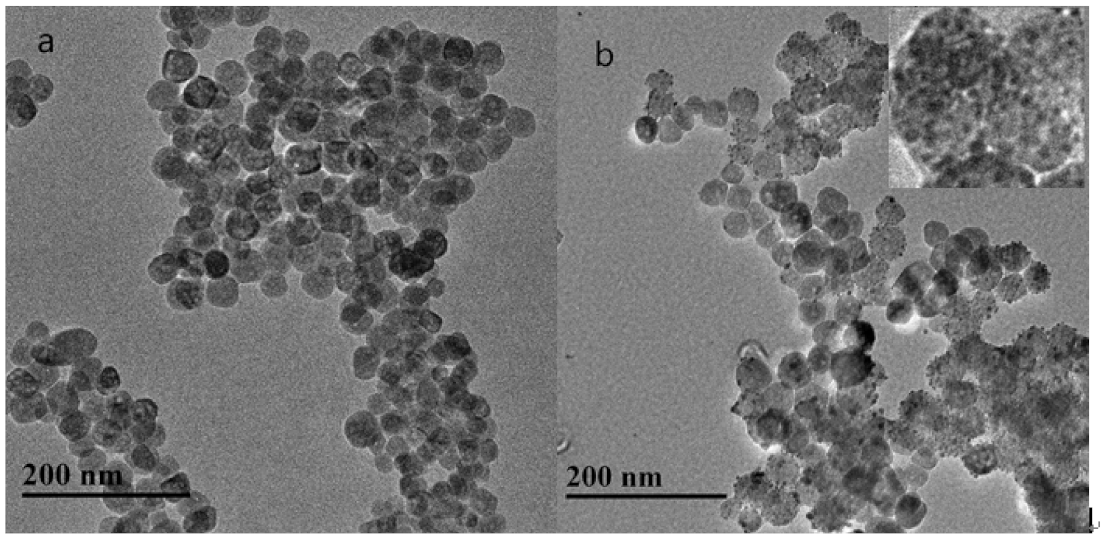
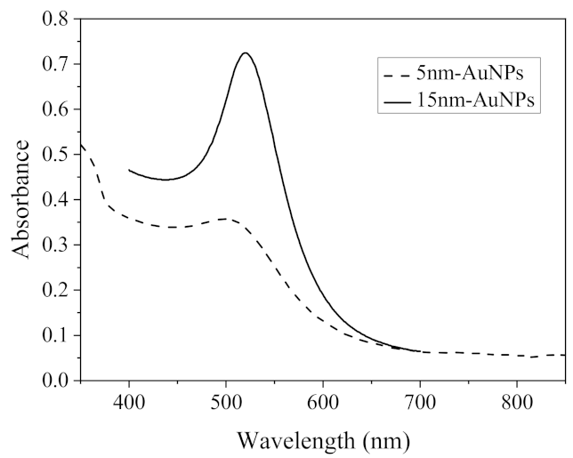
Figure 1a shows the TEM image of the Fe3O4, which displays a good dispersion with an average diameter of 30 nm. Since the Fe3O4 NPs modified with 3-Aminopropyltriethoxysilane (APTES) have abundant amino groups on the surface, the small AuNPs could be absorbed on the Fe3O4 NPs by the electrostatic interaction between citric acid and amino groups [22]. As shown in (Figure 1b), the AuNPs was dispersed around spherical Fe3O4 particles with an average diameter of 5.0 nm. The nanocomposites can be easily magnetically separated as shown in Figure S1. The nanocomposites can remain stable after storage for 1 month. As shown in the UV-vis spectra (Figure 2), strong characteristic absorption peaks at 498 nm and 520 nm of small AuNPs (5 nm) and AuNPs (15 nm) can be found [23,24].

2.2. Etching of Fe3O4@AuNPs–FITC by Lead and Thiosulfate Ions

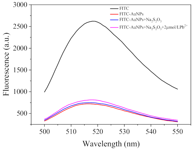
As shown in Figure 3, the fluorescence emission of FITC was almost completely quenched by AuNPs. With the addition of S2O32− and Pb2+ ions, the fluorescence intensity of the solution demonstrated slightly increasement. This may be because the above etching of AuNPs (15 nm) only slightly changed the physicochemical property and the etched AuNPs (15 nm) still were big enough to quench FITC.
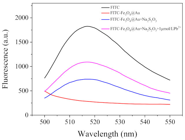
Thus, we prepared the Fe3O4@AuNPs–FITC to further explore whether the strategy of combing the etching of smaller AuNPs, and the AuNPs separation from FITC together has the potential to develop a sensitive sensor. As shown in Figure 4, very weak fluorescent emission is due to the quenching of FITC by AuNPs. In the presence of Na2S2O3, the fluorescent emission of the solution obviously increased. This may be due to the redox reaction between Na2S2O3 and AuNPs in Fe3O4@AuNPs–FITC, forming the Au(S2O3)23+ compound [25,26] and slightly impairing the adsorption of FITC on the surface of AuNPs. With the coexistence of S2O32− and Pb2+ ions, the fluorescent emission of the solution increased much more significantly. These results indicate that the etching of AuNPs by ions deserves further investigation for developing a Pb2+ sensing method.

2.3. Optimization of the Fluorescent Assay

To achieve a sensitive fluorescent assay for Pb2+, some factors including FITC concentration, incubation time, Na2S2O3 concentration, and pH value were investigated. As shown in Figure 5, the fluorescence intensity raises when the FITC concentration is higher than 0.5 μmol/L, which indicates excess FITC appears and separates from the Fe3O4@Au NPs (Figure 5). Thus, 0.5 μmol/L FITC was selected for further experiments.
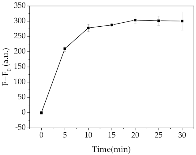
As shown in Figure 6, the recovered fluorescence intensity (F-F0) increased with the incubation time extension, and nearly leveled off after 10 min. Thus, incubating Fe3O4@Au–FITC with etching ions for 10 min was enough for sensing Pb2+.
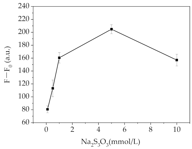
As shown in Figure 7, the recovered fluorescence intensity (F-F0) displayed a gradual increase in the range 0–5.0 mmol/L Na2S2O3 and a higher concentration decreased the fluorescence signal.
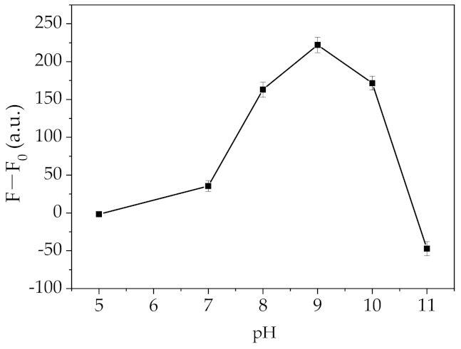
As shown in Figure 8, the pH value of glycine–NaOH buffer demonstrated big impact on the recovered fluorescence intensity (F-F0). However, good fluorescence response could be obtained with the glycine–NaOH buffer at the range of pH 8.0–10 and highest signal obtained at pH 9.0.

2.4. Selectivity of Pb2+ Sensing

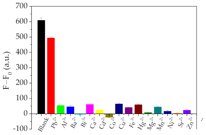
The selectivity of the assay was investigated by testing some common metal ions (Cu2+, Mg2+, Ca2+, Co2+, Fe3+, Sr2+, Al3+, Pb2+, Hg2+, Mn2+, Cd2+, Ni2+, Zn2+, Ba2+, and Bi2+, 10 µmol/L) under the same experimental conditions. As shown in Figure 9, all these metal ions produced negligible fluorescence signal to the Fe3O4@Au–FITC-based assay. The results indicated high selectivity toward Pb2+ of this Fe3O4@Au–FITC-based assay. The high selectivity should be ascribed to two aspects: (1) the magnetic separation and enrichment of Pb2+ ions; and (2) the selective etching of AuNPs by Pb2+ in the presence of Na2S2O3.

2.5. Analytical Performance of Pb2+ Sensing

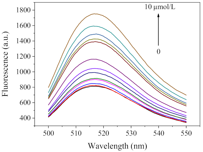
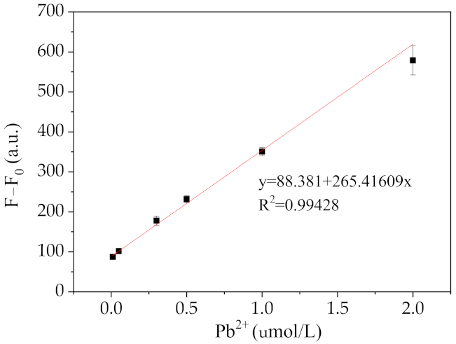
Under optimal conditions, the sensitivity of the fluorescence assay was evaluated. As shown in Figure 10, the fluorescence intensity increased gradually with the increasing Pb2+ concentration in the range of 0–10 µmol/L. A linear relationship between the fluorescence intensity and Pb2+ concentration could be achieved in the range 0.02 to 2.0 µmol/L (Figure 11). The limit of detection (LOD) was 5.2 nmol/L, estimated by a 3-fold signal-to-noise ratio (3S/N). The analytical performance of the Fe3O4@Au–FITC-based assay was much more sensitive and rapid than the other reported nanomaterial etching-based methods [25,26,27] (Table 1). In addition, the LOD of the proposed method was much lower than the maximum allowable levels of Pb2+ (72 nmol/L) in drinking water set by the U.S. EPA.

2.6. Application of Pb2+ Sensing

Samples collected from Tai lake (Wuxi, China) were filtered through a microfiltration membrane (0.22 μm) and spiked with different concentrations of Pb2+ (50, 100 and 150 nmol/L). The samples then were measured by the above fluorescent method. As demonstrated in Table 2, the recoveries from 98.2% to 106.4% were obtained with relative standard deviation (RSD) less than 10%. The above results demonstrated the practical potential of this Fe3O4@Au–FITC-based fluorescent method for Pb2+ in detecting environmental samples.

3. Materials and Methods

3.1. Chemicals and Reagents

All reagents were of analytical grade and used without further purification. Sodium borohydride (NaBH4) and 3-Aminopropyltriethoxysilane (APTES) were purchased from Shanghai Aladdin biochemical technology Co., Ltd. (Shanghai, China) Chloroauric acid hydrate (HAuCl4·3H2O) and sodium citrate were purchased from Sigma–Aldrich (Shanghai, China). Fluorescein isothiocyanate (isomeride I) (FITC > 95.0%) was purchased from TCI (Shanghai, China) chemical industry Co., Ltd. Copper, aluminum, nickel, cobalt, barium, cadmium, manganese, iron, zinc, strontium, bismuth, chromium were purchased from Guobiao (Beijing, China) Testing & Certification Co., Ltd. The nitrate, acetate, or chloride salts of other metal ions were purchased from Sinopharm Chemical Reagent Co, Ltd.(Shanghai, China). Ethylene glycol, ethanolamine, ethanol and Poly (ethylene glycol) (PEG2000) were purchased from Sinopharm Chemical Reagent Co, Ltd. (Shanghai, China).

3.2. Apparatus

UV-visible (UV-vis) absorption spectra of nanoparticles were obtained with a microplate reader (Bio-Tek, Elx800, USA) (Winooski, VT, USA). Steady-state fluorescence spectra were measured by a fluorospectrometric photometer (Lengguan F97, Shanghai Lengguan, China). Transmission electron microscopy (TEM) was performed on a JEOL JEM-2100 (Kyoto, Japan) at an accelerating voltage of 200 kV. Energy diffraction X-ray (EDX) spectrum was obtained using the TEM.

3.3. Synthesis of Au Nanoparticles (AuNPs)

AuNPs (5.0 nm) were synthesized by NaBH4 reduction method [23]. Briefly, HAuCl4 (0.5 mL, 4.0 g/L) and sodium citrate (2.0 mL, 0.025 mmol/L) were dissolved in 20 mL water under magnetic stirring. Then 0.6 mL NaBH4 (0.1 mol/L) solution was added and the mixture was stirred thoroughly. The color of the solution changed from light yellow to orange red, which indicated that the AuNPs were successfully synthesized.

3.4. Amine-Functionalization of Fe3O4 Nanoparticles Using APTES

Fe3O4 nanoparticles were synthesized according to the method with slight modifications [28]. FeCl3·6H2O (1.05 g), ethylene glycol (21 mL) and ethanolamine (7 mL) were added to the conical flask under magnetic stirring. Then, sodium acetate trihydrate (NaAc) (2.8 g) and PEG2000 (0.7 g) were added to the solution under vigorous stirring. The reaction was maintained at 200 °C for 8 h. Afterwards, the Fe3O4 NPs were obtained after the mixture was washed several times with water and ethanol. The Fe3O4 NPs were dried in a vacuum oven at 60 °C for 12 h and Fe3O4 NPs powder was obtained.
For APTES modification, the Fe3O4 NPs (150 mg) were suspended in a mixture of ethanol (40 mL) and APTES solution (1.0 mL). The mixture was gently stirred at room temperature for 3 h. The Fe3O4 NPs were separated using an external magnet, and thoroughly washed with ethanol and water. The amine-functionalization of Fe3O4 nanoparticles were finally suspended in 15 mL ethanol and stored at 4 °C for subsequent use.

3.5. Synthesis of Fe3O4@AuNCs-FITC

To prepare Fe3O4@AuNPs, 3.0 mL of amine-functionalization of Fe3O4 NPs was dissolved in 15 mL of water, and then 100 mL of AuNPs (5 nm) was added under stirring for 15 min. The Fe3O4@AuNPs were separated using an external magnet, washed with water and finally suspended in 25 mL of water [29]. The Fe3O4@AuNP-FITC nanocomposites were synthesized as follows. FITC (50 µL,50 µmol/L) onto Fe3O4@Au NPs (250 µL) in 4.7 mL water and stored at 4 °C overnight. FITC–AuNPs was prepared as follows. AuNPs (15 nm) were prepared using trisodium citrate reduction method [24]. Briefly, 100 mL of 0.01% HAuCl4 solution was slowly stirred, heated to a boil in the conical flask and kept boiling for 5 min. Then, 2 mL of 1% trisodium citrate solution was poured into the flask. The color of mixture changed from pale yellow to gray and finally to wine red. After boiling for 10 min, the obtained AuNPs (3.0 nmol/L) were cooled to room temperature and stored at 4 °C for subsequent use. FITC–AuNPs nanocomposites were synthesized according to the method reported by Wang et al. [30]. FITC solution (5.0 µL, 1.5 mmol/L) in ethanol was added into 5.0 mL of AuNPs solution and mixed gently. The mixture was stored at 4 °C overnight and then FITC–AuNPs nanocomposites were obtained.

3.6. Fluorescent Detection of Pb2+

A total of 10 µL of Fe3O4@AuNP–FITC composites, 40 µL of H2O, and 50 µL of sodium thiosulfate (20 mmol/L) as well as 50 µL of glycine–NaOH buffer (10 mmol/L, pH 9.0) were mixed gently. After incubation at room temperature for 5.0 min, Pb2+ solution was added and incubated at room temperature for 10 min. Then the Fe3O4@AuNPs were separated by an external magnet for 2 min. The fluorescence emission spectra of the supernatant were measured by a fluorescence spectrometer with 465 nm excitation.

4. Conclusions

A highly sensitive and selective fluorescent method for Pb2+ sensing was developed based on a new nanocomposite, Fe3O4@Au–FITC. This method combined the metal leaching by the Pb2+-S2O32− system with magnetic separation together. Considering that ion-induced etching of metal NPs has been applied in the development of analytical methods for the detection of metal ions, anions, small molecules, and proteins [31,32], the strategy proposed here is expected to be further explored in some challenging situations.

Supplementary Materials

The following are available online, Figure S1: Magnetic seperation of the Fe3O4@Au–FITC nanocomposites.

Author Contributions

Investigation, interpretation of data and writing: Y.C. and B.R.; Conceptualization and: C.P. and C.Z. project administration: X.W. All authors have read and agreed to the published version of the manuscript.

Funding

This research was funded by the National Key Research and Development Program (2018YFC1604400) and the National Natural Science Foundation of China (31871879).

Institutional Review Board Statement

Not applicable.

Informed Consent Statement

Not applicable.

Data Availability Statement

Not applicable.

Conflicts of Interest

The authors declare no conflict of interest.

References

  1. Knecht, M.R.; Sethi, M. Bio-inspired colorimetric detection of Hg2+ and Pb2+ heavy metal ions using Au nanoparticles. Anal. Bioanal. Chem. 2009, 394, 33–46. [Google Scholar] [CrossRef]
  2. Lan, Y.-J.; Lin, Y.-W. A non-aggregation colorimetric method for trace lead(II) ions based on the leaching of gold nanorods. Anal. Methods UK 2014, 6, 7234–7242. [Google Scholar] [CrossRef]
  3. Li, Y.F.; Chen, C.Y.; Li, B.; Sun, J.; Wang, J.X.; Gao, Y.X.; Zhao, Y.L.; Chai, Z.F. Elimination efficiency of different reagents for the memory effect of mercury using ICP-MS. J. Anal. Atom. Spectrom. 2006, 21, 94–96. [Google Scholar] [CrossRef]
  4. Mamatha, P.; Venkateswarlu, G.; Thangavel, S.; Sahayam, A.C.; Swamy, A.V.N. Methodology for the Speciation of Cr(III) and Cr(VI) in Leaf Powders Using the Sulfate Form of Dowex-1 After Microwave Extraction and ICP-OES Detection. Atom. Spectrosc. 2019, 40, 31–35. [Google Scholar] [CrossRef]
  5. Chen, J.L.; Zhang, Y.Y.; Cheng, M.P.; Mergny, J.L.; Lin, Q.M.; Zhou, J.; Ju, H.X. Highly active G-quadruplex/hemin DNAzyme for sensitive colorimetric determination of lead(II). Microchim. Acta 2019, 186, 786. [Google Scholar] [CrossRef]
  6. Tao, Z.; Zhou, Y.; Duan, N.; Wang, Z.P. A Colorimetric Aptamer Sensor Based on the Enhanced Peroxidase Activity of Functionalized Graphene/Fe3O4-AuNPs for Detection of Lead (II) Ions. Catalysts 2020, 10, 600. [Google Scholar] [CrossRef]
  7. Jiang, Y.L.; Wang, Y.X.; Meng, F.D.; Wang, B.X.; Cheng, Y.X.; Zhu, C.J. N-doped carbon dots synthesized by rapid microwave irradiation as highly fluorescent probes for Pb2+ detection. New J. Chem. 2015, 39, 3357–3360. [Google Scholar] [CrossRef]
  8. Chen, M.; Hassan, M.; Li, H.H.; Chen, Q.S. Fluorometric determination of lead(II) by using aptamer-functionalized upconversion nanoparticles and magnetite-modified gold nanoparticles. Microchim. Acta 2020, 187, 85. [Google Scholar] [CrossRef]
  9. Chen, S.H.; Li, Y.X.; Li, P.H.; Xiao, X.Y.; Jiang, M.; Li, S.S.; Zhou, W.Y.; Yang, M.; Huang, X.J.; Liu, W.Q. Electrochemical spectral methods for trace detection of heavy metals: A review. Trac Trend. Anal. Chem. 2018, 106, 139–150. [Google Scholar] [CrossRef]
  10. Li, P.H.; Song, Z.Y.; Yang, M.; Chen, S.H.; Xiao, X.Y.; Duan, W.C.; Li, L.N.; Huang, X.J. Electrons in Oxygen Vacancies and Oxygen Atoms Activated by Ce3+/Ce4+ Promote High-Sensitive Electrochemical Detection of Pb(II) over Ce-Doped alpha-MoO3 Catalysts. Anal. Chem. 2020, 92, 16089–16096. [Google Scholar] [CrossRef]
  11. Singh, H.; Bamrah, A.; Bhardwaj, S.K.; Deep, A.; Khatri, M.; Kim, K.-H.; Bhardwaj, N. Nanomaterial-based fluorescent sensors for the detection of lead ions. J. Hazard. Mater. 2021, 407, 124379. [Google Scholar] [CrossRef]
  12. Zamojc, K.; Kamrowski, D.; Zdrowowicz, M.; Wyrzykowski, D.; Wiczk, W.; Chmurzynski, L.; Makowska, J. A Pentapeptide with Tyrosine Moiety as Fluorescent Chemosensor for Selective Nanomolar-Level Detection of Copper(II) Ions. Int. J. Mol. Sci. 2020, 21, 743. [Google Scholar] [CrossRef] [Green Version]
  13. Lee, J.F.; Chen, H.L.; Lee, G.S.; Tseng, S.C.; Lin, M.H.; Liau, W.B. Photosensized Controlling Benzyl Methacrylate-Based Matrix Enhanced Eu3+ Narrow-Band Emission for Fluorescence Applications. Int. J. Mol. Sci. 2012, 13, 3718–3737. [Google Scholar] [CrossRef]
  14. Qi, Z.L.; Cheng, Y.H.; Xu, Z.; Chen, M.L. Recent Advances in Porphyrin-Based Materials for Metal Ions Detection. Int. J. Mol. Sci. 2020, 21, 5839. [Google Scholar] [CrossRef]
  15. Kurshanov, D.A.; Khavlyuk, P.D.; Baranov, M.A.; Dubavik, A.; Rybin, A.V.; Fedorov, A.V.; Baranov, A.V. Magneto-Fluorescent Hybrid Sensor CaCO3-Fe3O4-AgInS2/ZnS for the Detection of Heavy Metal Ions in Aqueous Media. Materials 2020, 13, 4373. [Google Scholar] [CrossRef]
  16. Kumar, A.; Chowdhuri, A.R.; Laha, D.; Mahto, T.K.; Karmakar, P.; Sahu, S.K. Green synthesis of carbon dots from Ocimum sanctum for effective fluorescent sensing of Pb2+ ions and live cell imaging. Sens. Actuat. B Chem. 2017, 242, 679–686. [Google Scholar] [CrossRef]
  17. Niu, X.F.; Zhong, Y.B.; Chen, R.; Wang, F.; Liu, Y.J.; Luo, D. A “turn-on” fluorescence sensor for Pb2+ detection based on graphene quantum dots and gold nanoparticles. Sens. Actuator B Chem. 2018, 255, 1577–1581. [Google Scholar] [CrossRef]
  18. Shu, T.; Su, L.; Wang, J.; Li, C.; Zhang, X. Chemical etching of bovine serum albumin-protected Au25 nanoclusters for label-free and separation-free detection of cysteamine. Biosens. Bioelectron. 2015, 66, 155–161. [Google Scholar] [CrossRef] [PubMed]
  19. Liu, R.L.; Chen, Z.P.; Wang, S.S.; Qu, C.L.; Chen, L.X.; Wang, Z. Colorimetric sensing of copper(II) based on catalytic etching of gold nanoparticles. Talanta 2013, 112, 37–42. [Google Scholar] [CrossRef]
  20. Liu, W.Q.; Hou, S.; Yan, J.; Zhang, H.; Ji, Y.L.; Wu, X.C. Quantification of proteins using enhanced etching of Ag coated Au nanorods by the Cu2+/bicinchoninic acid pair with improved sensitivity. Nanoscale 2016, 8, 780–784. [Google Scholar] [CrossRef] [PubMed]
  21. Shi, X.H.; Gu, W.; Peng, W.D.; Li, B.Y.; Chen, N.N.; Zhao, K.; Xian, Y.Z. Sensitive Pb2+ Probe Based on the Fluorescence Quenching by Graphene Oxide and Enhancement of the Leaching of Gold Nanoparticles. ACS Appl. Mater. Interfaces 2014, 6, 2568–2575. [Google Scholar] [CrossRef] [PubMed]
  22. Rahman, Z.U.; Ma, Y.H.; Hu, J.; Xu, Y.Y.; Wang, W.F.; Chen, X.G. Preparation and characterization of magnetic gold shells using different sizes of gold nanoseeds and their corresponding effects on catalysis. RSC Adv. 2014, 4, 5012–5020. [Google Scholar] [CrossRef]
  23. Jana, N.R.; Gearheart, L.; Murphy, C.J. Seeding growth for size control of 5–40 nm diameter gold nanoparticles. Langmuir 2001, 17, 6782–6786. [Google Scholar] [CrossRef]
  24. Peng, C.F.; Pan, N.; Xie, Z.J.; Wu, L.L. Highly sensitive and selective colorimetric detection of Hg2+ based on the separation of Hg2+ and formation of catalytic DNA-gold nanoparticles. Anal. Methods UK 2016, 8, 1021–1025. [Google Scholar] [CrossRef]
  25. Zhang, Y.J.; Leng, Y.M.; Miao, L.J.; Xin, J.W.; Wu, A.G. The colorimetric detection of Pb2+ by using sodium thiosulfate and hexadecyl trimethyl ammonium bromide modified gold nanoparticles. Dalton Trans. 2013, 42, 5485–5490. [Google Scholar] [CrossRef]
  26. Chen, Y.Y.; Chang, H.T.; Shiang, Y.C.; Hung, Y.L.; Chiang, C.K.; Huang, C.C. Colorimetric Assay for Lead Ions Based on the Leaching of Gold Nanoparticles. Anal. Chem. 2009, 81, 9433–9439. [Google Scholar] [CrossRef]
  27. Shi, X.H.; Gu, W.; Zhang, C.L.; Zhao, L.Y.; Peng, W.D.; Xian, Y.Z. A label-free colorimetric sensor for Pb2+ detection based on the acceleration of gold leaching by graphene oxide. Dalton Trans. 2015, 44, 4623–4629. [Google Scholar] [CrossRef]
  28. Deng, H.; Li, X.L.; Peng, Q.; Wang, X.; Chen, J.P.; Li, Y.D. Monodisperse magnetic single-crystal ferrite microspheres. Angew. Chem. Int. Edit. 2005, 44, 2782–2785. [Google Scholar] [CrossRef]
  29. Xia, Q.D.; Fu, S.S.; Ren, G.J.; Chai, F.; Jiang, J.J.; Qu, F.Y. Fabrication of magnetic bimetallic Fe3O4@Au-Pd hybrid nanoparticles with recyclable and efficient catalytic properties. RSC Adv. 2016, 6, 55248–55256. [Google Scholar] [CrossRef]
  30. Wang, S.S.; Wang, X.K.; Zhang, Z.Y.; Chen, L.X. Highly sensitive fluorescence detection of copper ion based on its catalytic oxidation to cysteine indicated by fluorescein isothiocyanate functionalized gold nanoparticles. Colloid Surf. A 2015, 468, 333–338. [Google Scholar] [CrossRef]
  31. Wu, S.; Li, D.; Wang, J.; Zhao, Y.; Dong, S.; Wang, X. Gold nanoparticles dissolution based colorimetric method for highly sensitive detection of organophosphate pesticides. Sens. Actuator B Chem. 2017, 238, 427–433. [Google Scholar] [CrossRef]
  32. Zhong, Y.P.; Wang, Q.P.; He, Y.; Ge, Y.L.; Song, G.W. A novel fluorescence and naked eye sensor for iodide in urine based on the iodide induced oxidative etching and aggregation of Cu nanoclusters. Sens. Actuator B Chem. 2015, 209, 147–153. [Google Scholar] [CrossRef]
Scheme 1. Schematic illustration of Pb2+ detection based on Fe3O4@Au–FITC nanocomposites.
Molecules 26 03180 sch001
Figure 1. TEM images of (a) Fe3O4 and (b) Fe3O4@Au NPs.
Molecules 26 03180 g001
Figure 2. UV-vis spectra of AuNPs.
Molecules 26 03180 g002
Figure 3. Fluorescence spectra of the FITC–AuNPs.
Molecules 26 03180 g003
Figure 4. Fluorescence spectra of Fe3O4@Au–FITC.
Molecules 26 03180 g004
Figure 5. Effect of FITC concentration on Pb2+ sensing.
Molecules 26 03180 g005
Figure 6. Effect of the incubation time on Pb2+ sensing.
Molecules 26 03180 g006
Figure 7. Effect of Na2S2O3 concentration on Pb2+ sensing.
Molecules 26 03180 g007
Figure 8. Effect of the pH value on Pb2+ sensing.
Molecules 26 03180 g008
Figure 9. Selectivity of the assay for various metal ions.
Molecules 26 03180 g009
Figure 10. Fluorescence spectra of Fe3O4@Au–FITC in the presence of various concentrations of Pb2+.
Molecules 26 03180 g010
Figure 11. The relationship between the difference value of fluorescence intensities and the different concentrations of Pb2+.
Molecules 26 03180 g011
Table 1. Comparison of several methods for Pb2+ detection.
NanoprobesLinear RangeLODTimeRef.
CTAB modified AuNPs1.0~6.0 µmol/L75 nmol/L30 min[25]
AuNPs and graphene oxide0.1~20 µmol/L50 nmol/L20 min[27]
AuNPs0.0025~10 µmol/L0.5 nmol/L2 h[26]
Fe3O4@Au–FITC0.02~2.0 µmol/L5.2 nmol/L12 minThis work
Table 2. Determination of Pb2+ in lake-water samples (n = 3).
Concentration (nmol/L)Result (nmol/L)Recovery (%)RSD (%)
5053.2106.48.3
10098.398.32.5
150159.0106.03.7
Publisher’s Note: MDPI stays neutral with regard to jurisdictional claims in published maps and institutional affiliations.