
Thiocoumarin Caged Nucleotides: Synthetic Access and Their Photophysical Properties

 , , , , , and
Abstract
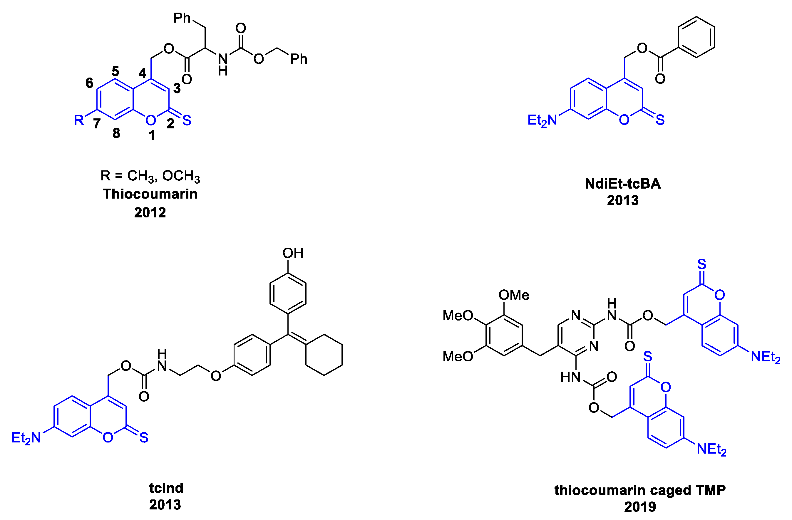
1. Introduction
2. Results
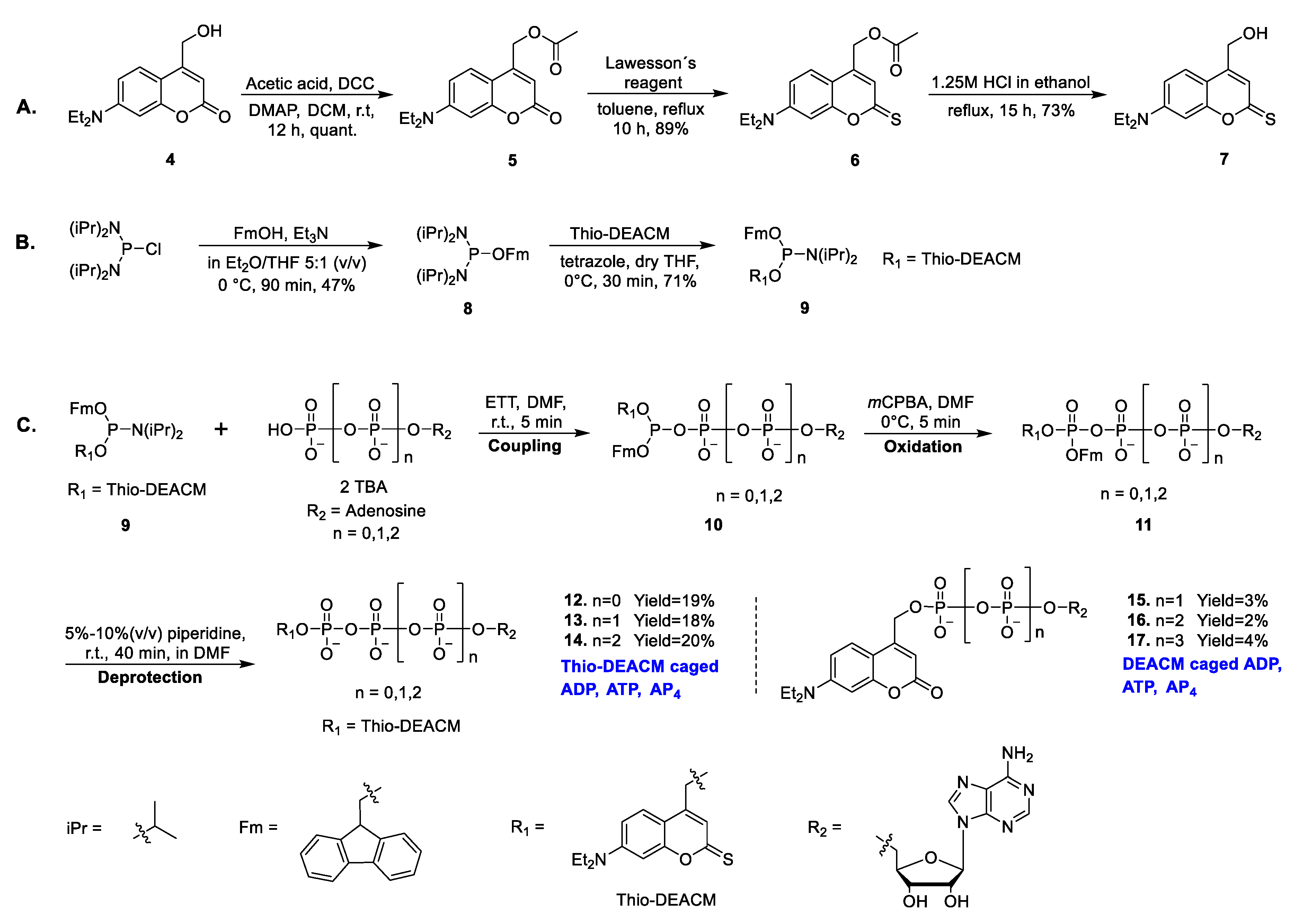
2.1. Chemistry
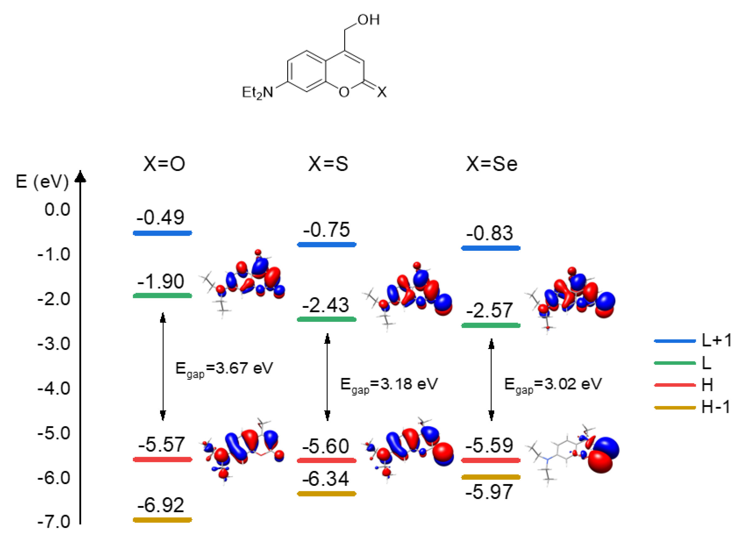
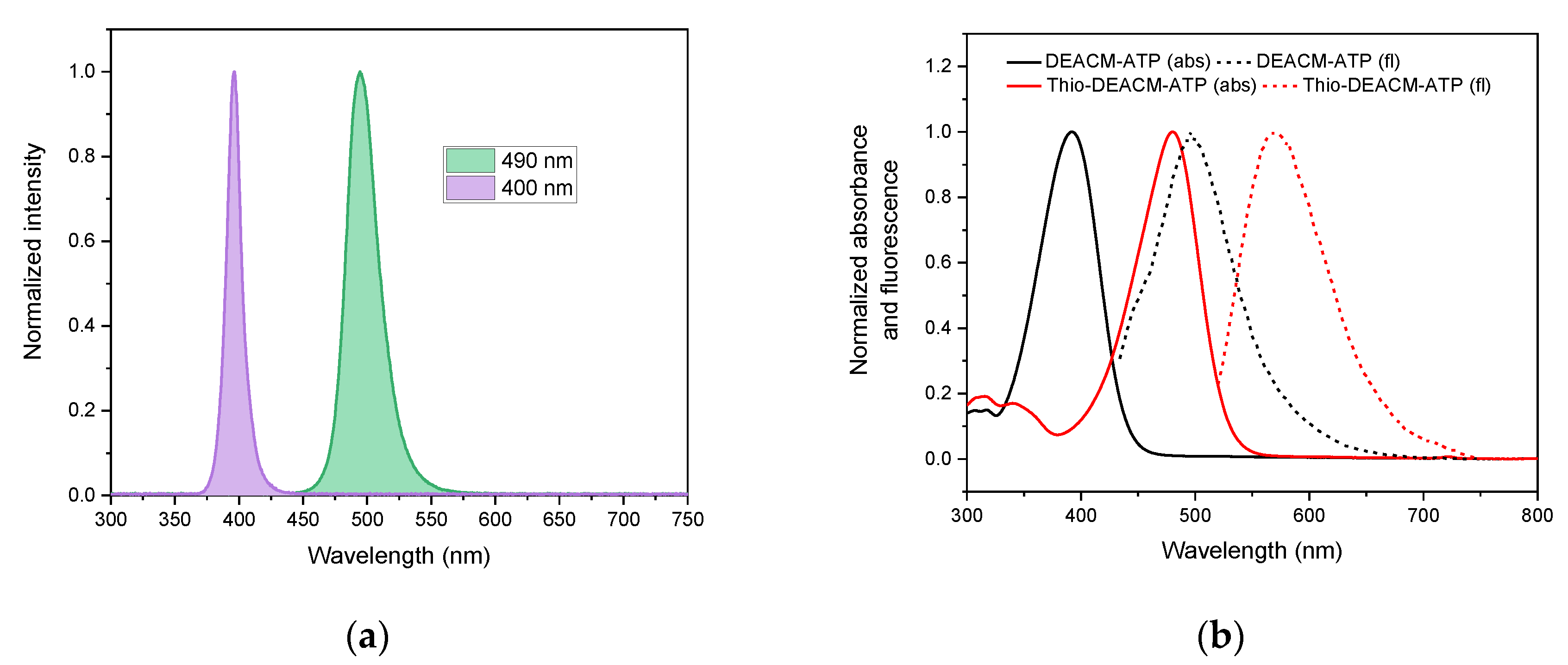
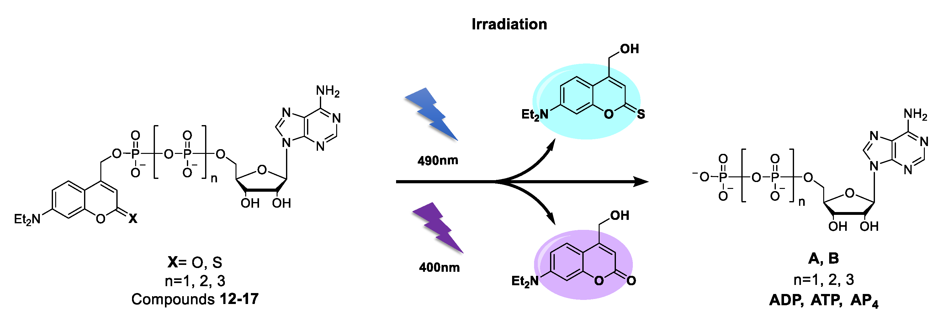
2.2. Photophysical Properties
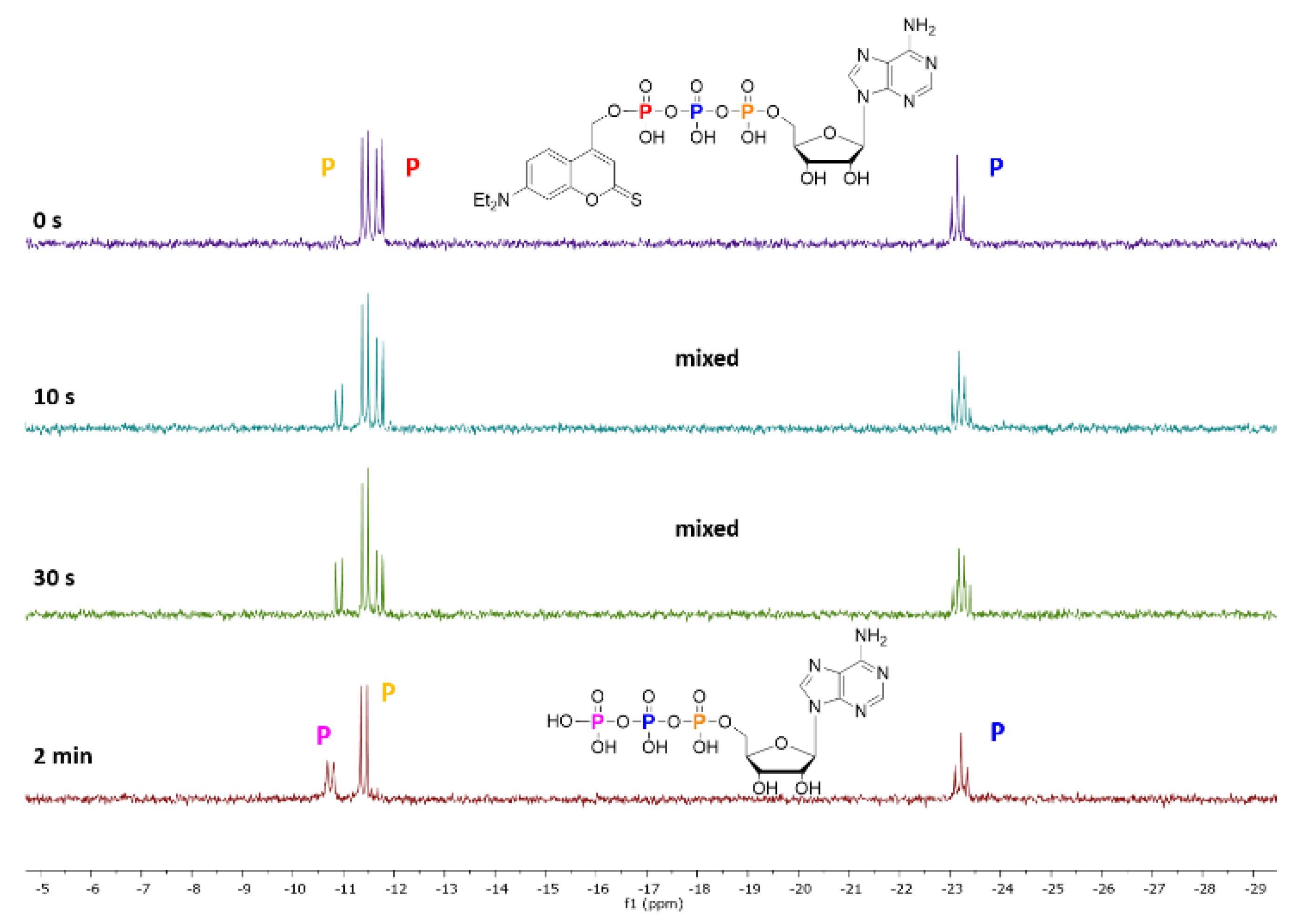
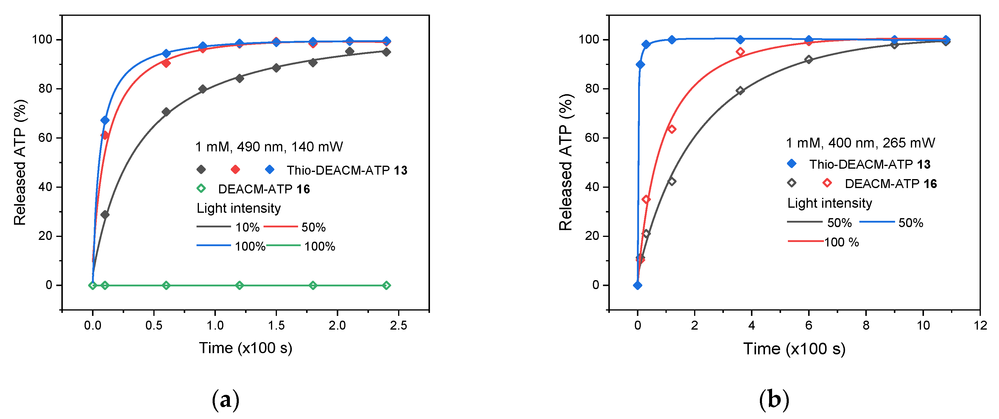
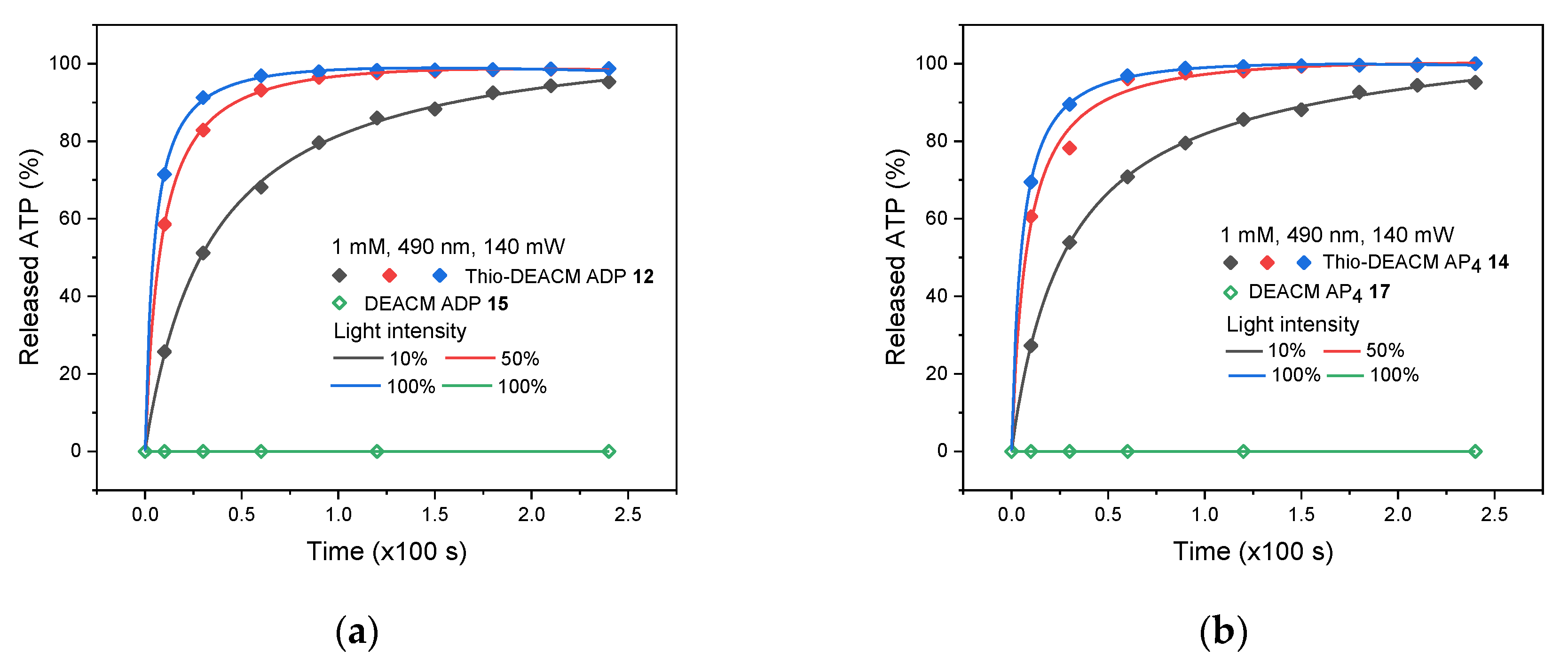
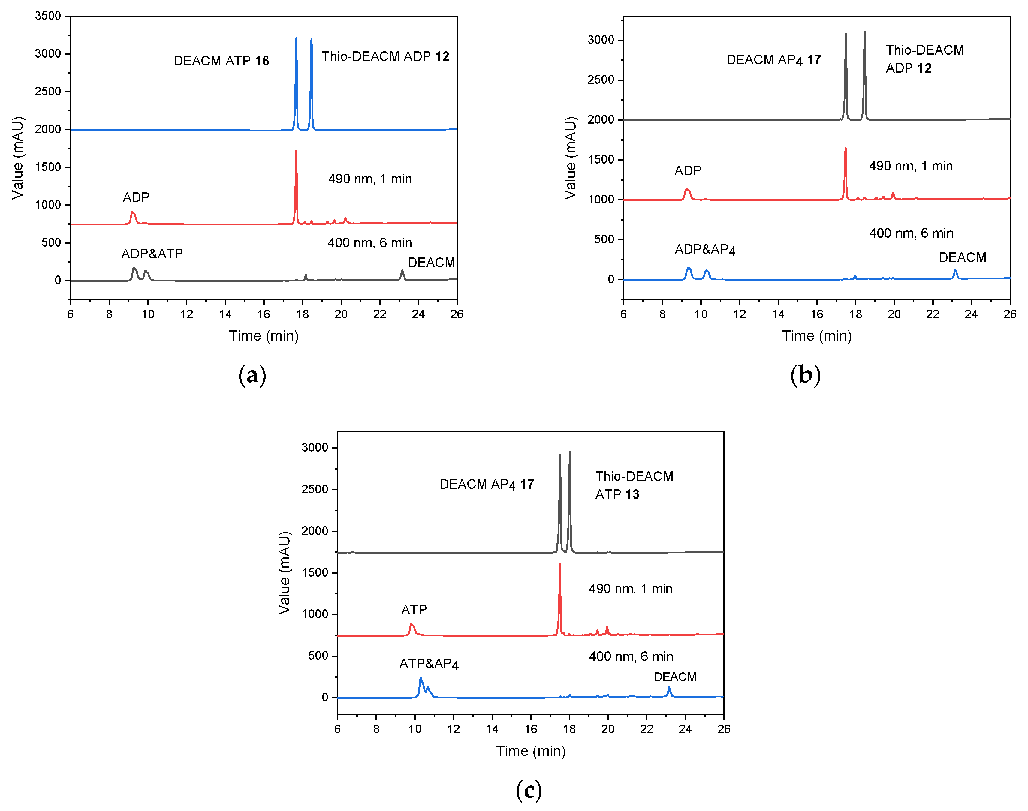
2.3. Photolysis Studies
3. Discussion
4. Conclusions
Supplementary Materials
Author Contributions
Funding
Acknowledgments
Conflicts of Interest
References
- Mayer, G.; Heckel, A. Biologically active molecules with a “light switch”. Angew. Chem. Int. Ed. 2006, 45, 4900–4921. [Google Scholar] [CrossRef]
- Kaplan, J.H.; Forbush, B.; Hoffman, J.F. Rapid Photolytic Release of Adenosine 5ʹ-Triphosphate from a Protected Analogue: Utilization by the Na:K Pump of Human Red Blood Cell Ghosts. Biochemistry 1978, 17, 1929–1935. [Google Scholar] [CrossRef] [PubMed]
- Hansen, M.J.; Velema, W.A.; Lerch, M.M.; Szymanski, W.; Feringa, B.L. Wavelength-selective cleavage of photoprotecting groups: Strategies and applications in dynamic systems. Chem. Soc. Rev. 2015, 44, 3358–3377. [Google Scholar] [CrossRef] [PubMed]
- Olson, J.P.; Banghart, M.R.; Sabatini, B.L.; Ellis-Davies, G.C.R. Spectral evolution of a photochemical protecting group for orthogonal two-color uncaging with visible light. J. Am. Chem. Soc. 2013, 135, 15948–15954. [Google Scholar] [CrossRef]
- Bojtár, M.; Kormos, A.; Kis-Petik, K.; Kellermayer, M.; Kele, P. Green-Light Activatable, Water-Soluble Red-Shifted Coumarin Photocages. Org. Lett. 2019, 21, 9410–9414. [Google Scholar] [CrossRef] [PubMed]
- Schade, B.; Hagen, V.; Schmidt, R.; Herbrich, R.; Krause, E.; Eckardt, T.; Bendig, J. Deactivation behavior and excited-state properties of (coumarin-4- yl)methyl derivatives. 1. Photocleavage of (7-methoxycoumarin-4-yl)methyl- caged acids with fluorescence enhancement. J. Org. Chem. 1999, 64, 9109–9117. [Google Scholar] [CrossRef]
- Schmidt, R.; Geissler, D.; Hagen, V.; Bendig, J. Kinetics study of the photocleavage of (coumarin-4-yl)methyl esters. J. Phys. Chem. A 2005, 109, 5000–5004. [Google Scholar] [CrossRef]
- Schmidt, R.; Geissler, D.; Hagen, V.; Bendig, J. Mechanism of photocleavage of (coumarin-4-yl)methyl esters. J. Phys. Chem. A 2007, 111, 5768–5774. [Google Scholar] [CrossRef]
- Pavlovic, I.; Thakor, D.T.; Vargas, J.R.; McKinlay, C.J.; Hauke, S.; Anstaett, P.; Camunã, R.C.; Bigler, L.; Gasser, G.; Schultz, C.; et al. Cellular delivery and photochemical release of a caged inositol-pyrophosphate induces PH-domain translocation in cellulo. Nat. Commun. 2016, 7, 1–8. [Google Scholar] [CrossRef]
- Nadler, A.; Yushchenko, D.A.; Müller, R.; Stein, F.; Feng, S.; Mulle, C.; Carta, M.; Schultz, C. Exclusive photorelease of signalling lipids at the plasma membrane. Nat. Commun. 2015, 6, 1–10. [Google Scholar] [CrossRef]
- Wagner, N.; Stephan, M.; Höglinger, D.; Nadler, A. A Click Cage: Organelle-Specific Uncaging of Lipid Messengers. Angew. Chem. Int. Ed. 2018, 57, 13339–13343. [Google Scholar] [CrossRef] [PubMed]
- Kantevari, S.; Matsuzaki, M.; Kanemoto, Y.; Kasai, H.; Ellis-Davies, G.C.R. Two-color, two-photon uncaging of glutamate and GABA. Nat. Methods 2010, 7, 123–125. [Google Scholar] [CrossRef] [PubMed]
- Amatrudo, J.M.; Olson, J.P.; Lur, G.; Chiu, C.Q.; Higley, M.J.; Ellis-Davies, G.C.R. Wavelength-selective one- and two-photon uncaging of Gaba. ACS Chem. Neurosci. 2014, 5, 64–70. [Google Scholar] [CrossRef] [PubMed]
- Olson, J.P.; Kwon, H.B.; Takasaki, K.T.; Chiu, C.Q.; Higley, M.J.; Sabatini, B.L.; Ellis-Davies, G.C.R. Optically selective two-photon uncaging of glutamate at 900 nm. J. Am. Chem. Soc. 2013, 135, 5954–5957. [Google Scholar] [CrossRef] [PubMed]
- Richers, M.T.; Amatrudo, J.M.; Olson, J.P.; Ellis-Davies, G.C.R. Cloaked Caged Compounds: Chemical Probes for Two-Photon Optoneurobiology. Angew. Chem. Int. Ed. 2017, 56, 193–197. [Google Scholar] [CrossRef]
- Rovira, A.; Gandioso, A.; Goñalons, M.; Galindo, A.; Massaguer, A.; Bosch, M.; Marchán, V. Solid-Phase Approaches for Labeling Targeting Peptides with Far-Red Emitting Coumarin Fluorophores. J. Org. Chem. 2019, 84, 1808–1817. [Google Scholar] [CrossRef] [PubMed]
- Novohradsky, V.; Rovira, A.; Hally, C.; Galindo, A.; Vigueras, G.; Gandioso, A.; Svitelova, M.; Bresolí-Obach, R.; Kostrhunova, H.; Markova, L.; et al. Towards Novel Photodynamic Anticancer Agents Generating Superoxide Anion Radicals: A Cyclometalated IrIII Complex Conjugated to a Far-Red Emitting Coumarin. Angew. Chem. Int. Ed. 2019, 58, 6311–6315. [Google Scholar] [CrossRef] [PubMed]
- Gandioso, A.; Palau, M.; Nin-Hill, A.; Melnyk, I.; Rovira, C.; Nonell, S.; Velasco, D.; García-Amorós, J.; Marchán, V. Sequential Uncaging with Green Light can be Achieved by Fine-Tuning the Structure of a Dicyanocoumarin Chromophore. ChemistryOpen 2017, 6, 375–384. [Google Scholar] [CrossRef]
- Gandioso, A.; Contreras, S.; Melnyk, I.; Oliva, J.; Nonell, S.; Velasco, D.; García-Amorós, J.; Marchán, V. Development of Green/Red-Absorbing Chromophores Based on a Coumarin Scaffold That Are Useful as Caging Groups. J. Org. Chem. 2017, 82, 5398–5408. [Google Scholar] [CrossRef]
- Gandioso, A.; Bresolí-Obach, R.; Nin-Hill, A.; Bosch, M.; Palau, M.; Galindo, A.; Contreras, S.; Rovira, A.; Rovira, C.; Nonell, S.; et al. Redesigning the Coumarin Scaffold into Small Bright Fluorophores with Far-Red to Near-Infrared Emission and Large Stokes Shifts Useful for Cell Imaging. J. Org. Chem. 2018, 83, 1185–1195. [Google Scholar] [CrossRef]
- Gandioso, A.; Palau, M.; Bresolí-Obach, R.; Galindo, A.; Rovira, A.; Bosch, M.; Nonell, S.; Marchán, V. High Photostability in Nonconventional Coumarins with Far-Red/NIR Emission through Azetidinyl Substitution. J. Org. Chem. 2018, 83, 11519–11531. [Google Scholar] [CrossRef] [PubMed]
- Bassolino, G.; Nançoz, C.; Thiel, Z.; Bois, E.; Vauthey, E.; Rivera-Fuentes, P. Photolabile coumarins with improved efficiency through azetidinyl substitution. Chem. Sci. 2018, 9, 387–391. [Google Scholar] [CrossRef] [PubMed]
- Fournier, L.; Gauron, C.; Xu, L.; Aujard, I.; Le Saux, T.; Gagey-Eilstein, N.; Maurin, S.; Dubruille, S.; Baudin, J.B.; Bensimon, D.; et al. A blue-absorbing photolabile protecting group for in vivo chromatically orthogonal photoactivation. ACS Chem. Biol. 2013, 8, 1528–1536. [Google Scholar] [CrossRef] [PubMed]
- Guan, Z.; Inscho, E.W. Role of adenosine 5′-triphosphate in regulating renal microvascular function and in hypertension. Hypertension 2011, 58, 333–340. [Google Scholar] [CrossRef] [PubMed]
- Thirlwell, H.; Corrie, J.E.; Reid, G.P.; Trentham, D.R.; Ferenczi, M.A. Kinetics of relaxation from rigor of permeabilized fast-twitch skeletal fibers from the rabbit using a novel caged ATP and apyrase. Biophys. J. 1994, 67, 2436–2447. [Google Scholar] [CrossRef]
- Sokolov, V.S.; Apell, H.J.; Corrie, J.E.T.; Trentham, D.R. Fast transient currents in Na,K-ATPase induced by ATP concentration jumps from the P3-[1-(3’,5’-dimethoxyphenyl)-2-phenyl-2-oxo]ethyl ester of ATP. Biophys. J. 1998, 74, 2285–2298. [Google Scholar] [CrossRef]
- Park, C.H.; Givens, R.S. New photoactivated protecting groups. 6. p-Hydroxyphenacyl: A phototrigger for chemical and biochemical probes. J. Am. Chem. Soc. 1997, 119, 2453–2463. [Google Scholar] [CrossRef]
- Geißler, D.; Kresse, W.; Wiesner, B.; Bendig, J.; Kettenmann, H.; Hagen, V. DMACM-caged adenosine nucleotides: Ultrafast phototriggers for ATP, ADP, and AMP activated by long-wavelength irradiation. ChemBioChem 2003, 4, 162–170. [Google Scholar] [CrossRef]
- Pinheiro, A.; Baptistap, P.; Lima, J.C. Light activation of transcription: Photocaging of nucleotides for control over RNA polymerization. Nucleic Acids Res. 2008, 36. [Google Scholar] [CrossRef]
- Fonseca, A.S.C.; Soares, A.M.S.; Gonçalves, M.S.T.; Costa, S.P.G. Thionated coumarins and quinolones in the light triggered release of a model amino acid: Synthesis and photolysis studies. Tetrahedron 2012, 68, 7892–7900. [Google Scholar] [CrossRef]
- Fournier, L.; Aujard, I.; Le Saux, T.; Maurin, S.; Beaupierre, S.; Baudin, J.B.; Jullien, L. Coumarinylmethyl caging groups with redshifted absorption. Chem. A Eur. J. 2013, 19, 17494–17507. [Google Scholar] [CrossRef] [PubMed]
- Chen, Z.; Sun, W.; Butt, H.J.; Wu, S. Upconverting-nanoparticle-assisted photochemistry induced by low-intensity near-infrared light: How low can we go? Chem. A Eur. J. 2015, 21, 9165–9170. [Google Scholar] [CrossRef] [PubMed]
- Manna, D.; Maji, B.; Gangopadhyay, S.A.; Cox, K.J.; Zhou, Q.; Law, B.K.; Mazitschek, R.; Choudhary, A. A Singular System with Precise Dosing and Spatiotemporal Control of CRISPR-Cas9. Angew. Chem. Int. Ed. 2019, 58, 6285–6289. [Google Scholar] [CrossRef] [PubMed]
- Weinrich, T.; Gränz, M.; Grünewald, C.; Prisner, T.F.; Göbel, M.W. Synthesis of a Cytidine Phosphoramidite with Protected Nitroxide Spin Label for EPR Experiments with RNA. Eur. J. Org. Chem. 2017, 2017, 491–496. [Google Scholar] [CrossRef]
- Caruthers, M.H. Chemical Synthesis of DNA and DNA Analogues. Acc. Chem. Res. 1991, 24, 278–284. [Google Scholar] [CrossRef]
- Hofer, A.; Cremosnik, G.S.; Müller, A.C.; Giambruno, R.; Trefzer, C.; Superti-Furga, G.; Bennett, K.L.; Jessen, H.J. A Modular Synthesis of Modified Phosphoanhydrides. Chem. A Eur. J. 2015, 21, 10116–10122. [Google Scholar] [CrossRef]
- Singh, J.; Ripp, A.; Haas, T.M.; Qiu, D.; Keller, M.; Wender, P.A.; Siegel, J.S.; Baldridge, K.K.; Jessen, H.J. Synthesis of Modified Nucleoside Oligophosphates Simplified: Fast, Pure, and Protecting Group Free. J. Am. Chem. Soc. 2019, 141, 15013–15017. [Google Scholar] [CrossRef]
- Haas, T.M.; Ebensperger, P.; Eisenbeis, V.B.; Nopper, C.; Dürr, T.; Jork, N.; Steck, N.; Jessen-Trefzer, C.; Jessen, H.J. Magic spot nucleotides: Tunable target-specific chemoenzymatic synthesis. Chem. Commun. 2019, 55, 5339–5342. [Google Scholar] [CrossRef]
- Lee, D.R.; Lee, K.H.; Shao, W.; Kim, C.L.; Kim, J.; Lee, J.Y. Heavy Atom Effect of Selenium for Metal-Free Phosphorescent Light-Emitting Diodes. Chem. Mater. 2020, 32, 2583–2592. [Google Scholar] [CrossRef]
- Becke, A.D. Density-functional thermochemistry. III. The role of exact exchange. J. Chem. Phys. 1993, 98, 5648–5652. [Google Scholar] [CrossRef]
- Stephens, P.J.; Devlin, F.J.; Chabalowski, C.F.; Frisch, M.J. Ab Initio Calculation of Vibrational Absorption and Circular Dichroism Spectra Using Density Functional Force Fields. J. Phys. Chem. 1994, 98, 11623–11627. [Google Scholar] [CrossRef]
- Weigend, F. Accurate Coulomb-fitting basis sets for H to Rn. Phys. Chem. Chem. Phys. 2006, 8, 1057–1065. [Google Scholar] [CrossRef] [PubMed]
- Weigend, F.; Ahlrichs, R. Balanced basis sets of split valence, triple zeta valence and quadruple zeta valence quality for H to Rn: Design and assessment of accuracy. Phys. Chem. Chem. Phys. 2005, 7, 3297–3305. [Google Scholar] [CrossRef] [PubMed]
- Mordhorst, S.; Singh, J.; Mohr, M.K.F.; Hinkelmann, R.; Keppler, M.; Jessen, H.J.; Andexer, J.N. Several Polyphosphate Kinase 2 Enzymes Catalyse the Production of Adenosine 5′-Polyphosphates. ChemBioChem 2019, 20, 1019–1022. [Google Scholar] [CrossRef] [PubMed]
- Bittner, T.; Wittwer, C.; Hauke, S.; Wohlwend, D.; Mundinger, S.; Dutta, A.K.; Bezold, D.; Dürr, T.; Friedrich, T.; Schultz, C.; et al. Photolysis of Caged Inositol Pyrophosphate InsP8Directly Modulates Intracellular Ca2+Oscillations and Controls C2AB Domain Localization. J. Am. Chem. Soc. 2020, 142, 10606–10611. [Google Scholar] [CrossRef]
- Peterson, J.A.; Wijesooriya, C.; Gehrmann, E.J.; Mahoney, K.M.; Goswami, P.P.; Albright, T.R.; Syed, A.; Dutton, A.S.; Smith, E.A.; Winter, A.H. Family of BODIPY Photocages Cleaved by Single Photons of Visible/Near-Infrared Light. J. Am. Chem. Soc. 2018, 140, 7343–7346. [Google Scholar] [CrossRef]
- Kand, D.; Liu, P.; Navarro, M.X.; Fischer, L.J.; Rousso-Noori, L.; Friedmann-Morvinski, D.; Winter, A.H.; Miller, E.W.; Weinstain, R. Water-Soluble BODIPY Photocages with Tunable Cellular Localization. J. Am. Chem. Soc. 2020, 142, 4970–4974. [Google Scholar] [CrossRef]
- Shrestha, P.; Dissanayake, K.C.; Gehrmann, E.J.; Wijesooriya, C.S.; Mukhopadhyay, A.; Smith, E.A.; Winter, A.H. Efficient Far-Red/Near-IR Absorbing BODIPY Photocages by Blocking Unproductive Conical Intersections. J. Am. Chem. Soc. 2020, 142, 15505–15512. [Google Scholar] [CrossRef]


| Compound | [nm] | [nm] | ε () [mM−1 cm−1] | Φfl (%) |
|---|---|---|---|---|
| DEACM (4) | 375 1 (374 6) | 472 1 | 16.8 1 | 25.6 1 |
| Thio-DEACM (7) | 457 1 (435 6) | 534 1 | 19.2 1 | <1 1 |
| DEACM ADP (15) 2 | 392 3 | 498 | 16.3 4 | 17.7 5 |
| Thio-DEACM ADP (12) 2 | 480 3 | 572 | 13.5 4 | 2.7 5 |
| DEACM ATP (16) 2 | 390 3 | 498 | 25.9 4 (24.7) 7 | 12.4 5 |
| Thio-DEACM ATP (13) 2 | 480 3 | 568 | 25.6 4 (4.6) 7 | 2.2 5 |
| DEACM AP4 (17) 2 | 392 3 | 498 | 16.6 4 | 13.3 5 |
| Thio-DEACM AP4 (14) 2 | 480 3 | 570 | 15.9 4 | 2.2 5 |
Publisher’s Note: MDPI stays neutral with regard to jurisdictional claims in published maps and institutional affiliations. |
© 2020 by the authors. Licensee MDPI, Basel, Switzerland. This article is an open access article distributed under the terms and conditions of the Creative Commons Attribution (CC BY) license (http://creativecommons.org/licenses/by/4.0/).
Share and Cite
Ma, J.; Ripp, A.; Wassy, D.; Dürr, T.; Qiu, D.; Häner, M.; Haas, T.; Popp, C.; Bezold, D.; Richert, S.; et al. Thiocoumarin Caged Nucleotides: Synthetic Access and Their Photophysical Properties. Molecules 2020, 25, 5325. https://doi.org/10.3390/molecules25225325
Ma J, Ripp A, Wassy D, Dürr T, Qiu D, Häner M, Haas T, Popp C, Bezold D, Richert S, et al. Thiocoumarin Caged Nucleotides: Synthetic Access and Their Photophysical Properties. Molecules. 2020; 25(22):5325. https://doi.org/10.3390/molecules25225325
Chicago/Turabian StyleMa, Jiahui, Alexander Ripp, Daniel Wassy, Tobias Dürr, Danye Qiu, Markus Häner, Thomas Haas, Christoph Popp, Dominik Bezold, Sabine Richert, and et al. 2020. "Thiocoumarin Caged Nucleotides: Synthetic Access and Their Photophysical Properties" Molecules 25, no. 22: 5325. https://doi.org/10.3390/molecules25225325
APA StyleMa, J., Ripp, A., Wassy, D., Dürr, T., Qiu, D., Häner, M., Haas, T., Popp, C., Bezold, D., Richert, S., Esser, B., & Jessen, H. J. (2020). Thiocoumarin Caged Nucleotides: Synthetic Access and Their Photophysical Properties. Molecules, 25(22), 5325. https://doi.org/10.3390/molecules25225325