Review

Friend or Foe: Lipid Droplets as Organelles for Protein and Lipid Storage in Cellular Stress Response, Aging and Disease

1
Department of Biosciences, University of Salzburg, 5020 Salzburg, Austria
2
Department of Laboratory Medicine, Paracelsus Medical University, 5020 Salzburg, Austria
3
First Department of Medicine, Paracelsus Medical University, 5020 Salzburg, Austria
*
Authors to whom correspondence should be addressed.
Molecules 2020, 25(21), 5053; https://doi.org/10.3390/molecules25215053
Submission received: 30 September 2020 / Revised: 20 October 2020 / Accepted: 22 October 2020 / Published: 30 October 2020
(This article belongs to the Special Issue Lipids in Health and Disease)

Abstract

:
Lipid droplets (LDs) were considered as a mere lipid storage organelle for a long time. Recent evidence suggests that LDs are in fact distinct and dynamic organelles with a specialized proteome and functions in many cellular roles. As such, LDs contribute to cellular signaling, protein and lipid homeostasis, metabolic diseases and inflammation. In line with the multitude of functions, LDs interact with many cellular organelles including mitochondria, peroxisomes, lysosomes, the endoplasmic reticulum and the nucleus. LDs are highly mobile and dynamic organelles and impaired motility disrupts the interaction with other organelles. The reduction of interorganelle contacts results in a multitude of pathophysiologies and frequently in neurodegenerative diseases. Contacts not only supply lipids for β-oxidation in mitochondria and peroxisomes, but also may include the transfer of toxic lipids as well as misfolded and harmful proteins to LDs. Furthermore, LDs assist in the removal of protein aggregates when severe proteotoxic stress overwhelms the proteasomal system. During imbalance of cellular lipid homeostasis, LDs also support cellular detoxification. Fine-tuning of LD function is of crucial importance and many diseases are associated with dysfunctional LDs. We summarize the current understanding of LDs and their interactions with organelles, providing a storage site for harmful proteins and lipids during cellular stress, aging inflammation and various disease states.

1. Introduction

LDs have been established as genuine organelles with a relevant role in the control of cellular stress and various diseases. In the past, LDs were considered to be inert „oil bodies“ inside a cell, but have gained attention as organelles with a distinct proteome [1]. LDs originate from the endoplasmic reticulum (ER) and consist of a neutral lipid core, which is surrounded by a phospholipid monolayer [2]. Phosphatidylcholines predominate the monolayer [3], whereas the hydrophobic core consists of triacylglycerols (TAG), sterol esters [4], diacylglycerols (DAG), and monoacylether-DAG [5]. Numerous proteins including perilipins (PLIN; LD associated coating proteins) or DGAT1/2 (diacylglycerol acyltransferase) are integrated into or attached to the monolayer [5]. LDs are also storage organelles for lipids and contribute to energy homeostasis [6]. Energy storing LDs serve as vehicles for the maintenance of essential metabolic functions inside the cell during times of low energy availability [7]. LDs also physically attach to and connect different organelles including mitochondria, peroxisomes, the nucleus, ER, vacuoles and lysosomes [1]. Shuttled to mitochondria or peroxisomes, LDs directly donate fatty acids (FAs) for beta oxidation in mammals [8]. LDs are not only involved in the utilization and exchange of lipids but also contribute to cellular protein transport and distribution [9].

2. Special Features of LDs

The involvement of this organelle in diverse cellular processes is based on two properties: (1) LDs are “interactive” and can get in contact with most other organelles in the cell; (2) LDs can act as a “sink”, implying the function as a transient storage site for harmful proteins and lipids. Both features give LDs a unique role in the cell and help to explain why LDs are at the heart of various diseases.

2.1. LD Motility

The high motility of LDs contributes to and enables contact with a variety of other cell organelles. A recent study correlates LD velocity along a network of microtubules with tumor aggressiveness [10]. Increased nutrient requirements and rates of cargo protein transport in cancer cells may result in elevated LD velocity. LDs reach mean velocities around 1.45 µm/s up to 3.4 µm/s in aggressive PC-3 cancer cells. LD velocity can dose-dependently decrease after nocodazole treatment, an antineoplastic agent that negatively affects microtubule polymerization. In comparison to the trafficking speed of other organelles (peroxisomes 0.4 µm/s in Yarrowia lipolytica [11], lysosomes 0.2 µm/s in monkey kidney epithelial cells [12]), LDs are among the fastest moving cellular compartments. While organelle velocity measurements are not comparable per se due to different model systems or treatments, they still illustrate that LDs are highly mobile and dynamic organelles.
LDs can slowly move by random free diffusion and limited Brownian motion. For high motility, LDs utilize the cytoskeleton and molecular motor proteins such as kinesin, dynein and myosin [13]. These movements on actin or microtubules are uni- or bidirectional, and ethanol-induced hyperacetylation of microtubules leads to LD immobility in a hepatoma derived cell line [14]. Actin dependent LD transport occurs in Drosophila melanogaster cells via Myo1, and in Arabidopsis thaliana via myosin XIs [15,16]. In Saccharomyces cerevisiae, Myo2 mediates the active redistribution of LDs along microtubules to daughter cells during cell division [17].
Schizosaccharomyes pombe are yeast cells, which form spores that can persist for years. Lack of nitrogen induces the redistribution of LDs to spores in a process that depends on actin-polymerization [18]. Cells devoid of TAG synthesizing enzymes fail to relocate LDs and produce spores that are incapable to germinate. The observation highlights the importance of active LD distribution. Other proteins such as VPS13A modulate LD motility in the human fibroblast MRC5 cell line and Drosophila cells. VPS13A negatively regulates LD motility acting as a tether that keeps the ER and LDs in close proximity. This is most probably a prerequisite for the transfer of lipids from the ER to LDs. Consequently, VPS13A knockout cells have less LD/ER contact sites and increased motility of LDs [19]. Additionally it has been shown that LDs are able to “hitchhike” on early endosomes and move along microtubules in the fungal model system Ustilago maydis [20]. Generally it can be stated that impaired LD motility also affects the contact to other organelles with an impact on several human diseases as shown in the following section.

2.2. LD-Organelle Interactions

The scope of LD interactions with other organelles is broad and some excellent reviews already cover this topic in depth [6,21]. We therefore only give a summary of LD interactions and focus on some of the involved key mechanisms and proteins. Membrane contact sites form between several organelles and are not specific for LDs (Figure 1).
Such interactions are essential to exchange signals, membrane lipids and proteins. One example for inter-organelle communication is via vesicles (e.g., COPII (coat protein complex II) vesicles that bud off from the ER and fuse with the Golgi apparatus) [22]. Another example is the close apposition (less than 80 nm) between membranes of two organelles and their stabilization by proteins. These contact sites can form between two identical or between different organelles [23]. More direct in its nature is the mitochondrial “kiss-and-run” mechanism in which two mitochondria transiently fuse to exchange lipids and proteins [24].
The formation of “lipidic bridges” is LD specific and occurs between the ER and LDs as observed by electron microscopy. Such bridges form a continuum between the phospholipid monolayer of LDs and the outer leaflet of the ER bilayer [8]. Importantly, “lipidic bridges” do not cause the inter-mixing of membranes, probably due to the high surface tension of LDs. The high surface tension of LDs originates in their unique lipid composition, where a dynamic phospholipid monolayer encloses neutral lipids such as TAGs and sterol esters. The lipid composition of the monolayer as well as the size of LDs modulate surface tension. Smaller-LDs have a significantly higher surface tension than larger ones [25]. LD specific proteins diffuse from the ER to LDs after their insertion into the ER bilayer. Lipidic bridges can also connect LDs and peroxisomes [26], and may be necessary for all LD-organelle interactions. The rigid bridges can persist for a long time, as in yeast cells where LDs mainly remain in close association with the ER [27]). To establish the required rigidness, protein tethers additionally stabilize the LD contact sites.
LDs originate from the ER, and various proteins can re-establish LD-ER contacts even after LD “bud off”. Such connectors are DGAT2 or FATP1, a FA transport protein with acyl-CoA synthetase activity. Both proteins promote LD growth by synthesizing and transferring TAG to LDs [28]. The Rab18-NRZ-SNARE complex is another connecting structure in preadipocytes [29]. Not only multi-protein complexes, but also some monomeric structures may act as a clamp between the ER and LDs. One ER-LD tether is sortin nexin 14 (SNX14), an ER associated protein that binds LDs via its transmembrane domains while still anchored to the ER bilayer [30]. Mutations in SNX14 lead to a neuronal disease known as Distinctive Autosomal-Recessive Cerebellar Ataxia (see Section 5) [31]. Another monomeric link is the ER and LD resident protein VPS13A [19], which may also localize to mitochondria. Consequently, deletion of VPS13A causes decreased ER-mitochondrial contacts. VPS13A contributes to many diverse interorganelle membrane contact sites [19]. Similar to mutations in genes that encode proteins involved in the maintenance of organelle tethers, mutations in VPS13A result in neuronal disorders. One example is autosomal recessive Chorea-acanthocytosis [32], which will be described later.
PLINs are members of a protein family that associate with the surface of LDs, and PLIN5 is of high importance for the interaction of LDs with mitochondria. PLIN5 is highly dynamic and attaches to LDs in dependence on the physiological state of the cell (see Section 3.2.2 and Section 4.2). Emphasizing the importance of LD junction deficiencies, PLIN5 dysfunction causes diseases such as neutral lipid storage disease (Section 5.2) and hepatocellular carcinoma (HCC) (Section 5.4).
Additional molecular connections occur in different cell types. PLIN1 interacts with mitofusin 2 (MFN2) in the outer mitochondrial membrane (OMM) [33], and is necessary to maintain transfer of FAs to mitochondria in brown adipose tissue. MFN2 associates with many common neuropathies including Parkinson’s disease [34,35].
Mitochondria in white adipose tissue contain mitoguardin 2 in the OMM, which also contributes to mitochondrial fusion. Mitoguardin 2 contains amphipathic segments that can target LDs and acts as a monomeric LD-mitochondrial tether [36]. Additional candidates involved in the formation of LD-mitochondrial connections were identified in yeast cells. LD proteins Erg6 and Pet10 physically interact with mitochondrial Mcr1. Pet10 is homologous to mammalian PLIN1, whereas Erg6 and Mcr1 are both involved in ergosterol biosynthesis, the yeast main sterol [37].
Connections between LDs and peroxisomes have long been observed, but the underlying mechanisms are unidentified [26]. Peroxisomes possess the ability to oxidize very long chain FAs through β-oxidation and branched-chain phytanic acid by α-oxidation. Only recently the M1 isoform of the ATPase spastin, was identified as a protein that facilitates LD-peroxisome contact [38]. Spastin localizes to LDs via an N-terminal hydrophobic hairpin motif and interacts with the long chain FA transporter ABCD1 via a peroxisome interacting region motif. Both proteins can contribute to the development of diseases, namely hereditary spastic paraplegia and X-linked adrenoleukodystrophy as will be discussed in the neurodegenerative disease section.
Of all interactions between LDs and organelles, the contact to the vacuole/lysosome (vacuole is the yeast lysosome) is least understood. LDs are internalized into the vacuole/lysosome in a process called lipophagy, the autophagic process for the mobilization of free FAs, lipid homeostasis and LD turnover. In yeast cells, LDs directly interact with the vacuolar membrane and stimulate its invagination. This strictly growth-phase dependent process was termed microlipophagy [39]. Protein tethers are not involved in this process, but LDs interact with sterol enriched microdomains in the vacuolar membrane [40]. Nonetheless, other proteins such as sterol transporters Ncr1 and Ncr2, MDM1 and autophagy-specific Atg14 modulate the LD invagination process [41,42]. Mdm1 is part of nucleus-vacuole junctions, a complex that tethers the ER to vacuoles. Mdm1 also associates with LDs, indicating a trimer organelle interaction (ER-LD-vacuole). In fact, a close association of LDs with the nucleus-vacuole junctions was observed [21,43].
Mammals have two alternative autophagic programs, termed macrolipophagy and chaperone-mediated autophagy. During macrolipophagy, a double-layered membrane coats LDs [44]. The resulting autophagosome is transported to and fuses with the lysosome before LDs are released. In addition to core autophagic components, some members of the Rab GTPase family (RAB7 and RAB10) on the LD surface are important for the process [45]. Of special interest is the protein huntingtin that modulates autophagy and upon deletion abolishes macrolipophagy [46]. In fact, mutations in huntingtin cause the accumulation of LDs and influences the development of Huntington’s disease (see Section 5.1).
The heat shock cognate 71 kDa protein (HSC70) is essential for chaperone mediated autophagy. HSC70 recognizes proteins that contain the pentapeptide motif KFERQ and mediates their degradation in the lysosome. PLIN2 and PLIN3 harbour such a motif and are substrates in this autophagic process. Degradation of PLIN2 and PLIN3 is a prerequisite for subsequent lipolysis [47]. These observations may support the general view that malfunctions in the establishment of protein tethers and “lipid bridges” between LDs and a broad variety of organelles contribute to several disease states, especially neuronal diseases.
LDs are actively involved in the cellular network and get in contact with diverse organelles. Both processes, the formation of contacts with other membranes as well as the directed transport of proteins onto LDs, require exquisite fine-tuning. During imbalance of cellular lipid homeostasis, LDs can also support cellular detoxification, as shown in the following section.

3. The Role of LDs in Cellular Detoxification

3.1. Lipotoxicity

Lipotoxicity displays a state of lipid dyshomeostasis, where the uptake, storage and distribution of lipids is impaired [48]. It results from the accumulation of specific lipid intermediates in several tissues, which cause cellular dysfunction and death. LDs can serve as reservoir for essential FAs, and as a deposit for damaged FAs. The LD monolayer mainly consists of phosphatidylcholine (PC), phosphatidylethanolamine (PE) and phosphatidylinositol (PI). Saturated and unsaturated FAs are present in these phospholipids, and the latter class includes monounsaturated (MUFAs) and polyunsaturated FAs (PUFAs). The composition of the LD monolayer, especially the distribution of MUFAs and PUFAs within this monolayer influences its fluidity as well as biophysical properties [49]. PUFAs within the monolayer also serve as precursors of lipid mediators such as eicosanoids [50], which modulate immune and inflammatory responses and their resolution. Furthermore, LDs provide an active site for eicosanoid synthesis out of arachidonyl lipids [50]. With losing their role as regulatory hubs in lipid homeostasis, they can also contribute to lipotoxcity.
Free FAs are generally toxic within cells, as they act like detergents and may disrupt membranes and proteins. Especially free saturated palmitate (C16:0) can have cytotoxic effects, which contribute to the development of atherosclerosis, type 2 diabetes as well as diabetic cardiomyopathy [51,52]. Here LDs can act as reservoir for palmitate to mitigate its lipotoxcic effects [53].
In contrast, some MUFAs seem less toxic or even are cytoprotective. Recently, the role of the MUFA oleate (C18:1) has been discussed controversially in literature. Some studies report oleate as a toxic FFA [54], whereas others report that oleate can attenuate palmitate induced apoptosis. Free palmitate can be used for TAG biosynthesis. In this way LDs act as a buffer for palmitate and due to its uptake into LDs lipotoxicity is reduced [9,48]. Metabolic intermediates of TAG metabolism, such as acyl-coenzyme A, lysophosphatidic acid (LPA), phosphatidic acid (PA) as well as di- and monoacylglycerol also contribute to lipotoxicity. These metabolites are signalling lipids and activate pathways involved in cellular stress and inflammation. LDs are a pool of TAGs and their metabolism exerts regulatory effects on these pathways via releasing or removing signalling lipids [55]. As such, LDs are able to positively as well as negatively regulate lipid homeostasis. In this context, LDs can facilitate MUFA trafficking via PLIN5 and induce SIRT1 expression for exerting anti-inflammatory effects (for greater detail see Section 3.2.2) [56].
MUFAs can protect against saturated FAs induced apoptosis by binding to G-protein coupled receptors (GPCRs), especially GPR40, 41,43 and 120 as direct ligands [57]. MUFAs are also ligands of peroxisome proliferator-activated receptors (PPARs) and other nuclear receptors. They positively regulate the expression of genes responsible for mitochondrial and peroxisomal beta-oxidation. Furthermore, they inhibit pro-inflammatory nuclear factor (NF)-kB signalling, and thereby attenuate lipotoxicity [57,58].
Specific lipotoxic effects depend on the cell type and the molecular lipid species, its cellular localization and time of appearance. The wrong lipids at the wrong cellular location at the wrong time may cause detrimental cellular effects. FAs with less than 12 carbons can enter mitochondria without the carnitine shuttle, whereas long chain FAs depend on the system. Very long chain FAs (>22 carbons) undergo a preliminary beta-oxidation in peroxisomes, before the shortened FAs are further oxidized in mitochondria. Due to the close interaction of LDs with mitochondria and peroxisomes, LDs and the available FAs have a crucial role in the modulation of lipotoxicity [59].
Lipotoxicity can become self-escalating, as the metabolization of FAs at mitochondria and peroxisomes increases reactive oxygen species (ROS) formation. Elevated intracellular levels of ROS increase lipid peroxidation and ER stress and can induce apoptosis. Mainly the lysosomal pathway induces lipoapoptosis [51,60].
Lipotoxicity contributes to diseases including NAFLD, diabetes, neurodegenerative diseases as well as cancer. In hepatic steatosis (see Section 5.4), overabundant palmitate together with pro-apoptotic Bax can permeabilize the lysosomal membrane, followed by the release of the lysosomal cysteine protease cathepsin B into the cytosol. Then cathepsin B contributes to the promotion of mitochondrial dysfunction through the enhancement of t-Bid generation. Then, t-Bid induces the oligomerization of BAX and causes the permeabilization of the OMM. Here LDs can act in an anti-apoptotic fashion via their ability in storing overabundant palmitate as well as pro-apoptotic proteins like Bax [9,48].
The LD core acts as a storage depot for harmful lipid species including acylcarnitines (ACs), DAGs, and ceramides (CEs). ACs with medium- and long chain FAs are usually fueled into mitochondria as an energy source [61]. Overabundance of ACs impairs mitochondrial function, and thus increases levels of oxidative stress and apoptosis [62]. Consequently, elevated AC levels are associated with metabolic disorders like insulin resistance and cardiovascular diseases. Besides ACs, other mediators of lipotoxicity can be stored safely in LDs upon metabolization.
The lipid composition of LDs including their buildup and breakdown products is a crucial step in the regulation of lipotoxicity [63]. Remodeling of LD proteins and lipids largely depends on COPI. COPI prevents full coverage of the LD surface by phospholipids [64]. Exposure of TAGs to the aqueous environment may prime LDs for organelle and protein interaction as well as binding events [65]. COPI also regulates the localization of key metabolic enzymes such as adipose triglyceride lipase (ATGL), glycerol-3-phosphate acyltransferase 4 (GPAT4) and DGAT2 to the surface of LDs [60].
Ectopic accumulation of specific lipids in liver and skeletal muscle cause metabolic derangements on several levels as exemplified for three molecular species:
(a)
Free FAs (in particular saturated ones) can activate pro-inflammatory pathways by Toll-like receptor 4 (TLR4) mediated mechanisms of the innate immune system [66]. TLR4 activation strongly contributes to the development of NAFLD and Alzheimer‘s disease (AD) [67].
(b)
DAGs and CEs are main molecular culprits involved in the development of insulin resistance [68]. DAGs activate protein kinase C (PKC) isoforms (among others PKCε), which phosphorylate the insulin receptor and inactivate its tyrosine kinase function.
(c)
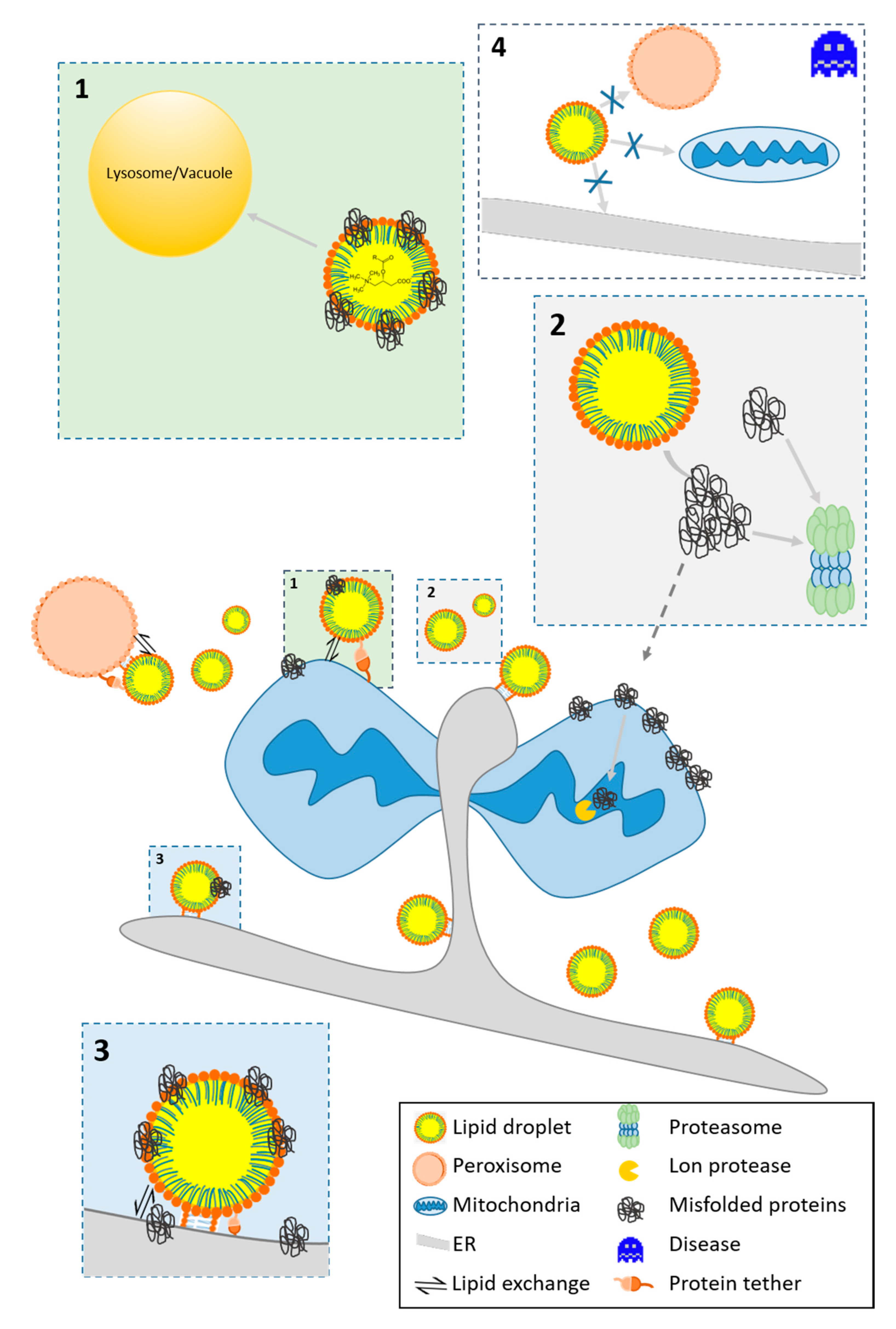
CEs activate other PKC isoforms (PKCζ) which interfere with AKT translocation and signaling downstream of the insulin receptor. Furthermore, CEs activate protein phosphatase 2A, which dephosphorylates and inactivates AKT signaling [69]. CEs negatively affect the permeability of the OMM and form channels for the release of pro-apoptotic proteins in the mitochondrial intermembrane space. The pro-apoptotic protein Bax stabilizes the channel [70]. Different Cer species within a cell have specific and diverse effects on the translocation of Bax to the OMM. There is evidence that excess CEs need to be stored in LDs. The conversion of CEs to acylceramides is catalyzed by DGAT2 [71], and thus storage in LDs may have a protective function (see Figure 1). Failure in the production of omega-O-acylceramides occurs in neutral lipid storage diseases (NLSD).
Besides CEs, PUFAs such as intracellular arachidonic acid (AA) can induce apoptosis via activation of caspase 3. To avoid cellular toxicity of non-esterified AA, cells can catabolize it by cyclooxygenase-2 (COX-2), LOX and FA-CoA ligase 4 (FACL4). The upregulation of pathways that activate these enzymes blocks apoptosis. Importantly, AA can be stored in LDs during inflammation with a concomitant reduction of apoptotic rates [55]. In addition, important mediators can be produced non-enzymatically by the interaction of lipids with ROS [72,73]. Bioactive lipids serve as precursors of specialized oxidative products during cell or tissue oxidative stress. These highly bioactive mediators specifically include prostaglandin-derived isoprostanes and arachidonic acid-derived isoketals. Rising evidence suggest that ROS-generated isoprostanes act as pro- or anti-inflammatory mediators in many diseases [72,74]. In detail isoprostanes extensively contribute to inflammatory processes in atherosclerotic cardiovascular diseases [75,76], neurodegenerative diseases [77,78] and NAFLD [79]. For further details on the role of LDs in inflammation see Section 5.5.
Lipid peroxidation is closely linked to apoptosis and lipotoxicity and PUFAs are highly susceptible for the process. Finally, hydroxyl radicals arise, which cause lipid peroxidation of FAs in lipoproteins and membranes. LDs can prevent lipid peroxidation at an early stage and store lipid peroxides to prevent further damage [80]. Lipid peroxidation contributes to the development of many diseases including NASH. Recent findings in yeast additionally underline the importance of LDs for detoxification of damaged as well as pro-apoptotic acting lipids [81]. Not only proteins and lipids but also hydrophobic xenobiotics can be safely stored in LDs [65,82].

3.2. Protein Homeostasis

3.2.1. The Fate of Misfolded Proteins

Protein homeostasis is of utmost importance for cellular integrity. During the folding process, proteins acquire a structure that is associated with minimal free energy and is thermodynamically stable. Sometimes proteins fail to fold correctly and gain a false conformation [83,84], and mutations as well as stressful situations increase misfolding. ROS originate from cellular metabolism or extracellular sources and can react with amino acids including histidine, leucine, methionine, and cysteine. These oxidative modifications either alter enzyme activity or result in protein unfolding. As another consequence, the formation of stable carbonyls can induce conformational changes in the polypeptide chain (for a detailed review see [85]. Cells use highly effective mechanisms to remove potentially dangerous proteins. An active and cytoskeleton dependent process translocates such proteins to distinct cellular aggregates. These aggregates are best described in yeast cells and include the JUxtaNuclear Quality Control Compartment (JUNQ; nuclear localization), the IntraNuclear Quality Control Compartment (INQ; nuclear localization), the Insoluble PrOtein Deposit (IPOD; phagophore assembly site localization close to the vacuole), as well as cytosolic Q-bodies (CytoQ) [86,87,88,89]. The fate of misfolded and damaged proteins at the aggregation sites differs. The presence of the molecular chaperone HSP104 at protein aggregates promotes refolding attempts [90,91]. The IPOD is mainly composed of misfolded proteins with low solubility and appears in stressed as well as unstressed cells. Its close proximity to the vacuole indicates the removal via autophagy [92]. JUNQ, INQ and CytoQ aggregates indicate cellular stress and harbour misfolded and aggregated proteins with some higher solubility [89]. The sorting signal for JUNQ is ubiquitination [87], and 26S proteasomes degrade terminally misfolded proteins [86,91]. A wide variety of diseases evolve due to the presence of such aggregates (e.g., amyloidosis and prion diseases). Under conditions of severe proteotoxic stress, LDs and mitochondria assist in removing of such aggregates as we will show in the following section (see Figure 1). Growing evidence also suggests a pivotal role of aggregates and LDs in neurodegenerative disorders [77]. Further details will be presented in Section 5.1.

3.2.2. LDs As A Protein Docking Site

When the proteasomal system is overwhelmed under severe stress conditions LDs have a supportive role. LDs and protein aggregates come into close proximity upon heat stress. Cells devoid of LDs and defective in sterol biosynthesis cannot effectively remove protein aggregates. LDs may supply the aggregates with sterol derivatives, which act as solvent and are crucial for the clearance of misfolded proteins [93].
Mitochondria also contribute to the removal of misfolded proteins. Deletion of the cytosolic chaperone Hsp70 results in the relocalization of misfolded proteins from the cytosol to the matrix of mitochondria. Degradation of misfolded proteins ensues at the matrix resident LON protease instead of the proteasome. This degradative process is known as MAGIC (mitochondria as guardian in cytosol) and may be supported by LDs. Mmi1 is a marker protein of MAGIC degradation and shuttles from mitochondria to LDs upon stress induction. In addition, certain pro-apoptotic proteins may shuttle from the cytosol to mitochondria. They have the capacity to permeabilize the OMM and can be removed by LDs after stress. Subsequently, these protein containing LDs reach the vacuole for degradation by lipophagy [9].
LDs can also remove misfolded proteins from the ER. Misfolded proteins in the lumen of the ER are ubiquitinated by ER resident enzymes, translocated back to the cytosol and are then degraded by the 26S proteasome. This pathway is known as ERAD (ER-associated (protein) degradation) [94]. The antibiotic tunicamycin is an agent that blocks the first step in the glycosylation of proteins and leads to unfolded proteins in the ER lumen [95]. ER stress initiation by tunicamycin also dramatically stimulates the number and size of LDs in the cell [96]. LDs specifically form at sites of stress induced ER aggregates that are marked by the chaperone Hsp104, and these LDs harbour polyubiquitinated proteins [97]. LDs may therefore assist the 26S proteasome during stress in the removal of damaged, unfolded and potentially dangerous proteins. Accordingly, a specific blockage of the proteasome by the inhibitor MG132 increased intracellular LD numbers [98]. Accumulating LDs are subsequently removed by the cell through microlipophagy [97]. The removal of unfolded proteins from the ER by LDs closely resembles the cytosolic and mitochondrial pathways. It is of interest that several components of ERAD, such as UBXD2, VCP, AUP1 and SELENOK are present in the human LD proteome, indicating a general mechanism of protein homeostasis [99]. More than 60 diseases are based upon defects in ERAD [100] and a considerable number of them show either an accumulation of LDs, LD malformations or altered LD distribution (e.g., [101,102,103,104].
Besides unfolded proteins, viral proteins occur at LDs and viruses can hijack LDs for their assembly. In addition, certain cellular proteins with regular conformation use LDs as a platform for storage and redistribution. Histones H2A, H2B and H2Av connect to LDs of oocytes in Drosophila melanogaster. This is of special importance for the growing embryo, as rapidly synthesized DNA needs to be packaged by histones. In this special scenario, LDs close at the nuclei act as a deposit for histones [105]. LDs can also influence the expression of certain genes. The transcription factor NFAT5 (Nuclear factor of activated T-cells 5) resides in the cytosol and translocates to the nucleus upon application of hyperosmotic stress. Upon binding to response elements on the DNA, an adaptive cellular response, which includes the production of osmolytes and expression of Hsp70 is initiated [106]. In adipocytes, LDs can attach to the cell death inducing DFFA like effector c (CIDEC) that among other functions promotes the growth of LDs. Yeast-2-hybrid analysis revealed a physical interaction between CIDEC and NFAT5. In fact, overexpression of CIDEC sequesters NFAT5 to LDs and thus prevents a stress induced translocation of NFAT5 to the nucleus and a proper stress response [107].
Expression levels of PLIN5 are high in tissues with augmented oxidative ATP production in mitochondria such as skeletal muscle, heart and liver. PLIN5 rich LDs physically interact with mitochondria [108], but PLIN5 was also found in the cytosol, at the ER and the nucleus. Starvation or catecholamine treatment induces nuclear localization and phosphorylation of PLIN5. In the nucleus, PLIN5 interacts with the transcriptional coactivator PGC-1α (peroxisome proliferator-activated receptor gamma coactivator 1-α) and the NAD+-dependent deacetylase SIRT1 (sirtuin 1). Stimulated activity of SIRT1 induces the deacetylation and activation of PGC-1α [109]. PCG1-1 is deeply involved in the biogenesis of mitochondria and respiration [110]. Several tissue specific isoforms of PGC1alpha may increase the complexity of the interaction, and some brain isoforms are associated with neurodegenerative diseases [111,112,113]. Therefore, PLIN5 can influence mitochondrial activity in several ways, either by mediating an interaction with LDs or by altering gene expression. Furthermore, LDs indirectly modulate the general stress response as well as inflammation via the PLIN5-SIRT1 axis [114].

3.2.3. Protein Motifs for LD Localization

Some proteins cannot reach LDs, as certain requirements are prerequisite for LD localization. [9]. LD proteins can enter either from the ER or from the cytosol. Accordingly, two distinct classes of LD resident proteins, namely Class I and II proteins can be distinguished [115]. Class I proteins are synthesized and inserted into the membrane at the ER. These proteins transfer via the lipid bridge that forms between the ER and the LD [64], and typical examples are GPAT4 and DGAT2. No classical consensus sequence or motif for LD localization has been reported so far, but some features for membrane insertion and shuttling start to emerge (Figure 2). Most of these proteins contain a hydrophobic, v-shaped hairpin sequence (two α-helical domains with a central proline, or “proline knot”). In many cases, positively charged amino acids with (a) hydrophobic tryptophan(s) in the middle flank the helices [116,117,118]. Most cytosolic class II proteins, such as PLINs target LDs with amphipathic helices or short hydrophobic sequences [115]. Due to their monolayer membrane, the surface of LDs is prone to phospholipid packaging defects. Proteins with amphipathic helices, such as CTP: phosphocholine cytidylyltransferase [118] are attracted by these imperfections and specifically attach to the surface of LDs [119]. All PLINs (PLIN1-PLIN5) contain several 11-mer repeats, which form amphipathic helices that have the capacity to bind LDs. Point mutations in these regions inhibit the localization to LDs [120,121].
Potentially harmful proteins, such as the mitochondrial anti-apoptotic TCTP, as well as pro-apoptotic BAX can relocalize to LDs A v-shaped hairpin structure in the BAX protein is required for the transfer process (Figure 2C). A GFP tagged version of the protein domain is sufficient to induce both, mitochondrial and LD localizationThe v-shaped hairpin in BAX closely resembles a 56 amino acid long hairpin domain in GPAT4, termed LiveDrop, which is required for LD localization (Figure 2C). The LiveDrop domain contains characteristic hydrophobic helices (Figure 2D) that contain a central tryptophan and positively charged amino acids at the hinges (Figure 2C). In the loop region, a proline residue causes a kink. Most elements are present in the BAX hairpin structure (Figure 2A,B). Due to its capacity to modulate apoptosis by redirecting proteins from the OMM, LDs have the capability to modulate diseases associated with alterations in apoptosis (e.g., cancer; several neurological disorders; several cardiovascular and autoimmune diseases) [122].

4. The Role of LDs in Cellular Stress and Aging

4.1. The Function of LDs in Modulating Stress

Different organelles have overlapping and diverse responses to cellular stressors. Stress response of mitochondria includes a widespread reconstruction of the mitochondrial network. In the first step, mitochondria execute hyperfusion to supply the cell with increased amounts of ATP for energy consuming stress response. Upon prolonged stress, the tubular network completely fragments. The fission process is a prerequisite for either OMM perturbation or the removal of damaged mitochondria via autophagy [123,124].
LDs stress responses differ to those of mitochondria and depend on LD size. In general two major subtypes of LDs can be distinguished as ER-associated LDs and cytosolic-LDs [125]. Large, ER-associated LDs are in constant contact with the ER and receive persistent lipid supply via the lipidic bridge. After detachment from the ER and disruption of the lipidic bridge, LDs decrease in size and transform into cytosolic LDs [126]. Furthermore, LDs do not equally distribute in healthy cells, but occur in clusters that are in close proximity to the nucleus [127]. It is possible that such LDs are ER-associated LDs that constantly bud off and are fed by the ER. Starvational stress induces LD redistribution and increased motion of the organelles. LDs move from the center of the cell to the periphery, where they get in close contact with mitochondria. The process is downstream of AMPK (AMP-activated protein kinase) activation that promotes LD dispersion on detyrosinated microtubules to increase mitochondrial FA oxidation [127]. Besides nutritional stimuli, physical exercise, stress and mitochondrial fragmentation induce the interaction between mitochondria and LDs [9,108,128]. The inter-organelle communication is preserved from yeast cells to mammals [37]. Some cell types (e.g., adipocytes) show profound interaction of mitochondria and LDs, and therefore these mitochondria are called “peridroplet mitochondria” [108]. A hallmark of stress is an increase in LD formation, as observed in a broad variety of cell types [9,80,96,129]. Several mechanisms cause the increase in LD numbers and inter organelle communication:
(a)
The high energy demand during stress response causes fueling of FAs from LD to mitochondria for β-oxidation [130]. Reverse transfer of lipids occurs, and lipid relocation from mitochondria to LDs may protect from lipotoxic settings [131] and aberrant lipid signaling (see Section 3.1).
(b)
LDs are involved in protein homeostasis and assist in the removal of damaged and misfolded proteins from mitochondria (see Section 3.2)
(c)
LDs modulate the apoptotic program by redirecting acylcarnitines from mitochondria (see Section 3.1). Similarly, mitochondrial proteins involved in the apoptotic program translocate [9,81]. In several studies, the pro-apoptotic protein BAX was found as a bona fide member of the LD proteome [9,99,132].
(d)
LDs fulfill antioxidative functions, reduce ROS levels and prevent the peroxidation of PUFAs. The mechanism involves the removal of these lipids from membranes to the core LDs, where they are less accessible to ROS [129]. Consequently, a stimulated increase in LD number can augment cell survival upon the onset of stressors [9].
Finally, cellular stress induces a changed lipid as well as proteome composition in LDs. In general, levels of proteins and lipids at LDs increase, indicative for their important role as a lipid as well as protein buffer [81]. Stress response induced alterations of LDs resembles changes that occur during cellular aging [81]. A prominent aging theory is the “mitochondrial free theory of aging” (MFRTA) proposed by Denham Harman in 1972 [133]. The electron transport chain (ETC) at the inner mitochondrial membrane is a prerequisite for the formation of the proton motive force and ATP production. The premature leakage of electrons at complex I and III causes the formation of superoxide [134]. MFRTA claims that ROS levels dramatically increase with aging and react with organic components of the cell, including DNA, proteins and lipids. In line with this, stress as well as aging induces the accumulation of LDs in yeast and mammalian cells [135,136,137]. There is growing evidence that LDs play a role in the regulation of longevity [138]. Results from the yeast S. cerevisiae strongly indicate the involvement of LDs in cellular aging. In yeast cells, two forms of aging processes, namely chronological and mother-cell specific (replicative) aging occurs [139,140]. Chronological aging that resembles aging in human differentiated cells reduces the viability in stationary phase when all nutrients are exhausted. Replicative aging that mimics the Hayflick limit in stem cells describes the mechanisms of asymmetric budding (the mother cells age and the daughter cells rejuvenate themselves). The deletion of either the diacylglycerol acyltransferase DGA1 or the Acyl-CoA:sterol acyltransferase ARE1 and ARE2 shortens the chronological lifespan. Both interventions reduce TAG as well as sterol ester levels and the number of LDs. Interestingly, the deletion of the TAG lipases TGL1-5 prolongs the chronological lifespan [141].

4.2. Emergence and Role of LDs During Aging

Since 1935 caloric restriction is a known and effective intervention to extend lifespan in rodents [142]. This finding holds true for many model organisms [143,144,145]. Caloric restriction initiates a metabolic change towards fatty-acid synthesis. Furthermore, caloric restriction enlarges LDs as well as influences the expression of genes involved in lipid metabolism, which is crucial for the life prolonging effect [146,147].
LDs may also affect longevity in mammals, as they can activate the sirtuin SIRT1. In many studies and in many model organisms, activation of this enzyme leads to a prolonged life, improved organ functions, a higher fitness and increased stress and disease resistance [135].
Several studies report a positive effect of olive oil on health and lifespan. This could partly explain the “French paradox”, the paradoxical epidemiological finding of a low incidence of coronary heart disease despite a diet rich in saturated FAs [148]. The most prevalent FA in olive oil is the MUFA 18:1 oleate (up to approximately 80%). Feeding of rats with olive oil increases their lifespan to 157% [149], and feeding mice with olive oil decreases body weight as well as fat mass [56]. Oleate generally stimulates LD formation [150], but hepatic LDs adapt their size from large to small, but PLIN5-rich LDs [56]. PLIN5 translocates into the nucleus upon binding of MUFAs, interacts with SIRT1 and PGC-1α, and induces the expression of genes such as UCP1, CPT1α, and complex I-IV of the respiratory chain. Consequently, oxygen consumption as well as heat production is increased [56]. Growing evidence suggests that altered lipid signaling could promote replicative senescence in yeast. It was shown that the amount of 19 polyunsaturated TAGs significantly changes during the aging process [151,152]. On the other hand, increased LD number is not always beneficial as some diseases and pathologies are associated with higher LD abundance, such as NLSD and atherosclerosis (for a detailed review see [153]). Therefore, balanced LD turnover is of utmost importance and strictly depends on the organism, tissue, cell types and growth phase of the cell. In the following, we will summarize selected diseases and pathologies that originate from imbalances in LD metabolism.

5. The Role of LDs in Disease

5.1. LDs and Neurodegenerative Diseases

Neurodegenerative diseases are associated with toxic protein accumulation, mitochondrial defects and elevated ROS levels that result in the decay of neurons (see Figure 1). Interestingly, these stressors also cause the accumulation of LD in glial cells [154]. One prevalent neurodegenerative disease with a close link to LDs is the neurodegenerative movement disorder Parkinson disease (PD). Defects in the proteins that connect the OMM with LDs, such as mitochondrial MFN2 and the LD resident PLIN1 have a role in the development of PD [33].
PD is characterized by the loss of dopaminergic neurons in certain areas of the substantia nigra. In PD, dysregulated accumulation of α-synuclein (aSyn) in the cytoplasm of distinct neurons occurs [155]. The accumulation is a multistep process and after the oligomerization of aSyn into small protofibrils, insoluble large aSyn fibrils are formed [156]. The N-terminal amphipathic region of aSyn enables binding to lipid membranes. This domain contains multiple lysine residues that contribute to electrostatic interactions with negatively charged membrane surfaces [157]. Strikingly, the LD monolayer attracts aSyn, where it preferentially accumulates [158]. PD patients additionally upregulate key enzymes of phospholipid metabolism in the substantia nigra, including phospholipase A2, phosphocholine cytidyltransferase and phosphoethanolamine cytidyltransferase [159]. Similarly, imbalances in aSyn homeostasis have severe effects on lipid metabolism. Toxic aSyn overexpression increases levels of DAGs, TAGs as well as oleate [160]. LD upregulation in PD may provide a “sponge/sink” for toxic aSyn proteins. Concomitant with that the role of LDs is ambiguous, as increased levels of LDs and Plin4 cause reduced mitophagy in mouse neurons and promote PD progression [161]. Reduction of LD formation is crucial for the toxicity of aSyn in yeast and induces apoptosis [160].
LDs are also associated with the development of additional neurological disorders such as Huntington’s disease (HD) and Alzheimer disease (AD). LDs are detectable in brain sections of HD models [162]. Changes in brain cholesterol homeostasis in HD increase the accumulation of cholesterol in striatal neurons [163], resulting in neuronal dysfunction [164]. In a HD model in Drosophila melanogaster, changed cellular lipid metabolism associates with altered LD size [165]. HD development largely depends on a CAG repeat expansion in the exon 1 of huntingtin gene, encoding polyglutamine repeats in the protein. The mutated huntingtin protein was associated with increased LD numbers in mouse models as well as cells derived from HD patients. Human striatal tissue of HD patients also shows increased Oil Red O staining intensities [162].
AD is the most common neurodegenerative disease and associated with microglia mediated inflammation and alterations of lipid metabolism. Already Alois Alzheimer described “adipose saccules” in many glial cells [166], but the discovery was ignored for almost a century. LD formation increases in murine and human AD samples and correlates with dysfunctional neurogenesis [167]. The connection between LDs and AD became also evident by findings on protein-O-linked hexosaminidase C (also known as N-Acetyl-β-D-glucosaminidase). Hexosaminidase C accumulates on the surface of nascent, PLIN2 containing LDs. The enzyme acts as a regulator of LD formation and pharmacological inhibition of hexosaminidase C slows neurodegeneration in various AD mouse models [168]. Imbalances in lipid metabolism and LD function play a major role in the development of AD [169]. Some data strongly suggest that LD functions may have an important role in development of AD, although the precise roles are yet unclear.
The accumulation of cholesterol and CEs in neurons is associated with neurodegeneration [170]. Furthermore, altered sphingolipid metabolism and their accumulation in the brain links to AD development [171,172]. In addition to neurons, glial cells and alterations in their lipid metabolic functions contribute to the development of neurodegeneration. The APOE E4 allele is a strong genetic risk factor for late onset of AD [173], while the APOE E2 allele lowers risk [174] in comparison to the common APOE E3 allele. APOE is a component of low-density lipoprotein (LDL) particles and astrocytes with E4 expression have small LDs compared to E3 expressing cells [77]. Astrocytes expressing E4 have increased PLIN2 levels and less efficient uptake and oxidation of FAs. Other glial cells contribute to the development of AD, and the activation of microglia and associated inflammatory processes are commonly present. Microglia also are becoming gradually activated and apparently dysfunctional during the ageing process. The activation of microglia includes the accumulation of LDs, in a process similar to the formation of foam cells in atherosclerosis. We recently reported the build-up of LDs in microglia in aging murine and human brains and denominated theses sells as LDAM (LD accumulating microglia) [175]. Mouse hippocampus microglial LDs contained PLIN3, whereas PLIN2 was present in microglia in human hippocampal brain sections and more abundant in aged individuals. The accumulation of LDs causes less efficient phagocytosis, higher levels of ROS and increased secretion of pro-inflammatory cytokines. LDAM have a unique transcriptional profile that supports inflammation [175].
Another disease featuring neurological decline is chorea acanthocytosis. This hereditary disease is exceptionally rare and caused by a mutation in the ER-LD connective protein VPS13A gene (see Section 2.2). Chorea acanthocytosis is an autosomal recessive disease, histologically characterized by acanthocytes, a pathological form of erythrocytes and shares similarities with HD [176].
Mutations in ER-LD tether protein SNX14 are associated with autosomal recessive Spinocerebellar Ataxia 20. The neurodevelopmental disorder belongs to a large heterogeneous group of spinocerebellar ataxias. Characterized by an early onset during infancy, the disease causes cerebellar atrophy and severely impaired psychomotor development [31].
Failure in the transfer of FAs from LDs to peroxisomes for beta-oxidation causes neuronal diseases such as spastic paraplegia and adrenoleukodystrophy. Mutations in the SPAST gene cause spastic paraplegia 4, an autosomal dominant and most prominent form of hereditary spastic paraplegias. The resulting protein product encodes an AAA ATPase involved in the severing of microtubules. Hereditary spastic paraplegias are generally defined by progressive spasticity and weakness in lower extremities [177]. Mutations in ABCD1, a member of the ABC transporter family cause the accumulation of very long chain FAs in the cell in adrenoleukodystrophy [178]. ABCD1 resides in the peroxisomal membrane and upon dimerization facilitates trafficking of very long chain FAs into peroxisomes. Failure of this transport severely damages the myelin sheath in neurons and may ultimately cause seizures, persistent vegetative state and death [31]. These genetic data strongly suggest that LD functions may have a causative role in development of neurodegenerative diseases.

5.2. Neutral Lipid Storage Disease (NLSD) and Rare Lipid Storage Diseases

NLSDs are rare autosomal recessive genetic disorders without specific therapies, and are associated with the accumulation of LDs in different cell types. Mutations occur either in adipose ATGL (encoded by PNPLA2) [179], or in α,β-hydrolase domain 5 (ABHD5, also known as CGI-58) [180], two genes with key functions in LD metabolism. NSLD phenotypes are characterized by the manifestation of either myopathy or ichtyosis [181]. NLSDs with myopathy develop progressive myopathy, cardiomyopathy, hepatomegaly, chronic pancreatitis and diabetes [182,183,184]. Large genetic heterogeneity in ATGL mutations causes wide variations in clinical phenotypes. The mutations affect either the binding to LDs or the catalytic activity of ATGL. Mutations in ABHD5 cause NLSDs with ichthyosis and additionally affect other organs, including liver, skeletal muscle, CNS and eyes [185]. ATGL activation is promoted by ABHD5, although ATGL mediated hydrolysis of TAG occurs in the absence of ABHD5 at low efficiency [186]. Lipolysis is a highly regulated process and ATGL catalyzes the rate-limiting step, followed by hormone sensitive lipase and monoacylglycerol lipase. FAs can be a substrate for mitochondrial beta-oxidation, synthesis of lipid mediators such as leukotrienes or membrane lipid synthesis. ATGL interacts with various cell-type enriched inhibitors or activators, frequently phosphorylated upon stimuli. In the context of NLSDs, adipocyte-enriched PLIN1 sequesters ABHD5 on the LD surface and inhibits lipolysis. Protein kinase A mediated phosphorylation of PLIN1 causes the release of ABHD5 and subsequent interaction with and activation of ATGL. In skeletal and cardiac muscle, the phosphorylation of PLIN5 similarly promotes lipolysis [1]. In NLSDs, hampered TAG degradation reduces the availability of FAs and causes the cytoplasmic accumulation of TAGs in many tissues. Consequently, the staining of neutral lipids in peripheral blood granulocytes confirms the clinical diagnosis [187,188]. In NLSDs with skeletal muscle myopathy, LD number increases in mainly oxidative muscle fibers between myofibrils and probably cause lipotoxic effects [189,190]. The accumulation of LDs in the heart mainly occurs in cardiomyocytes, but also affects endothelial cells, smooth muscle cells and intima media foam cells [191,192].
In NLSD forms with ichthyosis, the liver is frequently affected and manifestations include hepatomegaly and steatosis in more than 80% of cases. Liver biopsies reveal the accumulation of large LDs mainly in hepatocytes [193,194]. A common finding in NLSD forms with ichthyosis is also the accumulation of LDs in skin fibroblasts and keratinocytes [195]. ABHD5 also activates PNPLA1, which catalyzes the final step in omega-O-acylceramide biosynthesis [196]. Usually, Omega-O-acylceramides are essential for skin permeability and its barrier function [197]. Mutations in ABHD5 may therefore specifically cause the ichthyosis by the reduced availability of these lipids [198].
In contrast to NLSD, lipodystrophy is characterized by the inability to store TAGs in adipose tissue. Several genes participating in LD biology influence lipodystrophy. Some regulate de novo TAG synthesis like 1-acylglycerol-3-phosphate O-acyltransferase 2 (AGPAT2) and lipin1 (LPIN1). Others include the seipin BSCL2, which is a mayor-contributing factor in LD biogenesis [135,199].
Another disease manifestation associated with the loss of adipose tissue and therefore LDs is cachexia. Cachexia is a common comorbidity of cancer patients and manifests as an imbalance between catabolic and anabolic processes. Particularly changes in lipid metabolism cause rapid loss of adipose tissue and skeletal muscle [135,200]. The exact molecular connections between LDs and cachexia are yet unknown. Atgl knockout protects mice from skeletal muscle loss, and therefore increased lipolysis seems important for the pathophysiology of cachexia [201].

5.3. Atherosclerosis

Atherosclerosis is characterized by an accumulation of lipids and fibrotic material in the wall of large arteries. The accumulation of macrophages and their transition into LD-rich “foam cells” is a central component in development of atherosclerotic plaques. In addition to macrophages, smooth muscle cells can also convert into foam cells with large LDs [202,203]. Recent evidence suggests that up to 70% of foam cells in mouse atherosclerotic lesions originate from smooth muscle cells [204]. Such smooth muscle cell derived foam cells also significantly contribute to and are present in human lesions [205]. The formation of plaques in arterial walls occurs during all stages of atherosclerotic development and causes major cardiovascular diseases and stroke. Cytosolic LDs accumulate in foam cells and are rich in CEs, which originate from modified LDL particles. In addition, human atherosclerotic plaques also contain oxidized sterols and non-sterol lipid species [206]. Oxidized LDL stimulated a strong induction of isoprostane generation in human macrophages and LDs in foam cells may provide a storage site to prevent toxic consequences [207]. Macrophages take up oxidized LDL via scavenger receptor mediated endocytosis and other mechanisms [208]. Lysosomal acid lipase (LAL) hydrolytically cleaves LDL derived CEs into free cholesterol and FFAs. Free cholesterol may efflux from cells via ATP binding cassette (ABC) transporters or be re-esterified to FAs in the ER prior to its storage in LDs. Neutral cholesterol ester hydrolases can associate with LDs and mobilize stored CEs (for further details on CEs see Section 3.1). Hydrolysis of these CEs probably depends on several hydrolases, including LD-associated hydrolase that is highly abundant in human macrophages and atherosclerotic lesions [209]. Alternatively, LDs degradation follows autophagy and fusion with lysosomes prior to CE hydrolysis [210]. Free cholesterol efflux in macrophages occurs via the ABC transporters ABCA1, ABCG1 and ABCG4 [211], and transfer to high density lipoprotein (HDL) is an integral part of reverse cholesterol transport (RCT). RCT seems atheroprotective, and therefore increased hydrolysis of cholesterol-esters in LDs may be a therapeutic target. Foam cells derived from smooth muscle cells decrease their ABCA1 expression in comparison to macrophage derived foam cells, which probably contributes to less efficient cholesterol efflux [205]. Lipid acceptors, such as lipid poor apoA-I cannot only accept cholesterol, but also accept other lipid species from LDs of foam cell [212]. The expression of PLIN1 in macrophages remains controversial [213], but PLIN2 and PLIN3 are abundant in foam cells and atherosclerotic plaques [214,215]. PLIN2 is highly expressed in macrophages, and its expression increases upon lipid loading [216]. Mice with deleted PLIN2 exhibit decreased lesion formation in the ApoE knockout background [215]. PLIN2 deficiency does not increase cholesterol-associated toxicities in macrophages [217]. Furthermore, PLIN4 and PLIN5 expression is upregulated in THP-1 macrophages upon treatment with oxidatively damaged LDL [216]. The same study identifies LD proteins CIDEA, CIDEB and CIDEC (cell death activator proteins) in macrophages, although their contribution to atherosclerosis remains unclear. Foam cells also generate pro-inflammatory signals within the arterial wall. PLIN2 overexpression increases the secretion of chemokines and cytokines such as IL-6, TNF-alpha and MCP-1 [218]. The role of PLIN1 in the development of atherosclerosis remains unclear with conflicting evidence [213,219]. Overall, LDs have a causative role in the development of atherosclerosis.

5.4. Obesity and Non-Alcoholic Fatty Liver Disease

Obesity often associates with non-alcoholic fatty liver disease (NAFLD), and the chronic liver condition affects approximately 30% of the population in Western countries [220]. NAFLD comprises the accumulation of LDs in liver tissue and covers a spectrum from non-alcoholic fatty liver (NAFL) and non-alcoholic steatohepatitis (NASH) to fibrosis, and finally cirrhosis. Cirrhosis is an important risk factor for hepatocellular carcinoma (HCC) [221], one of the most frequent malignancies on a global scale. HCC has a poor prognosis and is the third most frequent cause of cancer deaths worldwide [222]. In NASH, significant inflammation and some scarring complements NAFL. Obesity and associated metabolic diseases frequently share insulin resistance as a common basis. NAFLD is thus widely considered as the hepatic manifestation of the metabolic syndrome and insulin resistance [223]. Excessive fat storage in LDs of hepatocytes and non-parenchymal liver cells promotes lipotoxicity and cellular damage. Attracted and activated Kupffer cells release ROS and other signals, which stimulate additional inflammatory cells. During NASH progression, lipid peroxidation and ROS augment liver steatosis by enhancing LD formation [224]. Kupffer cells activate stellate cells (also known as Ito cells) from a quiescent, vitamin A storing phenotype [225] into proliferating, fibrogenic and contractile myofibroblasts [226]. Strong evidence suggests that the accumulation of LDs and their metabolism contribute to NAFLD. Interestingly, LDs of stellate cells [227], Kupffer cells and hepatocytes contribute to NAFLD pathophysiology and sequelae [228]. A hallmark of NAFLD is the development of particularly large LDs in hepatocytes. The formation of giant-sized LDs arises from either coalescence or from facilitated diffusion of lipids from one LD to another [229]. Distinct proteins accumulate during NAFLD on the surface of LDs. One example is PNPLA3 (also known as adiponutrin), which encodes a TAG lipase homologous to adipose triglyceride lipase. The physiological roles of PNPLA3 are unclear, but a gene polymorphism is the predominant genetic risk factor for NAFL and its progression [230]. Additionally 17β-hydroxysteroid dehydrogenase 13 contributes to sex steroid synthesis, binds to LDs and promotes the development of NAFLD [231,232]. Other LD-specific proteins, such as CIDE proteins [233,234] and several PLINs also accumulate during NAFLD [228,235]. Members of the CIDE protein family promote larger LDs [236,237]. PLIN1 is present on LDs during the development of NAFL [238,239]. PLIN2 is strongly upregulated in NAFLD [239] and inhibits FA oxidation [240]. PLIN3 is upregulated in mice on high fat diet and reduction of PLIN3 levels improves NAFLD [241]. In addition, PLIN5 expression strongly increases in mouse NAFL [242]. High levels of PLIN5 can specifically recruit and attach LDs to mitochondria [108], as already discussed above. Emerging evidence suggests that mitochondria-LD interactions play critical roles in NAFLD pathogenesis and progression [243]. Mitochondria bound to LDs have distinct fusion-fission properties, enhanced bioenergetics capacities and support the expansion of LDs [244]. The direct interaction of mitochondria with LDs provides lipids for energy production via mitochondrial β-oxidation. The contact sites depend on PLIN2 and PLIN5, which additionally modify the efficiency of FA oxidation [240,245]. With the onset of hepatic steatosis, increases in mitochondrial FA oxidation, tricarboxylic acid cycle activity and oxidative phosphorylation ensue [246]. Increased mitochondrial activity initially seems to protect hepatocytes from lipotoxic effects of FFAs [246]. On the other hand, higher metabolic fluxes in mitochondria also increase ROS generation. ROS mediated damages intensify hepatic lipid accumulation, trigger inflammation and fibrosis, and cause cell death. LDs and their direct interaction with mitochondria also regulate apoptosis and cell death in NAFL. Hepatocellular death is a hallmark of NASH and reported for in vitro models and patients [247]. Higher apoptosis rates at earlier stages of NAFLD ensue prior to a reduced onset of apoptosis in HCC. Additional types of programmed cell death, such as pyroptosis partially overlap with classical apoptosis in NAFLD [248]. Increased levels of pro-apoptotic Bcl2 proteins, such as BAX and BAK appear in NASH [249]. On the other hand, expression levels of anti-apoptotic proteins such as Bcl-XL are reduced [247]. With the progression to HCC, rates of apoptosis strongly decrease and carcinoma cells become insensitive to the induction of apoptosis. The over-expression of anti-apoptotic Bcl-XL, MCL-1, XIAP and survivin seems to block the induction of apoptosis [250]. Specific proteins transfer from the ER to LDs through membrane bridges [64]. As such, LDs can specifically remove oxidatively damaged and aggregated proteins from the ER. Misfolded proteins also accumulate in inclusion bodies and LDs are essential for their efficient clearance [93]. In a recent work, we described a mechanism, by which damaged and apoptosis-related proteins relocate from the OMM to LDs in yeast and human hepatoma cells. The process probably promotes mitochondrial rejuvenation [9]. Others confirm the finding that BAX can also localize to LDs [99,132]. Additionally, mitochondria can transfer lipids to LDs to avoid lipotoxicity and aberrant lipid signaling [131]. An altered LD lipidome may also support the insertion of specific proteins. The size and curvature of LDs seems important for interactions with other organelles, as giant sized LDs typically develop in NASH [229], but LD size decreases during cirrhosis and progression to HCC [251]. Small LDs preferentially contain PLIN5, which facilitates the interaction with mitochondria [120]. Toxic lipids and proteins then shift via LDs to lysosomes, where degradation via lipophagy occurs. LDs in various hepatic cell types are crucial components in NAFLD pathophysiology.

5.5. LDs and Inflammation

LDs regulate the availability of FAs and derivatives thereof, which are essential for physiological signaling pathways. LD turnover increases during nutritional, metabolic and oxidative stress [80,81,252]. Inflammation and infection also induce LD formation in several exposed cell types of the immune system during their activation [175,253,254]. FFAs originate from the neutral core or the phospholipid monolayer and can bind to a variety of cell surface (mainly GPCRs) or intracellular receptors (nuclear receptors) [255]. LD derived PUFAs are precursors for lipid mediators such as eicosanoids, which modulate immune and inflammatory responses [256] and their resolution [257]. LD release eicosanoid precursors from membrane glycerophospholipids by phospholipase A2, or from the core by ATGL and HSL mediated lipolysis by [258,259,260]. In addition, cholesterol esters from LDs can contribute precursors upon the action of lysosomal acid lipase [261]. Furthermore, LDs may act as a sink for AA and reduce prolonged and extensive inflammatory responses in immune cells [262,263]. LDs can remove bioactive lipid mediators by inactivation to inert storage lipids, and thereby prevent lipotoxicity. Storage of saturated FAs in neutral TAGs reduces their availability for their inclusion and formation into CEs, thereby reducing inflammatory responses [48]. Similar alterations in neutral TAG levels occur in yeast during aging and apoptosis and provide a potential mechanism to buffer CE-induced stress [81].
Free cholesterol can form cytotoxic cholesterol crystals at high concentrations, which then generate inflammatory responses [264]. The formation and storage of cholesterol esters in LDs protects from cytotoxicity and inflammation [265]. In contrast, during atherosclerotic progression, macrophage LDs accumulate excess cholesterol esters and transform into foam cells, causing inflammation at the arterial walls. Signaling proteins that mediate inflammatory responses are present on LDs of leukocytes [253,266]. Cyclooxygenase-2 is present on LDs and can initiate the production of inflammatory eicosanoid mediators such as prostaglandins and leukotrienes [267,268]. LDs are also a reservoir for lipids involved in intracellular signaling cascades. Members of the PPAR nuclear receptor family are lipid ligand activated transcription factors and play a crucial role in lipotoxicity (see Section 3.1). PPARs control the expression of genes involved in intermediary metabolism, cell differentiation and proliferation as well as inflammation [269]. A fine balance between lipolysis and lipogenesis is required to avoid the accumulation of lipid products that foster IR and inflammation [270,271]. In addition, ATGL activity increases PPAR-α mediated transcription by positive regulation of the deacetylase SIRT1 [272]. SIRT1 can inactivate the transcription factor NF-kappaB and as such, reduce inflammation [273]. The action of ATLG on LDs may initiate additional downstream signaling to SIRT1that includes, FOXO1, PPARs and the transcriptional coactivator PGC1alpha for the coordination of inflammatory responses [274]. It is of special interest that PLIN5 not only regulates lipase activity at LDs [275], and interacts with mitochondria [108], but also binds to SIRT1/PGC1alpha in the nucleus as already discussed in Section 3.2.2 [109]. Among bioactive lipid mediators, omega-6 PUFA derived eicosanoids are important for the initiation and perpetuation of inflammation, although some may even possess anti-inflammatory properties [256]. AA and other omega-6 PUFA are substrates for various enzyme systems including LOX, COX and CYP450. The same enzymes use omega-3 PUFA, such as EPA or DHA for the production of specialized pro-resolving mediators (SPMs), important for the termination and resolution of inflammatory processes [257]. The balance between eicosanoids and SPMs is essential for tissue homeostasis and disturbed in chronic inflammatory diseases [276]. The availability of particular PUFAs and specific lipases in and at LDs, which liberate the substrates for enzymes is crucial for inflammatory processes, especially in cells of the immune system [256,259,277]. LDs may directly release signaling molecules, such as FAs, or precursor for the synthesis of other bioactive lipid mediators, including eicosanoids, isoprostanes, retinoic acid, endocannabinoids, and ceramides. On the other side, the transient accumulation of these lipid mediators in LDs may be important for balanced inflammatory reactions and their resolution [8]. The storage of isoprostanes in LDs could contribute to fine-tuning of the redox state in a response to elevated oxidative stress and prevent protein modifications. The close interaction with organelles that produce ROS, such as mitochondria may facilitate the transfer of oxidized lipid products. The non-enzymatic synthesis of isoprostanoids, inhibits growth and contributes to the induction of LD TAG accumulation in the diatom Phaeodactylum tricornutum in response to oxidative stress [9]. The role of LDs as possible storage sites of non-enzymatically synthesized lipid mediators needs further clarification. LDs are clearly at the central hub for controlling inflammatory processes from initiation to termination.

5.6. The Role of LDs in Viral, Bacterial and Protozoan Infections

LDs not only contribute to ageing and neurodegenerative diseases, but also in the process of viral infections. Some viruses, such as the hepatitis C virus (HCV) use LDs as a source for the production of infectious virus particles. Acute HCV infection can transform into a persistent infection, and chronic hepatitis may progress to hepatic cirrhosis that can result in hepatocellular carcinoma [278]. For viral replication, free viral core protein is transported to LDs [279]. Steatosis and the accumulation of intracellular LDs is a major complication associated with HCV infection [279].
Synthesis of the 191 amino acid HCV core protein initiates at the ER and involves a C-terminal signal peptide. After cleavage, the core protein transfers to LDs [280] in a mechanism that includes DGAT1. In addition, the prevention of TAG turnover increases the LD number and thus promotes viral assembly [280]. Also the nonstructural HCV protein 5A (NS5A) utilizes LDs as a vehicle. NS5A binds to CD2-associated protein, and the complex then translocates to LDs.
Besides HCV, the life cycle of rotaviruses (RVs) depends on LDs. Infections with RV play important roles in acute gastroenteritis among infants and children [281]. Infections depend on two nonstructural proteins, NSP2 and NSP5. Both proteins are crucial for the formation of viroplasms, the synthesis of dsRNA and virus replication [282]. In detail, viroplasms do interact with LDs via structural features [283]. Especially an amphipatic alpha helix (see Section 3.2.3) in NSP5 may be responsible for its translocation to the LD surface [118]. During RV infection, the lipidome of infected cells significantly changes and levels of CEs, free FAs, phosphatidic acid and sphingomyelins increase. It is interesting to mention, that during RV infection the lipid composition of LD itself remains unaffected [284]. Other classes of viruses utilize LDs to ensure their replication. Dengue virus (DENV), Zikavirus (ZIKV) as well as West Nile virus (WNV) all depend on the LD-associated acyltransferase ancient ubiquitous protein 1 (AUP1) [285]. TAG levels decrease, whereas CE levels increase in ZIKV-and DENV-infected cells. Flavivirus exploits the connection between apoptosis and LDs during late phases of infection. These viruses increase phosphatidylinositol levels to block the induction of apoptosis [286]. During all infections, a defined lipid microenvironment is established inside the cell. This ensures the transport, replication and assembly of core elements needed for pathogen maturation. This is also true for other host pathogen infections. During infections with Clamydia as well as the protozoan Toxoplasma changes in DGAT catalytic activity have been observed associated with an increased amount of TAGs [252].
All these examples exemplified above strongly suggest that LDs have a central role in a multitude of disease states due its influence on lipid metabolism and its role as a storage site for (harmful) proteins as well as lipids.

Author Contributions

Conceptualization, M.R. and T.K.F.; Writing—original draft preparation, M.R., F.G. and T.K.F.; Writing—review and editing, M.R., T.K.F., F.G., L.S., M.W., J.T. and E.A.; Illustration, M.R. and F.G.; Supervision and mentoring, M.R., T.K.F. All authors have read and agreed to the published version of the manuscript.

Funding

The work presented here was supported by the grant P33511 of the Austrian Science Fund FWF (to M.R.) and the research promotion fund PMU-FFF E-16/23/117-FEA (to T.F.).

Conflicts of Interest

The authors declare no conflict of interest.

References

  1. Gao, Q.; Goodman, J.M. The lipid droplet-a well-connected organelle. Front. Cell Dev. Biol. 2015, 3, 49. [Google Scholar] [CrossRef] [PubMed] [Green Version]
  2. Na, H.M.; Zhang, P.; Ding, Y.F.; Yang, L.; Wang, Y.; Zhang, H.N.; Xie, Z.S.; Yang, F.Q.; Cichello, S.; Liu, P.S. Proteomic Studies of Isolated Lipid Droplets from Bacteria, C-elegans, and Mammals. Method Cell Biol. 2013, 116, 1–14. [Google Scholar] [CrossRef]
  3. Penno, A.; Hackenbroich, G.; Thiele, C. Phospholipids and lipid droplets. BBA Mol. Cell Biol. Lipids 2013, 1831, 589–594. [Google Scholar] [CrossRef] [PubMed]
  4. Cohen, S. Lipid Droplets as Organelles. Int. Rev. Cell Mol. Bio. 2018, 337, 83–110. [Google Scholar] [CrossRef]
  5. Wolins, N.E.; Rubin, D.; Brasaemle, D.L. TIP47 associates with lipid droplets. J. Biol. Chem. 2001, 276, 5101–5108. [Google Scholar] [CrossRef] [Green Version]
  6. Olzmann, J.A.; Carvalho, P. Dynamics and functions of lipid droplets. Nat. Rev. Mol. Cell Biol. 2018, 20, 137–155. [Google Scholar] [CrossRef]
  7. Reue, K. A thematic review series: Lipid droplet storage and metabolism: From yeast to man. J. Lipid Res. 2011, 52, 1865–1868. [Google Scholar] [CrossRef] [Green Version]
  8. Schuldiner, M.; Bohnert, M. A different kind of love - Lipid droplet contact sites. BBA Mol. Cell Biol. Lipids 2017, 1862, 1188–1196. [Google Scholar] [CrossRef]
  9. Bischof, J.; Salzmann, M.; Streubel, M.K.; Hasek, J.; Geltinger, F.; Duschl, J.; Bresgen, N.; Briza, P.; Haskova, D.; Lejskova, R.; et al. Clearing the outer mitochondrial membrane from harmful proteins via lipid droplets. Cell Death Discov. 2017, 3, 17016. [Google Scholar] [CrossRef] [PubMed]
  10. Nardi, F.; Fitchev, P.; Brooks, K.M.; Franco, O.E.; Cheng, K.; Hayward, S.W.; Welte, M.A.; Crawford, S.E. Lipid droplet velocity is a microenvironmental sensor of aggressive tumors regulated by V-ATPase and PEDF. Lab. Investig. 2019, 99, 1822–1834. [Google Scholar] [CrossRef] [PubMed]
  11. Chang, J.L.; Fagarasanu, A.; Rachubinski, R.A. Peroxisomal peripheral membrane protein YlInp1p is required for peroxisome inheritance and influences the dimorphic transition in the yeast Yarrowia lipolytica. Eukaryot. Cell 2007, 6, 1528–1537. [Google Scholar] [CrossRef] [Green Version]
  12. Bandyopadhyay, D.; Cyphersmith, A.; Zapata, J.A.; Kim, Y.J.; Payne, C.K. Lysosome Transport as a Function of Lysosome Diameter. PLoS ONE 2014, 9. [Google Scholar] [CrossRef] [PubMed] [Green Version]
  13. Welte, M.A. Fat on the move: Intracellular motion of lipid droplets. Biochem. Soc. Trans. 2009, 37, 991–996. [Google Scholar] [CrossRef] [PubMed] [Green Version]
  14. Groebner, J.L.; Giron-Bravo, M.T.; Rothberg, M.L.; Adhikari, R.; Tuma, D.J.; Tuma, P.L. Alcohol-induced microtubule acetylation leads to the accumulation of large, immobile lipid droplets. Am. J. Physiol. Gastr. Liver 2019, 317, G373–G386. [Google Scholar] [CrossRef]
  15. Pfisterer, S.G.; Gateva, G.; Horvath, P.; Pirhonen, J.; Salo, V.T.; Karhinen, L.; Varjosalo, M.; Ryhanen, S.J.; Lappalainen, P.; Ikonen, E. Role for formin-like 1-dependent acto-myosin assembly in lipid droplet dynamics and lipid storage. Nat. Commun. 2017, 8. [Google Scholar] [CrossRef] [Green Version]
  16. Veerabagu, M.; Paul, L.K.; Rinne, P.L.; van der Schoot, C. Plant Lipid Bodies Traffic on Actin to Plasmodesmata Motorized by Myosin XIs. Int. J. Mol. Sci. 2020, 21, 1422. [Google Scholar] [CrossRef] [Green Version]
  17. Knoblach, B.; Rachubinski, R.A. Transport and Retention Mechanisms Govern Lipid Droplet Inheritance in Saccharomyces cerevisiae. Traffic 2015, 16, 298–309. [Google Scholar] [CrossRef]
  18. Yang, H.J.; Osakada, H.; Kojidani, T.; Haraguchi, T.; Hiraoka, Y. Lipid droplet dynamics during Schizosaccharomyces pombe sporulation and their role in spore survival. Biol. Open 2017, 6, 217–222. [Google Scholar] [CrossRef] [Green Version]
  19. Yeshaw, W.M.; van der Zwaag, M.; Pinto, F.; Lahaye, L.L.; Faber, A.I.E.; Gomez-Sanchez, R.; Dolga, A.M.; Poland, C.; Monaco, A.P.; van IJzendoorn, S.C.D.; et al. Human VPS13A is associated with multiple organelles and influences mitochondrial morphology and lipid droplet motility. Elife 2019, 8. [Google Scholar] [CrossRef]
  20. Guimaraes, S.C.; Schuster, M.; Bielska, E.; Dagdas, G.; Kilaru, S.; Meadows, B.R.A.; Schrader, M.; Steinberg, G. Peroxisomes, lipid droplets, and endoplasmic reticulum "hitchhike" on motile early endosomes. J. Cell Biol. 2015, 211, 945–954. [Google Scholar] [CrossRef] [PubMed] [Green Version]
  21. Bohnert, M. Tethering Fat: Tethers in Lipid Droplet Contact Sites. Contact 2020, 3, 2515256420908142. [Google Scholar] [CrossRef] [Green Version]
  22. Lee, M.C.; Miller, E.A. Molecular mechanisms of COPII vesicle formation. Semin. Cell Dev. Biol. 2007, 18, 424–434. [Google Scholar] [CrossRef]
  23. Scorrano, L.; De Matteis, M.A.; Emr, S.; Giordano, F.; Hajnoczky, G.; Kornmann, B.; Lackner, L.L.; Levine, T.P.; Pellegrini, L.; Reinisch, K.; et al. Coming together to define membrane contact sites. Nat. Commun. 2019, 10. [Google Scholar] [CrossRef] [PubMed]
  24. Liu, X.G.; Weaver, D.; Shirihai, O.; Hajnoczky, G. Mitochondrial ‘kiss-and-run’: Interplay between mitochondrial motility and fusion-fission dynamics. EMBO J. 2009, 28, 3074–3089. [Google Scholar] [CrossRef] [PubMed] [Green Version]
  25. Thiam, A.R.; Beller, M. The why, when and how of lipid droplet diversity. J. Cell Sci. 2017, 130, 315–324. [Google Scholar] [CrossRef] [PubMed] [Green Version]
  26. Binns, D.; Januszewski, T.; Chen, Y.; Hill, J.; Markin, V.S.; Zhao, Y.M.; Gilpin, C.; Chapman, K.D.; Anderson, R.G.W.; Goodman, J.M. An intimate collaboration between peroxisomes and lipid bodies. J. Cell Biol. 2006, 173, 719–731. [Google Scholar] [CrossRef] [Green Version]
  27. Kohlwein, S.D.; Veenhuis, M.; van der Klei, I.J. Lipid Droplets and Peroxisomes: Key Players in Cellular Lipid Homeostasis or A Matter of Fat-Store’em up or Burn’em down. Genetics 2013, 193, 1–50. [Google Scholar] [CrossRef] [Green Version]
  28. Xu, N.Y.; Zhang, S.B.O.; Cole, R.A.; McKinney, S.A.; Guo, F.L.; Haas, J.T.; Bobba, S.; Farese, R.V.; Mak, H.Y. The FATP1-DGAT2 complex facilitates lipid droplet expansion at the ER-lipid droplet interface. J. Cell Biol. 2012, 198, 895–911. [Google Scholar] [CrossRef] [Green Version]
  29. Xu, D.J.; Li, Y.Q.; Wu, L.Z.; Li, Y.; Zhao, D.Y.; Yu, J.H.; Huang, T.Z.; Ferguson, C.; Parton, R.G.; Yang, H.Y.; et al. Rab18 promotes lipid droplet (LD) growth by tethering the ER to LDs through SNARE and NRZ interactions. J. Cell Biol. 2018, 217, 975–995. [Google Scholar] [CrossRef] [Green Version]
  30. Datta, S.; Liu, Y.; Hariri, H.; Bowerman, J.; Henne, W.M. Cerebellar ataxia disease-associated Snx14 promotes lipid droplet growth at ER-droplet contacts. J. Cell Biol. 2019, 218, 1335–1351. [Google Scholar] [CrossRef] [Green Version]
  31. Thomas, A.C.; Williams, H.; Seto-Salvia, N.; Bacchelli, C.; Jenkins, D.; O’Sullivan, M.; Mengrelis, K.; Ishida, M.; Ocaka, L.; Chanudet, E.; et al. Mutations in SNX14 cause a distinctive autosomal-recessive cerebellar ataxia and intellectual disability syndrome. Am. J. Hum. Genet. 2014, 95, 611–621. [Google Scholar] [CrossRef] [PubMed] [Green Version]
  32. Shen, Y.; Liu, X.M.; Long, X.; Han, C.; Wan, F.; Fan, W.L.; Guo, X.F.; Ma, K.; Guo, S.Y.; Wang, L.X.; et al. Novel VPS13A Gene Mutations Identified in Patients Diagnosed with Chorea-acanthocytosis (ChAc): Case Presentation and Literature Review. Front. Aging Neuro Sci. 2017, 9. [Google Scholar] [CrossRef] [PubMed] [Green Version]
  33. Boutant, M.; Kulkarni, S.S.; Joffraud, M.; Ratajczak, J.; Valera-Alberni, M.; Combe, R.; Zorzano, A.; Canto, C. Mfn2 is critical for brown adipose tissue thermogenic function. EMBO J. 2017, 36, 1543–1558. [Google Scholar] [CrossRef] [Green Version]
  34. Escobar-Henriques, M.; Joaquim, M. Mitofusins: Disease Gatekeepers and Hubs in Mitochondrial Quality Control by E3 Ligases. Front. Physiol. 2019, 10. [Google Scholar] [CrossRef] [PubMed] [Green Version]
  35. Yang, Y.; Xue, L.J.; Xue, X.; Ou, Z.; Jiang, T.; Zhang, Y.D. MFN2 ameliorates cell apoptosis in a cellular model of Parkinson’s disease induced by rotenone. Exp. Ther. Med. 2018, 16, 3680–3685. [Google Scholar] [CrossRef] [Green Version]
  36. Freyre, C.A.C.; Rauher, P.C.; Ejsing, C.S.; Klemm, R.W. MIGA2 Links Mitochondria, the ER, and Lipid Droplets and Promotes De Novo Lipogenesis in Adipocytes. Mol. Cell 2019, 76, 811–825. [Google Scholar] [CrossRef]
  37. Pu, J.; Ha, C.W.; Zhang, S.Y.; Jung, J.P.; Huh, W.K.; Liu, P.S. Interactomic study on interaction between lipid droplets and mitochondria. Protein Cell 2011, 2, 487–496. [Google Scholar] [CrossRef] [Green Version]
  38. Chang, C.L.; Weigel, A.V.; Ioannou, M.S.; Pasolli, H.A.; Xu, C.S.; Peale, D.R.; Shtengel, G.; Freeman, M.; Hess, H.F.; Blackstone, C.; et al. Spastin tethers lipid droplets to peroxisomes and directs fatty acid trafficking through ESCRT-III. J. Cell Biol. 2019, 218, 2583–2599. [Google Scholar] [CrossRef]
  39. Van Zutphen, T.; Todde, V.; de Boer, R.; Kreim, M.; Hofbauer, H.F.; Wolinski, H.; Veenhuis, M.; van der Klei, I.J.; Kohlwein, S.D. Lipid droplet autophagy in the yeast Saccharomyces cerevisiae. Mol. Biol. Cell 2014, 25, 290–301. [Google Scholar] [CrossRef] [Green Version]
  40. Wang, C.W.; Miao, Y.H.; Chang, Y.S. A sterol-enriched vacuolar microdomain mediates stationary phase lipophagy in budding yeast. J. Cell Biol. 2014, 206, 357–366. [Google Scholar] [CrossRef] [Green Version]
  41. Tsuji, T.; Fujimoto, M.; Tatematsu, T.; Cheng, J.; Orii, M.; Takatori, S.; Fujimoto, T. Niemann-Pick type C proteins promote microautophagy by expanding raft-like membrane domains in the yeast vacuole. Elife 2017, 6. [Google Scholar] [CrossRef] [PubMed]
  42. Garcia, E.J.; Vevea, J.D.; Pon, L.A. Lipid droplet autophagy during energy mobilization, lipid homeostasis and protein quality control. Front. Biosci. 2018, 23, 1552–1563. [Google Scholar] [CrossRef]
  43. Hariri, H.; Rogers, S.; Ugrankar, R.; Liu, Y.L.; Feathers, J.R.; Henne, W.M. Lipid droplet biogenesis is spatially coordinated at ER-vacuole contacts under nutritional stress. EMBO Rep. 2018, 19, 57–72. [Google Scholar] [CrossRef]
  44. Zhang, X.; Evans, T.D.; Jeong, S.J.; Razani, B. Classical and alternative roles for autophagy in lipid metabolism. Curr. Opin. Lipidol. 2018, 29, 203–211. [Google Scholar] [CrossRef] [PubMed]
  45. Schulze, R.J.; Sathyanarayan, A.; Mashek, D.G. Breaking fat: The regulation and mechanisms of lipophagy. Biochim. Biophys. Acta Mol. Cell Biol. Lipids 2017, 1862, 1178–1187. [Google Scholar] [CrossRef]
  46. Rui, Y.N.; Xu, Z.; Patel, B.; Chen, Z.H.; Chen, D.S.; Tito, A.; David, G.; Sun, Y.M.; Stimming, E.F.; Bellen, H.J.; et al. Huntingtin functions as a scaffold for selective macroautophagy. Nat. Cell Biol. 2015, 17, 262. [Google Scholar] [CrossRef] [Green Version]
  47. Kaushik, S.; Cuervo, A.M. Degradation of lipid droplet-associated proteins by chaperone-mediated autophagy facilitates lipolysis. Nat. Cell Biol. 2015, 17, 759. [Google Scholar] [CrossRef] [Green Version]
  48. Listenberger, L.L.; Han, X.; Lewis, S.E.; Cases, S.; Farese, R.V., Jr.; Ory, D.S.; Schaffer, J.E. Triglyceride accumulation protects against fatty acid-induced lipotoxicity. Proc. Natl. Acad. Sci. USA 2003, 100, 3077–3082. [Google Scholar] [CrossRef] [Green Version]
  49. Arisawa, K.; Ichi, I.; Yasukawa, Y.; Sone, Y.; Fujiwara, Y. Changes in the phospholipid fatty acid composition of the lipid droplet during the differentiation of 3T3-L1 adipocytes. J. Biochem. 2013, 154, 281–289. [Google Scholar] [CrossRef]
  50. Bozza, P.T.; Bakker-Abreu, I.; Navarro-Xavier, R.A.; Bandeira-Melo, C. Lipid body function in eicosanoid synthesis: An update. Prostaglandins Leukot Essent Fatty Acids 2011, 85, 205–213. [Google Scholar] [CrossRef]
  51. Chen, Y.; Cruzat, V.F.; Newsholme, P. β-Cell Metabolism, Insulin Production and Secretion: Metabolic Failure Resulting in Diabetes. In Molecular Nutrition and Diabetes; Mauricio, D., Ed.; Academic Press: Cambridge, MA, USA, 2016; pp. 29–40. [Google Scholar]
  52. Dyntar, D.; Eppenberger-Eberhardt, M.; Maedler, K.; Pruschy, M.; Eppenberger, H.M.; Spinas, G.A.; Donath, M.Y. Glucose and palmitic acid induce degeneration of myofibrils and modulate apoptosis in rat adult cardiomyocytes. Diabetes 2001, 50, 2105–2113. [Google Scholar] [CrossRef] [PubMed] [Green Version]
  53. Plotz, T.; Hartmann, M.; Lenzen, S.; Elsner, M. The role of lipid droplet formation in the protection of unsaturated fatty acids against palmitic acid induced lipotoxicity to rat insulin-producing cells. Nutr. Metab. 2016, 13, 16. [Google Scholar] [CrossRef] [PubMed] [Green Version]
  54. Plotz, T.; Krummel, B.; Laporte, A.; Pingitore, A.; Persaud, S.J.; Jorns, A.; Elsner, M.; Mehmeti, I.; Lenzen, S. The monounsaturated fatty acid oleate is the major physiological toxic free fatty acid for human beta cells. Nutr. Diabetes 2017, 7. [Google Scholar] [CrossRef] [Green Version]
  55. Jarc, E.; Petan, T. A twist of FATe: Lipid droplets and inflammatory lipid mediators. Biochimie 2020, 169, 69–87. [Google Scholar] [CrossRef]
  56. Najt, C.P.; Khan, S.A.; Heden, T.D.; Witthuhn, B.A.; Perez, M.; Heier, J.L.; Mead, L.E.; Franklin, M.P.; Karanja, K.K.; Graham, M.J.; et al. Lipid Droplet-Derived Monounsaturated Fatty Acids Traffic via PLIN5 to Allosterically Activate SIRT1. Mol. Cell 2020, 77, 810–824. [Google Scholar] [CrossRef] [PubMed]
  57. Nolan, C.J.; Larter, C.Z. Lipotoxicity: Why do saturated fatty acids cause and monounsaturates protect against it? J. Gastroen. Hepatol. 2009, 24, 703–706. [Google Scholar] [CrossRef]
  58. Hihi, A.K.; Michalik, L.; Wahli, W. PPARs: Transcriptional effectors of fatty acids and their derivatives. Cell Mol. Life Sci. 2002, 59, 790–798. [Google Scholar] [CrossRef]
  59. Mesilati-Stahy, R.; Argov-Argaman, N. Changes in lipid droplets morphometric features in mammary epithelial cells upon exposure to non-esterified free fatty acids compared with VLDL. PLoS ONE 2018, 13. [Google Scholar] [CrossRef]
  60. Mari, M.; Fernandez-Checa, J.C. Damage Mediated by Dysfunction of Organelles and Cellular Systems: Lysosomes. Pathobiol. Hum. Disease A Dyn. Encycl. Disease Mech. 2014, 97–107. [Google Scholar] [CrossRef] [Green Version]
  61. Makrecka-Kuka, M.; Sevostjanovs, E.; Vilks, K.; Volska, K.; Antone, U.; Kuka, J.; Makarova, E.; Pugovics, O.; Dambrova, M.; Liepinsh, E. Plasma acylcarnitine concentrations reflect the acylcarnitine profile in cardiac tissues. Sci. Rep. 2017, 7. [Google Scholar] [CrossRef] [Green Version]
  62. Nguyen, T.B.; Olzmann, J.A. Lipid droplets and lipotoxicity during autophagy. Autophagy 2017, 13, 2002–2003. [Google Scholar] [CrossRef] [Green Version]
  63. Petan, T.; Jarc, E.; Jusovic, M. Lipid Droplets in Cancer: Guardians of Fat in a Stressful World. Molecules 2018, 23, 1941. [Google Scholar] [CrossRef] [Green Version]
  64. Wilfling, F.; Wang, H.J.; Haas, J.T.; Krahmer, N.; Gould, T.J.; Uchida, A.; Cheng, J.X.; Graham, M.; Christiano, R.; Frohlich, F.; et al. Triacylglycerol Synthesis Enzymes Mediate Lipid Droplet Growth by Relocalizing from the ER to Lipid Droplets. Dev. Cell 2013, 24, 384–399. [Google Scholar] [CrossRef] [Green Version]
  65. Thiam, A.R.; Antonny, B.; Wang, J.; Delacotte, J.; Wilfling, F.; Walther, T.C.; Beck, R.; Rothman, J.E.; Pincet, F. COPI buds 60-nm lipid droplets from reconstituted water-phospholipid-triacylglyceride interfaces, suggesting a tension clamp function. Proc. Natl. Acad. Sci. USA 2013, 110, 13244–13249. [Google Scholar] [CrossRef] [PubMed] [Green Version]
  66. Shi, H.; Kokoeva, M.V.; Inouye, K.; Tzameli, I.; Yin, H.; Flier, J.S. TLR4 links innate immunity and fatty acid-induced insulin resistance. J. Clin. Investig. 2006, 116, 3015–3025. [Google Scholar] [CrossRef] [PubMed]
  67. Roh, Y.S.; Seki, E. Toll-like receptors in alcoholic liver disease, non-alcoholic steatohepatitis and carcinogenesis. J. Gastroen. Hepatol. 2013, 28, 38–42. [Google Scholar] [CrossRef] [Green Version]
  68. Samuel, V.T.; Shulman, G.I. Mechanisms for Insulin Resistance: Common Threads and Missing Links. Cell 2012, 148, 852–871. [Google Scholar] [CrossRef] [Green Version]
  69. Petersen, M.C.; Shulman, G.I. Roles of Diacylglycerols and Ceramides in Hepatic Insulin Resistance. Trends Pharmacol. Sci. 2017, 38, 649–665. [Google Scholar] [CrossRef] [PubMed]
  70. Siskind, L.J. Mitochondrial ceramide and the induction of apoptosis. J. Bioenerg. Biomembr. 2005, 37, 143–153. [Google Scholar] [CrossRef]
  71. Senkal, C.E.; Salama, M.F.; Snider, A.J.; Allopenna, J.J.; Rana, N.A.; Koller, A.; Hannun, Y.A.; Obeid, L.M. Ceramide Is Metabolized to Acylceramide and Stored in Lipid Droplets. Cell Metab. 2017, 25, 686–697. [Google Scholar] [CrossRef] [Green Version]
  72. Li, P.L.; Gulbins, E. Bioactive Lipids and Redox Signaling: Molecular Mechanism and Disease Pathogenesis. Antioxid. Redox Signal. 2018, 28, 911–915. [Google Scholar] [CrossRef]
  73. Trachootham, D.; Lu, W.; Ogasawara, M.A.; Nilsa, R.D.; Huang, P. Redox regulation of cell survival. Antioxid Redox Signal. 2008, 10, 1343–1374. [Google Scholar] [CrossRef] [Green Version]
  74. Basu, S. F2-isoprostanes in human health and diseases: From molecular mechanisms to clinical implications. Antioxid. Redox Signal. 2008, 10, 1405–1434. [Google Scholar] [CrossRef] [PubMed]
  75. Gianazza, E.; Brioschi, M.; Fernandez, A.M.; Casalnuovo, F.; Altomare, A.; Aldini, G.; Banfi, C. Lipid Peroxidation in Atherosclerotic Cardiovascular Diseases. Antioxid. Redox Signal. 2020. [Google Scholar] [CrossRef] [PubMed]
  76. Zhong, S.; Li, L.; Shen, X.; Li, Q.; Xu, W.; Wang, X.; Tao, Y.; Yin, H. An update on lipid oxidation and inflammation in cardiovascular diseases. Free Radic. Biol. Med. 2019, 144, 266–278. [Google Scholar] [CrossRef]
  77. Farmer, B.C.; Walsh, A.E.; Kluemper, J.C.; Johnson, L.A. Lipid Droplets in Neurodegenerative Disorders. Front. Neurosci. Switz. 2020, 14. [Google Scholar] [CrossRef] [PubMed]
  78. Peng, Y.H.; Gao, P.P.; Shi, L.; Chen, L.; Liu, J.K.; Long, J.G. Central and Peripheral Metabolic Defects Contribute to the Pathogenesis of Alzheimer’s Disease: Targeting Mitochondria for Diagnosis and Prevention. Antioxid. Redox Signal. 2020, 32, 1188–1236. [Google Scholar] [CrossRef]
  79. Yang, J.; Fernandez-Galilea, M.; Martinez-Fernandez, L.; Gonzalez-Muniesa, P.; Perez-Chavez, A.; Martinez, J.A.; Moreno-Aliaga, M.J. Oxidative Stress and Non-Alcoholic Fatty Liver Disease: Effects of Omega-3 Fatty Acid Supplementation. Nutrients 2019, 11, 872. [Google Scholar] [CrossRef] [Green Version]
  80. Jarc, E.; Petan, T. Lipid Droplets and the Management of Cellular Stress. Yale J. Biol. Med. 2019, 92, 435–452. [Google Scholar]
  81. Geltinger, F.; Tevini, J.; Briza, P.; Geiser, A.; Bischof, J.; Richter, K.; Felder, T.; Rinnerthaler, M. The transfer of specific mitochondrial lipids and proteins to lipid droplets contributes to proteostasis upon stress and aging in the eukaryotic model system Saccharomyces cerevisiae. Geroscience 2020, 42, 19–38. [Google Scholar] [CrossRef] [Green Version]
  82. La Merrill, M.; Emond, C.; Kim, M.J.; Antignac, J.P.; Le Bizec, B.; Clement, K.; Birnbaum, L.S.; Barouki, R. Toxicological function of adipose tissue: Focus on persistent organic pollutants. Environ. Health Perspect. 2013, 121, 162–169. [Google Scholar] [CrossRef] [PubMed] [Green Version]
  83. Lindquist, S.L.; Kelly, J.W. Chemical and Biological Approaches for Adapting Proteostasis to Ameliorate Protein Misfolding and Aggregation Diseases-Progress and Prognosis. CSH Perspect. Biol. 2011, 3. [Google Scholar] [CrossRef] [PubMed]
  84. Valastyan, J.S.; Lindquist, S. Mechanisms of protein-folding diseases at a glance. Dis. Models Mech. 2014, 7, 9–14. [Google Scholar] [CrossRef] [Green Version]
  85. Rinnerthaler, M.; Bischof, J.; Streubel, M.K.; Trost, A.; Richter, K. Oxidative Stress in Aging Human Skin. Biomolecules 2015, 5, 545–589. [Google Scholar] [CrossRef] [Green Version]
  86. Kaganovich, D.; Kopito, R.; Frydman, J. Misfolded proteins partition between two distinct quality control compartments. Nature 2008, 454, 1088–1095. [Google Scholar] [CrossRef]
  87. Miller, S.B.M.; Ho, C.T.; Winkler, J.; Khokhrina, M.; Neuner, A.; Mohamed, M.Y.H.; Guilbride, D.L.; Richter, K.; Lisby, M.; Schiebel, E.; et al. Compartment-specific aggregases direct distinct nuclear and cytoplasmic aggregate deposition. EMBO J. 2015, 34, 778–797. [Google Scholar] [CrossRef] [PubMed] [Green Version]
  88. Gallina, I.; Colding, C.; Henriksen, P.; Beli, P.; Nakamura, K.; Offman, J.; Mathiasen, D.P.; Silva, S.; Hoffmann, E.; Groth, A.; et al. Cmr1/WDR76 defines a nuclear genotoxic stress body linking genome integrity and protein quality control. Nat. Commun. 2015, 6. [Google Scholar] [CrossRef]
  89. Rothe, S.; Prakash, A.; Tyedmers, J. The Insoluble Protein Deposit (IPOD) in Yeast. Front. Mol. Neurosci. 2018, 11. [Google Scholar] [CrossRef]
  90. Parsell, D.A.; Kowal, A.S.; Singer, M.A.; Lindquist, S. Protein disaggregation mediated by heat-shock protein Hsp104. Nature 1994, 372, 475–478. [Google Scholar] [CrossRef]
  91. Bagola, K.; Sommer, T. Protein quality control: On IPODs and other JUNQ. Curr. Biol. 2008, 18, R1019–R1021. [Google Scholar] [CrossRef] [Green Version]
  92. Kumar, R.; Nawroth, P.P.; Tyedmers, J. Prion Aggregates Are Recruited to the Insoluble Protein Deposit (IPOD) via Myosin 2-Based Vesicular Transport. PLoS Genet. 2016, 12, e1006324. [Google Scholar] [CrossRef]
  93. Moldavski, O.; Amen, T.; Levin-Zaidman, S.; Eisenstein, M.; Rogachev, I.; Brandis, A.; Kaganovich, D.; Schuldiner, M. Lipid Droplets Are Essential for Efficient Clearance of Cytosolic Inclusion Bodies. Dev. Cell 2015, 33, 603–610. [Google Scholar] [CrossRef] [Green Version]
  94. Lemus, L.; Goder, V. Regulation of Endoplasmic Reticulum-Associated Protein Degradation (ERAD) by Ubiquitin. Cells 2014, 3, 824–847. [Google Scholar] [CrossRef]
  95. Oslowski, C.M.; Urano, F. Measuring Er Stress and the Unfolded Protein Response Using Mammalian Tissue Culture System. Method EnzyMol. 2011, 490, 71–92. [Google Scholar] [CrossRef] [Green Version]
  96. Fei, W.; Wang, H.; Fu, X.; Bielby, C.; Yang, H. Conditions of endoplasmic reticulum stress stimulate lipid droplet formation in Saccharomyces cerevisiae. Biochem. J. 2009, 424, 61–67. [Google Scholar] [CrossRef] [PubMed] [Green Version]
  97. Vevea, J.D.; Garcia, E.J.; Chan, R.B.; Zhou, B.W.; Schultz, M.; Di Paolo, G.; McCaffery, J.M.; Pon, L.A. Role for Lipid Droplet Biogenesis and Microlipophagy in Adaptation to Lipid Imbalance in Yeast. Dev. Cell 2015, 35, 584–599. [Google Scholar] [CrossRef] [PubMed] [Green Version]
  98. Xu, G.; Sztalryd, C.; Lu, X.; Tansey, J.T.; Gan, J.; Dorward, H.; Kimmel, A.R.; Londos, C. Post-translational regulation of adipose differentiation-related protein by the ubiquitin/proteasome pathway. J. Biol. Chem. 2005, 280, 42841–42847. [Google Scholar] [CrossRef] [Green Version]
  99. Bersuker, K.; Peterson, C.W.H.; To, M.; Sahl, S.J.; Savikhin, V.; Grossman, E.A.; Nomura, D.K.; Olzmann, J.A. A Proximity Labeling Strategy Provides Insights into the Composition and Dynamics of Lipid Droplet Proteomes. Dev. Cell 2018, 44, 97–112. [Google Scholar] [CrossRef]
  100. Guerriero, C.J.; Brodsky, J.L. The Delicate Balance between Secreted Protein Folding and Endoplasmic Reticulum-Associated Degradation in Human Physiology. Physiol. Rev. 2012, 92, 537–576. [Google Scholar] [CrossRef]
  101. Callea, F.; Giovannoni, I.; Sari, S.; Guldal, E.; Dalgic, B.; Akyol, G.; Sogo, T.; Al-Hussaini, A.; Maggiore, G.; Bartuli, A.; et al. Fibrinogen Gamma Chain Mutations Provoke Fibrinogen and Apolipoprotein B Plasma Deficiency and Liver Storage. Int. J. Mol. Sci. 2017, 18, 2717. [Google Scholar] [CrossRef] [Green Version]
  102. Mazi, T.A.; Shibata, N.M.; Medici, V. Lipid and energy metabolism in Wilson disease. Liver Res. 2020, 4, 5–14. [Google Scholar] [CrossRef]
  103. Sobaniec-Lotowska, M.E.; Lebensztejn, D.M. Ultrastructure of Kupffer cells and hepatocytes in the Dubin-Johnson syndrome: A case report. World J. Gastroenterol. 2006, 12, 987–989. [Google Scholar] [CrossRef] [Green Version]
  104. Ramakrishnan, S.; Narayanappa, G.; Christopher, R. Lipid storage myopathy with clinical markers of Marfan syndrome: A rare association. Ann. Indian Acad. Neurol. 2012, 15, 332–335. [Google Scholar] [CrossRef] [PubMed]
  105. Li, Z.; Thiel, K.; Thul, P.J.; Beller, M.; Kuhnlein, R.P.; Welte, M.A. Lipid droplets control the maternal histone supply of Drosophila embryos. Curr. Biol. 2012, 22, 2104–2113. [Google Scholar] [CrossRef] [Green Version]
  106. Tong, E.H.; Guo, J.J.; Huang, A.L.; Liu, H.; Hu, C.D.; Chung, S.S.; Ko, B.C. Regulation of nucleocytoplasmic trafficking of transcription factor OREBP/TonEBP/NFAT5. J. Biol. Chem. 2006, 281, 23870–23879. [Google Scholar] [CrossRef] [Green Version]
  107. Ueno, M.; Shen, W.J.; Patel, S.; Greenberg, A.S.; Azhar, S.; Kraemer, F.B. Fat-specific protein 27 modulates nuclear factor of activated T cells 5 and the cellular response to stress. J. Lipid Res. 2013, 54, 734–743. [Google Scholar] [CrossRef] [PubMed] [Green Version]
  108. Wang, H.; Sreenivasan, U.; Hu, H.; Saladino, A.; Polster, B.M.; Lund, L.M.; Gong, D.W.; Stanley, W.C.; Sztalryd, C. Perilipin 5, a lipid droplet-associated protein, provides physical and metabolic linkage to mitochondria. J. Lipid Res. 2011, 52, 2159–2168. [Google Scholar] [CrossRef] [Green Version]
  109. Gallardo-Montejano, V.I.; Saxena, G.; Kusminski, C.M.; Yang, C.; McAfee, J.L.; Hahner, L.; Hoch, K.; Dubinsky, W.; Narkar, V.A.; Bickel, P.E. Nuclear Perilipin 5 integrates lipid droplet lipolysis with PGC-1alpha/SIRT1-dependent transcriptional regulation of mitochondrial function. Nat. Commun. 2016, 7, 12723. [Google Scholar] [CrossRef] [PubMed]
  110. Wu, Z.D.; Puigserver, P.; Andersson, U.; Zhang, C.Y.; Adelmant, G.; Mootha, V.; Troy, A.; Cinti, S.; Lowell, B.; Scarpulla, R.C.; et al. Mechanisms controlling mitochondrial biogenesis and respiration through the thermogenic coactivator PGC-1. Cell 1999, 98, 115–124. [Google Scholar] [CrossRef] [Green Version]
  111. Soyal, S.M.; Zara, G.; Ferger, B.; Felder, T.K.; Kwik, M.; Nofziger, C.; Dossena, S.; Schwienbacher, C.; Hicks, A.A.; Pramstaller, P.P.; et al. The PPARGC1A locus and CNS-specific PGC-1alpha isoforms are associated with Parkinson’s Disease. Neurobiol. Dis. 2019, 121, 34–46. [Google Scholar] [CrossRef]
  112. Soyal, S.M.; Felder, T.K.; Auer, S.; Hahne, P.; Oberkofler, H.; Witting, A.; Paulmichl, M.; Landwehrmeyer, G.B.; Weydt, P.; Patsch, W.; et al. A greatly extended PPARGC1A genomic locus encodes several new brain-specific isoforms and influences Huntington disease age of onset. Hum. Mol. Genet. 2012, 21, 3461–3473. [Google Scholar] [CrossRef] [Green Version]
  113. Felder, T.K.; Soyal, S.M.; Oberkofler, H.; Hahne, P.; Auer, S.; Weiss, R.; Gadermaier, G.; Miller, K.; Krempler, F.; Esterbauer, H.; et al. Characterization of novel peroxisome proliferator-activated receptor gamma coactivator-1alpha (PGC-1alpha) isoform in human liver. J. Biol. Chem. 2011, 286, 42923–42936. [Google Scholar] [CrossRef] [PubMed] [Green Version]
  114. Vachharajani, V.T.; Liu, T.; Wang, X.; Hoth, J.J.; Yoza, B.K.; McCall, C.E. Sirtuins Link Inflammation and Metabolism. J. Immunol. Res. 2016, 2016, 8167273. [Google Scholar] [CrossRef] [PubMed] [Green Version]
  115. Kory, N.; Farese, R.V., Jr.; Walther, T.C. Targeting Fat: Mechanisms of Protein Localization to Lipid Droplets. Trends Cell Biol. 2016, 26, 535–546. [Google Scholar] [CrossRef] [Green Version]
  116. Olarte, M.J.; Kim, S.; Sharp, M.E.; Swanson, J.M.J.; Farese, R.V., Jr.; Walther, T.C. Determinants of Endoplasmic Reticulum-to-Lipid Droplet Protein Targeting. Dev. Cell 2020. [Google Scholar] [CrossRef] [PubMed]
  117. Thiam, A.R.; Dugail, I. Lipid droplet-membrane contact sites - From protein binding to function. J. Cell Sci. 2019, 132. [Google Scholar] [CrossRef] [PubMed] [Green Version]
  118. Vieyres, G.; Pietschmann, T. HCV Pit Stop at the Lipid Droplet: Refuel Lipids and Put on a Lipoprotein Coat before Exit. Cells 2019, 8, 233. [Google Scholar] [CrossRef] [PubMed] [Green Version]
  119. Prevost, C.; Sharp, M.E.; Kory, N.; Lin, Q.Q.; Voth, G.A.; Farese, R.V.; Walther, T.C. Mechanism and Determinants of Amphipathic Helix-Containing Protein Targeting to Lipid Droplets. Dev. Cell 2018, 44, 73. [Google Scholar] [CrossRef]
  120. Itabe, H.; Yamaguchi, T.; Nimura, S.; Sasabe, N. Perilipins: A diversity of intracellular lipid droplet proteins. Lipids Health Dis. 2017, 16. [Google Scholar] [CrossRef] [Green Version]
  121. Rowe, E.R.; Mimmack, M.L.; Barbosa, A.D.; Haider, A.; Isaac, I.; Ouberai, M.M.; Thiam, A.R.; Patel, S.; Saudek, V.; Siniossoglou, S.; et al. Conserved Amphipathic Helices Mediate Lipid Droplet Targeting of Perilipins 1-3. J. Biol. Chem. 2016, 291, 6664–6678. [Google Scholar] [CrossRef] [Green Version]
  122. Favaloro, B.; Allocati, N.; Graziano, V.; Di Ilio, C.; De Laurenzi, V. Role of apoptosis in disease. Aging 2012, 4, 330–349. [Google Scholar] [CrossRef]
  123. Westermann, B. Bioenergetic role of mitochondrial fusion and fission. BBA Bioenerg. 2012, 1817, 1833–1838. [Google Scholar] [CrossRef] [Green Version]
  124. Shutt, T.E.; McBride, H.M. Staying cool in difficult times: Mitochondrial dynamics, quality control and the stress response. BBA Mol. Cell Res. 2013, 1833, 417–424. [Google Scholar] [CrossRef] [PubMed] [Green Version]
  125. Zhang, S.; Wang, Y.; Cui, L.; Deng, Y.; Xu, S.; Yu, J.; Cichello, S.; Serrero, G.; Ying, Y.; Liu, P. Morphologically and Functionally Distinct Lipid Droplet Subpopulations. Sci. Rep. 2016, 6, 29539. [Google Scholar] [CrossRef] [PubMed] [Green Version]
  126. Bohnert, M. Wrapping up the fats-a structure of the lipid droplet biogenesis protein seipin. J. Cell Biol. 2018, 217, 4053–4054. [Google Scholar] [CrossRef] [PubMed] [Green Version]
  127. Herms, A.; Bosch, M.; Reddy, B.J.; Schieber, N.L.; Fajardo, A.; Ruperez, C.; Fernandez-Vidal, A.; Ferguson, C.; Rentero, C.; Tebar, F.; et al. AMPK activation promotes lipid droplet dispersion on detyrosinated microtubules to increase mitochondrial fatty acid oxidation. Nat. Commun. 2015, 6, 7176. [Google Scholar] [CrossRef] [PubMed]
  128. Shaw, C.S.; Jones, D.A.; Wagenmakers, A.J.M. Network distribution of mitochondria and lipid droplets in human muscle fibres. Histochem. Cell Biol. 2008, 129, 65–72. [Google Scholar] [CrossRef]
  129. Bailey, A.P.; Koster, G.; Guillermier, C.; Hirst, E.M.; MacRae, J.I.; Lechene, C.P.; Postle, A.D.; Gould, A.P. Antioxidant Role for Lipid Droplets in a Stem Cell Niche of Drosophila. Cell 2015, 163, 340–353. [Google Scholar] [CrossRef] [Green Version]
  130. Rambold, A.S.; Cohen, S.; Lippincott-Schwartz, J. Fatty Acid Trafficking in Starved Cells: Regulation by Lipid Droplet Lipolysis, Autophagy, and Mitochondrial Fusion Dynamics. Dev. Cell 2015, 33, 489–490. [Google Scholar] [CrossRef] [Green Version]
  131. Wang, H.; Sztalryd, C. Oxidative tissue: Perilipin 5 links storage with the furnace. Trends Endocrinol. Metab. 2011, 22, 197–203. [Google Scholar] [CrossRef] [Green Version]
  132. Pataki, C.I.; Rodrigues, J.; Zhang, L.C.; Qian, J.Y.; Efron, B.; Hastie, T.; Elias, J.E.; Levitt, M.; Kopito, R.R. Proteomic analysis of monolayer-integrated proteins on lipid droplets identifies amphipathic interfacial alpha-helical membrane anchors. Proc. Natl. Acad. Sci. USA 2018, 115, Eb172–Eb180. [Google Scholar] [CrossRef] [PubMed] [Green Version]
  133. Harman, D. The biologic clock: The mitochondria? J. Am. Geriatr. Soc. 1972, 20, 145–147. [Google Scholar] [CrossRef]
  134. Turrens, J.F. Mitochondrial formation of reactive oxygen species. J. Physiol. 2003, 552, 335–344. [Google Scholar] [CrossRef]
  135. Beas, A.O.; Gordon, P.B.; Prentiss, C.L.; Olsen, C.P.; Kukurugya, M.A.; Bennett, B.D.; Parkhurst, S.M.; Gottschling, D.E. Independent regulation of age associated fat accumulation and longevity. Nat. Commun. 2020, 11, 2790. [Google Scholar] [CrossRef]
  136. Conte, M.; Vasuri, F.; Trisolino, G.; Bellavista, E.; Santoro, A.; Degiovanni, A.; Martucci, E.; D’Errico-Grigioni, A.; Caporossi, D.; Capri, M.; et al. Increased Plin2 Expression in Human Skeletal Muscle Is Associated with Sarcopenia and Muscle Weakness. PLoS ONE 2013, 8. [Google Scholar] [CrossRef] [Green Version]
  137. Flor, A.C.; Wolfgeher, D.; Wu, D.; Kron, S.J. A signature of enhanced lipid metabolism, lipid peroxidation and aldehyde stress in therapy-induced senescence. Cell Death Discov. 2017, 3, 17075. [Google Scholar] [CrossRef] [Green Version]
  138. Goldberg, A.A.; Bourque, S.D.; Kyryakov, P.; Boukh-Viner, T.; Gregg, C.; Beach, A.; Burstein, M.T.; Machkalyan, G.; Richard, V.; Rampersad, S.; et al. A novel function of lipid droplets in regulating longevity. Biochem. Soc. Trans. 2009, 37, 1050–1055. [Google Scholar] [CrossRef]
  139. Longo, V.D.; Shadel, G.S.; Kaeberlein, M.; Kennedy, B. Replicative and Chronological Aging in Saccharomyces cerevisiae. Cell Metab. 2012, 16, 18–31. [Google Scholar] [CrossRef] [Green Version]
  140. Laun, P.; Rinnerthaler, M.; Bogengruber, E.; Heeren, G.; Breitenbach, M. Yeast as a model for chronological and reproductive aging - A comparison. Exp. Gerontol. 2006, 41, 1208–1212. [Google Scholar] [CrossRef] [PubMed]
  141. Arlia-Ciommo, A.; Leonov, A.; Beach, A.; Richard, V.R.; Bourque, S.D.; Burstein, M.T.; Kyryakov, P.; Gomez-Perez, A.; Koupaki, O.; Feldman, R.; et al. Caloric restriction delays yeast chronological aging by remodeling carbohydrate and lipid metabolism, altering peroxisomal and mitochondrial functionalities, and postponing the onsets of apoptotic and liponecrotic modes of regulated cell death. Oncotarget 2018, 9, 16163–16184. [Google Scholar] [CrossRef] [Green Version]
  142. McCay, C.M.; Crowell, M.F.; Maynard, L.A. The effect of retarded growth upon the length of life span and upon the ultimate body size. J. Nutr. 1935, 10, 63–79. [Google Scholar] [CrossRef]
  143. Pifferi, F.; Terrien, J.; Marchal, J.; Dal-Pan, A.; Djelti, F.; Hardy, I.; Chahory, S.; Cordonnier, N.; Desquilbet, L.; Hurion, M.; et al. Caloric restriction increases lifespan but affects brain integrity in grey mouse lemur primates. Commun. Biol. 2018, 1. [Google Scholar] [CrossRef] [Green Version]
  144. Lin, S.J.; Kaeberlein, M.; Andalis, A.A.; Sturtz, L.A.; Defossez, P.A.; Culotta, V.C.; Fink, G.R.; Guarente, L. Calorie restriction extends Saccharomyces cerevisiae lifespan by increasing respiration. Nature 2002, 418, 344–348. [Google Scholar] [CrossRef] [PubMed]
  145. Lee, G.D.; Wilson, M.A.; Zhu, M.; Wolkow, C.A.; de Cabo, R.; Ingram, D.K.; Zou, S.G. Dietary deprivation extends lifespan in Caenorhabditis elegans. Aging Cell 2006, 5, 515–524. [Google Scholar] [CrossRef] [Green Version]
  146. Katewa, S.D.; Demontis, F.; Kolipinski, M.; Hubbard, A.; Gill, M.S.; Perrimon, N.; Melov, S.; Kapahi, P. Intramyocellular Fatty-Acid Metabolism Plays a Critical Role in Mediating Responses to Dietary Restriction in Drosophila melanogaster. Cell Metab. 2012, 16, 97–103. [Google Scholar] [CrossRef] [Green Version]
  147. Palgunow, D.; Klapper, M.; Doring, F. Dietary Restriction during Development Enlarges Intestinal and Hypodermal Lipid Droplets in Caenorhabditis elegans. PLoS ONE 2012, 7. [Google Scholar] [CrossRef] [PubMed] [Green Version]
  148. Fernandez del Rio, L.; Gutierrez-Casado, E.; Varela-Lopez, A.; Villalba, J.M. Olive Oil and the Hallmarks of Aging. Molecules 2016, 21, 163. [Google Scholar] [CrossRef] [Green Version]
  149. Baati, T.; Bourasset, F.; Gharbi, N.; Njim, L.; Abderrabba, M.; Kerkeni, A.; Szwarc, H.; Moussa, F. The prolongation of the lifespan of rats by repeated oral administration of [60] fullerene. Biomaterials 2012, 33, 6292–6294. [Google Scholar] [CrossRef]
  150. Rohwedder, A.; Zhang, Q.F.; Rudge, S.A.; Wakelam, M.J.O. Lipid droplet formation in response to oleic acid in Huh-7 cells is mediated by the fatty acid receptor FFAR4. J. Cell Sci. 2014, 127, 3104–3115. [Google Scholar] [CrossRef] [Green Version]
  151. Bustos, V.; Partridge, L. Good Ol’ Fat: Links between Lipid Signaling and Longevity. Trends Biochem. Sci. 2017, 42, 812–823. [Google Scholar] [CrossRef]
  152. Lizardo, D.Y.; Lin, Y.L.; Gokcumen, O.; Atilla-Gokcumen, G.E. Regulation of lipids is central to replicative senescence. Mol. Biosyst. 2017, 13, 498–509. [Google Scholar] [CrossRef]
  153. Krahmer, N.; Farese, R.V.; Walther, T.C. Balancing the fat: Lipid droplets and human disease. EMBO Mol. Med. 2013, 5, 973–983. [Google Scholar] [CrossRef] [PubMed]
  154. Liu, L.; Zhang, K.; Sandoval, H.; Yamamoto, S.; Jaiswal, M.; Sanz, E.; Li, Z.H.; Hui, J.; Graham, B.H.; Quintana, A.; et al. Glial Lipid Droplets and ROS Induced by Mitochondrial Defects Promote Neurodegeneration. Cell 2015, 160, 177–190. [Google Scholar] [CrossRef] [Green Version]
  155. Poewe, W.; Seppi, K.; Tanner, C.M.; Halliday, G.M.; Brundin, P.; Volkmann, J.; Schrag, A.E.; Lang, A.E. Parkinson disease. Nat. Rev. Dis. Primers 2017, 3. [Google Scholar] [CrossRef]
  156. Guerrero-Ferreira, R.; Taylor, N.M.I.; Mona, D.; Ringler, P.; Lauer, M.E.; Riek, R.; Britschgi, M.; Stahlberg, H. Cryo-EM structure of alpha-synuclein fibrils. Elife 2018, 7. [Google Scholar] [CrossRef] [PubMed]
  157. Suzuki, M.; Sango, K.; Wada, K.; Nagai, Y. Pathological role of lipid interaction with alpha-synuclein in Parkinson’s disease. Neurochem. Int. 2018, 119, 97–106. [Google Scholar] [CrossRef]
  158. Halliday, G.M.; Ophof, A.; Broe, M.; Jensen, P.H.; Kettle, E.; Fedorow, H.; Cartwright, M.I.; Griffiths, F.M.; Shepherd, C.E.; Double, K.L. Alpha-synuclein redistributes to neuromelanin lipid in the substantia nigra early in Parkinson’s disease. Brain 2005, 128, 2654–2664. [Google Scholar] [CrossRef] [Green Version]
  159. Ross, B.M.; Mamalias, N.; Moszczynska, A.; Rajput, A.H.; Kish, S.J. Elevated activity of phospholipid biosynthetic enzymes in substantia nigra of patients with Parkinson’s disease. Neuroscience 2001, 102, 899–904. [Google Scholar] [CrossRef]
  160. Fanning, S.; Haque, A.; Imberdis, T.; Baru, V.; Barrasa, M.I.; Nuber, S.; Termine, D.; Ramalingam, N.; Ho, G.P.H.; Noble, T.; et al. Lipidomic Analysis of alpha-Synuclein Neurotoxicity Identifies Stearoyl CoA Desaturase as a Target for Parkinson Treatment. Mol. Cell 2018. [Google Scholar] [CrossRef] [Green Version]
  161. Han, X.; Zhu, J.; Zhang, X.; Song, Q.; Ding, J.; Lu, M.; Sun, S.; Hu, G. Plin4-Dependent Lipid Droplets Hamper Neuronal Mitophagy in the MPTP/p-Induced Mouse Model of Parkinson’s Disease. Front. Neurosci. 2018, 12, 397. [Google Scholar] [CrossRef] [Green Version]
  162. Martinez-Vicente, M.; Talloczy, Z.; Wong, E.; Tang, G.M.; Koga, H.; Kaushik, S.; de Vries, R.; Arias, E.; Harris, S.; Sulzer, D.; et al. Cargo recognition failure is responsible for inefficient autophagy in Huntington’s disease. Nat. Neurosci. 2010, 13, U567–U574. [Google Scholar] [CrossRef] [Green Version]
  163. Karasinska, J.M.; Hayden, M.R. Cholesterol metabolism in Huntington disease. Nat. Rev. Neurol. 2011, 7, 561–572. [Google Scholar] [CrossRef] [PubMed]
  164. Boussicault, L.; Alves, S.; Lamaziere, A.; Planques, A.; Heck, N.; Moumne, L.; Despres, G.; Bolte, S.; Hu, A.; Pages, C.; et al. CYP46A1, the rate-limiting enzyme for cholesterol degradation, is neuroprotective in Huntington’s disease. Brain 2016, 139, 953–970. [Google Scholar] [CrossRef] [PubMed] [Green Version]
  165. Aditi, K.; Shakarad, M.N.; Agrawal, N. Altered lipid metabolism in Drosophila model of Huntington’s disease. Sci. Rep. 2016, 6, 31411. [Google Scholar] [CrossRef] [PubMed]
  166. Alzheimer, A.; Stelzmann, R.A.; Schnitzlein, H.N.; Murtagh, F.R. An English translation of Alzheimer’s 1907 paper, “Uber eine eigenartige Erkankung der Hirnrinde”. Clin. Anat. 1995, 8, 429–431. [Google Scholar] [CrossRef]
  167. Hamilton, L.K.; Dufresne, M.; Joppe, S.E.; Petryszyn, S.; Aumont, A.; Calon, F.; Barnabe-Heider, F.; Furtos, A.; Parent, M.; Chaurand, P.; et al. Aberrant Lipid Metabolism in the Forebrain Niche Suppresses Adult Neural Stem Cell Proliferation in an Animal Model of Alzheime’s Disease. Cell Stem Cell 2015, 17, 397–411. [Google Scholar] [CrossRef] [PubMed] [Green Version]
  168. Wani, W.Y.; Chatham, J.C.; Darley-Usmar, V.; McMahon, L.L.; Zhang, J. O-GlcNAcylation and neurodegeneration. Brain Res. Bull. 2017, 133, 80–87. [Google Scholar] [CrossRef] [PubMed]
  169. Puglielli, L.; Tanzi, R.E.; Kovacs, D.M. Alzheimer’s disease: The cholesterol connection. Nat. Neurosci. 2003, 6, 345–351. [Google Scholar] [CrossRef]
  170. Cutler, R.G.; Kelly, J.; Storie, K.; Pedersen, W.A.; Tammara, A.; Hatanpaa, K.; Troncoso, J.C.; Mattson, M.P. Involvement of oxidative stress-induced abnormalities in ceramide and cholesterol metabolism in brain aging and Alzheimer’s disease. Proc. Natl. Acad. Sci. USA 2004, 101, 2070–2075. [Google Scholar] [CrossRef] [Green Version]
  171. Pralhada Rao, R.; Vaidyanathan, N.; Rengasamy, M.; Mammen Oommen, A.; Somaiya, N.; Jagannath, M.R. Sphingolipid metabolic pathway: An overview of major roles played in human diseases. J. Lipids 2013, 2013, 178910. [Google Scholar] [CrossRef] [PubMed] [Green Version]
  172. Siddique, M.M.; Li, Y.; Chaurasia, B.; Kaddai, V.A.; Summers, S.A. Dihydroceramides: From Bit Players to Lead Actors. J. Biol. Chem. 2015, 290, 15371–15379. [Google Scholar] [CrossRef] [PubMed] [Green Version]
  173. Liu, C.C.; Liu, C.C.; Kanekiyo, T.; Xu, H.; Bu, G. Apolipoprotein E and Alzheimer disease: Risk, mechanisms and therapy. Nat. Rev. Neurol. 2013, 9, 106–118. [Google Scholar] [CrossRef] [Green Version]
  174. Corder, E.H.; Saunders, A.M.; Risch, N.J.; Strittmatter, W.J.; Schmechel, D.E.; Gaskell, P.C., Jr.; Rimmler, J.B.; Locke, P.A.; Conneally, P.M.; Schmader, K.E.; et al. Protective effect of apolipoprotein E type 2 allele for late onset Alzheimer disease. Nature Genet. 1994, 7, 180–184. [Google Scholar] [CrossRef] [PubMed]
  175. Marschallinger, J.; Iram, T.; Zardeneta, M.; Lee, S.E.; Lehallier, B.; Haney, M.S.; Pluvinage, J.V.; Mathur, V.; Hahn, O.; Morgens, D.W.; et al. Lipid-droplet-accumulating microglia represent a dysfunctional and proinflammatory state in the aging brain. Nat. Neurosci. 2020, 23, 194–208. [Google Scholar] [CrossRef] [PubMed]
  176. Rubio, J.P.; Danek, A.; Stone, C.; Chalmers, R.; Wood, N.; Verellen, C.; Ferrer, X.; Malandrini, A.; Fabrizi, G.M.; Manfredi, M.; et al. Chorea-acanthocytosis: Genetic linkage to chromosome 9q21. Am. J. Hum. Genet. 1997, 61, 899–908. [Google Scholar] [CrossRef] [Green Version]
  177. Blackstone, C. Converging cellular themes for the hereditary spastic paraplegias. Curr. Opin. Neurobiol. 2018, 51, 139–146. [Google Scholar] [CrossRef]
  178. Engelen, M.; Kemp, S.; de Visser, M.; van Geel, B.M.; Wanders, R.J.A.; Aubourg, P.; Poll-The, B.T. X-linked adrenoleukodystrophy (X-ALD): Clinical presentation and guidelines for diagnosis, follow-up and management. Orphanet J. Rare Dis. 2012, 7. [Google Scholar] [CrossRef]
  179. Fischer, J.; Lefevre, C.; Morava, E.; Mussini, J.M.; Laforet, P.; Negre-Salvayre, A.; Lathrop, M.; Salvayre, R. The gene encoding adipose triglyceride lipase (PNPLA2) is mutated in neutral lipid storage disease with myopathy. Nat. Genet. 2007, 39, 28–30. [Google Scholar] [CrossRef]
  180. Lefevre, C.; Jobard, F.; Caux, F.; Bouadjar, B.; Karaduman, A.; Heilig, R.; Lakhdar, H.; Wollenberg, A.; Verret, J.L.; Weissenbach, J.; et al. Mutations in CGI-58, the gene encoding a new protein of the esterase/lipase/thioesterase subfamily, in Chanarin-Dorfman syndrome. Am. J. Hum. Genet. 2001, 69, 1002–1012. [Google Scholar] [CrossRef] [Green Version]
  181. Igal, R.A.; Rhoads, J.M.; Coleman, R.A. Neutral lipid storage disease with fatty liver and cholestasis. J. Pediatric Gastroenterol. Nutr. 1997, 25, 541–547. [Google Scholar] [CrossRef]
  182. Kaneko, K.; Kuroda, H.; Izumi, R.; Tateyama, M.; Kato, M.; Sugimura, K.; Sakata, Y.; Ikeda, Y.; Hirano, K.; Aoki, M. A novel mutation in PNPLA2 causes neutral lipid storage disease with myopathy and triglyceride deposit cardiomyovasculopathy: A case report and literature review. Neuromuscul. Disord. 2014, 24, 634–641. [Google Scholar] [CrossRef] [PubMed]
  183. Laforet, P.; Stojkovic, T.; Bassez, G.; Carlier, P.G.; Clement, K.; Wahbi, K.; Petit, F.M.; Eymard, B.; Carlier, R.Y. Neutral lipid storage disease with myopathy: A whole-body nuclear MRI and metabolic study. Mol. Genet. Metab. 2013, 108, 125–131. [Google Scholar] [CrossRef] [PubMed]
  184. Reilich, P.; Horvath, R.; Krause, S.; Schramm, N.; Turnbull, D.M.; Trenell, M.; Hollingsworth, K.G.; Gorman, G.S.; Hans, V.H.; Reimann, J.; et al. The phenotypic spectrum of neutral lipid storage myopathy due to mutations in the PNPLA2 gene. J. Neurol. 2011, 258, 1987–1997. [Google Scholar] [CrossRef]
  185. Pennisi, E.M.; Arca, M.; Bertini, E.; Bruno, C.; Cassandrini, D.; D’Amico, A.; Garibaldi, M.; Gragnani, F.; Maggi, L.; Massa, R.; et al. Neutral Lipid Storage Diseases: Clinical/genetic features and natural history in a large cohort of Italian patients. Orphanet J. Rare Dis. 2017, 12, 90. [Google Scholar] [CrossRef] [PubMed] [Green Version]
  186. Lass, A.; Zimmermann, R.; Haemmerle, G.; Riederer, M.; Schoiswohl, G.; Schweiger, M.; Kienesberger, P.; Strauss, J.G.; Gorkiewicz, G.; Zechner, R. Adipose triglyceride lipase-mediated lipolysis of cellular fat stores is activated by CGI-58 and defective in Chanarin-Dorfman Syndrome. Cell Metab. 2006, 3, 309–319. [Google Scholar] [CrossRef] [Green Version]
  187. Durdu, M.; Missaglia, S.; Moro, L.; Tavian, D. Clinical and genetic characterization of a Chanarin Dorfman Syndrome patient born to diseased parents. BMC Med. Genet. 2018, 19, 88. [Google Scholar] [CrossRef] [Green Version]
  188. Nur, B.G.; Gencpinar, P.; Yuzbasioglu, A.; Emre, S.D.; Mihci, E. Chanarin-Dorfman syndrome: Genotype-Phenotype Correlation. Eur. J. Med. Genet. 2015, 58, 238–242. [Google Scholar] [CrossRef]
  189. Akman, H.O.; Davidzon, G.; Tanji, K.; Macdermott, E.J.; Larsen, L.; Davidson, M.M.; Haller, R.G.; Szczepaniak, L.S.; Lehman, T.J.; Hirano, M.; et al. Neutral lipid storage disease with subclinical myopathy due to a retrotransposal insertion in the PNPLA2 gene. Neuromuscul. Disord. 2010, 20, 397–402. [Google Scholar] [CrossRef]
  190. Fiorillo, C.; Brisca, G.; Cassandrini, D.; Scapolan, S.; Astrea, G.; Valle, M.; Scuderi, F.; Trucco, F.; Natali, A.; Magnano, G.; et al. Subclinical myopathy in a child with neutral lipid storage disease and mutations in the PNPLA2 gene. Biochem. Biophys. Res. Commun. 2013, 430, 241–244. [Google Scholar] [CrossRef]
  191. Hirano, K.; Ikeda, Y.; Zaima, N.; Sakata, Y.; Matsumiya, G. Triglyceride deposit cardiomyovasculopathy. N. Engl. J. Med. 2008, 359, 2396–2398. [Google Scholar] [CrossRef]
  192. Li, M.; Hirano, K.I.; Ikeda, Y.; Higashi, M.; Hashimoto, C.; Zhang, B.; Kozawa, J.; Sugimura, K.; Miyauchi, H.; Suzuki, A.; et al. Triglyceride deposit cardiomyovasculopathy: A rare cardiovascular disorder. Orphanet J. Rare Dis. 2019, 14, 134. [Google Scholar] [CrossRef] [PubMed] [Green Version]
  193. Missaglia, S.; Valadares, E.R.; Moro, L.; Faguntes, E.D.; Quintao Roque, R.; Giardina, B.; Tavian, D. Early onset of Chanarin-Dorfman syndrome with severe liver involvement in a patient with a complex rearrangement of ABHD5 promoter. BMC Med. Genet. 2014, 15, 32. [Google Scholar] [CrossRef] [Green Version]
  194. Ronchetti, A.; Prati, D.; Pezzotta, M.G.; Tavian, D.; Colombo, R.; Callea, F.; Colli, A. Severe steatohepatitis in a patient with a rare neutral lipid storage disorder due to ABHD5 mutation. J. Hepatol. 2008, 49, 474–477. [Google Scholar] [CrossRef] [PubMed] [Green Version]
  195. Redaelli, C.; Coleman, R.A.; Moro, L.; Dacou-Voutetakis, C.; Elsayed, S.M.; Prati, D.; Colli, A.; Mela, D.; Colombo, R.; Tavian, D. Clinical and genetic characterization of Chanarin-Dorfman syndrome patients: First report of large deletions in the ABHD5 gene. Orphanet J. Rare Dis. 2010, 5, 33. [Google Scholar] [CrossRef] [Green Version]
  196. Kien, B.; Grond, S.; Haemmerle, G.; Lass, A.; Eichmann, T.O.; Radner, F.P.W. ABHD5 stimulates PNPLA1-mediated omega-O-acylceramide biosynthesis essential for a functional skin permeability barrier. J. Lipid Res. 2018, 59, 2360–2367. [Google Scholar] [CrossRef] [PubMed] [Green Version]
  197. Uchida, Y.; Holleran, W.M. Omega-O-acylceramide, a lipid essential for mammalian survival. J. Dermatol. Sci. 2008, 51, 77–87. [Google Scholar] [CrossRef]
  198. Uchida, Y.; Cho, Y.; Moradian, S.; Kim, J.; Nakajima, K.; Crumrine, D.; Park, K.; Ujihara, M.; Akiyama, M.; Shimizu, H.; et al. Neutral lipid storage leads to acylceramide deficiency, likely contributing to the pathogenesis of Dorfman-Chanarin syndrome. J. Investig. Dermatol. 2010, 130, 2497–2499. [Google Scholar] [CrossRef] [Green Version]
  199. Chung, J.; Wu, X.; Lambert, T.J.; Lai, Z.W.; Walther, T.C.; Farese, R.V., Jr. LDAF1 and Seipin Form a Lipid Droplet Assembly Complex. Dev. Cell 2019, 51, 551–563. [Google Scholar] [CrossRef]
  200. Bielecka-Dabrowa, A.; Ebner, N.; Dos Santos, M.R.; Ishida, J.; Hasenfuss, G.; von Haehling, S. Cachexia, muscle wasting, and frailty in cardiovascular disease. Eur. J. Heart Fail. 2020. [Google Scholar] [CrossRef]
  201. Das, S.K.; Eder, S.; Schauer, S.; Diwoky, C.; Temmel, H.; Guertl, B.; Gorkiewicz, G.; Tamilarasan, K.P.; Kumari, P.; Trauner, M.; et al. Adipose triglyceride lipase contributes to cancer-associated cachexia. Science 2011, 333, 233–238. [Google Scholar] [CrossRef] [Green Version]
  202. Gown, A.M.; Tsukada, T.; Ross, R. Human atherosclerosis. II. Immunocytochemical analysis of the cellular composition of human atherosclerotic lesions. Am. J. Pathol. 1986, 125, 191–207. [Google Scholar]
  203. Katsuda, S.; Boyd, H.C.; Fligner, C.; Ross, R.; Gown, A.M. Human atherosclerosis. III. Immunocytochemical analysis of the cell composition of lesions of young adults. Am. J. Pathol. 1992, 140, 907–914. [Google Scholar] [PubMed]
  204. Wang, Y.; Dubland, J.A.; Allahverdian, S.; Asonye, E.; Sahin, B.; Jaw, J.E.; Sin, D.D.; Seidman, M.A.; Leeper, N.J.; Francis, G.A. Smooth Muscle Cells Contribute the Majority of Foam Cells in ApoE (Apolipoprotein E)-Deficient Mouse Atherosclerosis. Arterioscler. Thromb. Vasc. Biol. 2019, 39, 876–887. [Google Scholar] [CrossRef]
  205. Allahverdian, S.; Chehroudi, A.C.; McManus, B.M.; Abraham, T.; Francis, G.A. Contribution of intimal smooth muscle cells to cholesterol accumulation and macrophage-like cells in human atherosclerosis. Circulation 2014, 129, 1551–1559. [Google Scholar] [CrossRef] [Green Version]
  206. Stegemann, C.; Drozdov, I.; Shalhoub, J.; Humphries, J.; Ladroue, C.; Didangelos, A.; Baumert, M.; Allen, M.; Davies, A.H.; Monaco, C.; et al. Comparative lipidomics profiling of human atherosclerotic plaques. Circ. Cardiovasc. Genet. 2011, 4, 232–242. [Google Scholar] [CrossRef] [PubMed]
  207. Lara-Guzman, O.J.; Gil-Izquierdo, A.; Medina, S.; Osorio, E.; Alvarez-Quintero, R.; Zuluaga, N.; Oger, C.; Galano, J.M.; Durand, T.; Munoz-Durango, K. Oxidized LDL triggers changes in oxidative stress and inflammatory biomarkers in human macrophages. Redox Biol. 2018, 15, 1–11. [Google Scholar] [CrossRef] [PubMed]
  208. Moore, K.J.; Sheedy, F.J.; Fisher, E.A. Macrophages in atherosclerosis: A dynamic balance. Nat. Rev. Immunol. 2013, 13, 709–721. [Google Scholar] [CrossRef]
  209. Goo, Y.H.; Son, S.H.; Kreienberg, P.B.; Paul, A. Novel lipid droplet-associated serine hydrolase regulates macrophage cholesterol mobilization. Arterioscler. Thromb. Vasc. Biol. 2014, 34, 386–396. [Google Scholar] [CrossRef] [Green Version]
  210. Ouimet, M.; Franklin, V.; Mak, E.; Liao, X.; Tabas, I.; Marcel, Y.L. Autophagy regulates cholesterol efflux from macrophage foam cells via lysosomal acid lipase. Cell Metab. 2011, 13, 655–667. [Google Scholar] [CrossRef] [Green Version]
  211. Wang, N.; Lan, D.; Chen, W.; Matsuura, F.; Tall, A.R. ATP-binding cassette transporters G1 and G4 mediate cellular cholesterol efflux to high-density lipoproteins. Proc. Natl. Acad. Sci. USA 2004, 101, 9774–9779. [Google Scholar] [CrossRef] [Green Version]
  212. Paul, A.; Lydic, T.A.; Hogan, R.; Goo, Y.H. Cholesterol Acceptors Regulate the Lipidome of Macrophage Foam Cells. Int. J. Mol. Sci. 2019, 20, 3784. [Google Scholar] [CrossRef] [Green Version]
  213. Zhao, X.; Gao, M.; He, J.; Zou, L.; Lyu, Y.; Zhang, L.; Geng, B.; Liu, G.; Xu, G. Perilipin1 deficiency in whole body or bone marrow-derived cells attenuates lesions in atherosclerosis-prone mice. PLoS ONE 2015, 10, e0123738. [Google Scholar] [CrossRef] [Green Version]
  214. Becker, L.; Gharib, S.A.; Irwin, A.D.; Wijsman, E.; Vaisar, T.; Oram, J.F.; Heinecke, J.W. A macrophage sterol-responsive network linked to atherogenesis. Cell Metab. 2010, 11, 125–135. [Google Scholar] [CrossRef] [PubMed] [Green Version]
  215. Paul, A.; Chang, B.H.; Li, L.; Yechoor, V.K.; Chan, L. Deficiency of adipose differentiation-related protein impairs foam cell formation and protects against atherosclerosis. Circ. Res. 2008, 102, 1492–1501. [Google Scholar] [CrossRef] [Green Version]
  216. Li, H.; Song, Y.; Li, F.; Zhang, L.; Gu, Y.; Zhang, L.; Jiang, L.; Dong, W.; Ye, J.; Li, Q. Identification of lipid droplet-associated proteins in the formation of macrophage-derived foam cells using microarrays. Int. J. Mol. Med. 2010, 26, 231–239. [Google Scholar] [CrossRef] [PubMed]
  217. Son, S.H.; Goo, Y.H.; Chang, B.H.; Paul, A. Perilipin 2 (PLIN2)-deficiency does not increase cholesterol-induced toxicity in macrophages. PLoS ONE 2012, 7, e33063. [Google Scholar] [CrossRef] [PubMed] [Green Version]
  218. Chen, F.L.; Yang, Z.H.; Wang, X.C.; Liu, Y.; Yang, Y.H.; Li, L.X.; Liang, W.C.; Zhou, W.B.; Hu, R.M. Adipophilin affects the expression of TNF-alpha, MCP-1, and IL-6 in THP-1 macrophages. Mol. Cell Biochem. 2010, 337, 193–199. [Google Scholar] [CrossRef]
  219. Langlois, D.; Forcheron, F.; Li, J.Y.; del Carmine, P.; Neggazi, S.; Beylot, M. Increased atherosclerosis in mice deficient in perilipin1. Lipids Health Dis. 2011, 10, 169. [Google Scholar] [CrossRef] [Green Version]
  220. Pereira, K.; Salsamendi, J.; Casillas, J. The Global Nonalcoholic Fatty Liver Disease Epidemic: What a Radiologist Needs to Know. J. Clin. Imaging Sci. 2015, 5, 32. [Google Scholar] [CrossRef]
  221. Walker, M.; El-Serag, H.B.; Sada, Y.; Mittal, S.; Ying, J.; Duan, Z.; Richardson, P.; Davila, J.A.; Kanwal, F. Cirrhosis is under-recognised in patients subsequently diagnosed with hepatocellular cancer. Aliment. Pharmacol. Ther. 2016, 43, 621–630. [Google Scholar] [CrossRef] [Green Version]
  222. Bosetti, C.; Turati, F.; La Vecchia, C. Hepatocellular carcinoma epidemiology. Best Pract. Res. Clin. Gastroenterol. 2014, 28, 753–770. [Google Scholar] [CrossRef]
  223. Marchesini, G.; Brizi, M.; Bianchi, G.; Tomassetti, S.; Bugianesi, E.; Lenzi, M.; McCullough, A.J.; Natale, S.; Forlani, G.; Melchionda, N. Nonalcoholic fatty liver disease: A feature of the metabolic syndrome. Diabetes 2001, 50, 1844–1850. [Google Scholar] [CrossRef] [PubMed] [Green Version]
  224. Li, X.; Wang, T.X.; Huang, X.; Li, Y.; Sun, T.; Zang, S.; Guan, K.L.; Xiong, Y.; Liu, J.; Yuan, H.X. Targeting ferroptosis alleviates methionine-choline deficient (MCD)-diet induced NASH by suppressing liver lipotoxicity. Liver Int. 2020. [Google Scholar] [CrossRef] [PubMed]
  225. Senoo, H.; Kojima, N.; Sato, M. Vitamin A-storing cells (stellate cells). Vitam. Horm. 2007, 75, 131–159. [Google Scholar] [CrossRef] [PubMed]
  226. Friedman, S.L. Hepatic fibrosis—Overview. Toxicology 2008, 254, 120–129. [Google Scholar] [CrossRef]
  227. O’Mahony, F.; Wroblewski, K.; O’Byrne, S.M.; Jiang, H.; Clerkin, K.; Benhammou, J.; Blaner, W.S.; Beaven, S.W. Liver X receptors balance lipid stores in hepatic stellate cells through Rab18, a retinoid responsive lipid droplet protein. Hepatology 2015, 62, 615–626. [Google Scholar] [CrossRef] [Green Version]
  228. Minehira, K.; Gual, P. Role of Lipid Droplet Proteins in the Development of NAFLD and Hepatic Insulin Resistance. In Non-Alcoholic Fatty Liver Disease—Molecular Bases, Prevention and Treatment; Baez, R.V., Ed.; Intech: Rijeka, Croatia, 2017. [Google Scholar]
  229. Gluchowski, N.L.; Becuwe, M.; Walther, T.C.; Farese, R.V., Jr. Lipid droplets and liver disease: From basic biology to clinical implications. Nat. Rev. Gastroenterol. Hepatol. 2017, 14, 343–355. [Google Scholar] [CrossRef]
  230. Romeo, S.; Kozlitina, J.; Xing, C.; Pertsemlidis, A.; Cox, D.; Pennacchio, L.A.; Boerwinkle, E.; Cohen, J.C.; Hobbs, H.H. Genetic variation in PNPLA3 confers susceptibility to nonalcoholic fatty liver disease. Nature Genet. 2008, 40, 1461–1465. [Google Scholar] [CrossRef] [Green Version]
  231. Su, W.; Wang, Y.; Jia, X.; Wu, W.; Li, L.; Tian, X.; Li, S.; Wang, C.; Xu, H.; Cao, J.; et al. Comparative proteomic study reveals 17beta-HSD13 as a pathogenic protein in nonalcoholic fatty liver disease. Proc. Natl. Acad. Sci. USA 2014, 111, 11437–11442. [Google Scholar] [CrossRef] [PubMed] [Green Version]
  232. Zhang, X.; Wang, Y.; Liu, P. Omic studies reveal the pathogenic lipid droplet proteins in non-alcoholic fatty liver disease. Protein Cell 2017, 8, 4–13. [Google Scholar] [CrossRef] [Green Version]
  233. Wang, R.; Kong, X.; Cui, A.; Liu, X.; Xiang, R.; Yang, Y.; Guan, Y.; Fang, F.; Chang, Y. Sterol-regulatory-element-binding protein 1c mediates the effect of insulin on the expression of Cidea in mouse hepatocytes. Biochem. J. 2010, 430, 245–254. [Google Scholar] [CrossRef] [PubMed] [Green Version]
  234. Zhou, L.; Xu, L.; Ye, J.; Li, D.; Wang, W.; Li, X.; Wu, L.; Wang, H.; Guan, F.; Li, P. Cidea promotes hepatic steatosis by sensing dietary fatty acids. Hepatology 2012, 56, 95–107. [Google Scholar] [CrossRef] [PubMed]
  235. Pawella, L.M.; Hashani, M.; Eiteneuer, E.; Renner, M.; Bartenschlager, R.; Schirmacher, P.; Straub, B.K. Perilipin discerns chronic from acute hepatocellular steatosis. J. Hepatol. 2014, 60, 633–642. [Google Scholar] [CrossRef]
  236. Chen, F.J.; Yin, Y.; Chua, B.T.; Li, P. CIDE family proteins control lipid homeostasis and the development of metabolic diseases. Traffic 2020, 21, 94–105. [Google Scholar] [CrossRef] [PubMed]
  237. Gao, G.; Chen, F.J.; Zhou, L.; Su, L.; Xu, D.; Xu, L.; Li, P. Control of lipid droplet fusion and growth by CIDE family proteins. Biochim. Biophys. Acta Mol. Cell Biol. Lipids 2017, 1862, 1197–1204. [Google Scholar] [CrossRef]
  238. Greco, D.; Kotronen, A.; Westerbacka, J.; Puig, O.; Arkkila, P.; Kiviluoto, T.; Laitinen, S.; Kolak, M.; Fisher, R.M.; Hamsten, A.; et al. Gene expression in human NAFLD. Am. J. Physiol. Gastrointest. Liver Physiol. 2008, 294, G1281–G1287. [Google Scholar] [CrossRef] [Green Version]
  239. Straub, B.K.; Stoeffel, P.; Heid, H.; Zimbelmann, R.; Schirmacher, P. Differential pattern of lipid droplet-associated proteins and de novo perilipin expression in hepatocyte steatogenesis. Hepatology 2008, 47, 1936–1946. [Google Scholar] [CrossRef]
  240. Magnusson, B.; Asp, L.; Bostrom, P.; Ruiz, M.; Stillemark-Billton, P.; Linden, D.; Boren, J.; Olofsson, S.O. Adipocyte differentiation-related protein promotes fatty acid storage in cytosolic triglycerides and inhibits secretion of very low-density lipoproteins. Arterioscler. Thromb. Vasc. Biol. 2006, 26, 1566–1571. [Google Scholar] [CrossRef] [Green Version]
  241. Carr, R.M.; Patel, R.T.; Rao, V.; Dhir, R.; Graham, M.J.; Crooke, R.M.; Ahima, R.S. Reduction of TIP47 improves hepatic steatosis and glucose homeostasis in mice. Am. J. Physiol. Regul. Integr. Comp. Physiol. 2012, 302, R996–R1003. [Google Scholar] [CrossRef] [Green Version]
  242. Hall, A.M.; Brunt, E.M.; Chen, Z.; Viswakarma, N.; Reddy, J.K.; Wolins, N.E.; Finck, B.N. Dynamic and differential regulation of proteins that coat lipid droplets in fatty liver dystrophic mice. J. Lipid Res. 2010, 51, 554–563. [Google Scholar] [CrossRef] [Green Version]
  243. Li, Z.; Li, Y.; Zhang, H.X.; Guo, J.R.; Lam, C.W.K.; Wang, C.Y.; Zhang, W. Mitochondria-Mediated Pathogenesis and Therapeutics for Non-Alcoholic Fatty Liver Disease. Mol. Nutr. Food Res. 2019, 63, e1900043. [Google Scholar] [CrossRef]
  244. Benador, I.Y.; Veliova, M.; Mahdaviani, K.; Petcherski, A.; Wikstrom, J.D.; Assali, E.A.; Acin-Perez, R.; Shum, M.; Oliveira, M.F.; Cinti, S.; et al. Mitochondria Bound to Lipid Droplets Have Unique Bioenergetics, Composition, and Dynamics that Support Lipid Droplet Expansion. Cell Metab. 2018, 27, 869. [Google Scholar] [CrossRef] [PubMed] [Green Version]
  245. Kimmel, A.R.; Sztalryd, C. Perilipin 5, a lipid droplet protein adapted to mitochondrial energy utilization. Curr. Opin. Lipidol. 2014, 25, 110–117. [Google Scholar] [CrossRef] [Green Version]
  246. Koliaki, C.; Szendroedi, J.; Kaul, K.; Jelenik, T.; Nowotny, P.; Jankowiak, F.; Herder, C.; Carstensen, M.; Krausch, M.; Knoefel, W.T.; et al. Adaptation of hepatic mitochondrial function in humans with non-alcoholic fatty liver is lost in steatohepatitis. Cell Metab. 2015, 21, 739–746. [Google Scholar] [CrossRef] [Green Version]
  247. Kanda, T.; Matsuoka, S.; Yamazaki, M.; Shibata, T.; Nirei, K.; Takahashi, H.; Kaneko, T.; Fujisawa, M.; Higuchi, T.; Nakamura, H.; et al. Apoptosis and non-alcoholic fatty liver diseases. World J. Gastroenterol. 2018, 24, 2661–2672. [Google Scholar] [CrossRef] [PubMed]
  248. Beier, J.I.; Banales, J.M. Pyroptosis: An inflammatory link between NAFLD and NASH with potential therapeutic implications. J. Hepatol. 2018, 68, 643–645. [Google Scholar] [CrossRef] [PubMed] [Green Version]
  249. Alkhouri, N.; Carter-Kent, C.; Feldstein, A.E. Apoptosis in nonalcoholic fatty liver disease: Diagnostic and therapeutic implications. Expert Rev. Gastroenterol. Hepatol. 2011, 5, 201–212. [Google Scholar] [CrossRef] [Green Version]
  250. Fabregat, I. Dysregulation of apoptosis in hepatocellular carcinoma cells. World J. Gastroenterol. 2009, 15, 513–520. [Google Scholar] [CrossRef]
  251. Berndt, N.; Eckstein, J.; Heucke, N.; Gajowski, R.; Stockmann, M.; Meierhofer, D.; Holzhutter, H.G. Characterization of Lipid and Lipid Droplet Metabolism in Human HCC. Cells 2019, 8, 512. [Google Scholar] [CrossRef] [Green Version]
  252. Henne, W.M.; Reese, M.L.; Goodman, J.M. The assembly of lipid droplets and their roles in challenged cells (vol. 37, e98947, 2018). EMBO J. 2019, 38. [Google Scholar] [CrossRef]
  253. Bozza, P.T.; Magalhaes, K.G.; Weller, P.F. Leukocyte lipid bodies—Biogenesis and functions in inflammation. Biochim. Biophys. Acta 2009, 1791, 540–551. [Google Scholar] [CrossRef] [PubMed] [Green Version]
  254. Libbing, C.L.; McDevitt, A.R.; Azcueta, R.P.; Ahila, A.; Mulye, M. Lipid Droplets: A Significant but Understudied Contributor of Host(-)Bacterial Interactions. Cells 2019, 8, 354. [Google Scholar] [CrossRef] [Green Version]
  255. Papackova, Z.; Cahova, M. Fatty acid signaling: The new function of intracellular lipases. Int. J. Mol. Sci. 2015, 16, 3831–3855. [Google Scholar] [CrossRef] [PubMed] [Green Version]
  256. Dennis, E.A.; Norris, P.C. Eicosanoid storm in infection and inflammation. Nat. Rev. Immunol. 2015, 15, 511–523. [Google Scholar] [CrossRef] [Green Version]
  257. Serhan, C.N. Pro-resolving lipid mediators are leads for resolution physiology. Nature 2014, 510, 92–101. [Google Scholar] [CrossRef] [PubMed] [Green Version]
  258. Gartung, A.; Zhao, J.; Chen, S.; Mottillo, E.; VanHecke, G.C.; Ahn, Y.H.; Maddipati, K.R.; Sorokin, A.; Granneman, J.; Lee, M.J. Characterization of Eicosanoids Produced by Adipocyte Lipolysis: Implication of cyclooxygenase-2 in adipose inflammation. J. Biol. Chem. 2016, 291, 16001–16010. [Google Scholar] [CrossRef] [Green Version]
  259. Schlager, S.; Goeritzer, M.; Jandl, K.; Frei, R.; Vujic, N.; Kolb, D.; Strohmaier, H.; Dorow, J.; Eichmann, T.O.; Rosenberger, A.; et al. Adipose triglyceride lipase acts on neutrophil lipid droplets to regulate substrate availability for lipid mediator synthesis. J. Leukoc. Biol. 2015, 98, 837–850. [Google Scholar] [CrossRef] [Green Version]
  260. Sohn, J.H.; Lee, Y.K.; Han, J.S.; Jeon, Y.G.; Kim, J.I.; Choe, S.S.; Kim, S.J.; Yoo, H.J.; Kim, J.B. Perilipin 1 (Plin1) deficiency promotes inflammatory responses in lean adipose tissue through lipid dysregulation. J. Biol. Chem. 2018, 293, 13974–13988. [Google Scholar] [CrossRef] [Green Version]
  261. Schlager, S.; Vujic, N.; Korbelius, M.; Duta-Mare, M.; Dorow, J.; Leopold, C.; Rainer, S.; Wegscheider, M.; Reicher, H.; Ceglarek, U.; et al. Lysosomal lipid hydrolysis provides substrates for lipid mediator synthesis in murine macrophages. Oncotarget 2017, 8, 40037–40051. [Google Scholar] [CrossRef] [Green Version]
  262. Johnson, M.M.; Vaughn, B.; Triggiani, M.; Swan, D.D.; Fonteh, A.N.; Chilton, F.H. Role of arachidonyl triglycerides within lipid bodies in eicosanoid formation by human polymorphonuclear cells. Am. J. Respir. Cell Mol. Biol. 1999, 21, 253–258. [Google Scholar] [CrossRef]
  263. Triggiani, M.; Oriente, A.; Marone, G. Differential roles for triglyceride and phospholipid pools of arachidonic acid in human lung macrophages. J. Immunol. 1994, 152, 1394–1403. [Google Scholar]
  264. Duewell, P.; Kono, H.; Rayner, K.J.; Sirois, C.M.; Vladimer, G.; Bauernfeind, F.G.; Abela, G.S.; Franchi, L.; Nunez, G.; Schnurr, M.; et al. NLRP3 inflammasomes are required for atherogenesis and activated by cholesterol crystals. Nature 2010, 464, 1357–1361. [Google Scholar] [CrossRef] [Green Version]
  265. Maxfield, F.R.; Tabas, I. Role of cholesterol and lipid organization in disease. Nature 2005, 438, 612–621. [Google Scholar] [CrossRef] [PubMed]
  266. Bozza, P.T.; Melo, R.C.; Bandeira-Melo, C. Leukocyte lipid bodies regulation and function: Contribution to allergy and host defense. Pharmacol. Ther. 2007, 113, 30–49. [Google Scholar] [CrossRef]
  267. Accioly, M.T.; Pacheco, P.; Maya-Monteiro, C.M.; Carrossini, N.; Robbs, B.K.; Oliveira, S.S.; Kaufmann, C.; Morgado-Diaz, J.A.; Bozza, P.T.; Viola, J.P. Lipid bodies are reservoirs of cyclooxygenase-2 and sites of prostaglandin-E2 synthesis in colon cancer cells. Cancer Res. 2008, 68, 1732–1740. [Google Scholar] [CrossRef] [Green Version]
  268. Chen, X.; Xu, S.; Wei, S.; Deng, Y.; Li, Y.; Yang, F.; Liu, P. Comparative Proteomic Study of Fatty Acid-treated Myoblasts Reveals Role of Cox-2 in Palmitate-induced Insulin Resistance. Sci. Rep. 2016, 6, 21454. [Google Scholar] [CrossRef] [Green Version]
  269. Wahli, W.; Michalik, L. PPARs at the crossroads of lipid signaling and inflammation. Trends Endocrinol. Metab. 2012, 23, 351–363. [Google Scholar] [CrossRef]
  270. Chitraju, C.; Mejhert, N.; Haas, J.T.; Diaz-Ramirez, L.G.; Grueter, C.A.; Imbriglio, J.E.; Pinto, S.; Koliwad, S.K.; Walther, T.C.; Farese, R.V., Jr. Triglyceride Synthesis by DGAT1 Protects Adipocytes from Lipid-Induced ER Stress during Lipolysis. Cell Metab. 2017, 26, 407–418. [Google Scholar] [CrossRef]
  271. Schreiber, R.; Xie, H.; Schweiger, M. Of mice and men: The physiological role of adipose triglyceride lipase (ATGL). Biochim. Biophys. Acta Mol. Cell Biol. Lipids 2019, 1864, 880–899. [Google Scholar] [CrossRef]
  272. Khan, S.A.; Sathyanarayan, A.; Mashek, M.T.; Ong, K.T.; Wollaston-Hayden, E.E.; Mashek, D.G. ATGL-catalyzed lipolysis regulates SIRT1 to control PGC-1alpha/PPAR-alpha signaling. Diabetes 2015, 64, 418–426. [Google Scholar] [CrossRef] [Green Version]
  273. Chen, X.; Lu, Y.; Zhang, Z.; Wang, J.; Yang, H.; Liu, G. Intercellular interplay between Sirt1 signalling and cell metabolism in immune cell biology. Immunology 2015, 145, 455–467. [Google Scholar] [CrossRef] [PubMed] [Green Version]
  274. Zechner, R.; Madeo, F.; Kratky, D. Cytosolic lipolysis and lipophagy: Two sides of the same coin. Nat. Rev. Mol. Cell Biol. 2017, 18, 671–684. [Google Scholar] [CrossRef]
  275. Granneman, J.G.; Moore, H.P.; Krishnamoorthy, R.; Rathod, M. Perilipin controls lipolysis by regulating the interactions of AB-hydrolase containing 5 (Abhd5) and adipose triglyceride lipase (Atgl). J. Biol. Chem. 2009, 284, 34538–34544. [Google Scholar] [CrossRef] [PubMed] [Green Version]
  276. Chiurchiu, V.; Leuti, A.; Maccarrone, M. Bioactive Lipids and Chronic Inflammation: Managing the Fire Within. Front. Immunol. 2018, 9, 38. [Google Scholar] [CrossRef] [PubMed] [Green Version]
  277. Guijas, C.; Rodriguez, J.P.; Rubio, J.M.; Balboa, M.A.; Balsinde, J. Phospholipase A2 regulation of lipid droplet formation. Biochim. Biophys. Acta 2014, 1841, 1661–1671. [Google Scholar] [CrossRef] [Green Version]
  278. Ogawa, K.; Hishiki, T.; Shimizu, Y.; Funami, K.; Sugiyama, K.; Miyanari, Y.; Shimotohno, K. Hepatitis C virus utilizes lipid droplet for production of infectious virus. Proc. Jpn. Acad. B Phys. 2009, 85, 217–228. [Google Scholar] [CrossRef] [Green Version]
  279. McLauchlan, J.; Lemberg, M.K.; Hope, G.; Martoglio, B. Intramembrane proteolysis promotes trafficking of hepatitis C virus core protein to lipid droplets. EMBO J. 2002, 21, 3980–3988. [Google Scholar] [CrossRef] [PubMed]
  280. Harris, C.; Herker, E.; Farese, R.V.; Ott, M. Hepatitis C Virus Core protein decreases lipid droplet turnover a mechanism for core-induced steatosis. J. Biol. Chem. 2011, 286, 42615–42625. [Google Scholar] [CrossRef] [Green Version]
  281. Senecal, M.; Brisson, M.; Lebel, M.H.; Yaremko, J.; Wong, R.; Gallant, L.A.; Garfield, H.A.; Ableman, D.J.; Ward, R.L.; Samjalis, J.S.; et al. Measuring the Impact of Rotavirus Acute Gastroenteritis Episodes (MIRAGE): A prospective community-based study. Can. J. Infect. Dis. Med. 2008, 19, 397–404. [Google Scholar] [CrossRef] [Green Version]
  282. Contin, R.; Arnoldi, F.; Mano, M.; Burrone, O.R. Rotavirus replication requires a functional proteasome for effective assembly of viroplasms. J. Virol. 2011, 85, 2781–2792. [Google Scholar] [CrossRef] [Green Version]
  283. Cheung, W.; Gill, M.; Esposito, A.; Kaminski, C.F.; Courousse, N.; Chwetzoff, S.; Trugnan, G.; Keshavan, N.; Lever, A.; Desselberger, U. Rotaviruses associate with cellular lipid droplet components to replicate in viroplasms, and compounds disrupting or blocking lipid droplets inhibit viroplasm formation and viral replication. J. Virol. 2010, 84, 6782–6798. [Google Scholar] [CrossRef] [PubMed] [Green Version]
  284. Gaunt, E.R.; Zhang, Q.F.; Cheung, W.; Wakelam, M.J.O.; Lever, A.M.L.; Desselberger, U. Lipidome analysis of rotavirus-infected cells confirms the close interaction of lipid droplets with viroplasms. J. Gen. Virol. 2013, 94, 1576–1586. [Google Scholar] [CrossRef]
  285. York, A. Oiling the Flavivirus replication machinery. Nat. Rev. Microbiol. 2018, 16, 455. [Google Scholar] [CrossRef]
  286. Leier, H.C.; Weinstein, J.B.; Kyle, J.E.; Lee, J.Y.; Bramer, L.M.; Stratton, K.G.; Kempthorne, D.; Navratil, A.R.; Tafesse, E.G.; Hornemann, T.; et al. A global lipid map defines a network essential for Zika virus replication. Nat. Commun. 2020, 11, 3652. [Google Scholar] [CrossRef]
Figure 1. LDs as a storage organelle. In a trimeric complex LDs show a strong association with the ER and mitochondria. LDs constantly bud off the ER (birth of LDs) and are degraded in the vacuole/lysosome in an autophagic process called lipophagy (death of LDs) (1). During its life, LDs fulfil a multitude of functions. Among them is the role of LDs in protein homeostasis. LDs assist the 26S proteasome in dissolving protein aggregates by sequestering sterol derivatives (2). Stabilized by a protein tether and a lipidic bridge LDs interact with the ER and take up misfolded proteins after ERAD malfunction. Additionally, toxic lipids (e.g., lipid peroxides, acylceramides) accumulate in LDs (3). Similarly, harmful mitochondrial proteins can be relocalized from the OMM to LDs. Finally, these loaded LDs degrade in the lysosome/vacuole (3). A failure in interorganelle contacts results in a multitude of disease (especially neurodegenerative diseases) (4).
Molecules 25 05053 g001
Figure 2. (A) Structure prediction of LiveDrop is based on the residues 163–206 of Drosophila GPAT4 (isoform B) [64] mounted on the v-shaped hairpin motif of the BAX protein [9] so that proline residues are aligned (residues 108–151; coordinates from PDB 1f16, chain A). Labels show proline, positively charged residues (H, K, R) and tryptophans. In (B) Helical wheel plots for the N-terminal helix (residues 163–184) and C-terminal helix (residues 186–206) of LiveDrop are presented. Positively charged residues are shown in octagons, tryptophans in squares (EMBOSS pepwheel). The first amino acid in helix one is marked by a red triangle, the first amino acid in helix 2 is marked by a blue triangle. Amino acids 1–18 are located in the inner circle, amino acid 19 and the following in the outer circle. (C) shows a structure of the v-shaped hairpin motif of the BAX protein. Labels as in (A). In (D) Helical wheel plots for the N-terminal helix (residues 108-129) and C-terminal helix (residues 131–151) of the BAX hairpin are displayed. Symbols as in (B).
Molecules 25 05053 g002
Publisher’s Note: MDPI stays neutral with regard to jurisdictional claims in published maps and institutional affiliations.

