Article

Synthesis, Biological Evaluation and Stability Studies of Some Novel Aza-Acridine Aminoderivatives

by
Maria Karelou
1,
Vasileios Kourafalos
1,
Athanasia P. Tragomalou
2,†,
Panagiotis Marakos
1,
Nicole Pouli
1,
Ourania E. Tsitsilonis
2,
Evangelos Gikas
3,* and
Ioannis K. Kostakis
1,*
1
Department of Pharmacy, Division of Pharmaceutical Chemistry, National and Kapodistrian University of Athens, Panepistimiopolis, Zografou, 15771 Athens, Greece
2
Section of Animal & Human Physiology, Department of Biology, National & Kapodistrian University of Athens, Panepistimiopolis, Ilissia, 15771 Athens, Greece
3
Laboratory of Analytical Chemistry, Department of Chemistry, National and Kapodistrian University of Athens, Panepistimiopolis, Zografou, 15771 Athens, Greece
*
Authors to whom correspondence should be addressed.
Current address: Division of Endocrinology, Metabolism and Diabetes, 1st Department of Pediatrics, Medical School, “Aghia Sophia” Children’s Hospital, National and Kapodistrian University of Athens, 15771 Athens, Greece.
Molecules 2020, 25(19), 4584; https://doi.org/10.3390/molecules25194584
Submission received: 8 September 2020 / Revised: 30 September 2020 / Accepted: 5 October 2020 / Published: 8 October 2020
(This article belongs to the Section Bioorganic Chemistry)

Abstract

:
Several new amino-substituted aza-acridine derivatives bearing a basic side chain have been designed and synthesized. The antiproliferative activity of the target compounds has been evaluated against three cancer cell lines—namely HCT-116 (colorectal), the uterine sarcoma MES-SA, and its doxorubicin-resistant variant MES-SA/Dx5. A limited number of the new acridines showed marginal cytotoxicity against the tested cell lines; nevertheless, these analogues possessed a similar substitution pattern. The moderate biological activity of these derivatives was attributed to their instability in aqueous media, which has been studied by mass spectrometry and computational chemistry experiments at the density functional level of theory (DFT).

Graphical Abstract

1. Introduction

Acridine derivatives have been extensively studied due to their wide range of biological activities and pharmaceutical properties. Regarding their anticancer activities, substantial efforts have been directed at designing and synthesizing novel compounds with improved pharmacological and toxicological profiles. Acridines are characterized by a unique, semi-planar, and hydrophobic scaffold that interacts with diverse biomolecular targets. These properties are mainly attributed to the effectiveness of the π conjugated structure to intercalate between double-stranded DNA, thereby inhibiting DNA replication in the rapidly growing cancer cells [1,2,3,4], However, intercalation alone cannot fully support the cytotoxicity shown by acridine derivatives; thus, the inhibition of various DNA-related enzymes such as topoisomerases [1,5], telomerases [6], and poly(ADP-ribose)polymerases (PARPs) [7], is implicated in their mode of action.
It has been reported that the effectiveness of acridin-9-amines is related to their ability to exist in two tautomeric forms; the heterocyclic nitrogen adopts an acceptor or donor conformation, which has a radical effect on the binding properties of the molecule [8,9]. On the other hand, structure–activity relationship (SAR) research indicates that the substitution with a 7-methoxy group, and/or an electron-deficient nitro group are crucial for their activity and selectivity. Furthermore, the incorporation of a flexible dialkylaminoalkylamino-substituted side chain results in a noticeable increase in potency. The distance between the two amino groups is essential and the optimal length is equal to two or three methylene units. Interestingly, if the ability of the amino group to participate in hydrogen bonding is hindered, the antitumor activity is significantly reduced or demolished [10,11,12].
In order to further improve the pharmacodynamic and pharmacokinetic properties of acridines and develop more efficient analogues, several structural modifications have been reported. Many of these derivatives exhibited noticeable cytotoxic activity and many have been proved to be clinically effective. The most characteristic examples include pyrazoloacridines [10,13,14,15], thiadiazinoacridines [12], triazoloacridones [16], and imidazoloacridones [17]; among them, the pyrazoloacridine PZA [13,14], an amino-substituted 5-nitropyrazolo [3,4,5-kl] acridine derivative, the imidazoloacridine C-1311 [11,18], the acridine carboxamide DACA [4], and the natural product acronycine [19,20] are of great interest (Figure 1).
Although considerable progress has been made in this field, more work is needed to further improve the potency and selectivity of these compounds. In the course of our involvement in this area, we had previously studied several amino-substituted acridine derivatives with interesting anticancer properties [15,21,22,23]. These compounds share common structural features with the compounds described above, bearing a basic side chain and a nitro group, and exhibit substantial cytotoxic activities against a panel of cancer cell lines, presumably due to DNA binding and intercalation.
In an effort to explore the optimal structural requirements for acridines, in order to express biological activity and since there are only limited examples of acridines with an additional nitrogen participating in the ring junction, we designed a number of new amino-substituted aza-acridines and present herein the study of their synthesis, biological evaluation and stability. The rationale for this modification was prompted by the correlation of these compounds with potent acridines. In the new derivatives, the orientation of the amino chain substitution is strictly defined due to the strong intramolecular hydrogen bond between the acridone carbonyl and the adjacent amino group. On the other hand, due to the ring-junction nitrogen, the resulting scaffold is locked in one of the tautomeric forms, in which the acridine nitrogen adopts only the acceptor conformation. This alteration could induce a radical effect on the binding properties of the molecule.

2. Results and Discussion
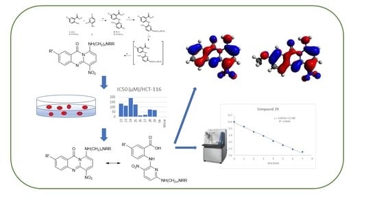
2.1. Chemistry

Commercially available methyl anthranilate (1) or methyl 5-methoxyanthranilate (2) were used as starting materials (Scheme 1). Treatment of esters 1 and 2 with 2,6-dichloro-3-nitropyridine (3) afforded only the desired methyl esters 4 and 5, respectively. The structure of these derivatives was elucidated using 1H and 13C NMR spectral data, using both direct and long-range homonuclear and heteronuclear correlation experiments (HMBC, HMQC, and NOESY sequences). Each one of the esters 4 and 5 was then reacted with the appropriately substituted N,N-dialkylethylenediamines or N,N-dialkylpropylenediamines providing the target amines 613. Mild saponification of the esters 613 resulted in the carboxylic acids 1421, respectively, which, without further purification, were ring closed upon treatment with a 1/2 v/v mixture of trifluoroacetic anhydride and trifluoroacetic acid, providing the title compounds 2229.
Each compound’s structure was unambiguously established by 1H and 13C NMR spectroscopy, using both direct and long-range heteronuclear correlation experiments. Structural discrimination resulted from the observation that H-8 of the aza-acridines 2229 exhibits 4J coupling with the carbonyl group, while in the case of esters 613 such a correlation is not possible. In general, the 1H NMR spectra of the esters 6–13 as well as the aza-acridines 2229 showed slight differences, especially in the chemical shifts of the pyridine moiety. H-7 appears downfield as a doublet at approximately 8.30 ppm, while H-8 shifts upfield around 5.50 ppm.

2.2. Biological Evaluation

The in vitro antiproliferative activities of the new aza-acridine aminoderivatives were evaluated against three human tumor cell lines—namely, HCT-116 (colorectal), the uterine sarcoma MES-SA and its variant MES-SA/Dx5, reported to be 100 times more resistant to doxorubicin (Dx) [24]. The results of the MTT dye reduction assay, expressed as 50% inhibitory concentrations (IC50) in μM, are summarized in Table 1. For comparison, Dx and mitoxantrone (Mitox) were used as positive controls.
Most of the new compounds showed low cytotoxic activity and only three of them, namely 26, 27 and 29, were detected to possess a certain degree of cytotoxic activity which is enhanced against the two uterine sarcoma cell lines (MES-SA and MES-SA/Dx5). Even so, it is noticeable that all three compounds possess the methoxy substitution, providing evidence that the presence of the methoxy group is favorable for the cytotoxicity of this class of compounds. This finding is consistent with previous observations [2,11,12] concerning the improved cytotoxic activity of methoxy-substituted analogues.
From a direct comparison of the cytotoxic activity against the Dx-sensitive (MES-SA) and Dx-resistant (MES-SA/Dx5) cell lines, it is observed that derivatives 2225 exhibit equal cytotoxicity against both cell lines, as indicated by the relevant resistant factor (RF) values (~1.0). On the contrary, the methoxy-substituted compounds 2629, proved to be more active against the Dx-resistant (MES-SA/Dx5) cell line and thus retain full antiproliferative activity against P-glycoprotein-overexpressing cells, with a relevant resistant factor of ~0.5. It must be pointed that even if the overall activity of the derivatives is moderate, the potency of compounds 2629 is enhanced against the two uterine sarcoma cell lines (MES-SA and MES-SA/Dx5). This probably indicates that the methoxy-substituted derivatives could overcome Multi Drug Resistance (MDR).
Overall, the activity of the new compounds was remarkably reduced when compared to the structurally related lead compounds (Figure 1). This is most likely due to the insertion of the additional nitrogen atom, which blocks the tautomerism of the acridine scaffold and could also interfere with the strength of the intramolecular hydrogen bond. On the other hand, it has been reported that aza-acridines are amenable to ring-opening [25] and we considered that the diminished activity of the new aza-acridines could potentially be due to hydrolytic instability. A hydrolysis mechanism should involve the nucleophilic attack of H2O to the carbonyl group, with concomitant ring-opening resulting in their precursor acids (1421, Scheme 2). Therefore, the observed antiproliferative activity could be attributed to a mixture of aza-acridines and ring-opened compounds.
In order to verify our hypothesis, we performed a series of computational chemistry- and mass spectrometry-based hydrolysis experiments. Two compounds of the synthesized set of derivatives were selected as representative examples; i.e., 25 and its methoxy-substituted analogue 29, aiming to evaluate the effect of the methoxy group on the stability and therefore on the activity.

2.3. Mass Spectrometry

In order to reveal the stability of compounds 25 and 29, an initial estimation of the corresponding hydrolysis rates was performed. Both derivatives were found to be very unstable in an aqueous environment, albeit with different rates of hydrolysis, with the methoxy derivative 29 being far more stable. The results are presented in Figure 2.
In order to assimilate the conditions used for the biological testing of the substances, the substances were analyzed at the 20 μg/mL level (~60 μΜ). It should be pointed out that the use of mass spectrometry was chosen since the low concentration could make detection through NMR spectroscopy difficult. Furthermore, such a concentration is expected not to induce any matrix effect. Due to the fact that the hydrolysis products (i.e., 17 and 21) afforded much cleaner spectra, the negative ion mode was employed. The kinetic equation used has been transformed to fit the data linearly. Thus, the equation used was
ln[A0 − B] = A0 − kt
where A0 is the initial concentration of each molecule, B the concentration of the respective product after hydrolysis and k is the hydrolysis constant. The two hydrolysis reactions were evaluated as linear models, based on the equation shown above. The statistical evaluation is presented in Table 2. The results are consistent with a first-order degradation reaction, as the fitting employing this equation order is linear. Rapid degradation was observed for the non-methoxy derivative under the conditions examined, whereas substantial degradation was observed for the methoxy analogue. Comparing the two slopes, which are the degradation coefficients, it was found that the degradation of 25 is 26 times faster than the corresponding hydrolysis of the methoxy derivative 29. Assuming that the reactions are pseudo first-order the corresponding t1/2 values are 10.6 min−1 for 25 and 277 min−1 for 29.

2.4. Computational Chemistry

The two derivatives exhibit roughly the same geometry features. As the region of interest is located at the internal amide bond, the geometry features of the involved bonds are presented in Table 3. Both structures are planar, denoting a high aromaticity degree, whereas the bond lengths are essentially the same. Therefore, the difference in reactivity should be revealed by the electronic properties of the two molecules. Calculation of the electrostatic potential (ESP) (see Supplementary Materials, Figure S1) shows that the carbonyl C is depleted of electronic charge and therefore it is susceptible to an electrophile attack, which is consistent with the first step of hydrolysis (nucleophilic attack of water). Nevertheless, no appreciable difference has been observed between the two ESP surfaces, denoting that the differences calculated by the ESP methodology are too subtle to be detected.
The electronic charge on carbonyl C as well as that of the connected N belonging to the internal amide bond also act as a measure of reactivity of the two derivatives. The Merz-Kollmann (MK) ESP-fitting atomic charges have been employed to estimate the electron partitioning scheme and showed that carbonyl C of the derivatives is the second most electron deficient carbon of the molecule with a charge of 0.761, slightly less than the pyridine carbon that is connected to the amine group, which bears a charge of 0.763. The attachment of the methoxy group renders carbonyl C the most electrophilic site of the molecule with a charge of 0.828 with the aforementioned pyridine carbon having an atomic charge of 0.797. Furthermore, the bond order of the C-N bond could also help explain the reactivity difference. The Mayer bond order analysis scheme that has been employed showed that the bond order increased by nearly 0.07 to the methoxy-bearing molecule, rendering the derivative more stable and thus resistant to hydrolysis. The delocalization of the HOMO orbital over the molecular skeleton provides visual assessment of the bonding across the atoms (Figure 3A,B). The spread of electronic density across the C-N bond of the internal amide in the case of the methoxy derivative indicates that the bond exhibits a higher overlap between the atomic orbitals (AOs) and therefore should be less prone to hydrolysis. The ELF function has been calculated for both molecules and the results can be visualized in Supplementary Figures S2A and S2B. In order to evaluate the concentration of electronic density the ELF value at the CP of the C-N bond has been assessed and was found to be 0.898 in both cases. Values larger than 0.7 indicate substantial π-character, but the ELF theory was not able to differentiate between the two molecules. Furthermore, the condensed Fukui function analysis was employed in order to pinpoint the electrophilic reactivity difference of the two carbonyl carbon atoms. The corresponding values were found to be 0.0207 and 0.0211 for the non-methoxy and methoxy derivative, respectively. The slight difference observed does not reflect their hydrolytic stability. Furthermore, as expected, their electrophilicity indices (e*eV) are also substantially similar (0.066 and 0.065, respectively). Interestingly, the condensed local softness (Hartree*e) for electrophilic attack is substantially different between the species. Thus, the corresponding values are 0.0874 for the non-methoxy derivative and 0.0933 for its methoxy counterpart. It is noteworthy that the computed local electrophilicity indexes (based on the Frozen Molecular Orbitals (FMOs) approximation) are also substantially different with values of 0.9608 and 0.7619 for the two molecules, but their order is opposite to the hydrolysis rate showing that this approach is not consistent with the experimental results in this case. Figure S3a,b shows the distribution of the Fukui function between the two molecules. Finally, Bader’s QTAIM (quantum theory of atoms-in-molecules) theory has been employed in order to study the electronic density partitioning between atoms. The AIM charges were calculated as well as the electron density Rho at the bond critical point connecting the carbonyl C to the pyridine N. The values obtained are for the carbon and nitrogen—+0.991 and −0.911, respectively—whereas for the BCP (bond critical point) a value of +0.253 was obtained for the non-methoxylated molecule. The corresponding values were +0.997, −0.911 and +0.255 for the methoxy derivative. The partitioning scheme does not indicate appreciable differences between the two molecular species. Finally, in order to estimate the energy difference between each molecule and its hydrolyzed counterpart, a full geometry optimization with the same level of theory has been performed. The corresponding values are −568.95 kcal/mol for the non-methoxylated derivative and -616.91 kcal/mol for its hydrolyzed derivative leading to an energy difference of 47.96 kcal/mol. The corresponding values for the methoxy molecule are −640.81 and -688.76 kcal/mol, leading to an energy difference of 47.95 kcal/mol. These results show that the thermodynamic result of the reaction is negligible and thus could not be the driving force of the more extensive degradation of the methoxy-bearing molecule. Overall it seems that the differentiation of the carbonyl carbon electronic charge as also depicted by the shape of the HOMO orbital could explain the difference of the hydrolytic stability, whereas the aromatic character as calculated by the topological analysis is not affected to a large extent.

3. Materials and Methods

3.1. General Information

All commercially available chemicals and solvents were used as received without any further purification. Melting points were determined on a Büchi apparatus and are uncorrected. 1H NMR spectra and 2D spectra were recorded on a Bruker Avance DRX 400 instrument (Bruker BioSpin GmbH, Rheinstetten, Germany), whereas 13C NMR spectra were recorded on a Bruker AC 200 spectrometer (Bruker BioSpin GmbH, Rheinstetten, Germany) in deuterated solvents and were referenced to TMS (δ scale). The signals of 1H and 13C spectra were unambiguously assigned by using 2D NMR techniques: 1H-1H COSY, NOESY, HMQC, and HMBC. Mass spectra were recorded with a LTQ Orbitrap Discovery instrument (Thermo Scientific, Brehmen, Germany), possessing an Ionmax ionization source. Flash chromatography was performed on Merck silica gel 60 (0.040–0.063 mm) (Merck KGa, Darmstadt, Germany). Analytical thin layer chromatography (TLC) was carried out on precoated (0.25 mm) Merck silica gel F-254 plates. Stock solutions of compounds for biological experiments were prepared in DMSO (St. Louis, MO, USA) and stored at 20 °C. Working dilutions contained up to 0.1% v/v DMSO.

3.2. Synthesis

Methyl 2-((6-chloro-3-nitropyridin-2-yl)amino)benzoate (4): NaH (460 mg, 11.5 mmol, 60% in mineral oil) was added to a solution of methyl-2-aminobenzoate (1.63 g, 10.78 mmol, 1) in dry THF (100 mL) at 0 °C under argon and the resulting suspension was stirred at room temperature for 30 min, followed by dropwise addition of a 2,6-dichloro-3-nitropyridine (2.26 g, 10.78 mmol, 3) solution in dry THF (10 mL). The reaction mixture was then stirred at 60 °C for 2 h; an excess of NaH was quenched with methanol and the solvents were vacuum evaporated. The residue was dissolved in CH2Cl2, washed with a 10% Na2CO3 solution and water, dried over Na2SO4, and the solvent was evaporated to dryness. Flash chromatography on silica gel, using a mixture of cyclohexane–CH2Cl2 3:1 as the eluent, afforded the title compound (2.22 g, 67 %) as an oil. 1H NMR (400 MHz, CDCl3) δ 12.46 (brs, 1H, D2O exch., NH), 8.54 (d, J = 8.6 Hz, 1H, H-4’), 8.50 (dd, J = 8.5, 1.1 Hz, 1H, H-3), 8.08 (d, J = 7.82 Hz, 1H, H-6), 7.59 (t, J = 7.82 Hz, 1H, H-4), 7.18 (t, J = 7.82 Hz, 1H, H-5), 6.87 (d, J = 8.61 Hz, 1H, H-5’), 3.98 (s, 3H, CH3). 13C NMR (50 MHz, CDCl3) δ 167.6 (C=O), 155.1 (C-2’), 148.5 (C-6’), 139.6 (C-2), 138.0 (C-4’), 133.5 (C-4), 131.2 (C-6), 128.8 (C-3’), 123.4 (C-3), 122.3 (C-5), 118.6 (C-1), 114.7 (C-5’), 52.5 (CH3). HR-MS (ESI) m/z: calcd. for C13H11ClN3O4+: [M1 + H]+ = 308.0433, found 308.0438.
Methyl 2-((6-chloro-3-nitropyridin-2-yl)amino)-5-methoxybenzoate (5): This compound was prepared by an analogous procedure as described for the preparation of 4 using compound 2. Yield: 76 %. Oil. 1H NMR (400 MHz, CDCl3) δ 12.11 (brs, 1H, D2O exch., NH), 8.48 (d, J = 8.76 Hz, 1H, H-4’), 8.38 (d, J = 9.19 Hz, 1H, H-3), 7.55 (d, J = 3.01 Hz, 1H, H-6), 7.15 (dd, J = 9.19, 3.02 Hz, 1H, H-4), 6.81 (d, J = 8.76 Hz, 1H, H-5’), 3.98 (s, 3H, COOCH3), 3.86 (s, 3H, OCH3). 13C NMR (50 MHz, CDCl3) δ 167.1 (C=O), 155.4 (C-5), 155.3 (C-2’), 148.6 (C-6’), 138.0 (C-4’), 132.6 (C-2), 128.2 (C-3’), 124.4 (C-3), 120.1 (C-1), 119.8 (C-4), 114.9 (C-6), 114.1 (C-5’), 55.6 (OCH3), 52.5 (COOCH3). HR-MS (ESI) m/z: calcd. for C14H13ClN3O5+: [M1 + H]+ = 338.0538, found 338.0545.
Methyl 2-((6-((2-(diethylamino)ethyl)amino)-3-nitropyridin-2-yl)amino)benzoate (6): To a solution of chloride 4 (200 mg, 0.65 mmol) in absolute ethanol (20 mL) was added N,N-diethylethylenediamine (0.36 mL, 2.6 mmol) and the mixture was refluxed for 12 h. Upon cooling, the mixture was vacuum-evaporated, extracted with ethyl acetate-water, the organic layer was dried (Na2SO4) and evaporated to dryness. The residue was purified by column chromatography (silica gel, EtOAc-MeOH 9:1) to afford the title compound (230 mg, 91%). Mp 114–116 °C (CH2Cl2). 1H NMR (400 MHz, CDCl3) δ 12.38 (brs, 1H, D2O exch., NH), 8.52 (d, J = 7.85 Hz, 1H, H-3), 8.21 (d, J = 8.53 Hz, 1H, H-4’), 8.00 (d, J = 7.51 Hz, 1H, H-6), 7.46 (t, J = 7.51 Hz, 1H, H-4), 7.08 (t, J = 7.51 Hz, 1H, H-5), 6.05 (brs, 1H, D2O exch., NHCH2CH2), 5.96 (d, J = 8.53 Hz, 1H, H-5’), 3.94 (s, 3H, COOCH3), 3.43 (m, 2H, NHCH2CH2), 2.61 (t, J = 5.80 Hz, 2H, NHCH2CH2), 2.51 (q, J = 6.83 Hz, 4H, N(CH2CH3)2), 0.97 (t, J = 6.83 Hz, 6H, N(CH2CH3)2). 13C NMR (50 MHz, CDCl3) δ 167.3 (CO), 159.6 (C-6’), 151.0 (C-1′), 140.0 (C-4’), 136.3 (C-2), 132.3 (C-4), 131.2 (C-6), 123.7 (C-3), 122.6 (C-5), 120.7 (C-3′), 119.7 (C-1), 102.1 (C-5′), 52.3 (COOCH3), 51.1 (NHCH2CH2), 46.5 (N(CH2CH3)2), 39.1 (NHCH2CH2), 11.7 (N(CH2CH3)2). HR-MS (ESI) m/z: calcd. for C19H26N2O4+: [M1 + H]+ = 388.1979, found 388.1987.
Methyl 2-((6-((2-(dimethylamino)ethyl)amino)-3-nitropyridin-2-yl)amino)benzoate (7): This compound was prepared by an analogous procedure as described for the preparation of 6 using compound 4 and N,N-dimethylethylenediamine. Yield: 79 %. Mp 122–124 °C (CH2Cl2). 1H NMR (400 MHz, CDCl3) δ 12.37 (brs, 1H, D2O exch., NH), 8.52 (d, J = 7.85 Hz, 1H, H-3), 8.24 (d, J = 8.53 Hz, 1H, H-4’), 8.02 (d, J = 7.85 Hz, 1H, H-6), 7.49 (t, J =7.85 Hz, 1H, H-4), 7.10 (t, J = 7.85 Hz, 1H, H-5), 5.98 (m, 2H, NHCH2CH2, D2O exch., H-5’), 3.96 (s, 3H, COOCH3), 3.46 (m, 2H, NHCH2CH2), 2.52 (t, J = 5.80 Hz, 2H, NHCH2CH2), 2.25 (s, 6H, N(CH3)2). 13C NMR (50 MHz, CDCl3) δ 167.3 (CO), 159.6 (C-6’), 150.9 (C-2’), 140.0 (C-4’), 136.4 (C-2), 132.3 (C-4), 131.1 (C-6), 123.7 (C-3), 122.6 (C-5), 120.8 (C-3′), 119.7 (C-1), 101.8 (C-6’), 57.4 (NHCH2CH2), 52.3 (COOCH3), 45.0 (N(CH3)2)), 39.0 (NHCH2CH2). HR-MS (ESI) m/z: calcd. for C17H22N5O4+: [M1 + H]+ = 360.1666, found 360.1672.
Methyl 2-((6-((3-(diethylamino)propyl)amino)-3-nitropyridin-2-yl)amino)benzoate (8): This compound was prepared by an analogous procedure as described for the preparation of 6 using compound 4 and N,N-diethyl-1,3-propanediamine. Yield: 80%. Mp 116–117 °C (CH2Cl2). 1H NMR (400 MHz, CDCl3) δ 12.38 (brs, 1H, D2O exch., NH), 8.61 (d, J = 8.87 Hz, 1H, H-3), 8.18 (d, J = 8.77 Hz, 1H, H-4’), 8.01 (d, J = 7.60 Hz, 1H, H-6), 7.97 (brs, 1H, D2O exch., NHCH2 CH2CH2), 7.46 (t, J = 7.60 Hz, 1H, H-4), 7.08 (t, J = 7.60 Hz, 1H, H-5), 5.87 (d, J = 8.7 Hz, 1H, H-5’), 3.95 (s, 3H, COOCH3), 3.51 (m, 2H, NHCH2CH2CH2), 2.63 (t, J = 5.55 Hz, 2H, NHCH2CH2CH2), 2.52 (q, J = 7.89 Hz, 4H, N(CH2CH3)2), 1.73 (quintet, J = 5.55 Hz, 2H, NHCH2CH2CH2), 1.03 (t, J =7.89 Hz, 6H, N(CH2CH3)2). 13C NMR (50 MHz, CDCl3) δ 167.3 (CO), 159.7 (C-6’), 151.2 (C-2’), 140.2 (C-4’), 136.0 (C-2), 132.3 (C-4), 131.1 (C-6), 123.8 (C-3), 122.4 (C-5), 120.4 (C-3’), 119.4 (C-1), 102.2 (C-5’) 52.7 (COOCH3), 52.2 (NHCH2CH2CH2), 46.7 (N(CH2CH3)2), 42.6 (NHCH2CH2CH2), 24.9 (NHCH2CH2CH2), 11.7 (N(CH2CH3)2). HR-MS (ESI) m/z: calcd. for C20H28N5O4+: [M1 + H]+ = 402.2136, found 402.2133.
Methyl 2-((6-((3-(dimethylamino)propyl)amino)-3-nitropyridin-2-yl)amino)benzoate (9): This compound was prepared by an analogous procedure as described for the preparation of 6 using compound 4 and N,N-dimethyl-1,3-propanediamine. Yield: 90%. Mp 115–117 °C (CH2Cl2). 1H NMR (400 MHz, CDCl3) δ 12.43 (brs, 1H, D2O exch., NH), 8.58 (d, J = 8.62 Hz, 1H, H-3), 8.17 (brs, 1H, H-4’), 8.00 (d, J = 8.62 Hz, 1H, H-6), 7.46 (t, J = 8.62 Hz, 1H, H-4), 7.24 (brs, 1H, D2O exch., NHCH2CH2CH2), 7.08 (t, J = 8.62 Hz, 1H, H-5), 5.93 (d, J = 8.62 Hz, 1H, H-5’), 3.94 (s, 3H, COOCH3), 3.49 (m, 2H, NHCH2CH2CH2), 2.38 (t, J = 6.74 Hz, 2H, NCH2CH2CH2), 2.21 (s, 6H, N(CH3)2)), 1.73 (quintet, J = 6.74 Hz, 2H, NHCH2CH2CH2). 13C NMR (50 MHz, CDCl3) δ 167.3 (CO), 159.9 (C-6’), 151.1 (C-2’), 140.2 (C-4’), 135.8 (C-2), 132.3 (C-4), 131.1 (C-6), 123.8 (C-3), 122.5 (C-5), 120.3 (C-3’), 119.4 (C-1), 102.6 (C-5’), 58.5 (NHCH2CH2CH2), 52.3 (COOCH3), 45.2 (N(CH3)2), 41.8 (NHCH2CH2CH2), 25.9 NH(CH2CH2CH2). HR-MS (ESI) m/z: calcd. for C18H24N5O4+: [M1 + H]+ = 374.1823, found 374.1818.
Methyl 2-((6-((2-(diethylamino)ethyl)amino)-3-nitropyridin-2-yl)amino)-5-methoxybenzoate (10): This compound was prepared by an analogous procedure as described for the preparation of 6 using compound 5 and N,N-diethylethylenediamine. Yield: 97 %. Mp 153–155 °C (CH2Cl2). 1H NMR (400 MHz, CDCl3) δ 12.12 (brs, 1H, D2O exch., NH), 8.34 (d, J = 9.33 Hz, 1H, H-3), 8.23 (d, J = 8.87 Hz, 1H, H-4’), 7.50 (d, J = 3.11 Hz, 1H, H-6), 7.06 (dd, J = 9.33, 3.11 Hz, 1H, H-4), 5.93 (m, 2H, D2O exch, NHCH2CH2, H-5’), 3.95 (s, 3H, COOCH3), 3.85 (s, 3H, OCH3), 3.40 (m, 2H, NHCH2CH2), 2.46–2.66 (m, 6H, NHCH2CH2, N(CH2CH3)2), 1.00 (t, J = 7.23 Hz, 6H, N(CH2CH3)2). 13C NMR (50 MHz, CDCl3) δ 167.0 (CO), 159.7 (C-6’), 154.9 (C-5), 151.2 (C-2’), 136.3 (C-4’), 133.1 (C-2), 125.9 (C-3), 121.3 (C-2’), 120.1 (C-4), 119.3 (C-1), 114.1 (C-6), 101.9 (C-5’), 55.6 (OCH3), 52.4 (COOCH3), 51.1 (NHCH2CH2), 46.5 (N(CH2CH3)2), 39.0 (NHCH2CH2), 11.6 (N(CH2CH3)2). HR-MS (ESI) m/z: calcd. for C20H28N5O5+: [M1 + H]+ = 418.2085, found 418.2089.
Methyl 2-((6-((2-(dimethylamino)ethyl)amino)-3-nitropyridin-2-yl)amino)-5-methoxybenzoate (11): This compound was prepared by an analogous procedure as described for the preparation of 6 using compound 5 and N,N-dimethylethylenediamine. Yield: 94 %. Mp 158–160 °C (CH2Cl2). 1H NMR (400 MHz, CDCl3) δ 12.11 (brs, 1H, D2O exch., NH), 8.33 (d, J = 9.22 Hz, 1H, H-3), 8.20 (d, J = 9.22 Hz, 1H, H-4’), 7.50 (d, J = 3.07 Hz, 1H, H-6), 7.06 (dd, J = 9.22, 3.07 Hz, 1H, H-4), 5.92 (m, 2H, D2O exch., NHCH2CH2, H-5’), 3.94 (s, 3H, COOCH3), 3.84 (s, 3H, OCH3), 3.42 (m, 2H, NHCH2CH2), 2.48 (t, J = 5.80 Hz, 2H, NHCH2CH2), 2.23 (s, 6H, N(CH3)2). 13C NMR (50 MHz, CDCl3) δ 166.9 (CO), 159.7 (C-6’), 154.9 (C-5), 151.1 (C-2’), 136.3 (C-4’), 133.0 (C-2’), 131.1 (C-2), 125.9 (C-3), 121.2 (C-3’), 120.2 (C-4), 119.3 (C-1), 114.2 (C-6), 102.3 (C-5’), 57.4 (OCH3), 55.5 (COOCH3), 52.3 (NHCH2CH2), 44.9 (N(CH3)2), 38.9 (NHCH2CH2). HR-MS (ESI) m/z: calcd. for C18H24N5O5+: [M1 + H]+ = 390.1772, found 390.1766.
Methyl 2-((6-((3-(diethylamino)propyl)amino)-3-nitropyridin-2-yl)amino)-5-methoxybenzoate (12): This compound was prepared by an analogous procedure as described for the preparation of 6 using compound 5 and N,N-diethyl-1,3-propanediamine. Yield: 89 %. Mp 140–142 °C (CH2Cl2). 1H NMR (400 MHz, CDCl3) δ 12.18 (brs, 1H, D2O exch., NH), 8.40 (d, J = 9.39 Hz, 1H, H-3), 8.18 (brs, 1H, H-4’), 7.96 (brs, 1H, D2O exch., NHCH2CH2CH2,), 7.50 (d, J = 2.47 Hz, 1H, H-6), 7.05 (dd, J = 9.22, 3.07 Hz, 1H, H-4), 5.83 (d, J = 8.61 Hz, 1H, H-5’), 3.95 (s, 3H, COOCH3), 3.84 (s, 3H, OCH3), 3.49 (m, 2H, NHCH2CH2CH2,), 2.50–2.59 (m, 6H, NHCH2CH2CH2, and N(CH2CH3)2), 1.72 (quintet, 2H, NHCH2CH2CH2), 1.04 (t, J = 7.04 Hz, 6H, N(CH2CH3)2). 13C NMR (50 MHz, CDCl3) δ 167.0 (CO), 159.8 (C-6’), 154.9 (C-5), 151.5 (C-2’), 136.0 (C-4’), 133.3 (C-2), 126.0 (C-3), 121.2 (C-3’), 119.3 (C-1, C-4), 114.1 (C-6), 102.5 (C-5’), 55.6 (OCH3), 52.9 (COOCH3), 52.4 (NHCH2CH2CH2,), 46.9 (N(CH2CH3)2), 42.6 (NHCH2CH2CH2), 25.0 (NHCH2CH2CH2), 11.6 (N(CH2CH3)2)). HR-MS (ESI) m/z: calcd. for C21H30N5O5+: [M1 + H]+ = 432.2241, found 432.2245.
Methyl 2-((6-((3-(dimethylamino)propyl)amino)-3-nitropyridin-2-yl)amino)-5-methoxybenzoate (13): This compound was prepared by an analogous procedure as described for the preparation of 6 using compound 5 and N,N-dimethyl-1,3-propanediamine. Yield: 95%. Mp 163–164 °C (CH2Cl2). 1H NMR (400 MHz, CDCl3) δ 12.15 (brs, 1H, D2O exch., NH), 8.37 (d, J = 9.23 Hz, 1H, H-3), 8.18 (brs, 1H, H-4’), 7.49 (d, J = 3.07 Hz, 1H, H-6), 7.16 (brs, 1H, D2O exch., NHCH2CH2CH2), 7.05 (dd, J = 9.22, 3.07 Hz, 1H, H-4), 5.86 (d, J = 8.87 Hz, 1H, H-5’), 3.94 (s, 3H, COOCH3), 3.83 (s, 3H, OCH3), 3.47 (m, 2H, NHCH2CH2CH2), 2.39 (t, J = 6.14 Hz, 2H, NHCH2CH2CH2), 2.22 (s, 6H, N(CH3)2), 1.71 (m, 2H, NHCH2CH2CH2). 13C NMR (50 MHz, CDCl3) δ 167.0 (CO), 159.9 (C-6’), 154.9 (C-5), 151.3 (C-2’), 136.1 (C-4’), 133.1 (C-2), 126.0 (C-3), 121.2 (C-3’), 119.8 (C-1), 119.3 (C-4), 114.1 (C-6), 101.9 (C-6’), 60.3 (NHCH2CH2CH2), 55.6 (OCH3), 52.4 (COOCH3), 45.4 (N(CH3)2)), 41.9 (NHCH2CH2CH2), 25.7 (NHCH2CH2CH2). HR-MS (ESI) m/z: calcd. for C19H26N5O5+: [M1 + H]+ = 404.1928, found 404.1923.
9-((2-(Diethylamino)ethyl)amino)-6-nitro-11H-pyrido[2,1-b]quinazolin-11-one (22): To a solution of ester 6 (160 mg, 0.413 mmol) in methanol (20 mL) was added dropwise, at room temperature, a cold 20% NaOH solution (1 mL). The mixture was stirred for 14 h at room temperature and then poured into water and neutralized (pH 7) with a 4 % HCl solution. The resulting carboxylic acid 14 was filtered, dried over P2O5 and then, dissolved in a 1:2 v/v mixture of trifluoroacetic anhydride and trifluoroacetic acid (1.5 mL). The resulting mixture was heated at 60 °C for 6 h and, upon cooling, it was poured into ice-water and neutralized with solid sodium carbonate. The precipitate was filtered and air-dried to give crude 22, which was purified by column chromatography (silica gel) using a mixture of CH2Cl2-MeOH 10:1, as the eluent, to provide the title compound as a yellow solid (110 mg, 75%). Mp 100–101°C (EtOAc). 1H NMR (400 MHz, CDCl3) δ 11.31 (brs, 1H, D2O exch., NH), 8.32 (d, J = 9.39 Hz, 1H, H-7), 8.25 (d, J = 8.22 Hz, 1H, H-1), 7.79 (t, J = 8.22 Hz, 1H, H-3), 7.70 (d, J= 8.22 Hz, 1H, H-4), 7.38 (t, J = 8.22 Hz, 1H, H-2), 5.58 (d, J = 9.39 Hz, 1H, H-8), 3.37 (m, 2H, NHCH2CH2), 2.38 (m, 2H, NHCH2CH2), 2.66 (m, 4H, N(CH2CH3)2), 1.12 (t, J = 6.93 Hz, 6H, N(CH2CH3)2). 13C NMR (50 MHz, CDCl3) δ 165.5 (CO), 154.8 (C-5a), 146.8 (C-4a), 141.7 (C-9), 137.5 (C-7), 136.0 (C-3), 128.2 (C-6), 127.3 (C-1), 127.0 (C-4), 125.4 (C-2), 117.3 (C-1), 86.7 (C-8), 49.9 (NCH2CH2), 46.6 (N(CH2CH3)2), 42.0 (NHCH2CH2), 11.7 (N(CH2CH3)2). HR-MS (ESI) m/z: calcd. for C18H22N5O3+: [M1 + H]+ = 356.1717, found 356.1725.
9-((2-(Dimethylamino)ethyl)amino)-6-nitro-11H-pyrido[2,1-b]quinazolin-11-one (23): This compound was prepared by an analogous procedure as described for the preparation of 22 using compound 7. Yield: 71 %. Mp 127–129 °C (EtOAc). 1H NMR (400 MHz, CDCl3) δ 11.28 (brs, 1H, D2O exch., NH), 8.29 (d, J = 8.88 Hz, 1H, H-7), 8.22 (d, J = 7.85 Hz, 1H, H-1), 7.77 (t, J = 7.85 Hz, 1H, H-3), 7.67 (d, J = 7.85 Hz, 1H, H-4), 7.36 (t, J =7.85 Hz, 1H, H-2), 5.54 (d, J = 8.88 Hz, 1H, H-8), 3.35 (m, 2H, NHCH2CH2), 2.67 (t, J = 6.14 Hz, 2H, NHCH2CH2), 2.35 (s, 6H, (N(CH3)2). 13C NMR (50 MHz, CDCl3) δ 165.7 (CO), 154.8 (C-5a), 146.8 (C-4a), 141.6 (C-9), 137.5 (C-7), 136.1 (C-3), 128.5 (C-6), 127.3 (C-1), 127.0 (C-4), 125.6 (C-2), 117.2 (C-11a), 86.5 (C-8), 56.4 (NHCH2CH2), 45.1 (N(CH3)2), 41.6 (NHCH2CH2). HR-MS (ESI) m/z: calcd. for C18H22N5O3+: [M1 + H]+ = 328.1404, found 328.1410.
9-((3-(Diethylamino)propyl)amino)-6-nitro-11H-pyrido[2,1-b]quinazolin-11-one (24): This compound was prepared by an analogous procedure as described for the preparation of 22 using compound 8. Yield: 73 %. Mp 110–111 °C (EtOAc). 1H NMR (400 MHz, CDCl3) δ 11.30 (brs, 1H, D2O exch., NH), 8.31 (d, J = 9.21 Hz, 1H, H-7), 8.24 (d, J = 8.09 Hz, 1H, H-1), 7.78 (t, J = 8.32 Hz, 1H, H-3), 7.70 (d, J = 8.30 Hz, 1H, H-4), 7.37 (t, J = 7.11 Hz, 1H, H-2), 5.56 (d, J = Hz, 1H, H-8), 3.34 (q, J = 6.7 Hz, 3H, NHCH2CH2CH2), 2.81 (t, J = 6.5 Hz, 2H, NHCH2CH2CH2), 2.64 (q, 4H, N(CH2CH3)2), 1.58 (quintet, J = 6.6 Hz, 2H, NHCH2CH2CH2), 1.11 (t, 6H, N(CH2CH3)2). 13C NMR (50 MHz, CDCl3) δ 165.7 (CO), 154.8 (C-5a), 146.8 (C-4a), 141.5 (C-9), 137.3 (C-7), 136.0 (C-3), 128.2 (C-6), 127.0 (C-1), 126.9 (C-4), 125.5 (C-2), 117.1 (C-11a), 86.5 (C-8), 50.3 (NHCH2CH2CH2), 46.7 (N(CH2CH3)2), 42.7 (NHCH2CH2CH2), 25.6 (NHCH2CH2CH2), 11.3 (N(CH2CH3)2. HR-MS (ESI) m/z: calcd. for C19H24N5O3+: [M1 + H]+ = 370.1874, found 370.1867.
9-((3-(Dimethylamino)propyl)amino)-6-nitro-11H-pyrido[2,1-b]quinazolin-11-one (25): This compound was prepared by an analogous procedure as described for the preparation of 22 using compound 9. Yield: 74 %. Mp 106–107 °C (EtOAc). 1H NMR (400 MHz, CDCl3) δ 11.34 (brs, 1H, D2O exch., NH), 8.28 (d, J = 9.2 Hz, 1H, H-7), 8.18 (d, J = 8.1 Hz, 1H, H-1), 7.77 (t, J = 7.1 Hz, 1H, H-3), 7.69 (d, J = 8.2 Hz, 1H, H-4), 7.36 (t, J = 7.9 Hz, 1H, H-2), 5.61 (d, J = 9.3 Hz, 1H, H-8), 3.39 (q, J = 6.7 Hz, 2H, NHCH2CH2CH2), 2.48 (t, J = 6.5 Hz, 2H, NHCH2CH2CH2), 2.29 (s, 6H, N(CH3)2), 1.92 (quintet, J= 6.6 Hz, 2H, NHCH2CH2CH2). 13C NMR (50 MHz, CDCl3) δ 166.0 (CO), 155.0 (C-5a), 146.9 (C-4a), 141.6 (C-9), 137.4 (C-7), 136.2 (C-3), 128.4 (C-6), 127.1 (C-1, C-4), 125.6 (C-2), 117.1 (C-11a), 86.5 (C-8), 56.8 (NHCH2CH2CH2), 45.3 (N(CH3)2), 42.5 (NHCH2CH2CH2), 25.7 (NHCH2CH2CH2). HR-MS (ESI) m/z: calcd. for C17H20N5O3+: [M1 + H]+ = 342.1561, found 342.1555.
9-((2-(Diethylamino)ethyl)amino)-2-methoxy-6-nitro-11H-pyrido[2,1-b]quinazolin-11-one (26): This compound was prepared by an analogous procedure as described for the preparation of 22 using compound 10. Yield: 71 %. Mp 105–107 °C (EtOAc). 1H NMR (400 MHz, CDCl3) δ 11.24 (brs, 1H, D2O exch., NH), 8.19 (d, J = 9.22 Hz, 1H, H-7), 7.53 (d, J = 8.87 Hz, 1H, H-4), 7.34 (d, J = 2.73 Hz, 1H, H-1), 7.30 (dd, J = 8.87, 2.73 Hz, 1H, H-3), 5.44 (d, J = 9.22 Hz, 1H, H-8), 3.83 (s, 3H, OCH3), 3.29 (t, J = 6.14 Hz, 2H, NHCH2CH2), 2.77 (t, J = 6.14 Hz, 2H, NHCH2CH2), 2.62 (q, J = 7.1 Hz, 4H, N(CH2CH3)2), 1.08 (t, J = 7.17 Hz, 6H, N(CH2CH3)2). 13C NMR (50 MHz, CDCl3) δ 164.7 (CO), 157.3 (C-2), 154.5 (C-5a), 141.4 (C-4a), 139.7 (C-9), 136.8 (C-7), 128.5 (C-4), 127.7 (C-6), 127.0 (C-3), 117.6 (C-11a), 105.3 (C-1), 86.6 (C-8), 55.6 (OCH3), 49.8 (NHCH2CH2), 46.5 (N(CH2CH3)2), 41.9 (NHCH2CH2), 11.6 (N(CH2CH3)2). HR-MS (ESI) m/z: calcd. for C19H24N5O4+: [M1 + H]+ = 386.1823, found 386.1826.
9-((2-(Dimethylamino)ethyl)amino)-2-methoxy-6-nitro-11H-pyrido[2,1-b]quinazolin-11-one (27): This compound was prepared by an analogous procedure as described for the preparation of 22 using compound 11. Yield: 79 %. Mp 118–120 °C (EtOAc). 1H NMR (400 MHz, CDCl3) δ 11.24 (brs, 1H, D2O exch., NH), 8.26 (d, J = 9.39 Hz, 1H, H-7), 7.64 (d, J = 8.61 Hz, 1H, H-4), 7.52 (d, J = 2.74 Hz, 1H, H-1), 7.40 (dd, J = 8.61, 2.47 Hz, 1H, H-3), 5.51 (d, J = 9.39 Hz, 1H, H-8), 3.90 (3H, s, OCH3), 3.36 (m, 2H, NHCH2CH2), 2.69 (t, J = 5.48 Hz, 2H, NHCH2CH2), 2.37 (s, 6H, N(CH3)2). 13C NMR (50 MHz, CDCl3) δ 165.2 (CO), 157.6 (C-2), 154.6 (C-5a), 141.7 (C-4a), 139.9 (C-9), 136.7 (C-7), 128.7 (C-4), 128.5 (C-6), 127.4 (C-3), 117.7 (C-11a), 105.4 (C-1), 86.2 (C-8), 56.4 (NHCH2CH2), 55.8 (OCH3), 45.1 (N(CH3)2), 41.6 (NHCH2CH2). HR-MS (ESI) m/z: calcd. for C17H20N5O4+: [M1 + H]+ = 358.1510, found 358.1517.
9-((3-(Diethylamino)propyl)amino)-2-methoxy-6-nitro-11H-pyrido[2,1-b]quinazolin-11-one (28): This compound was prepared by an analogous procedure as described for the preparation of 22 using compound 12. Yield: 80 %. Mp 113–114 °C (EtOAc). 1H NMR (400 MHz, CDCl3) δ 11.30 (brs, 1H, D2O exch., NH), 8.26 (d, J = 9.39 Hz, 1H, H-7), 7.65 (d, J = 9.00 Hz, 1H, H-4), 7.48 (d, J = 2.47 Hz, 1H, H-1), 7.41 (dd, J = 9.00, 2.74 Hz, 1H, H-3), 5.58 (d, J = 9.39 Hz, 1H, H-8), 3.90 (s, 3H, OCH3), 3.38 (m, 2H, NHCH2CH2CH2), 2.59 (m, 6H, NHCH2CH2CH2 and N(CH2CH3)2), 1.91 (quintet, 2H, NHCH2CH2CH2), 1.05 (t, J = 7.11 Hz, 6H, N(CH2CH3)2). 13C NMR (50 MHz, CDCl3) δ 165.4 (CO), 157.7 (C-2), 154.7 (C-5a), 141.9 (C-4a), 139.9 (C-9), 136.6 (C-7), 128.9 (C-4), 128.6 (C-6), 127.4 (C-3), 117.7 (C-11a), 105.3 (C-1), 86.1 (C-8), 55.7 (OCH3), 50.2 (NHCH2CH2CH2), 46.8 (N(CH2CH3)2), 42.5 (NHCH2CH2CH2), 25.9 (NHCH2CH2CH2), 11.5 (N(CH2CH3)2). HR-MS (ESI) m/z: calcd. for C20H26N5O4+: [M1 + H]+ = 400.1979, found 400.1984.
9-((3-(Dimethylamino)propyl)amino)-2-methoxy-6-nitro-11H-pyrido[2,1-b]quinazolin-11-one (29): This compound was prepared by an analogous procedure as described for the preparation of 22 using compound 13. Yield: 71%. Mp 130–131 °C (EtOAc). 1H NMR (400 MHz, CDCl3) δ 11.31 (brs, 1H, D2O exch., NH), 8.23 (d, J = 9.2 Hz, 1H, H-7), 7.61 (d, J = 8.9 Hz, 1H, H-4), 7.44 (d, J = 2.6 Hz, 1H, H-1), 7.38 (dd, J = 8.9, 2.62 Hz, 1H, H-3), 5.55 (d, J = 9.2 Hz, 1H, H-8), 3.88 (s, 3H, OCH3), 3.37 (m, 2H, NHCH2CH2CH2), 2.45 (t, J = 6.6 Hz, 2H, NHCH2CH2CH2), 2.27 (s, 6H, (N(CH3)2), 1.92 (quintet, 2H, J = 6.7 Hz, NHCH2CH2CH2). 13C NMR (50 MHz, CDCl3) δ 165.3 (CO), 157.6 (C-2), 154.7 (C-5a), 141.8 (C-4a), 139.9 (C-9), 136.6 (C-7), 128.8 (C-4), 128.4 (C-6), 127.3 (C-3), 117.6 (C-11a), 105.2 (C-1), 86.1 (C-8), 56.7 (NHCH2CH2CH2), 55.7 (OCH3), 45.4 (N(CH3)2), 42.3 (NHCH2CH2CH2), 25.9 (NHCH2CH2CH2). HR-MS (ESI) m/z: calcd. for C18H22N5O4+: [M1 + H]+ = 372.1666, found 372.1660.

3.3. Biological Assays and Experiments

The cytotoxic activity of the synthetic compounds was tested against the human cancer cell lines HCT-116 (colorectal), MES-SA (uterine sarcoma) and its variant MES-SA/Dx5, which is 100 times more resistant to Dx. HCT-116 was purchased from the American Tissue Culture Collection (ATCC, Manassas, VA, USA); MES-SA and MES-SA/Dx5 were kindly donated by Doctors D. Kletsas and H. Pratsinis (Institute of Biosciences and Applications, NCSR “Demokritos”, Athens, Greece). Cells were grown as monolayer in culture medium RPMI-1640, supplemented with 10% heat-inactivated fetal bovine serum, 10 mM Hepes, 10 U/mL penicillin, 10 μg/mL streptomycin and 5 mg/mL gentamycin (all from Lonza, Cologne, Germany) at 37 °C in a humidified 5% CO2 incubator and passaged by trypsinization every 2 to 3 days.
Stock concentrations of the compounds (10.0 mg/mL) were prepared in DMSO (Sigma-Aldrich, Darmstadt, Germany) and stored at −20 °C. Cytotoxicity was evaluated by the MTT reduction assay [26], allowing to estimate the number of metabolically active/viable cells present in culture. Briefly, cells were seeded in 96-well plates (Greiner Bio-One GmbH, Frickenhausen, Germany; 5 × 10³ cells/well) and allowed to adhere overnight. On the day of the experiment, stock solutions were serially diluted in culture medium (200.0 to 6.25 μM), added to the plates and incubated at 37 °C for 72 h. Cells incubated in culture medium or in medium containing the equivalent amount of DMSO, and cells incubated in the presence of Dx or Mitox (Sigma-Aldrich) were used as negative and positive controls, respectively. The MTT reagent (Sigma-Aldrich; 1 mg/mL in phosphate buffered saline; 100 μL/well) was added to each well during the last 4 h of incubation. The formazan crystals formed were dissolved by adding 0.1 M HCl in 2-propanol (Sigma-Aldrich; 100 μL/well) and absorption was measured using an ELISA reader (Denley WeScan, Finland) at 545 nm with the reference filter set at 690 nm. All cultures were set in triplicate. The IC50 values were calculated by plotting the sigmoidal curves (response vs. log concentration) fitted to the 4-parameter logistic equation using non-linear regression (GraphPad Prism version 6.0 for Windows from GraphPad Software, Inc., La Jolla, CA, USA).

3.4. Computational Chemistry

In order to explain the difference of the derivatives’ hydrolysis rates, a series of electronic property calculations were carried out. All electronic structure calculations were performed with the Gaussian09 computational chemistry package [27]. The drawn structure was initially optimized at the semi-empirical theory employing the PM3 Hamiltonian and at the final geometry employing DFT calculation with B3LYP 6-31G basis set. A population analysis was performed by the NBO 3 program as embedded in the Gaussion software, writing the fchk files as well as the .47 file. The estimation of the Fukui functions as well as the philicities were achieved using the UCA-FUKUI 2.1 program [28]. The finite difference approximation based on BO analysis as well as the FMO approximation based on the NBO coefficients have been used. The electronic properties of the molecule—i.e., the atomic charges—are based on the MK ESP-fitting atomic charge scheme [29], whereas the bond order analysis was based on the Mayer method. Furthermore, the QTAIM theory of Bader et al. was employed in order to investigate the partitioning of the electrostatic density between the atoms as well as to investigate the formation of bonds that are not detected by the classical methodologies [30]. The MutiWFN software [31] was employed for the population, the bond order, and the QTAIM analyses.

3.5. Chemical Analysis

The hydrolysis rate of the two products was studied by mass spectrometry employing a Waters QToF premiere (Waters, Manchester, UK). The introduction of the sample was done using the embedded syringe. Solutions of the two molecules were freshly prepared in acetonitrile at the 1 mg/mL level and the samples were diluted in water at the 20 μg/mL level and were directly infused into the instrument. The quasimolecular ions of the hydrolysis products were monitored in the (-) ion mode. The parameters used for the mass spectrometric analysis electrospray voltage at 1.5 kV, sample cone voltage at 35 V, extraction cone voltage at 3 V and the MCP plates were operated at 1850 V. Nitrogen was used both as the desolvation gas and was set at 650 L·h−1 and heated at 350 °C. The TOF analyzer was operated in the V optics mode affording a resolution of 9500. The intensity of each ion was monitored over time.

4. Conclusions

A series of new aza-acridine analogues bearing a basic side chain have been synthesized and evaluated for their antiproliferative activity. The compounds possessed low biological activity compared to their acridine analogues against three cancer cell lines tested. It was assumed that this could probably be attributed to the hydrolytic instability of the compounds in aqueous media. Thus, a study of two selected analogues—i.e., 25 and 29—differing by a methoxy group at position 2 of their core, was performed using mass spectrometry and computational chemistry experiments employing the DFT theory. It was proved that the aza-acridines exist in a mixture with their corresponding ring-opened counterparts and the ratio is dependent on the substitution of the aza-acridine core. The combination of approaches followed herein to explain the loss of anticipated activity could lead to the redesign of more stable and thus more potent aza-acridine analogues.

Supplementary Materials

The following are available online, Figure S1: ESP electrostatic potential; Figure S2: ELF values; Figure S3: Fukui function of compounds 25 and 29; 1H and 13C-NMR spectra are available online.

Author Contributions

All authors contributed to the writing and gave approval to the final version of the manuscript. M.K., V.K. and I.K.K. performed chemical synthesis experiments and analyzed results and wrote the manuscript. A.P.T. and O.E.T. performed cell viability assays and wrote the manuscript. E.G performed mass spectrometry, computational chemistry experiments and wrote the manuscript. P.M., N.P., E.G. and I.K.K. designed chemical synthesis, analyzed results, and wrote the manuscript. All authors have read and agreed to the published version of the manuscript.

Funding

This research received no external funding.

Conflicts of Interest

The authors declare no conflict of interest.

References

  1. Belmont, P.; Bosson, J.; Godet, T.; Tiano, M. Acridine and Acridone Derivatives, Anticancer Properties and Synthetic Methods: Where Are We Now? Anticancer Agents Med. Chem. 2007, 7, 139–169. [Google Scholar] [CrossRef] [PubMed]
  2. Zhang, B.; Li, X.; Li, B.; Gao, C.; Jiang, Y. Acridine and its derivatives: A patent review (2009–2013). Expert Opin. Ther. Pat. 2014, 24, 647–664. [Google Scholar] [CrossRef] [PubMed]
  3. Kaur, J.; Singh, P. Acridine derivatives: A patent review (2009–2010). Expert Opin. Ther. Pat. 2011, 21, 437–454. [Google Scholar] [CrossRef] [PubMed]
  4. Demeunynck, M. Antitumour acridines. Expert Opin. Ther. Pat. 2004, 14, 55–70. [Google Scholar] [CrossRef]
  5. Pommier, Y. DNA Topoisomerases and Cancer; Springer Science & Business Media: Berlin, Germany, 2011. [Google Scholar]
  6. Gunaratnam, M.; Greciano, O.; Martins, C.; Reszka, A.P.; Schultes, C.M.; Morjani, H.; Riou, J.-F.; Neidle, S. Mechanism of acridine-based telomerase inhibition and telomere shortening. Biochem. Pharmacol. 2007, 74, 679–689. [Google Scholar] [CrossRef]
  7. Yuan, Z.; Chen, S.; Chen, C.; Chen, J.; Chen, C.; Dai, Q.; Gao, C.; Jiang, Y. Design, synthesis and biological evaluation of 4-amidobenzimidazole acridine derivatives as dual PARP and Topo inhibitors for cancer therapy. Eur. J. Med. Chem. 2017, 138, 1135–1146. [Google Scholar] [CrossRef]
  8. Ebead, Y.; Roshal, A.D.; Wroblewska, A.; Doroshenko, A.O.; Błazejowski, J. Tautomerism of acridin-9-amines substituted at the exocyclic nitrogen atom: Spectroscopic investigations and theoretical studies. Spectrochim. Acta A 2007, 66, 1016–1023. [Google Scholar] [CrossRef]
  9. Capomacchia, C.; Casper, J.; Schulman, S.G. Valence Tautomerism of Singly Protonated 9-Aminoacridine and Its Implications for Intercalative Interactions with Nucleic Acids. J. Pharm. Sci. 1974, 63, 1272–1276. [Google Scholar] [CrossRef]
  10. Bu, X.; Chen, J.; Deady, L.W.; Denny, W.A. Synthesis and cytotoxicity of potential anticancer derivatives of pyrazolo[3,4,5-kl]acridine and indolo[2,3-a]acridine. Tetrahedron 2002, 58, 175–181. [Google Scholar] [CrossRef]
  11. Cholody, W.M.; Martelli, S.; Paradziej-Lukowicz, J.; Konopa, J. 5-[(Aminoalkyl)amino]imidazo[4,5,1-de]acridin-6-ones as a novel class of antineoplastic agents. Synthesis and biological activity. J. Med. Chem. 1990, 33, 49–52. [Google Scholar] [CrossRef]
  12. Antonini, I.; Polucci, P.; Magnano, A.; Cacciamani, D.; Konieczny, M.T.; Paradziej-Łukowicz, J.; Martelli, S. Rational design, synthesis and biological evaluation of thiadiazinoacridines: A new class of antitumor agents. Bioorg. Med. Chem. 2003, 11, 399–405. [Google Scholar] [CrossRef]
  13. Zalupski, M.M.; Shields, A.F.; Philip, P.A.; Kraut, M.; LoRusso, P.; Heilbrun, L.K.; Vaitkevicius, V. Evaluation of pyrazoloacridine in patients with advanced pancreatic carcinoma. Invest. New Drugs 1998, 16, 93–96. [Google Scholar] [CrossRef] [PubMed]
  14. Ramaswamy, B.; Mrozek, E.; Kuebler, J.P.; Bekaii-Saab, T.; Kraut, E.H. Phase II trial of pyrazoloacridine (NSC#366140) in patients with metastatic breast cancer. Invest. New Drugs 2011, 29, 347–351. [Google Scholar] [PubMed] [Green Version]
  15. Kostakis, I.K.; Magiatis, P.; Pouli, N.; Marakos, P.; Skaltsounis, A.-L.; Pratsinis, H.; Léonce, S.; Pierré, A. Design, Synthesis, and Anti-proliferative Activity of Some New Pyrazole-Fused Amino Derivatives of the Pyranoxanthenone, Pyranothioxanthenone, and Pyranoacridone Ring Systems:  A New Class of Cytotoxic Agents. J. Med. Chem. 2002, 45, 2599–2609. [Google Scholar] [CrossRef] [PubMed]
  16. Lemke, K.; Poindessous, V.; Skladanowski, A.; Larsen, A.K. The Antitumor Triazoloacridone C-1305 Is a Topoisomerase II Poison with Unusual Properties. Mol. Pharmacol. 2004, 66, 1035–1042. [Google Scholar] [CrossRef] [PubMed] [Green Version]
  17. Mazerska, Z.; Sowiński, P.; Konopa, J. Molecular mechanism of the enzymatic oxidation investigated for imidazoacridinone antitumor drug, C-1311. Biochem. Pharmacol. 2003, 66, 1727–1736. [Google Scholar] [CrossRef]
  18. Wiśniewska, A.; Chrapkowska, A.; Kot-Wasik, A.; Konopa, J.; Mazerska, Z. Metabolic transformations of antitumor imidazoacridinone, C-1311, with microsomal fractions of rat and human liver. Acta Biochim. Pol. 2007, 54, 831–838. [Google Scholar] [CrossRef] [Green Version]
  19. Guilbaud, N.; Léonce, S.; Tillequin, F.; Koch, M.; Hickman, J.A.; Pierré, A. Acronycine derivatives as promising antitumor agents. Anticancer Drugs 2002, 13, 445–449. [Google Scholar] [CrossRef]
  20. Elomri, A.; Skaltsounis, A.L.; Michel, S.; Tillequin, F.; Koch, M.; Rolland, Y.; Pierré, A.; Atassi, G. Synthesis and cytotoxic activity of acronycine derivatives modified at the pyran ring. Chem. Pharm. Bull. 1996, 44, 2165–2168. [Google Scholar] [CrossRef] [Green Version]
  21. Kostakis, I.K.; Pouli, N.; Marakos, P.; Skaltsounis, A.L.; Pratsinis, H.; Kletsas, D. Design and synthesis of novel amino-substituted xanthenones and benzo[b]xanthenones: Evaluation of their antiproliferative activity and their ability to overcome multidrug resistance toward MES-SA/Dx5 cells. Bioorg. Med. Chem. 2006, 14, 2910–2934. [Google Scholar] [CrossRef]
  22. Kostakis, I.K.; Tenta, R.; Pouli, N.; Marakos, P.; Skaltsounis, A.L.; Pratsinis, H.; Kletsas, D. Design, synthesis, and antiproliferative activity of some novel aminosubstituted xanthenones, able to overcome multidrug resistance toward MES-SA/Dx5 cells. Bioorg. Med. Chem. Lett. 2005, 15, 5057–5060. [Google Scholar] [CrossRef] [PubMed]
  23. Giannouli, V.; Kostakis, I.K.; Pouli, N.; Marakos, P.; Kousidou, O.C.; Tzanakakis, G.N.; Karamanos, N.K. Design, Synthesis, and Evaluation of the Antiproliferative Activity of a Series of Novel Fused Xanthenone Aminoderivatives in Human Breast Cancer Cells. J. Med. Chem. 2007, 50, 1716–1719. [Google Scholar] [CrossRef] [PubMed]
  24. Giannouli, V.; Kostakis, I.K.; Pouli, N.; Marakos, P.; Samara, P.; Tsitsilonis, O. Synthesis and anti-proliferative activity of some novel benzo-fused imidazo[1,8]naphthyridinones. Bioorg. Med. Chem. Lett. 2015, 25, 2621–2623. [Google Scholar] [CrossRef] [PubMed]
  25. Timári, G.; Hajós, G.; Messmer, A. Synthesis, alkylation and ring opening of two differently fused pyridoquinazolones. J. Heterocycl. Chem. 1990, 27, 2005–2009. [Google Scholar] [CrossRef]
  26. Samara, P.; Christoforidou, N.; Lemus, C.; Argyropoulou, A.; Ioannou, K.; Vougogiannopoulou, K.; Aligiannis, N.; Paronis, E.; Gaboriaud-Kolar, N.; Tsitsilonis, O.; et al. New semi-synthetic analogs of oleuropein show improved anticancer activity in vitro and in vivo. Eur. J. Med. Chem. 2017, 137, 11–29. [Google Scholar] [CrossRef] [PubMed]
  27. Frisch, M.J.; Trucks, G.W.; Schlegel, H.B.; Scuseria, G.E.; Robb, M.A.; Cheeseman, J.R.; Scalmani, G.; Barone, V.; Mennucci, B.; Petersson, G.A.; et al. Gaussian 09 Citation | Gaussian.com; Gaussian, Inc.: Wallingford, CT, USA, 2009. [Google Scholar]
  28. Sánchez-Márquez, J.; Zorrilla, D.; Sánchez-Coronilla, A.; de los Santos, D.M.; Navas, J.; Fernández-Lorenzo, C.; Alcántara, R.; Martín-Calleja, J. Introducing “UCA-FUKUI” software: Reactivity-index calculations. J. Mol. Model. 2014, 20, 2492. [Google Scholar] [CrossRef]
  29. Singh, U.C.; Kollman, P.A. An approach to computing electrostatic charges for molecules. J. Comput. Chem. 1984, 5, 129–145. [Google Scholar] [CrossRef]
  30. Bader, R.F.W. A Quantum Theory of Molecular Structure and Its Applications. Chem. Rev. 1991, 91, 893–928. [Google Scholar] [CrossRef]
  31. Lu, T.; Chen, F. Multiwfn: A multifunctional wavefunction analyzer. J. Comput. Chem. 2012, 33, 580–592. [Google Scholar] [CrossRef]
Sample Availability: Samples of the compounds 2229 are available from the authors.
Figure 1. Structures of acronycine, DACA, PZA and C-1311.
Molecules 25 04584 g001
Scheme 1. Reagents and conditions: a: NaH, THF dry, 60 °C, 2 h; b: suitable amine, ethanol abs., reflux, 12 h; c: NaOH 20%, MeOH, room temperature, 14 h; d: CF3COOH, (CF3CO)2O, 60 °C, 6 h.
Molecules 25 04584 sch001
Scheme 2. Hydrolysis of compounds 25 and 29.
Molecules 25 04584 sch002
Figure 2. Pseudo first-order degradation of compound 29.
Molecules 25 04584 g002
Figure 3. The HOMO orbitals of the non-methoxy-substituted analogue (A) and the methoxy-substituted analogue (B).
Molecules 25 04584 g003
Table 1. Inhibition of proliferation (IC50 values in μM) of compounds 2229 against HCT-116, MES-SA, and MES-SA/DX5 human tumor cell lines determined after 72 h of exposure.
CompoundsIC50 (μM) a
HCT-116MES-SAMES-SA/Dx5RF b
22136.03 ± 4.8072.10 ± 1.9065.07 ± 3.930.90
23114.93 ± 5.9949.90 ± 3.0350.30 ± 1.351.01
24192.43 ± 10.47188.10 ± 1.82164.47 ± 4.200.87
25126.30 ± 5.3751.73 ± 3.4747.93 ± 1.990.93
2615.37 ± 2.3437.03 ± 3.4221.27 ± 0.760.57
2720.47 ± 3.7522.27 ± 1.6010.33 ± 0.550.46
2873.13 ± 3.1847.43 ± 2.5634.40 ± 1.410.73
2967.67 ± 1.3936.83 ± 1.3719.47 ± 1.250.53
Dx0.183 ± 0.0160.037 + 0.0112.680 ± 0.26172.43
Mitox0.020 ± 0.0060.007 ± 0.0030.081± 0.01311.57
a The results presented are means ± standard deviation (SD) of three independent experiments and are expressed as IC50; i.e., the effective concentration reducing viability by 50% compared to unexposed control cells; b IC50 of resistant cells (MES-SA/Dx5)/IC50 of sensitive cells (MES-SA).
Table 2. Equations of the hydrolysis reactions.
CompoundEquation y = intercept (±Standard Error) + slope (±Standard Error) × tCorrelation Coefficient
25y = 11.49 (±0.001) − 0.065 (±0.0001) × t0.992
29y = 11.59 (±0.008) − 0.0025 (±0.002) × t0.994
Table 3. Selected geometry characteristics of compounds 25 and 29 describing the lactam as well as its adjacent bonds based on DFT B3LYP 6-31G+. The two molecular structures show only subtle differences.
Molecules 25 04584 i001 Molecules 25 04584 i002
Compound 25Compound 29
Bond Length (Å)9-104-1010-16 *10-15 *8-94-5
251.4451.4491.261 1.4611.415
291.4411.448 1.2611.464931.416
Angle (°)11-9-109-10-49-10-169-10-154-10-164-10-1510-4-1
25121.032116.810120.886 122.299 120.034
29120.975116.838 121.133 122.023119.779
Dihedral Angle (°)12-11-9-1011-9-10-1611-9-10-159-10-4-110-4-1-3
25177.747352.686 179.446−178.913
29−177.38246 −2.483−179.522179.078
Atomic Charges (e)N9C10C11C4O16O15C1
25−0.4450.7610.763−0.374−0.641 0.004
29−0.5220.8280.797−0.319 −0.635−0.283
* Numbering is based on the Gaussian 09 numbering. Atom 15 for compound 25 and 16 for compound 29 refer to the carbonyl O.

