Article

Antileishmanial Compounds Isolated from Psidium Guajava L. Using a Metabolomic Approach

by
Chiobouaphong Phakeovilay
1,2,
Sandra Bourgeade-Delmas
1,
Pierre Perio
1,
Alexis Valentin
1,
François Chassagne
1,3,
Eric Deharo
1,
Karine Reybier
1 and
Guillaume Marti
1,*
1
UMR 152 Pharma Dev, Université de Toulouse, IRD, UPS, 31000 Toulouse, France
2
Faculty of Pharmacy, University of Health Sciences, Ministry of Health, Vientiane 01000, Lao People’s Democratic Republic
3
Center for the Study of Human Health, Emory University, 615 Michael Street, Whitehead Building, Atlanta, GA 30322, USA
*
Author to whom correspondence should be addressed.
Molecules 2019, 24(24), 4536; https://doi.org/10.3390/molecules24244536
Submission received: 27 November 2019 / Revised: 6 December 2019 / Accepted: 9 December 2019 / Published: 11 December 2019
(This article belongs to the Section Natural Products Chemistry)

Abstract

:
With an estimated annual incidence of one million cases, leishmaniasis is one of the top five vector-borne diseases. Currently available medical treatments involve side effects, including toxicity, non-specific targeting, and resistance development. Thus, new antileishmanial chemical entities are of the utmost interest to fight against this disease. The aim of this study was to obtain potential antileishmanial natural products from Psidium guajava leaves using a metabolomic workflow. Several crude extracts from P. guajava leaves harvested from different locations in the Lao People’s Democratic Republic (Lao PDR) were profiled by liquid chromatography coupled to high-resolution mass spectrometry, and subsequently evaluated for their antileishmanial activities. The putative active compounds were highlighted by multivariate correlation analysis between the antileishmanial response and chromatographic profiles of P. guajava mixtures. The results showed that the pooled apolar fractions from P. guajava were the most active (IC50 = 1.96 ± 0.47 µg/mL). Multivariate data analysis of the apolar fractions highlighted a family of triterpenoid compounds, including jacoumaric acid (IC50 = 1.318 ± 0.59 µg/mL) and corosolic acid (IC50 = 1.01 ± 0.06 µg/mL). Our approach allowed the identification of antileishmanial compounds from the crude extracts in only a small number of steps and can be easily adapted for use in the discovery workflows of several other natural products.

1. Introduction

Leishmaniasis is in the top five of vector-bone diseases identified in the Global Health Estimates 2016 summary table of the World Health Organization (WHO). It is one of 20 neglected tropical diseases that infect one billion people in low socioeconomic populations in 149 countries [1]. Leishmaniasis is caused by the genus Leishmania spp., which includes 29 species. Among them, Leishmania donovani and Leishmania infantum cause visceral leishmaniasis, the lethal form of the disease [2]. Leishmaniasis is transmitted to mammalians by the bites of the female phlebotomine sandfly. The epidemiology of leishmaniasis depends on a variety of factors including the between host, reservoir and vector (human, animal and sandfly), the local ecological characteristics of the transmission sites such as alterations in temperature and water storage, irrigation habits, deforestation, climate changes, exposure of the human population to the parasite, and human behavior [3]. The disease is characterized by four forms; visceral leishmaniasis, post-kala-azar dermal leishmaniasis, cutaneous leishmaniasis, and mucocutaneous leishmaniasis [4]. Available medical treatments are not sufficient because of their extensive toxicity (e.g., the nephrotoxicity of amphotericin B, the teratogenicity of miltefosine), their lack of efficacy, their expensive cost (amphotericin B), the development of drug resistance (sodium stibogluconate and meglumine antimoniate), and non-specific targeting [4,5]. Fexinidazole, a 5-nitroimidazole, was recently in phase II of clinical trials on visceral leishmaniasis but exhibited a lack of efficacy, and in 2018 no new lead molecules were included in clinical trials [4].
Control of leishmaniasis by traditional approaches (vector control and treatment of patients) is facing the difficulties of identifying new cost-effectively produced bioactive molecules able to be produced on an industrial scale, rapid development of drug resistance, and the lack of access to drugs by the threatened populations. These issues are at the heart of concerns of the WHO regarding the control of tropical diseases [6]. Therefore, combination therapies are used to improve the efficacy of antileishmanial therapies and can shorten treatment duration [7]. Furthering our fundamental knowledge through original approaches, herein concerning the activity of natural compounds from a promising plant species active against Leishmania spp., is vital to overcome the challenges involved in treating leishmaniasis [8].
Psidium guajava Linn., or guava, is a tree belonging to the Myrtaceae family. Guava is native to Mexico [9], and mainly grows in tropical or subtropical areas. Guava is widely used for food and traditional medicine around the world. The guava leaves can be used in the form of an infusion or decoction for the treatment of diarrhea (especially in Lao People’s Democratic Republic (PDR) [10]), pain, fever, cough, diabetes, hypertension, wounds [9], spasms, and rheumatism [11]. Biological assays revealed that leaf extracts exhibit antibacterial and antifungal activity against Staphylococcus aureus, Streptococcus pyogenes, Escherichia coli, Pseudomonas aeruginosa, and Candida albicans [12]. Previous studies have described its antileishmanial potential, showing a 90% and 75% inhibition of L. donovani and L. infantum promastigotes, respectively [13,14]. However, to date, antileishmanial activity of purified active compounds from guava has not been reported.
The purpose of this study was to find new antileishmanial compounds from leaf extracts of P. guajava L. using a metabolomic approach and correlation analysis. Our group has previously described the use of this workflow to decipher the most active redox compounds in crude extracts of Viola alba subsp. Dehnhardii, Violaceae [15] and to identify antihepatocarcinogenic compounds from plants used to treat liver cancer in Cambodia [16]. Our approach aims to link the chemical profile variability of several extracts or fractions to bioassay results using multivariate data analysis. This holistic method has been implemented to rapidly understand the diversity and role of chemical components involved in bioactivity and is in contrast to the reductionist approach that involves successive fractionation steps for the purification of compounds responsible for the activity [17]. The use of partial least square (PLS) regression models allowed the ranking of putative active features detected from liquid chromatography–mass spectrometry (LC–MS) profiles. The annotation of peaks of interests was based on high-resolution mass spectrometry (HRMS) and tandem mass spectrometry (MS/MS) patterns mirrored to in silico fragmentation and confirmed by purification or commercial authentic standards [18].

2. Results

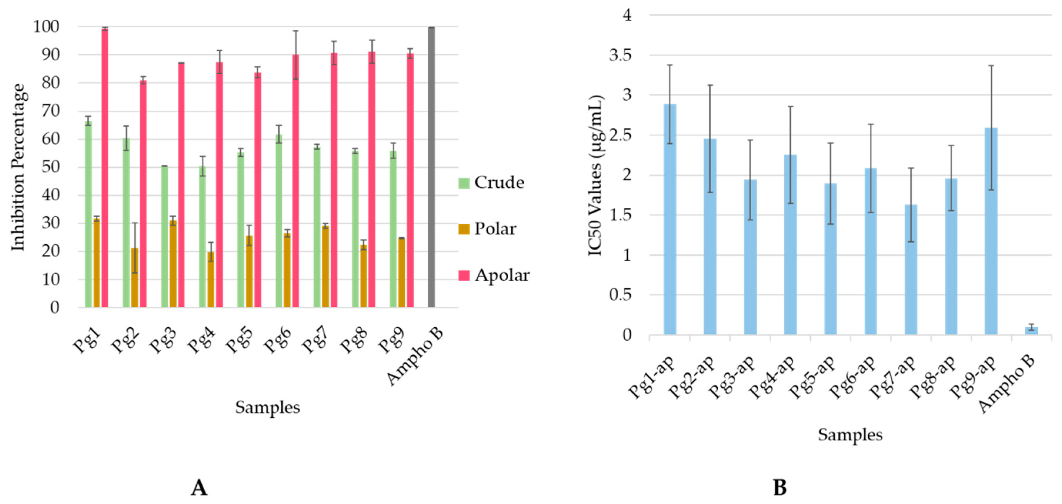
2.1. Antileishmanial Activity

Antileishmanial activities of the nine crude extracts and fractions from P. guajava leaves collected in different areas of Lao PDR were tested on the promastigote and amastigote forms of L. infantum at concentrations of 25 and 50 µg/mL. The results indicated that crude extracts, and polar and apolar fractions were not active on promastigote forms, whereas crude extracts and apolar fractions inhibited over 50% of L. infantum amastigotes at 25 µg/mL (Figure 1A). The apolar fractions showed the highest antileishmanial potential with more than 80% growth inhibition. The IC50 values of the nine apolar fractions ranged from 1.62 ± 0.04 µg/mL to 2.886 ± 0.24 µg/mL (Figure 1B). These results are not so far from the values obtained with amphothericin B, taking into account that we assessed an extract and not a pure molecule (Figure 1B).

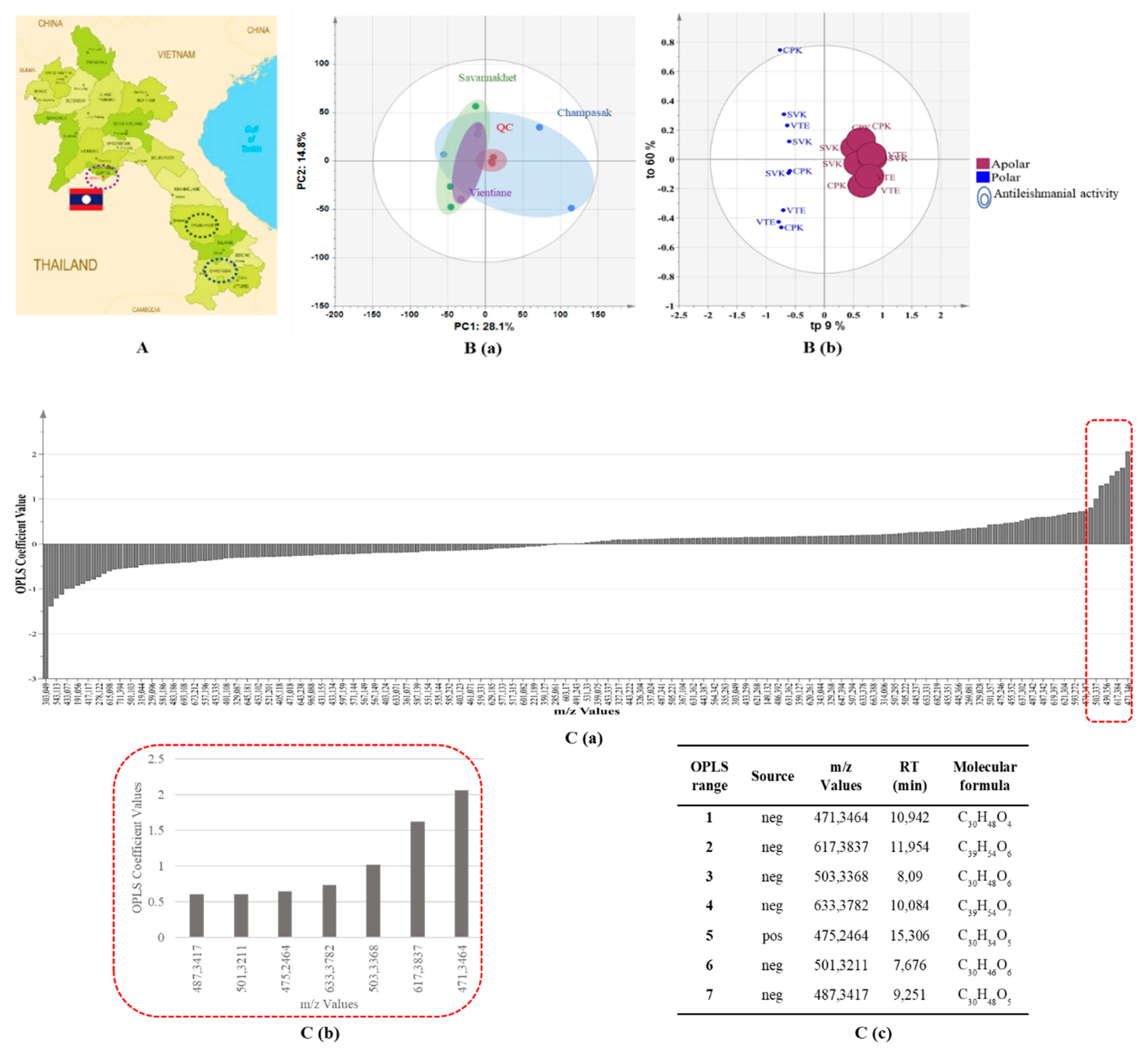
2.2. UHPLC–HRMS-based Metabolomics Approach

UHPLC–HRMS (ultra-high-performance liquid chromatography–high resolution mass spectrometry) profiles of nine extracts (three crude extracts from the Champasak province, three crude extracts from the Savannakhet province, and three crude extracts from the Vientiane province (Figure 2A) provided 448 features (m/z-RT pairs) in negative ionization (NI) mode and 163 features (m/z-RT pairs) in positive ionization (PI) mode. To obtain an unsupervised overview of sample closeness, a principal component analysis (PCA) score plot was performed to project crude extracts and quality control (QC) samples (an aliquot of each sample used in the study) on the latent variable space. This PCA score plot revealed that the analytical workflow was accurate and reproducible due to the central clustering of QC samples. In addition, samples did not cluster according to collection areas; only two samples from the Champasak province did not cluster with the other samples (Figure 2B(a)).
We then built an orthogonal projection to latent structures (OPLS) regression model to rank the features (m/z-RT pairs) toward the IC50 values of antileishmanial activity (Y-input) for polar and apolar fractions. The quality of model prediction was adequate (R2Y = 0.985, Q2Y = 0.97) and a permutation test assessed its validity (Supplementary Figure S1). The model revealed that the liquid–liquid extraction method clearly separated polar and apolar fractions in terms of chemical constituents and antileishmanial activity, and the apolar phases showed the most promising antileishmanial potential (Figure 2B(c)). This supervised technique classified potentially antileishmanial compounds according to their coefficient values (Figure 2C(a,b)), with positive coefficients relating to a high correlation with antileishmanial potential and negative coefficients to non-active compounds. The first seven hits (Figure 2C(c)) were identified based on their respective accurate mass and MS/MS analysis (Supplementary Figure S2).

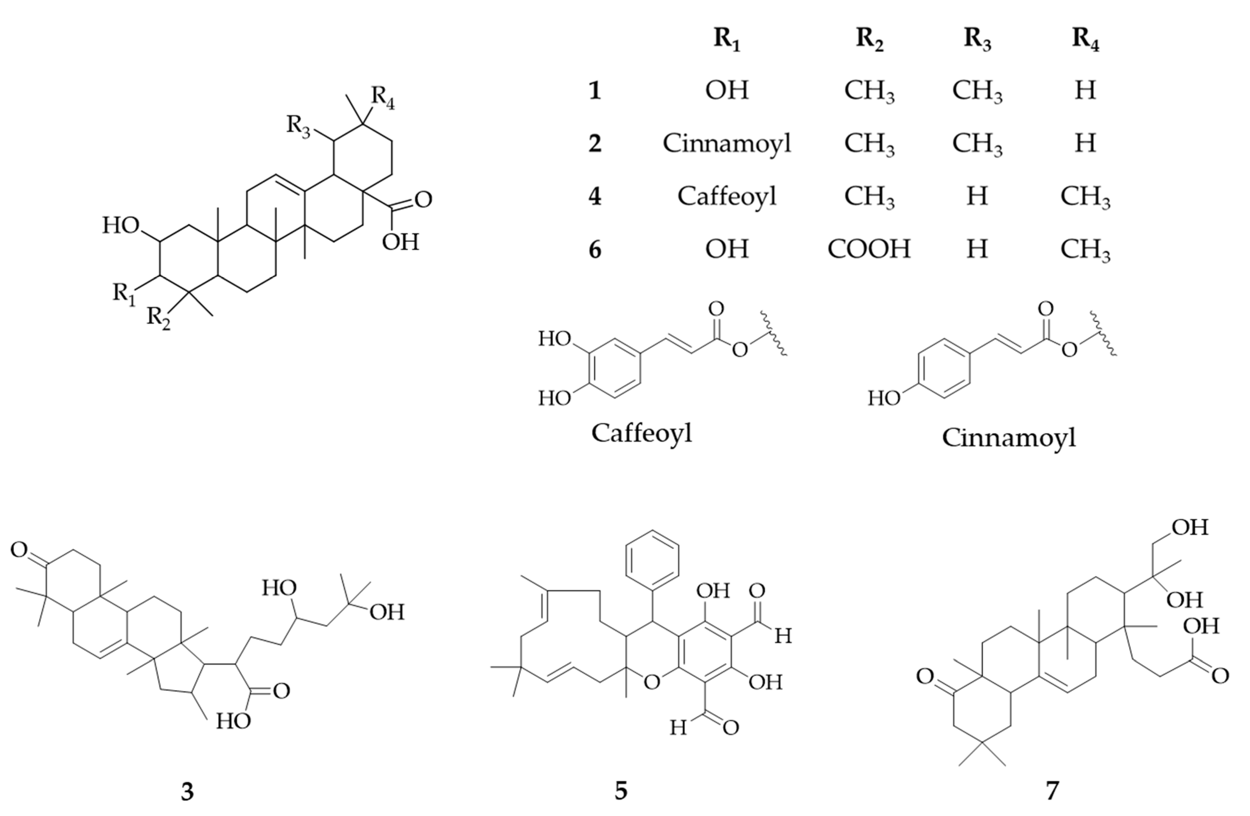
2.3. Identification of Putative Antileishmanial Compounds Based on Liquid–liquid Extraction

The first seven ranked compounds from the OPLS regression analysis were putatively annotated by matching compounds in the Dictionary of National Products (DNP) database (CRC press, v27.2). Compounds belonging to the Myrtaceae family and the Psidium genus were prioritized to narrow down the possibilities. For each compound, some candidates were ranked and proposed based on their similarity score according to the comparison between experimental MS/MS fragmentation and the in silico spectra of candidates. This process resulted in the annotation of seven candidates belonging to the Psidium genus, and all were classified as triterpenoid compounds. Annotation results were supported UV spectra of each peak (Table 1; column “UV”). The UV spectra of three triterpenoids (compounds 2, 4, 5) showed an absorption band between 278 and 310 nm highlighting the presence of a conjugated radical (Figure 3). If available, commercial authentic standards were used to confirm feature annotation (compounds 2, 5, 6) or purified from crude apolar fraction and confirmed by 1D and 2D nuclear magnetic resonance (NMR) (compound 1, Supplementary Table S1).

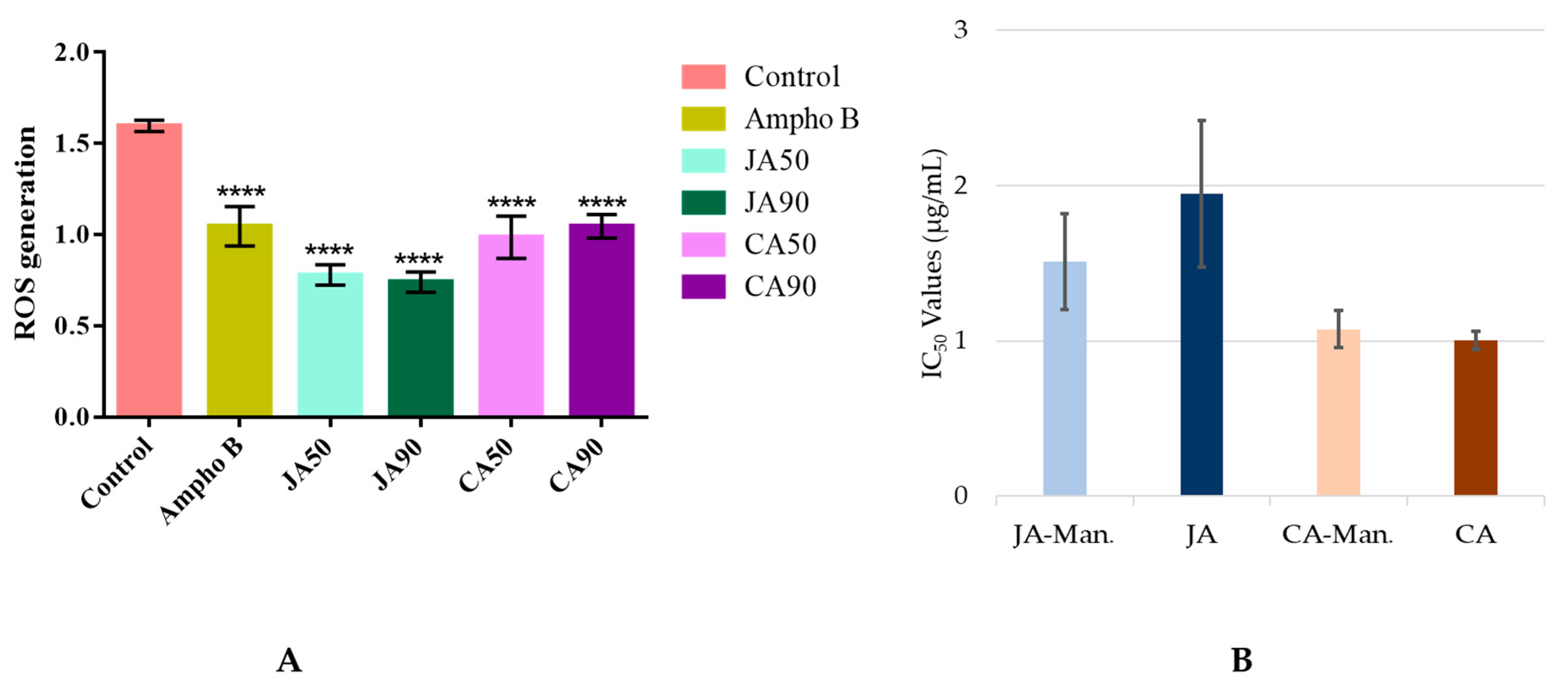
2.4. Putative Mechanism of Action

Antileishmanial assays revealed that pooled apolar fractions had a good activity with an IC50 value of 1.96 ± 0.47 µg/mL and selectivity index (SI) of around 26. The first two ranked compounds, corosolic acid (1) and jacoumaric acid (2) were more active than guajadial B (5) and medicagenic acid (6), with an IC50 of 1.32 ± 0.59 µg/mL for jacoumaric acid and 1.01 ± 0.06 µg/mL for corosolic acid (Table 2).
Since most antileishmanial drugs act by the production of reactive oxygen species (ROS) that lead to the death of parasites [23,24], we measured the production of ROS induced by jacoumaric acid and corosolic acid at the IC50 (1.32 µg/mL and 1.06 µg/mL, respectively) and IC90 (1.90 µg/mL and 1.43 µg/mL, respectively) using H2DCFDA (2′,7′-dichlorodihydrofluorescein diacetate). The corresponding graph presented in Figure 4A clearly demonstrates that jacoumaric acid and corosolic acid have an antioxidant capacity, since the fluorescence proportional to the production of ROS significantly decreased compared to the control by addition of either compound regardless of the concentration used. We also measured the IC50 of these compounds when associated with mannitol, a strong antioxidant (Figure 4B). We found no significant differences in IC50 values, confirming that the activity of jacoumaric acid and corosolic acid does not involve the production of ROS.

3. Discussion

The aim of this study was to identify antileishmanial compounds from the leaves of P. guajava and their putative mechanism of action. According to the inhibition percentage at 25 µg/mL, the ethanolic leaf extracts of P. guajava killed more than 50% of parasites, which is more than the reported antileishmanial activity of seven Brazilian plant species [25] and plants from the northwest of Morocco [26]. Moreover, the apolar fractions have interesting antileishmanial activity, with IC50 values ranging from 1.62 ± 0.46 µg/mL to 2.89 ± 0.49 µg/mL and an SI measured on L. infantum amastigotes of about 26. This result indicates that this plant is more active than Mangifera indica and Digera muricate, plants that have previously been reported to have antileishmanial activity [27]. To identify compounds responsible for this activity, we compared the in vitro antileishmanial activity and chromatographic profiles of several P. guajava leaf extracts using a metabolomic workflow. The approach depicted here allowed the ranking of detected features based on their correlation to a putative antileishmanial activity in a small number of steps. The annotation results were confirmed by commercial authentic standards (Table 1, compounds 2, 5, and 6) or a purification process (1). Guajadial B exhibited antitumor activity in five human cancer cell lines (HCT116, CCRFCEM, DU145, Huh7, and A549) at an IC50 value of 150 nM [28]. Medicagenic acid has been identified as an antifungal agent against Pyricularia pryzae (minimum inhibitory concentration (MIC) = 0.01 mg/mL and minimum fungicidal concentration (MFC) = 0.03 mg/mL) [29]. In our study, corosolic acid and jacoumaric acid displayed the most interesting antileishmanial activity in the amastigotes stage with respective IC50 values of 1.01 ± 0.06 µg/mL and 1.32 ± 0.59 µg/mL and an SI above five. This makes these two compounds more active than other triterpenoids such as ursolic acid (IC50 value 27 µg/mL) [30], E-caryophyllene (IC50 value 10.7 ± 0.6 µg/mL) [31], and spergulin-A (IC50 value 6.22 µg/mL) [32]. Both compounds have previously been identified in P. guajava and suggested to be good candidates for the development of new treatments for sickle cell anemia [33]. Furthermore, we have demonstrated their role in ROS scavenging activity, with antioxidant capacity comparable to rosmarinic acid and caffeic acid [34]. Corosolic acid has been shown to prevent inflammation and hypertension in rats [35] and to reduce the plasma glucose level in humans [19]. Corosolic acid is also known for its antibacterial activity against Paenibacillus larvae or Melissococcus plutonius [36], its anticancer activity [37], and its antitumor effect on human cervical adenocarcinoma Hela cells [38]. Jacoumaric acid also displayed antitumor effects in many carcinoma cell lines, such as leukemia [39], colorectal cancer, and human breast cancer cell lines [40]. To the best of our knowledge, this is the first report demonstrating antileishmanial activity of both compounds at the amastigotes stage. However, the close derivatives maslinic and oleanoic acid displayed antileishmanial activity on the promastigote and amastigote forms of L. infantum and L. amazonensis in the same concentration range as recorded in this study (IC50 on L. infantum amastigote: 0.99 ± 0.09 µg/mL and 2.91 ± 0.066 µg/mL, respectively) [41]. This could highlight a common mechanism of action of these triterpenoid compounds, probably related to ROS scavenging and radical positioning. According to our biological assay results and those obtained by the teams of Torres-Santos et al. and Sifaoui et al. [30,41], the presence of a carboxy group in position C-4 decreases the antileishmanial activity. Overall, the metabolomic workflow depicted in this study allowed us to rapidly target compounds responsible for the antileishmanial activity and limit multiple fractionation steps.

4. Materials and Methods

4.1. Plant Material

On November 2016, nine samples of P. guajava L. (Pg) leaves were collected from middle and southern parts of Lao PDR (Champasak: Pg1 to Pg3, Savannakhet: Pg4 to Pg6 and Vientiane province: Pg7 to Pg9). Samples were washed and air-dried, protected from the sunlight, before being ground into powder to obtain 1 kg of each. A sample specimen of each sample was collected and deposited at the herbarium of the Institute of Traditional Medicine of Lao, Vientiane, Lao PDR.

4.2. Leaf Extraction

Each accession of P. guajava leaves (250 g) sample were extracted two times using a ratio of 1 g of dried leaves for 10 mL of of 80% ethanol under agitation at room temperature for 24 h. The filtrate solutions were evaporated under reduced pressure (Buchi rotavapor R-114, Paris, France) and yield61 ± 2 g for each extract. Crude extracts (1 g) were submitted to a liquid–liquid extraction using 200 mL of a biphasic mixture composed of M1 and M2 (M1: methyl tert-butyl ether: water, 75:25; M2, water: methanol, 75:25), to obtain nine polar and nine apolar fractions. The 18 fractions were dried under reduced pressure and aliquoted for UHPLC–HRMS profiling at 2 mg/mL in a MeOH/Water 80/20 of LC–MS grade (Fisher Scientific, Schwerte, Germany) and for antileishmanial bioassays at 10 mg/mL in DMSO (Sigma-Aldrich, 99.5%, St. Louis, MO, USA).

4.3. Purification of Compound 1

To purify compound 1, 20 g of crude extract was partitioned between M1:M2 at 1:1 ratio to obtained 11.34 g of apolar fraction. Then, 6.39 g of the apolar fraction was separated using flash chromatography instrument (Spot Ultimate, Armen, France) on a silica column (CHROMABOND ® Flash RS 80 SiOH 40–63 µm) and eluted using a gradient of dichloromethane and MeOH (100% CH2Cl2 to CH2Cl2/MeOH 8/2 then 100% MeOH), to obtain six fractions. Fraction 5 (1.33 g) was selected based on its LC–MS profile and chromatographed on a silica column (CHROMABOND ® Flash RS 25 SiOH) with a gradient of chloroform and methanol (100% CHCl3 to CHCl3/MeOH 8/2), to yield compound 1 (195 mg, 2.02 g/100 g of dried leaves).
The structure of compound 1 was determined by 1D and 2D NMR spectroscopy on a Bruker 500 MHz (Avance 500, Billerica, MA, USA) using dimethylsulfoxide-d6. Chemical shifts (relative to tetramethylsilane (TMS)) are in ppm, and coupling constants in Hz. The compound was identified as “corosolic acid” by comparing with data from the literature [40] (Supplementary Table S1).

4.4. UHPLC–HRMS Profiling

All extracts (2 mg/mL) were profiled using a UHPLC-DAD-LTQ Orbitrap XL instrument (Ultimate 3000, Thermo Fisher Scientific, Hemel Hempstead, UK). The UV detection from 210 to 400 nm was performed with a diode array detector (DAD) (Hemel Hempstead, UK). Mass detection was performed using an atmospheric pressure chemical ionization (APCI) source in both NI and PI modes at 15,000 resolving power (full width at half maximum at 400 m/z). The mass scanning range was m/z 100–1500 Da. The capillary temperature was 300 °C and the spray voltage was fixed at 3.0 kV. Mass measurements were externally calibrated before starting the experiment. Each full MS scan was followed by data-dependent MS/MS on the four most intense peaks using collision-induced dissociation (35% normalized collision energy, isolation width 2 Da, activation Q 0.250). The LC–MS system was run in binary gradient mode using a BEH C18 Acquity column (100 × 2.1 mm i.d., 1.7 µm, 130 Å, Waters, MA, USA) equipped with a guard column. Mobile phase A (MPA) was 0.1% formic acid (FA) in water and mobile phase B (MPB) was 0.1% FA in acetonitrile. Gradient conditions were: 0 min, 95% MPA; 0.5 min 95% MPA; 12 min, 5% MPA; 15 min, 5% MPA, 15.5 min, 95% MPA, and 19 min, 95% MPA. The flow rate was 0.3 mL/min, column temperature 40 °C, and injection volume was 2 µL.

4.5. Data Processing

The UHPLC–HRMS raw data were converted to abf files (Reifycs, Japan) and processed with MS-DIAL version 3.90 [42] for mass signal extraction between 100 and 1500 Da from 0.5 to 16.5 min. Respective tolerances for MS1 and MS2 were set to 0.01 and 0.2 Da in centroid mode. The optimized detection threshold was set to 105 (negative) and 2 × 10⁵ (positive) for MS1. The peaks were aligned on a quality control sample (an aliquot of each fraction) reference file with a retention time tolerance of 0.15 min and a mass tolerance of 0.025 Da. Adducts and complexes were identified to exclude them from the final peak list along with features from blanks sample (injection of dilution solvent). Additionally, features with a relative standard deviation above 30% in QC sample were also deleted. The resulting peak list was then exported to comma-separated value (CSV) format prior to multivariate data analysis using SIMCA-P+ (version 15.0.2, Umerics, Umea, Sweden).

4.6. Statistical Analysis

CSV files were directly imported into SIMCA-P+ (version 15.0.2, Umerics, Umea, Sweden). For multivariate data analysis, all data were pareto scaled. The OPLS regression analysis was carried out with IC50 of antileishmanial activity as the Y input. Coefficient scores were used to rank variables according to their antileishmanial potential.

4.7. Identification of Significant Features

Molecular formulae of significant features were calculated with MS-FINDER 3.24 [18]. Various parameters were used in order to reduce the number of potential candidates, such as exclusive selection of the elements C, H, and O; mass tolerance fixed to MS1:0.01 Da and MS2:0.2 Da; and the isotopic ratio tolerance set to 20%. Only natural product databases focused on plants were selected from the DNP (CRC press, v27.2). Compounds from the Psidium genus or Myrtaceae family were prioritized. The results were presented as a list of compounds sorted according to the score value of the match. This value encompassed uncertainty on accurate mass, the isotopic pattern score, and the experimental MS/MS fragmentation mirrored to in silico matches. Only chemical identities with a final score above five were retained.
If available, commercial authentic standards were ordered from Wuhan ChemFaces Biochemical Co., ltd. (Wuhan, Hubei, China) to confirm identity. Compound 2 (jacoumaric acid), 5 (guajadial B), and 6 (medicagenic acid) were ordered with a purity ≥ 98% (measured by 13C-NMR spectroscopy and 1H-NMR spectroscopy).

4.8. Antileishmanial Evaluation

The Leishmania species used in this study was the L. infantum strain MHOM/MA/67/ITMAP-263 (CNR Leishmania, Montpellier, France) expressing luciferase activity.

4.8.1. Antileishmanial Activity on Promastigotes

We use luciferase assays to evaluate the effect of the tested compounds on the growth of L. infantum promastigotes. Briefly, logarithmic phase promastigotes suspended in RPMI 1640 medium supplemented with 10% fetal calf serum, 2 mM l-glutamine, and antibiotics (100 U/mL penicillin, 100 mg/mL streptomycin, and 50 µg/mL geneticin) were incubated at a density of 106 parasites/mL in sterile 96-well plates with crude extract and fractions (diluted at 50 and 25 µg/mL) in duplicate. Amphotericin B (purchased from Sigma-Aldrich) was used as a positive control. After a 72-h incubation period at 24 °C, we examined each well plate under a microscope to detect possible precipitate formation. To estimate the luciferase activity of promastigotes, 80 µL of each well was transferred into white 96-well plates after mild resuspension. Steady Glow® reagent (Promega, Madison, WI, USA) was added according to the manufacturer’s instructions, and plates were incubated for 2 min at room temperature. The luminescence was measured by a MicroBeta luminescence counter (PerkinElmer, Waltham, MA, USA). For the IC50 evaluation, the most active fractions or compounds were measured by eight dosage dilutions (0.39–50 µg/mL). IC50 was calculated by non-linear regression analysis processed on dose-response curves, using the GraphPad Prism 6.0. (San Diego, CA, USA). IC50 values represent the mean value calculated from three independent experiments.

4.8.2. Antileishmanial Activity on Axenic Amastigotes

Logarithmic phase L. infantum promastigotes were centrifuged at 900 g for 10 min. The supernatant was removed carefully and was replaced by the same volume of RPMI 1640 complete medium at pH 5.4 and incubated for 24 h at 24 °C. The acidified promastigotes were incubated for 24 h at 37 °C in a ventilated flask and were transformed into amastigotes. The effect of the tested compounds on the growth of L. infantum axenic amastigotes was assessed as follows: L. infantum amastigotes were incubated at a density of 2 × 10⁶ parasites/mL in sterile 96-well plates with crude extract and fractions (diluted at 50 and 25 µg/mL) in duplicate. Amphotericin B was used as a positive control. After a 48-h incubation period at 37 °C, each well plate was then examined under a microscope to detect any precipitate formation. Subsequently, to determine the IC50, we used the same method described in Section 4.8.1.

4.9. Cytotoxicity Evaluation

An MTT (3-(4,5-Dimethylthiazol-2-yl)-2,5-Diphenyltetrazolium Bromide) assay with the J774A.1 cell line (mouse macrophage cell line, Sigma-Aldrich) was performed to evaluate the cytotoxicity of the tested compounds. Briefly, cells (5 × 10⁴ cells/mL) in 100 µL of complete medium (DMEM high glucose supplemented with 10% fetal calf serum (FSC), 2 mM l-glutamine and antibiotics (100 U/mL penicillin and 100 µg/mL streptomycin)) were seeded into each well of 96-well plates and incubated at 37 °C and 5% CO2. After incubation for 24 h, 100 µL of medium with various concentrations of jacoumaric acid, carasolic acid, and appropriate controls (doxorubicin and amphotericin B, purchased from Sigma-Aldrich) were added and the plates were incubated for 72 h at 37 °C and 5% CO2. Each 96-well plate was then examined under a microscope to detect possible precipitate formation before the medium was aspirated from the well. 100 µL of MTT solution (0.5mg/mL in DMEM) was then added to each well. Cells were incubated for 2 h at 37 °C and 5% CO2. Then, the MTT solution was removed and DMSO (100 µL/well) was added to dissolve the resulting formazan crystals. Plates were shaken vigorously (300 rpm) for 5 min. The absorbance was measured at 570 nm with a microplate spectrophotometer (Eon Bio Tek, Winooski, VT, USA). DMSO was used as blank. CC50 values were calculated by non-linear regression analysis processed on dose-response curves, using GraphPad Prism 6.0. (San Diego, CA, USA). CC50 values represent the mean value calculated from three independent experiments.

4.10. Determination of Intracellular ROS Generation

ROS levels in treated and untreated L. infantum amastigotes were monitored using the cell permeable fluorogenic dye H2DCFDA [43]. 1 × 10⁶ cells/mL amastigotes were treated with IC50 and IC90 doses of jacoumaric acid and corosolic acid for 24 h. The cells were then centrifuged, washed with PBS, resuspended in PBS, and incubated for 30 min in the dark with 10 µM H2DCFDA at 37 °C. ROS were measured as an increase in fluorescence caused by the conversion of non-fluorescent dye to highly fluorescence H2DCFDA (excitation wavelength 490 nm, emission wavelength 525 nm) in a fluorescence microplate reader (SAFAS Xenius XM, Monaco, France).

5. Conclusions

We set up a metabolomic workflow to rapidly decipher antileishmanial compounds from P. guajava L. leaves extract. Our dereplication approach lead to the identification of corosolic acid and jacoumaric acid as the most active compounds against amastigotes L. infantum, along with the ROS scavenging in the antileishmanial mechanism. This is the first report in antileishmanial activity of corosolic acid and jacoumaric acid. Further studies would be required to understand the mechanism of action of these triterpenoids’ compounds on leishmaniasis parasites.

Supplementary Materials

The following are available online, Figure S1: Permutation plot of the OPLS correlation model between antileishmanial activity and the liquid chromatography–mass spectrometry dataset; Figure S2: MS/MS fragmentation pattern of standard compound 1, 2, 5, 6 versus identified peaks in crude extract chromatograms.; Table S1: 1H-NMR and 13C-NMR data (DMSO-d6) of compound 1. LC–MS raw files and treated data spreadsheet has been archived to zenodo with the linked DOI: 10.5281/zenodo.3555529.

Author Contributions

G.M. proposed the study and designed the experiments. C.P., E.D., and F.C. collected the plants in Lao PDR. C.P. performed the extraction of crude material, the antileishmanial assays under the supervision of S.B.-D. and A.V., processed the LC–HRMS profiles acquired, performed multivariate data analysis, under the supervision of G.M., and processed the determination of intracellular ROS generation under the supervision of K.R. and P.P. Finally, C.P. analyzed the results and wrote the manuscript, which was reviewed by all the collaborators of the work.

Funding

This research received no external funding.

Acknowledgments

The authors would like to thank the Pierre Fabre Foundation for sponsoring the PhD fellowship.

Conflicts of Interest

The authors declare no conflict of interest.

References

  1. Burza, S.; Croft, S.L.; Boelaert, M. Leishmaniasis. Lancet 2018, 392, 951–970. [Google Scholar] [CrossRef]
  2. Chappuis, F.; Sundar, S.; Hailu, A.; Ghalib, H.; Rijal, S.; Peeling, R.W.; Alvar, J.; Boelaert, M. Visceral leishmaniasis: What are the needs for diagnosis, treatment and control? Nat. Rev. Microbiol. 2007, 5, 873–882. [Google Scholar] [CrossRef] [PubMed]
  3. Oryan, A.; Akbari, M. Worldwide risk factors in leishmaniasis. Asian Pac. J. Trop. Med. 2016, 9, 925–932. [Google Scholar] [CrossRef] [PubMed] [Green Version]
  4. Zulfiqar, B.; Shelper, T.B.; Avery, V.M. Leishmaniasis drug discovery: recent progress and challenges in assay development. Drug Discov. Today 2017, 22, 1516–1531. [Google Scholar] [CrossRef] [PubMed]
  5. Tiuman, T.S.; Santos, A.O.; Ueda-Nakamura, T.; Filho, B.P.D.; Nakamura, C.V. Recent advances in leishmaniasis treatment. Int. J. Infect. Dis. 2011, 15, 525–532. [Google Scholar] [CrossRef] [Green Version]
  6. Jannin, J.; Solano, P.; Quick, I.; Debre, P. The francophone network on neglected tropical diseases. PLoS Negl. Trop. Dis. 2017, 11, e0005738. [Google Scholar] [CrossRef] [Green Version]
  7. Kip, A.E.; Schellens, J.H.M.; Beijnen, J.H.; Dorlo, T.P.C. Clinical pharmacokinetics of systemically administered antileishmanial drugs. Clin. Pharm. 2018, 57, 151–176. [Google Scholar] [CrossRef] [Green Version]
  8. de Mesquita, M.L.; Desrivot, J.; Bories, C.; Fournet, A.; de Paula, J.E.; Grellier, P.; Espindola, L.S. Antileishmanial and trypanocidal activity of Brazilian Cerrado plants. Mem. Inst. Oswaldo Cruz. Rio De Janerio 2005, 100, 783–787. [Google Scholar] [CrossRef]
  9. Gutiérrez, R.M.P.; Mitchell, S.; Solis, R.V. Psidium guajava: A review of its traditional uses, phytochemistry and pharmacology. J. Ethnopharmacol. 2008, 117, 1–27. [Google Scholar] [CrossRef]
  10. Dubost, J.M.; Phakeovilay, C.; Her, C.; Bochaton, A.; Elliott, E.; Deharo, E.; Xayvue, M.; Bouamanivong, S.; Bourdy, G. Hmong herbal medicine and herbalists in Lao PDR: pharmacopeia and knowledge transmission. J. Ethnobiol. Ethnomed. 2019, 15, 1–15. [Google Scholar] [CrossRef]
  11. Sajjekhan, S.; Jha, P.K.; Dakappa, S.S. Pharmacognostic evaluation of Psidium guajava Linn. leaves (Mrytaceae). Med. Aromat. Plant Sci. Biotechnol. 2011, 5, 156–159. [Google Scholar]
  12. Growther, L.; Sukirtha, K. Phytochemical analysis and antimicrobial properties of leaves and Psidium guajava bark extracts. Asian J. Pharm. Pharm. 2018, 4, 318–323. [Google Scholar] [CrossRef]
  13. Gawad, S.M.A.; Hetta, M.H.; Ross, S.A.; Badria, F.A.E.R. Antiprotozoal and antimicrobial activity of selected medicinal plants growing in Upper Egypt, Beni-Suef Region. World J. Pharm. Pharm. Sci. 2015, 4, 1720–1740. [Google Scholar]
  14. Souza, C.E.S.; Silva, A.R.P.; Gomez, M.C.V.; Rolóm, M.; Coronel, C.; Costa, J.G.M.; Sousa, A.K.; Rolim, L.A.; Souza, F.H.S.; Coutinho, H.D.M. Anti-trypanosoma, anti-leishmania and cytotoxic activities of natural products from Psidium brownianum Mart. ex DC. and Psidium guajava var. Pomifera analysed by LC–MS. Acta Trop. 2017, 176, 380–384. [Google Scholar] [CrossRef]
  15. Chervin, J.; Perio, P.; Martins-Froment, N.; Pharkeovilay, C.; Reybier, K.; Nepveu, F.; Fabre, N.; Talou, T.; Bonzon-Ponnet, V.; Marti, G. Dereplication of natural products from complex extracts by regression analysis and molecular networking: Case study of redox-active compounds from Viola alba subsp. dehnhardtii. Metabolomics 2017, 13, 1–12. [Google Scholar] [CrossRef]
  16. Chassagne, F.; Haddad, M.; Amiel, A.; Phakeovilay, C.; Manithip, C.; Bourdy, G.; Deharo, E.; Marti, G. A metabolomic approach to identify anti-hepatocarcinogenic compounds from plants used traditionally in the treatment of liver diseases. Fitoterapia 2018, 127, 226–236. [Google Scholar] [CrossRef]
  17. Ayouni, K.; Berboucha-Rahmani, M.; Kim, H.K.; Atmani, D.; Verpoorte, R.; Choi, Y.H. Metabolomic tool to identify antioxidant compounds of Fraxinus angustifolia leaf and stem bark extracts. Ind. Crops Prod. 2016, 88, 65–77. [Google Scholar] [CrossRef]
  18. Tsugawa, H.; Kind, T.; Nakabayashi, R.; Yukihira, D.; Tanaka, W.; Cajka, T.; Saito, K.; Fiehn, O.; Arita, M. Hydrogen rearrangement rules: Computational MS/MS fragmentation and structure elucidation using ms-finder software. Anal. Chem. 2016, 88, 7946–7958. [Google Scholar] [CrossRef]
  19. Fukushima, M.; Matsuyama, F.; Ueda, N.; Egawa, K.; Takemoto, J.; Kajimoto, Y.; Yonaha, N.; Miura, T.; Kaneko, T.; Nishi, Y.; et al. Effect of corosolic acid on postchallenge plasma glucose levels. Diabetes Res. Clin. Pract. 2006, 73, 174–177. [Google Scholar] [CrossRef]
  20. Ogura, M.; Cordell, G.A.; Farnsworth, N.R. Jacoumaric acid, a new triterpene ester from Jacaranda caucana. Phytochemistry 1977, 16, 286–287. [Google Scholar] [CrossRef]
  21. Gao, Y.; Wang, G.Q.; Wei, K.; Hai, P.; Wang, F.; Liu, J.-K. Isolation and Biomimetic Synthesis of (±)-Guajadial B, a Novel Meroterpenoid from Psidium guajava. Org. Lett. 2012, 14, 5936–5939. [Google Scholar] [CrossRef]
  22. Maestrini, M.; Tava, A.; Mancini, S.; Salari, F.; Perrucci, S. In vitro anthelmintic activity of saponins derived from medicago spp. plants against donkey gastrointestinal nematodes. Vet. Sci. 2019, 6, 35. [Google Scholar] [CrossRef] [Green Version]
  23. Martins, R.C.; Dorneles, G.P.; Teixeira, V.O.N.; Antonello, A.M.; Couto, J.L.; Rodrigues Júnior, L.C.; Monteiro, M.C.; Peres, A.; Schrekker, H.S.; Romão, P.R.T. Imidazolium salts as innovative agents against Leishmania amazonensis. Int. Immunopharmacol. 2018, 63, 101–109. [Google Scholar] [CrossRef]
  24. Pramanik, P.K.; Paik, D.; Pramanik, A.; Chakraborti, T. White jute (Corchorus capsularis L.) leaf extract has potent leishmanicidal activity against Leishmania donovani. Parasitol. Int. 2019, 71, 41–45. [Google Scholar] [CrossRef]
  25. Paula, R.C.; Silva, S.M.; Faria, K.F.; Frézard, F.; Moreira, C.P.S.; Foubert, K.; Lopes, J.C.D.; Campana, P.R.V.; Rocha, M.P.; Silva, A.F.; et al. In vitro antileishmanial activity of leaf and stem extracts of seven Brazilian plant species. J. Ethnopharmacol. 2019, 232, 155–164. [Google Scholar] [CrossRef]
  26. Bouyahya, A.; Et-Touys, A.; Dakka, N.; Fellah, H.; Abrini, J.; Bakri, Y. Antileishmanial potential of medicinal plant extracts from the North-West of Morocco. Beni-Suef Univ. J. Basic Appl. Sci. 2018, 7, 50–54. [Google Scholar] [CrossRef]
  27. Mehwish, S.; Islam, A.; Ullah, I.; Wakeel, A.; Qasim, M.; Khan, M.A.; Ahmad, A.; Ullah, N. In vitro antileishmanial and antioxidant potential, cytotoxicity evaluation and phytochemical analysis of extracts from selected medicinally important plants. Biocatal. Agric. Biotechnol. 2019, 19, 1–9. [Google Scholar] [CrossRef]
  28. Qin, X.J.; Yu, Q.; Yan, H.; Khan, A.; Feng, M.-Y.; Li, P.-P.; Hao, X.J.; An, L.K.; Liu, H.Y. Meroterpenoids with antitumor activities from Guava (Psidium guajava). J. Agric. Food Chem. 2017, 65, 4993–4999. [Google Scholar] [CrossRef]
  29. Abbruscato, P.; Tosi, S.; Crispino, L.; Biazzi, E.; Menin, B.; Picco, A.M.; Pecetti, L.; Avato, P.; Tava, A. Triterpenoid glycosides from Medicago sativa as antifungal agents against Pyricularia oryzae. J. Agric. Food Chem. 2014, 62, 11030–11036. [Google Scholar] [CrossRef]
  30. Torres-Santos, E.C.; Lopes, D.; Rodrigues Oliveira, R.; Carauta, J.P.P.; Bandeira Falcao, C.A.; Kaplan, M.A.C.; Rossi-Bergmann, B. Antileishmanial activity of isolated triterpenoids from Pourouma guianensis. Phytomedicine 2004, 11, 114–120. [Google Scholar] [CrossRef]
  31. Moreira, R.R.D.; Santos, A.G.D.; Carvalho, F.A.; Perego, C.H.; Crevelin, E.J.; Crotti, A.E.M.; Cogo, J.; Cardoso, M.L.C.; Nakamura, C.V. Antileishmanial activity of Melampodium divaricatum and Casearia sylvestris essential oils on Leishmania amazonensis. Rev. Inst. Med. Trop. Sao Paulo 2019, 61, 1–7. [Google Scholar] [CrossRef] [PubMed]
  32. Banerjee, S.; Mukherjee, N.; Gajbhiye, R.L.; Jaisankar, P.; Datta, S.; Das Saha, K. Intracellular anti-leishmanial effect of Spergulin-A, a triterpenoid saponin of Glinus oppositifolius. Infect. Drug Resistance 2019, 12, 2933–2942. [Google Scholar] [CrossRef] [PubMed] [Green Version]
  33. Ngbolua, K.N.; Lufuluabo, L.G.; Moke, L.E.; Bango, G.N.; Liyongo, C.I.; Ashande, C.M.; Sapo, B.S.; Zoawe, B.G.; Mpiana, P.T. A review on the phytochemistry and pharmacology of psidium guajava L. (Myrtaceae) and future direction. Discov. Phytomed. 2018, 5, 7–13. [Google Scholar] [CrossRef]
  34. Garcia, A.R.; Oliveira, D.M.P.; Amaral, A.C.F.; Jesus, J.B.; Sodero, A.C.R.; Souza, A.M.T.; Supuran, C.T.; Vermelho, A.B.; Rodrigues, I.A.; Pinheiro, A.S. Leishmania infantum arginase: biochemical characterization and inhibition by naturally occurring phenolic substances. J. Enzym. Inhib. Med. Chem. 2019, 34, 1100–1109. [Google Scholar] [CrossRef] [PubMed] [Green Version]
  35. Yamaguchi, Y.; Yamada, K.; Yoshikawa, N.; Nakamura, K.; Haginaka, J.; Kunitomo, M. Corosolic acid prevents oxidative stress, inflammation and hypertension in SHR/NDmcr-cp rats, a model of metabolic syndrome. Life Sci. 2006, 79, 2474–2479. [Google Scholar] [CrossRef] [PubMed]
  36. Kim, J.; Park, S.; Shin, Y.K.; Kang, H.; Kim, K.Y. In vitro antibacterial activity of macelignan and corosolic acid against the bacterial bee pathogens Paenibacillus larvae and Melissococcus plutonius. Acta Vet. Brno 2018, 87, 277–284. [Google Scholar] [CrossRef]
  37. Chen, Y.; Zhang, Q.W.; Li, S.L.; Yi, Y.; Zhao, J.; Wang, Y.; Ye, W.C. Psidium guajava, a potential resource rich in corosolic acid revealed by high performance liquid chromatography. J. Med. Plants Res. 2011, 5, 4261–4266. [Google Scholar]
  38. Hsiao, Y.H.; Lin, C.W.; Wang, P.H.; Hsin, M.C.; Yang, S.F. The potential of chinese herbal medicines in the treatment of cervical cancer. Integr. Cancer Ther. 2019, 18, 1–9. [Google Scholar] [CrossRef] [Green Version]
  39. Numata, A.; Yang, P.; Takahashi, C.; Fujiki, R.; Nabae, M.; Fujita, E. Cytotoxic triterpenes from a chinese medicine, Goreishi. Chem. Pharm. Bull. 1989, 37, 648–651. [Google Scholar] [CrossRef]
  40. Hou, W.; Li, Y.; Zhang, Q.; Wei, X.; Peng, A.; Chen, L.; Wei, Y. Triterpene acids isolated from Lagerstroemia speciosa leaves as α-glucosidase inhibitors. Phytother. Res. 2009, 23, 614–618. [Google Scholar] [CrossRef]
  41. Sifaoui, I.; López-Arencibia, A.; Martín-Navarro, C.M.; Reyes-Batlle, M.; Mejri, M.; Valladares, B.; Lorenzo-Morales, J.; Abderabba, M.; Enrique, J. Selective activity of oleanolic and maslinic acids on the amastigote form of Leishmania spp. Iran J. Pharm. Res. 2017, 16, 1190–1193. [Google Scholar] [PubMed]
  42. Tsugawa, H.; Cajka, T.; Kind, T.; Ma, Y.; Higgins, B.; Ikeda, K.; Kanazawa, M.; VanderGheynst, J.; Fiehn, O.; Arita, M. MS-DIAL: data-independent MS/MS deconvolution for comprehensive metabolome analysis. Nat. Methods 2015, 12, 523–526. [Google Scholar] [CrossRef] [PubMed]
  43. Fonseca-Silva, F.; Inacio, J.D.F.; Canto-Cavalheiro, M.M.; Almeida-Amaral, E.E. Reactive oxygen species production and mitochondrial dysfunction contribute to quercetin induced death in Leishmania amazonensis. PLoS ONE 2011, 6, e0014666. [Google Scholar] [CrossRef] [PubMed]
Sample Availability: Samples of the compounds corosolic acid are available from the authors.
Figure 1. Antileishmanial activity of guava leaf extracts: (A) Inhibition percentage at 25 µg/mL of crude extracts and of the corresponding polar and apolar fractions measured on L. infantum amastigotes. (B) IC50 of nine apolar fractions from P. guajava (Pg1–9) compared to amphotericin B (Ampho B).
Molecules 24 04536 g001
Figure 2. Multivariate data analysis workflow: A P. guajava collection areas. B (a) PCA (principal component analysis) score plot of the APCI-NI&PI (atmospheric pressure chemical ionization – negative and positive ionization) dataset colored according to the collection areas; (b) OPLS regression (orthogonal projections to latent structures) score plot of the polar and apolar phase dataset correlated to their antileishmanial activity (High circle radius for low IC50). C (a) Coefficient plot obtained by OPLS regression; (b) Emphasis on the first loadings; (c) Details of the first seven features according to the putative antileishmanial activity. CPK, Champasak; QC, quality control; SVK, Savannakhet; VTE, Vientiane.
Molecules 24 04536 g002
Figure 3. Chemical structures of annotated compounds (See Table 1 for details).
Molecules 24 04536 g003
Figure 4. Antioxidant capacity of jacoumaric acid and corosolic acid (JA) and (CA): A Reactive oxygen species (ROS) measured using the H2DCFDA (2′,7′-dichlorodihydrofluorescein diacetate) assay on axenic amastigotes in the presence of JA and CA at IC50 and IC90 concentrations compared to amphotericin B (AmphoB) (2 µg/mL). **** indicates significant difference relative to the control group (p < 0.0001)). B IC50 comparison of JA and CA when associated with mannitol 50 µM (JA-Man and CA-Man) (JA: p = 0.816 and CA: p = 0.403).
Molecules 24 04536 g004
Table 1. Summary of all the compounds identified or dereplicated in this work.
OPLS Rank *Sourcem/z ValuesRT (min)Adduct TypeMFΔm/z (mDa)Main MS/MS FragmentsUV (nm)Putative AnnotationChemical ClassBiology Source
1neg4,713,46410.942[M − H]C30H48O41.5836451.2516
393.3562
289.2787
NDCorosolic acid a,c
(2α-hydroxyursolic acid)
Ursane triterpenoidsLagerstroemia speciosa [19]
2neg6,173,83711.954[M − H]C39H54O61.063597.0866
537.0319
497.4617
453.3008
310Jacoumaric acid a,bUrsane triterpenoidsJacaranda caucana [20]
3neg5,033,3688.090[M − H]C30H48O61.0128485.3478
437.4027
389.4317
183.3482
ND16,24,25-Trihydroxy-3-oxoeuph-7-en-21-oic acid aDammarane triterpenoidsPsidium guajava
4neg6,333,78210.084[M − H]C39H54O71.4776563.7110
513.4430
469.5226
392.2423
3002,3-Dihydroxy-12-oleanen-28-oic acid; 3-O-(3,4-Dihydroxy-E-cinnamoyl) aOleanane triterpenoidsPsidium guajava
5pos4,752,46415.306[M + H]+C30H34O51.5006323.2012
293.3159
271.0567
205.2643
278Guajadial B a,bMeroterpenoidsPsidium guajava [21]
6neg5,013,2117.676[M − H]C30H46O61.0627483.6488
389.3652
321.4315
189.4521
NDMedicagenic acid a,bUrsane triterpenoidsMedicago sativa [22]
7neg4,873,4179.251[M − H]C30H48O51.1982469.5041
424.4036
268.196
ND4,23-Dihydroxy-22-oxo-3,4-seco-12-oleanen-3-oic acid aOleanane triterpenoidsStreptomyces sp.
* Ranking based on PLS regression coefficients, served as compound number. a Determined by in silico MS/MS fragmentation with MS-FINDER. b Confirmed by commercial authentic standard compounds. c Confirmed by NMR spectroscopy. ND, not detected; NMR, nuclear magnetic resonance; MF, molecular formula; MS/MS, tandem mass spectrometry; OPLS, orthogonal partial least squares/projections to latent structures; UV, ultraviolet.
Table 2. Confirmation of the antileishmanial potential of putative annotations.
CompoundsPromastigotesAxenic AmastigotesJ774A1SI
IC50 (µg/mL)IC50 (µg/mL)CC50 (µg/mL)PromastigotesAmastigotes
Pooled apolar fraction>501.96 ± 0.4751.19 ± 9.21ND26.12
Corosolic acid (1)18.43 ± 1.201.01 ± 0.065.77 ± 0.500.315.71
Jacoumaric acid (2)>501.32 ± 0.5912.88 ± 2.50ND9.76
Guajadial B (5)>50>50NTNDND
Medicagenic acid (6)>50>50NTNDND
Amphotericin B0.09 ± 0.010.09 ± 0.045.79 ± 0.6564.3364.33
DoxorubicinNTNT0.04 ± 0.004NDND
NT, None Tested; ND, not Detected; SI, Selectivity Index.

