Article

A Convenient Synthesis of Pentaporphyrins and Supramolecular Complexes with a Fulleropyrrolidine

by
Joana I. T. Costa
1,
Andreia S. F. Farinha
2,
Filipe A. Almeida Paz
3 and
Augusto C. Tomé
1,*
1
QOPNA and LAQV-REQUIMTE, Department of Chemistry, University of Aveiro, 3810-193 Aveiro, Portugal
2
King Abdullah University of Science and Technology (KAUST), Water Desalination and Reuse Center (WDRC), Division of Biological and Environmental Sciences (BESE), Thuwal, Saudi Arabia
3
Department of Chemistry, CICECO—Aveiro Institute of Materials, University of Aveiro, 3810-193 Aveiro, Portugal
*
Author to whom correspondence should be addressed.
Molecules 2019, 24(17), 3177; https://doi.org/10.3390/molecules24173177
Submission received: 30 July 2019 / Revised: 29 August 2019 / Accepted: 31 August 2019 / Published: 1 September 2019

Abstract

:
A simple and straightforward synthesis of diporphyrins and pentaporphyrins is reported here. The supramolecular interactions of the new porphyrin derivatives with C60 and PyC60 (a pyridyl [60]fulleropyrrolidine) were evaluated by absorption and fluorescence titrations in toluene. While no measurable modifications of the absorption and fluorescence spectra were observed upon addition of C60 to the porphyrin derivatives, the addition of PyC60 to the corresponding mono-Zn(II) porphyrins resulted in the formation of Zn(porphyrin)–PyC60 coordination complexes and the binding constants were calculated. Results show that the four free-base porphyrin units in pentaporphyrin 6 have a significant contribution in the stabilization of the 6–PyC60 complex. The crystal and molecular features of the pentaporphyrin Zn5 were unveiled using single-crystal X-ray diffraction studies.

Graphical Abstract

1. Introduction

Porphyrins are extremely versatile compounds from structural and chemical viewpoints. They display a rigid planar geometry, high stability, intense electronic absorption and emission, a small HOMO–LUMO energy gap, and flexible tunability of their optical and redox properties by changing the metal center [1]. Porphyrins can be efficiently modified at the meso or beta positions, or at the meso-(hetero)aryl substituents. Consequently, these compounds are one of the most attractive building blocks for the formation of covalent or supramolecular assemblies [2,3,4,5,6]. The design and synthesis of multiporphyrin arrays is an important research topic due to their potential application as artificial light-harvesting systems [7,8,9,10,11,12,13], functional materials (near infrared dyes, electron-conducting molecular wires, nonlinear optics, molecular recognition and sensing) [14,15,16,17], and also as agents for medical imaging and photodynamic therapy [18]. Several synthetic strategies have been developed to produce multiporphyrin oligomers, including directly mesomeso-, mesoβ- and ββ-linked multiporphyrin systems, oligoporphyrins with fused π-systems and arrays bearing rigid or flexible spacers. In this context, different types of covalent or supramolecular porphyrin arrays with a large structural diversity [19,20,21,22], including linear [23,24,25], zig-zag [26], and dendritic arrays [27,28], tapes [29,30,31], belts [32], barrels [33], rings [34,35,36,37,38,39,40,41,42,43,44] and boxes [45] and balls [46], have been synthesized.
Fullerenes are another group of attractive building blocks, frequently used in association with porphyrins. In fact, architectures consisting of porphyrin derivatives (donors) and fullerenes (acceptors) are of particular interest, either as models for natural photosynthesis or for the conversion of light into electricity. Because of the unique three-dimensional structure of fullerenes, their low reduction potentials and small reorganization energy, providing the formation of long-lived charge separated states [47], porphyrin–fullerene photoactive systems have provided promising materials for photovoltaic applications [48,49,50,51,52,53,54]. In many cases, the photoactive system is a supramolecular complex formed by a multiporphyrin receptor and a fullerene guest [55,56,57,58,59,60,61,62,63]. Combining π–π interactions with hydrogen bonding [64] or axial coordination [65] is another strategy that results in the assembly of stable and robust architectures.
5,10,15,20-Tetrakis(pentafluorophenyl)porphyrin (2) reacts with a range of nucleophiles, namely amines, thiols, alcohols and phenols, leading to the formation of nucleophilic aromatic substitution products under mild conditions [66]. The para-fluorine atoms are selectively substituted by the nucleophile and mono- to tetra-substituted porphyrin derivatives may be obtained. We have reported the synthesis of multiporphyrin compounds via nucleophilic aromatic substitution of fluorine atoms in porphyrin 2 [20] or hexafluorobenzene [67,68]. Using a similar approach, herein we present a simple procedure for the preparation of diporphyrins and pentaporphyrins. The interaction of pentaporphyrins 5 and Zn5 with C60 and pentaporphyrin 6 with 1-methyl-2-(4-pyridyl)[60]fullero[c]pyrrolidine (PyC60) is also discussed.

2. Results and Discussion

2.1. Synthesis and Characterization

We have shown that 5-(4-hydroxyphenyl)-10,15,20-triphenylporphyrin (1) reacts as a nucleophile with hexafluorobenzene or pentafluorophenyl groups to give multiporphyrin derivatives under very mild conditions [67]. As indicated above, porphyrin 2 reacts with nucleophiles to yield nucleophilic aromatic substitution products. Thus, it seemed obvious to us that we could access multiporphyrin compounds by reacting porphyrins 1 and 2 (Scheme 1 and Figure 1). In fact, the reaction of porphyrins 1 and 2 (in excess) afforded diporphyrin 3 in 55% yield; minor amounts of triporphyrin derivatives were also formed. Similarly, the reaction of porphyrin 1 with the Zn2 complex, previously prepared by metalation of 2 with zinc acetate, produced diporphyrin 4 in 32% yield. When porphyrin 2 was treated with 5 equivalents of porphyrin 1, the pentaporphyrin 5 (Figure 1) was obtained in 76% yield as the main product. Similarly, pentaporphyrin 6 (Figure 1) was obtained in 64% yield using 1 and Zn2 as the starting porphyrins. Metalation of pentaporphyrin 5 with zinc acetate afforded Zn5 in quantitative yield.
The structures of the new multiporphyrin compounds 36 and Zn5 were confirmed by NMR, UV–Vis, and mass spectrometry. Comparing the 1H NMR spectra of diporphyrin 3 and pentaporphyrin 5, the main differences are in the signals due to the resonances of the β-pyrrolic and NH protons. For diporphyrin 3, the resonances of the β-pyrrolic protons of the fluorinated porphyrin appear as a singlet (at δ 8.93 ppm), and two doublets. However, due to the symmetry of pentaporphyrin 5, only one singlet is observed (at 9.16 ppm) for the eight β-pyrrolic protons of the central porphyrin unit. Both spectra show two singlets assigned to the NH protons: −2.87 (2H) and −2.73 ppm (2H) for diporphyrin 3, and −2.78 (8H) and −2.69 (2H) for pentaporphyrin 5. The integration of those signals (1:1 or 1:4) agrees with the presence of two or five porphyrin units, for 3 and 5, respectively. The 1H NMR spectra of diporphyrin 4 and pentaporphyrin 6 exhibit only one singlet at high field assigned to the NH protons (−2.74 ppm and −2.78 ppm, respectively). The 19F NMR spectra of the diporphyrins 3 and 4 and the pentaporphyrins 5 and 6 further confirm the proposed structures, particularly the number of the para-fluorine atoms substituted by porphyrin units. The 19F NMR spectra of diporphyrins 3 and 4 show a multiplet between δ −174.82 and −174.65 ppm for diporphyrin 3, and between δ −175.03 and −174.85 ppm for diporphyrin 4, assigned to the three para-fluorine atoms. On the other hand, the absence of signals due to the para-fluorine atoms in the 19F NMR spectra of pentaporphyrins 5 and 6 confirm the tetra-substitution. The mass spectra of diporphyrins 3 and 4 show the expected [M + H]+ ions (m/z = 1585.3 for diporphyrin 3 and m/z = 1647.3 for diporphyrin 4). The HRMS (ESI) of diporphyrin 3 also shows a peak at m/z = 793.2 corresponding to the [M + 2H]2+ ion, which results from multiple protonation of the molecule (Figure S4). The MALDI-TOF mass spectra of pentaporphyrins 5 and 6 reveal the peaks corresponding to the monoisotopic masses of the [M + H]+ ions (m/z = 3416.0 for pentaporphyrin 5 and m/z = 3477.9 for pentaporphyrin 6). However, the HRMS (ESI) of pentaporphyrin 5 shows only the peaks corresponding to the [M + 2H]2+ and [M + 3H]3+.
Comparing the UV–Vis spectra of diporphyrins and pentaporphyrins with those of their porphyrin precursors (Figure 2), the sole difference is the intensity of the Soret and Q bands, ultimately confirming that there is no conjugation between the porphyrin units.
Pentaporphyrin Zn5 was isolated as large red single-crystals through recrystallization from a mixture of chloroform and methanol (among other solvents in minor quantity). Figure 3 depicts the centrosymmetric molecular unit present in the crystal structure of Zn5·Solvent (side and top views), clearly reflecting the high level of conformational flexibility of the large molecule. The three crystallographically independent Zn2+ metal centers exhibit identical coordination environments which strongly resemble a distorted square pyramid: while the basal plane is formed by the four nitrogen atoms arising from the porphyrinic core [Zn–N distances ranging from 2.040(3) to 2.089(4) Å (for the three environments)], the apical position is always occupied by a coordinated methanol molecule with the Zn–Omethanol distance being found in the 2.118(5)–2.197(5) Å range. As also depicted in Figure 3, the four peripheral porphyrin molecules adopt a conformation so they are close together, mutually interacting via weak supramolecular contacts most certainly also involving the solvent molecules of crystallization (not shown).

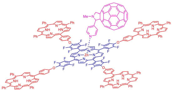
2.2. Formation of Porphyrin–Fullerene Complexes

The binding capabilities of pentaporphyrins 5 and Zn5 to form complexes with pristine C60 was investigated by absorption and fluorescence titrations in toluene. Surprisingly, the addition of a toluene solution of C60 to pentaporphyrins 5 and Zn5 did not induce a quantifiable alteration in their absorption and fluorescence spectra. This indicates that the eventual π-π interactions established between the host and C60 are too weak, in solution, to induce spectral modifications. However, the titration of pentaporphyrin 6 with PyC60 revealed a markedly distinct behavior. The addition of PyC60 to a solution of 6 in toluene induced a decrease of the Soret band (at 422 nm) along with the appearance of an isosbestic point at 428 nm (Figure 4). This suggests the formation of a 6–PyC60 complex, presumably via axial coordination of the pyridyl group to the zinc porphyrin (Figure 5), as reported in similar systems [56,68,69,70,71,72].
To evaluate the contribution of the four free porphyrin units in the stabilization of the 6–PyC60 complex, the precursor Zn2 and the diporphyrin 4 were also titrated with PyC60. As expected, the addition of PyC60 to Zn2 induced a red shift of the Soret and Q bands with the formation of an isosbestic point at 423 nm (Figure S14 in the ESI), probably as a result of axial coordination of the pyridyl group of PyC60 to Zn2. D’Souza already observed the complex formation between porphyrin 2 and PyC60 in dichloromethane [73]. Similar behavior was observed when a solution of diporphyrin 4 in toluene was titrated with PyC60. The incremental addition of PyC60 caused a decrease and a red shift of the Soret with the formation of an isosbestic point at 425 nm (Figure S16 in the ESI). A red shift was observed in the Q bands, which is most likely a consequence of coordination complex formation. As discussed below, the binding constant for the 6–PyC60 complex is ca. 5.8 times higher than the binding constant for the Zn2–PyC60 complex, which confirms that the four free-base porphyrin units in pentaporphyrin 6 have a significant contribution in the stabilization of the 6–PyC60 complex. The role of the four peripheral porphyrin units in stabilizing the 6–PyC60 complex may be compared to the synergic effect observed in C60 and C70 hosting behavior of corannulene- and pyrene-substituted porphyrins [74,75,76].
The steady state fluorescence spectrum of pentaporphyrin 6 displays two bands centered at 650 and 715 nm (Figure 6). Upon addition of PyC60 to a solution of 6 in toluene, the fluorescence intensity decreased significantly when exciting at 428 nm, as depicted in Figure 6. These changes are fully consistent with strong interactions between the photoexcited 6 and PyC60 in the 6–PyC60 complex [68,70,73]. The fluorescence quenching of the precursor Zn2 and diporphyrin 4 upon addition of PyC60 was also observed (Figures S15 and S17 in the ESI).
The binding constants (K) were obtained from the absorption and fluorescence spectral data by using a non-linear 1:1 binding model (Figure 4 and Figure 6) and are listed in Table 1. The average values for the binding constants (Kav) increase in the order Zn2 < 4 < 6. In particular, the Kav value of the 6–PyC60 complex (Kav = 1.53 × 10⁵ M−1) was found to be ca. 5.8 times higher than the Kav for the Zn2–PyC60 complex (Kav = 2.66 × 10⁴ M−1). As discussed above, the enhanced binding ability of the pentaporphyrin 6 arises presumably from the synergic effect of the π–π interactions between the fullerene unit and the four free-base porphyrin units.

3. Materials and Methods

3.1. Chemicals and Instrumentation

1H, 13C and 19F NMR spectra were recorded on Bruker AVANCE 300 or Bruker AVANCE 500 spectrometers. CDCl3 was use as solvent and tetramethylsilane (TMS) as internal reference. The chemical shifts are expressed in δ (ppm) and the coupling constants (J) in hertz (Hz). UV–Vis spectra were recorded on a Shimadzu UV-2501PC spectrophotometer using toluene or CHCl3 as solvent. UV−vis absorption spectral wavelengths (λ) are reported in nanometers (nm), and molar absorption coefficients (ε) are reported in M−1 cm−1. Fluorescence emission spectra were recorded on a JASCO FP-8300 spectrofluorometer. Mass spectra were recorded using a MALDI TOF/TOF 4800 Applied Biosystems MDS Sciex mass spectrometer, CHCl3 as solvent and 3-nitrobenzyl alcohol (NBA) as matrix. High-resolution mass spectra (HRMS) were recorded on a Bruker Apex-Qe FTICR mass spectrometer or on a LTQ Orbitrap XL mass spectrometer using CHCl3 as solvent. Melting points were measured on a Büchi B-540 apparatus and are uncorrected. Column chromatography was carried out using silica gel (Merck, 35-70 mesh). Analytical TLC was carried out on precoated sheets with silica gel (Merck 60, 0.2 mm thick). Solvents were purified or dried according to the literature procedures [77]. Compounds 1 [78], 2 [78] and PyC60 [79] were prepared according to published procedures.

3.2. Synthesis

3.2.1. 5,10,15,20-Tetrakis(pentafluorophenyl)porphyrinatozinc(II) (Zn2)

Prepared as in [73]. Zinc acetate (28.2 mg, 0.154 mmol) was added to a solution of 2 (50.0 mg, 51.3 μmol) in chloroform/methanol (2:1) and the resulting mixture was stirred at 60 °C for 15 min. After cooling down to ambient temperature, the reaction mixture was washed with distilled water. The organic phase was dried (Na2SO4) and the solvent was evaporated under reduced pressure. The Zn2 complex was obtained in quantitative yield. UV–Vis (toluene): λmax (log ε) 421 (5.5), 546 (4.2), 581 (3.7) nm. MS (MALDI-TOF): m/z 1036.0 [M]+.

3.2.2. Diporphyrin 3

A solution of 1 (20.0 mg, 31.7 μmol), 2 (92.7 mg, 95.1 μmol) and potassium carbonate (13.1 mg, 95.1 μmol) in dry DMSO (3 mL) was stirred under a nitrogen atmosphere at 50 °C for 3 h. After cooling to ambient temperature, the porphyrinic compounds were precipitated with an aqueous solution of citric acid, filtered and washed with water. The solid was dissolved in dichloromethane and then washed with water. The organic phase was dried (Na2SO4) and the solvent was evaporated under reduced pressure. The residue was purified by column chromatography on silica gel using a gradient of dichloromethane/hexane. The first fraction was identified as the unreacted porphyrin 2. The following fraction afforded diporphyrin 3 (28 mg, 55% yield) after crystallization from dichloromethane/methanol. mp > 300 °C. 1H NMR (CDCl3, 300 MHz): δ, ppm −2.87 (s, 2H, NH of moiety 1), −2.73 (s, 2H, NH of moiety 2), 7.71–7.82 (m, 11H, C6H4-m-H and Ph-m,p-H), 8.22–8.26 (m, 6H, Ph-o-H), 8.37 (d, J = 8.6 Hz, 2H, C6H4-o-H), 8.87 (s, 4H, β-H of moiety 1), 8.91–8.97 (3 overlapped d, 6H, β-H), 8.93 (s, 4H, β-H of moiety 2), 9.09 (d, J = 4.8 Hz, 2H, β-H of moiety 2). 13C NMR (CDCl3, 126 MHz): δ, ppm 103.6, 104.4, 114.4, 118.6, 120.3, 120.4, 126.7, 127.8, 131.1 (br s, β-C), 134.6, 135.9, 138.4, 142.1, 157.2. 19F NMR (CDCl3, 282 MHz): δ, ppm −184.83 (dt, J = 23.2 and 7.6 Hz, 6F, C6F5-m-F), −176.86 (dd, J = 23.1 and 9.3 Hz, 2F, C6F4-m-F), −174.82 to −174.65 (m, 3F, C6F5-p-F), −160.40 (dd, J = 23.1 and 9.3 Hz, 2F, C6F4-o-F), −160.00 (dd, J = 23.2 and 7.6 Hz, 6F, C6F5-o-F). UV–Vis (CHCl3): λmax (log ε) 418 (5.8), 510 (4.3), 548 (4.2), 586 (3.8), 644 (3.7) nm. MS (MALDI-TOF): m/z 1585.3 [M + H]+. MS (HRESI): m/z calcd. for C88H40F19N8O [M + H]+ 1585.3016, found 1585.3037, calcd. for C88H41F19N8O [M + 2H]2+ 793.1545, found 793.1534.

3.2.3. Diporphyrin 4

A solution of 1 (20.0 mg, 31.7 μmol), Zn2 (84.2 mg, 95.1 μmol) and potassium carbonate (13.1 mg, 95.1 μmol) in dry DMSO (3 mL) was stirred under a nitrogen atmosphere at 50 ºC for 4 h. The workup procedures were the same as previously described for diporphyrin 3. The residue was purified by column chromatography on silica gel using a gradient of dichloromethane/hexane. The first fraction was identified as the starting porphyrin Zn2. The following fraction afforded diporphyrin 4 (23 mg, 32% yield) after crystallization from dichloromethane/methanol. mp > 300 ºC. 1H NMR (CDCl3, 300 MHz): δ, ppm −2.74 (s, 2H, NH), 7.72–7.83 (m, 11H, C6H4-m-H and Ph-m,p-H), 8.23–8.27 (m, 6H, Ph-o-H), 8.38 (d, J = 8.5 Hz, 2H, C6H4-o-H), 8.88 (s, 4H, β-H of moiety 1), 8.93 and 8.98 (AB, J = 4.8 Hz, β-H of moiety 1), 9.01 (s, 4H, β-H of moiety Zn2), 9.04 and 9.17 (AB, J = 4.7 Hz, 4H, β-H of moiety Zn2). 19F NMR (CDCl3, 282 MHz): δ, ppm −184.75 (dt, J = 23.6 and 8.0 Hz, 6F, C6F5-m-F), −176.77 (dd, J = 23.4 and 9.4 Hz, 2F, C6F4-m-F), −175.03 to −174.85 (m, 3F, C6F5-p-F), −160.21 (dd, J = 23.4 and 9.4 Hz, 2F, C6F4-o-F), −159.85 (dd, J = 23.6 and 8.0 Hz, 6F, C6F5-o-F). UV–Vis (toluene): λmax (log ε) 421 (5,8), 513 (4,3), 546 (4,4), 584 (3,9), 646 (3,7) nm. MS (MALDI-TOF): m/z 1647.3 [M + H]+. MS (HRESI): m/z calcd. for C88H38F19N8OZn [M + H]+ 1647.2078, found 1647.2110.

3.2.4. Pentaporphyrin 5

A solution of 2 (10.0 mg, 10.2 μmol), 1 (32.4 mg, 51.3 μmol) and potassium carbonate (17.0 mg, 0.123 mmol) in dry DMSO (1 mL) was stirred under a nitrogen atmosphere at 80 °C for 2 h. After cooling to ambient temperature, the porphyrinic compounds were precipitated with an aqueous solution of citric acid, filtered and washed with water. The solid was dissolved in dichloromethane and then washed with water. The organic phase was dried (Na2SO4) and the solvent was evaporated under reduced pressure. The residue was purified by column chromatography on silica gel using dichloromethane/hexane (2:1) as the eluent. A major faction was eluted and then the unreacted porphyrin 1 was recovered. The major fraction afforded pentaporphyrin 5 (27 mg, 76% yield) after crystallization from dichloromethane/methanol. mp > 300 °C. 1H NMR (CDCl3, 300 MHz): δ, ppm −2.78 (s, 8H, NH of moiety 1), −2.69 (s, 2H, NH of moiety 2), 7.63–7.78 (m, 44H, C6H4-m-H and Ph-m,p-H), 8.15 (dd, J = 7.5 and 1.8 Hz, 16H, 10,20-Ph-o-H), 8.22 (dd, J = 7.3 and 1.7 Hz, 8H, 15-Ph-o-H), 8.33 (d, J = 8.7 Hz, 8H, C6H4-o-H), 8.81 (d, J = 6 Hz, 8H, β-H of moiety 1), 8.83–8.86 (m, 16H, β-H of moiety 1), 8.81 (d, J = 6 Hz, 8H, β-H of moiety 1), 9.16 (s, 8H, β-H of moiety 2). 13C NMR (CDCl3, 75 MHz): δ, ppm 104.3, 114.3, 116.7, 118.6, 120.2, 120.3, 126.6, 126.7, 127.7, 129.8–132.4 (β-C), 134.5, 135.9, 138.3, 142.0, 142.1, 157.1. 19F NMR (CDCl3, 282 MHz): δ, ppm -176.92 (dd, J = 23.1 and 9.2 Hz, 8F, C6F4-m-F), −160.33 (dd, J = 23.1 and 9.2 Hz, 8F, C6F4-o-F). UV–Vis (toluene): λmax (log ε) 420 (6.2), 513 (5.0), 547 (4.6), 590 (4.5), 648 (4.3) nm. MS (MALDI-TOF): m/z 3416.0 [M + H]+. MS (HRESI): m/z calcd. for C220H128F16N20O4 [M + 2H]2+ 1708.5081, found 1708.5129, calcd. for C220H129F16N20O4 [M + 3H]3+ 1139.3411, found 1139.3427.

3.2.5. Pentaporphyrin Zn5

Zinc acetate (16.1 mg, 87.8 mmol) was added to a solution of 5 (20.0 mg, 5.85 μmol) in chloroform/methanol (2:1) and the resulting mixture was stirred at 60 °C for 1 h. After cooling down to ambient temperature, the reaction mixture was washed with distilled water. The organic phase was dried (Na2SO4) and the solvent was evaporated under reduced pressure. The Zn5 complex was obtained in quantitative yield. 1H NMR (CDCl3, 300 MHz): δ, ppm 7.57–7.74 (m, 44H, C6H4-m-H and Ph-m,p-H), 8.09–8.18 (m, 24H, Ph-o-H), 8.28 (d, J = 8.6 Hz, 8H, C6H4-o-H), 8.80–8.86 (m, 24H, β-H of moiety 1), 8.92 (d, J = 4.7 Hz, 8H, β-H of moiety Zn1), 9.13 (s, 8H, β-H of moiety Zn2). UV–Vis (toluene): λmax (log ε) 424 (6.1), 548 (4.9), 588 (4.4) nm. MS (MALDI-TOF): m/z 3734,3 [M]+·.

3.2.6. Pentaporphyrin 6

A solution of Zn2 (10.0 mg, 11.3 μmol), 1 (35.6 mg, 56.4 μmol) and potassium carbonate (18.7 mg, 0.135 mmol) in dry DMSO (1 mL) was stirred under a nitrogen atmosphere at 80 ºC for 2 h. The workup procedures were the same as described for pentaporphyrin 5. The residue was purified by column chromatography on silica gel using dichloromethane/hexane (2:1) as the eluent. A major faction was eluted and then unreacted porphyrin 1 was recovered. The major fraction afforded pentaporphyrin 6 (25 mg, 64% yield) after crystallization from dichloromethane/hexane. mp > 300 ºC. 1H NMR (CDCl3, 300 MHz): δ, ppm -2.78 (s, 8H, NH), 7.60–7.78 (m, 44H, 5-C6H4-m-H and 10,15,20-Ph-m,p-H), 8.15 (dd, J = 7.4 and 1.8 Hz, 16H, 10,20-Ph-o-H), 8.22 (dd, J = 7.4 and 1.7 Hz, 8H, 15-Ph-o-H), 8.34 (d, J = 8.5 Hz, 8H, 5-C6H4-o-H), 8.80–8.86 (m, 24H, β-H of moiety 1), 8.93 (d, J = 4.8 Hz, 8H, β-H of moiety 1), 9.25 (s, 8H, β-H of moiety Zn2). 13C NMR (CDCl3, 126 MHz): δ, ppm 105.0, 114.3, 117.4, 117.6, 117.7, 118.7, 120.28, 120.34, 126.65, 126.73, 127.7, 127.8, 129.8–132.3 (β-C), 132.4, 134.5, 134.6, 135.9, 138.3, 140.6, 140.8, 142.0, 142.1, 142.7, 142.8, 146.0, 148.1, 150.4, 157.2. 19F NMR (CDCl3, 282 MHz): δ, ppm -176.81 (dd, J = 23.4 and 9.2 Hz, 8F, C6F4-m-F), -160.14 (dd, J = 23.4 and 9.2 Hz, 8F, C6F4-o-F). UV–Vis (toluene): λmax (log ε) 422 (6.1), 513 (4.8), 549 (4.7), 589 (4.4), 645 (4.3) nm. MS (MALDI-TOF): m/z 3477.9 [M + H]+.

3.3. Absorption and Fluorescence Titrations

The UV–Vis and fluorescence titrations were carried out using a stock solution of each compound in toluene in a quartz cuvette (1 cm path length). A PyC60 stock solution was added in aliquots and the spectra were recorded after each addition. Absorption spectra were corrected for the contribution of the added PyC60. In the fluorescence measurements the sample was excited at the isosbestic point. Measurements were repeated 2–3 times and found to be reproducible within a margin of error of ca. 15%. All measurements were performed at 23 °C.
The binding constants (K) were assessed from the UV–Vis and fluorescence titrations. The variation of the absorbance at the Soret band caused by consecutive additions of PyC60 was used to determine the binding constants. The spectral changes at the Soret band were fit using non-linear least-squares procedures using the following equation for a 1:1 binding:
A = ε Por [ Por ] + ε Por Ful K [ Por ] [ Ful ] 1 + K [ Ful ]
in which A is the absorbance, [Por] is the total concentration of the porphyrin derivative, [Ful] is the concentration of PyC60, εPor and εPor-Ful are constants associated with the molar extinction coefficients of the porphyrin derivative and the complex formed, respectively.
In a similar way, the spectral changes at maximum fluorescence intensity were fit using non-linear least-squares procedures using the following equation for 1:1 binding:
I = Φ Por [ Por ] + Φ Por Ful K [ Por ] [ Ful ] 1 + K [ Ful ]
where I is the fluorescence intensity, ΦPor and ΦPor-Ful are constants associated with the emission quantum yields of the porphyrin derivative and of the complex formed, respectively [80].

3.4. Single-Crystal X-Ray Diffraction Studies of Zn5·Solvent

Single crystals of compound Zn5·Solvent were manually harvested from the crystallization vial and immersed in highly viscous FOMBLIN Y perfluoropolyether vacuum oil (LVAC 140/13, Sigma-Aldrich) to avoid degradation caused by the evaporation of the solvent [81]. Crystals were mounted on Hampton Research CryoLoops with the help of a Stemi 2000 stereomicroscope equipped with Carl Zeiss lenses. X-ray diffraction data were collected at 179(2) K on a Bruker D8 QUEST equipped with Mo Kα sealed tube (λ = 0.71073 Å), a multilayer TRIUMPH X-ray mirror, a PHOTON 100 CMOS detector, and an Oxford Instruments Cryostrem 700+ Series low temperature device. Diffraction images were processed using the software package SAINT+ [82], and data were corrected for absorption by the multiscan semi-empirical method implemented in SADABS [83]. The structure was solved using the algorithm implemented in SHELXT-2014 [84], which allowed for the immediate location of almost all of the heaviest atoms composing the molecular unit. The remaining missing and misplaced non-hydrogen atoms were located from difference Fourier maps calculated from successive full-matrix least-squares refinement cycles on F2 using the latest SHELXL from the 2017/1 release [85]. All structural refinements were performed using the graphical interface ShelXle [86].
The crystal diffracted poorly throughout the entire angular range, this despite a long acquisition time per frame (ca. 1 min) and the use of a wide frame strategy (0.5° ϕ scans). Indeed, below 1.0 Å of resolution the mean I/σ drops below 5.0 with the Rmerge value being concomitantly high (for example, for a resolution of 1.20 Å the Rmerge is already above 10%). The poor quality of the overall diffraction has important consequences in the modeled structure, giving rise to a number of alerts in PLATON [87,88]. Nevertheless, structure solution immediately showed the presence of the most important chemical moiety in the crystal structure.
Besides the pentaporphyrin Zn5 derivative, the crystal structure contains one large void and two smaller ones (total volume of about 2446 Å3 as estimated by PLATON) [87,88] centred at (0.385 0.3305 0.000), (0.263 −0.819 0.417), and (0.736 0.181 0.583), which most likely contain highly disordered solvent molecules. From difference Fourier maps it was possible to discern the presence of a considerable smeared-out electron density in these locations. Nevertheless, several attempts to locate and model solvent molecules proved to be unproductive. The original data set was treated using the SQUEEZE [89] subroutines implemented in PLATON [87,88] in order to remove the contribution of these highly disordered molecules. It was estimated that the aforementioned cavities would contain a total of ca. 632 electrons. The calculated solvent-free reflection list was used for subsequent structural refinements that converged to the solvent-free structure reported in this manuscript and having the reliability factors listed below.
The asymmetric unit is composed of half of the pentaporphyrin in which the metallic centres are coordinated to a methanol molecule. These three crystallographically independent methanol molecules were found to be severely affected by positional disorder and were included in the final structural model with the distances restrained to common (refineable) distances and with isotropic displacement parameters for the non-hydrogen atoms, so as to ensure chemically reasonable geometries for these moieties.
Hydrogen atoms bound to carbon were placed at their idealized positions using appropriate HFIX instructions in SHELXL: 43 (aromatic carbon atoms and coordinated hydroxyl groups) and 137 (for all methyl groups). These hydrogen atoms were included in subsequent refinement cycles with isotropic thermal displacements parameters (Uiso) fixed at 1.2 (for the former family of hydrogen atoms) or 1.5×Ueq (solely for those associated with the methyl groups) of the parent atoms.
The last difference Fourier map synthesis showed the highest peak (1.947 eÅ−3) and the deepest hole (-1.354 eÅ−3) located at 0.39 and 0.07 Å from H2M and C2M (associated with the coordinated methanol molecule), respectively. Structural drawings have been created using the software package Crystal Impact Diamond [90].
Crystal data for Zn5·Solvent (SQUEEZE Data): C225H136F16N20O9Zn5, M = 3894.38, triclinic, space group Pī, Z = 1, a = 12.995(2) Å, b = 15.034(3) Å, c = 34.607(7) Å, α = 78.338(10)°, β = 81.133(9)°, γ = 79.449(10)°, V = 6461(2) Å3, μ(Mo-Kα) = 0.518 mm−1, Dc = 1.001 g cm−3, red block with crystal size of 0.20 × 0.10 × 0.05 mm3. Of a total of 127,498 reflections collected, 23,500 were independent (Rint = 0.0640). Final R1 = 0.0769 [I > 2σ(I)] and wR2 = 0.2353 (all data). Data completeness to theta = 25.24°, 99.5%.
Crystallographic data (including structure factors) for the crystal structure of compound Zn5·Solvent have been deposited with the Cambridge Crystallographic Data Centre as supplementary publication No. CCDC-1825479. Copies of the data can be obtained free of charge on application to CCDC, 12 Union Road, Cambridge CB2 2EZ, UK. FAX: (+44) 1223 336033. E-mail: [email protected].

4. Conclusions

The reaction of 5-(4-hydroxyphenyl)-10,15,20-triphenylporphyrin (1) with 5,10,15,20-tetrakis(pentafluorophenyl)porphyrin (2), under mild conditions affords a diporphyrin (3) or a pentaporphyrin (5) selectively and in good yields. Similarly, the reaction of 1 with Zn2 leads to the nono-Zn(II) diporphyrin 4 or to the mono-Zn(II) pentaporphyrin 6.
Absorption and fluorescence titrations of the new porphyrin derivatives with C60 in toluene did not show the formation of supramolecular complexes. However, titrations with PyC60 lead to the formation of Zn(porphyrin)–PyC60 coordination complexes and the corresponding binding constants were calculated. Comparing the binding constants for the Zn2–PyC60 complex (Kav = 2.66 × 10⁴ M−1) and the 6–PyC60 complex (Kav = 1.53 × 10⁵ M−1), it is evident that the four free-base porphyrin units in pentaporphyrin 6 have a significant contribution in the stabilization of the 6–PyC60 complex.
The method reported here for the synthesis of pentaporphyrins 5 and 6 may be useful to decorate a central porphyrin with one to four structural units with specific functions. Considering that porphyrin–fullerene systems are potentially useful for photovoltaic applications, this method may be used to decorate a porphyrin with other photoactive units or substituents able to make strong binding interactions with fullerenes (pyrene of corannulene units, for instance).

Supplementary Materials

The following are available online: 1H, 13C and 19F NMR spectra and absorption and fluorescence titrations with PyC60.

Author Contributions

Conceptualization, A.C.T.; synthesis of the porphyrin derivatives, J.I.T.C.; absorption and fluorescence titrations, A.S.F.F.; single-crystal X-ray diffraction studies, F.A.A.P.

Funding

This research was funded by Fundação para a Ciência e a Tecnologia (FCT), through project PTDC/QEQ-QOR/6160/2014 and the research units UID/QUI/00062/2019 and UID/CTM/50011/2019.

Acknowledgments

Thanks are due to the University of Aveiro and Fundação para a Ciência e a Tecnologia (FCT, Portugal) for the financial support to the project PTDC/QEQ-QOR/6160/2014 and the research units UID/QUI/00062/2019 and UID/CTM/50011/2019 through national funds and where applicable co-financed by the FEDER, within the PT2020 Partnership Agreement, and also to the Portuguese NMR Network.

Conflicts of Interest

The authors declare no conflict of interest.

References

  1. Aratani, N.; Takagi, A.; Yanagawa, Y.; Matsumoto, T.; Kawai, T.; Yoon, Z.S.; Kim, D.; Osuka, A. Giant meso–meso-linked porphyrin arrays of micrometer molecular length and their fabrication. Chem. Eur. J. 2005, 11, 3389–3404. [Google Scholar] [CrossRef] [PubMed]
  2. Beletskaya, I.; Tyurin, V.S.; Tsivadze, A.Y.; Guilard, R.; Stern, C. Supramolecular chemistry of metalloporphyrins. Chem. Rev. 2009, 109, 1659–1713. [Google Scholar] [CrossRef] [PubMed]
  3. Drain, C.M.; Varotto, A.; Radivojevic, I. Self-organized porphyrinic materials. Chem. Rev. 2009, 109, 1630–1658. [Google Scholar] [CrossRef] [PubMed]
  4. Balaban, T.S. Self-assembling porphyrins and chlorins as synthetic mimics of the chlorosomal bacteriochlorophylls. In Handbook of Porphyrin Science; Kadish, K.M., Smith, K.M., Guilard, R., Eds.; World Scientific: Singapore, 2010; Volume 1, Chapter 3; pp. 221–306. [Google Scholar]
  5. Morisue, M.; Kobuke, Y. Supramolecular organization of porphyrins and phthalocyanines by use of biomimetic coordination methodology. In Handbook of Porphyrin Science; Kadish, K.M., Smith, K.M., Guilard, R., Eds.; World Scientific: Singapore, 2014; Volume 32, Chapter 166; pp. 1–126. [Google Scholar]
  6. Bessmertnykh-Lemeune, A.G.; Guilard, R.; Stern, C.; Enakieva, Y.Y.; Gorbunova, Y.G.; Tsivadze, A.Y.; Nefedov, S.E. Biomimetic studies of porphyrin self-assembled systems. In Supramolecular Systems: Chemistry, Types and Applications; Pena, C., Ed.; Nova Science Publishers: Hauppauge, NY, USA, 2017; Chapter 4; pp. 213–278. [Google Scholar]
  7. Jeong, Y.-H.; Son, M.; Yoon, H.; Kim, P.; Lee, D.-H.; Kim, D.; Jang, W.-D. Guest-induced photophysical property switching of artificial light-harvesting dendrimers. Angew. Chem. Int. Ed. 2014, 53, 6925–6928. [Google Scholar] [CrossRef] [PubMed]
  8. Lee, H.; Jeong, Y.-H.; Kim, J.-H.; Kim, I.; Lee, E.; Jang, W.-D. Supramolecular coordination polymer formed from artificial light-harvesting dendrimer. J. Am. Chem. Soc. 2015, 137, 12394–12399. [Google Scholar] [CrossRef] [PubMed]
  9. Hasobe, T.; Ida, K.; Sakai, H.; Ohkubo, K.; Fukuzumi, S. Coronenetetraimide-centered cruciform pentamers containing multiporphyrin units: Synthesis and sequential photoinduced energy- and electron-transfer dynamics. Chem. Eur. J. 2015, 21, 11196–11205. [Google Scholar] [CrossRef] [PubMed]
  10. Yim, D.; Sung, J.; Kim, S.; Oh, J.; Yoon, H.; Sung, Y.M.; Kim, D.; Jang, W.-D. Guest-induced modulation of the energy transfer process in porphyrin-based artificial light harvesting dendrimers. J. Am. Chem. Soc. 2017, 139, 993–1002. [Google Scholar] [CrossRef]
  11. Terazono, Y.; Kodis, G.; Chachisvilis, M.; Cherry, B.R.; Fournier, M.; Moore, A.; Moore, T.A.; Gust, D. Multiporphyrin arrays with π-π interchromophore interactions. J. Am. Chem. Soc. 2015, 137, 245–258. [Google Scholar] [CrossRef]
  12. Delavaux-Nicot, B.; Aziza, H.B.; Nierengarten, I.; Trinh, T.M.N.; Meichsner, E.; Chessé, M.; Holler, M.; Abidi, R.; Maisonhaute, E.; Nierengarten, J.-F. A rotaxane scaffold for the construction of multiporphyrinic light-harvesting devices. Chem. Eur. J. 2018, 24, 133–144. [Google Scholar] [CrossRef]
  13. Amati, A.; Cavigli, P.; Demitri, N.; Natali, M.; Indelli, M.T.; Iengo, E. Sn(IV) multiporphyrin arrays as tunable photoactive systems. Inorg. Chem. 2019, 58, 4399–4411. [Google Scholar] [CrossRef]
  14. Tanaka, T.; Osuka, A. Conjugated porphyrin arrays: Synthesis, properties and applications for functional materials. Chem. Soc. Rev. 2015, 44, 943–969. [Google Scholar] [CrossRef] [PubMed]
  15. Iengo, E.; Cavigli, P.; Milano, D.; Tecilla, P. Metal mediated self-assembled porphyrin metallacycles: Synthesis and multipurpose applications. Inorg. Chim. Acta 2014, 417, 59–78. [Google Scholar] [CrossRef]
  16. Wytko, J.A.; Ruppert, R.; Jeandon, C.; Weiss, J. Metal-mediated linear self-assembly of porphyrins. Chem. Commun. 2018, 54, 1550–1558. [Google Scholar] [CrossRef] [PubMed] [Green Version]
  17. Li, Y.; Wang, L.; Gao, Y.; Yang, W.; Li, Y.; Guo, C. Porous metalloporphyrinic nanospheres constructed from metal 5,10,15,20-tetraksi(4’-ethynylphenyl)porphyrin for efficient catalytic degradation of organic dyes. RSC Adv. 2018, 8, 7330–7339. [Google Scholar] [CrossRef]
  18. Mongin, O.; Sankar, M.; Charlot, M.; Mir, Y.; Blanchard-Desce, M. Strong enhancement of two-photon absorption properties in synergic ‘semi-disconnected’ multiporphyrin assemblies designed for combined imaging and photodynamic therapy. Tetrahedron Lett. 2013, 54, 6474–6478. [Google Scholar] [CrossRef]
  19. Aratani, N.; Osuka, A. Exploration of giant functional porphyrin arrays. Bull. Chem. Soc. Jpn. 2015, 88, 1–27. [Google Scholar] [CrossRef]
  20. Lacerda, P.S.S.; Silva, A.M.G.; Tomé, A.C.; Neves, M.G.P.M.S.; Silva, A.M.S.; Cavaleiro, J.A.S.; Llamas-Saiz, A.L. [1,2,3]Triazolo[4,5-b]porphyrins: New building blocks for porphyrinic materials. Angew. Chem. Int. Ed. 2006, 45, 5487–5491. [Google Scholar] [CrossRef]
  21. Pedro-Hernández, L.D.; Cortez-Maya, S.; Calderón-Pardo, J.; Hernández-Ortega, S.; Martínez-García, M. Nanostructured multiporphyrin dendrimers: Synthesis, characterization and their spectroscopic properties. Curr. Org. Chem. 2018, 22, 2308–2314. [Google Scholar] [CrossRef]
  22. Martin, M.M.; Dill, M.; Langer, J.; Jux, N. Porphyrin−hexaphenylbenzene conjugates via mixed cyclotrimerization reactions. J. Org. Chem. 2019, 84, 1489–1499. [Google Scholar] [CrossRef]
  23. Yang, J.; Lee, J.-E.; Lee, C.Y.; Aratani, N.; Osuka, A.; Hupp, J.T.; Kim, D. The role of electronic coupling in linear porphyrin arrays probed by single-molecule fluorescence spectroscopy. Chem. Eur. J. 2011, 17, 9219–9225. [Google Scholar] [CrossRef]
  24. Morisue, M.; Hoshino, Y.; Shimizu, M.; Uemura, S.; Sakurai, S. A tightly stretched ultralong supramolecular multiporphyrin array propagated by double-strand formation. Chem. Eur. J. 2016, 22, 13019–13022. [Google Scholar] [CrossRef] [PubMed]
  25. Kamonsutthipaijit, N.; Anderson, H.L. Template-directed synthesis of linear porphyrin oligomers: Classical, Vernier and mutual Vernier. Chem. Sci. 2017, 8, 2729–2740. [Google Scholar] [CrossRef] [PubMed]
  26. Zhang, S.; Kim, J.O.; Li, Y.; Wen, B.; Zhou, M.; Liu, S.; Aratani, N.; Xu, L.; Kim, D.; Song, J. Meso-to-meso 2,5-Pyrrolylene bridged zig-zag porphyrin arrays. Chem. Commun. 2017, 53, 11488–11491. [Google Scholar] [CrossRef]
  27. He, Z.; Ishizuka, T.; Jiang, D. Dendritic architectures for design of photo- and spin-functional nanomaterials. Polymer J. 2007, 39, 889–922. [Google Scholar] [CrossRef]
  28. Li, W.-S.; Aida, T. Dendrimer porphyrins and phthalocyanines. Chem. Rev. 2009, 109, 6047–6076. [Google Scholar] [CrossRef] [PubMed]
  29. Nakamura, Y.; Jang, S.Y.; Tanaka, T.; Aratani, N.; Lim, J.M.; Kim, K.S.; Kim, D.; Osuka, A. Two-dimensionally extended porphyrin tapes: Synthesis and shape-dependent two-photon absorption properties. Chem. Eur. J. 2008, 14, 8279–8289. [Google Scholar] [CrossRef]
  30. Tanaka, T.; Lee, B.S.; Aratani, N.; Yoon, M.-C.; Kim, D.; Osuka, A. Synthesis and properties of hybrid porphyrin tapes. Chem. Eur. J. 2011, 17, 14400–14412. [Google Scholar] [CrossRef] [PubMed]
  31. Mori, H.; Tanaka, T.; Lee, S.; Lim, J.M.; Kim, D.; Osuka, A. Meso-meso Linked porphyrin−[26]hexaphyrin−porphyrin hybrid arrays and their triply linked tapes exhibiting strong absorption bands in the NIR region. J. Am. Chem. Soc. 2015, 137, 2097–2106. [Google Scholar] [CrossRef]
  32. Xue, S.; Kuzuhara, D.; Aratani, N.; Yamada, H. Synthesis of a porphyrin(2.1.2.1) nanobelt and its ability to bind fullerene. Org. Lett. 2019, 21, 2069–2072. [Google Scholar] [CrossRef]
  33. Song, J.; Aratani, N.; Shinokubo, H.; Osuka, A. A porphyrin nanobarrel that encapsulates C60. J. Am. Chem. Soc. 2010, 132, 16356–16357. [Google Scholar] [CrossRef]
  34. Aratani, N.; Kim, D.; Osuka, A. Discrete cyclic porphyrin arrays as artificial light-harvesting antenna. Acc. Chem. Res. 2009, 42, 1922–1934. [Google Scholar] [CrossRef] [PubMed]
  35. O’Sullivan, M.C.; Sprafke, J.K.; Kondratuk, D.V.; Rinfray, C.; Claridge, T.D.W.; Saywell, A.; Blunt, M.O.; O’Shea, J.N.; Beton, P.H.; Malfois, M.; et al. Vernier templating and synthesis of a 12-porphyrin nano-ring. Nature 2011, 469, 72–75. [Google Scholar] [CrossRef] [PubMed]
  36. Jiang, H.-W.; Ham, S.; Aratani, N.; Kim, D.; Osuka, A. A 1,3-phenylene-bridged hexameric porphyrin wheel and efficient excitation energy transfer along the wheel. Chem. Eur. J. 2013, 19, 13328–13336. [Google Scholar] [CrossRef] [PubMed]
  37. Rousseaux, S.A.L.; Gong, J.Q.; Haver, R.; Odell, B.; Claridge, T.D.W.; Herz, L.M.; Anderson, H.L. Self-assembly of russian doll concentric porphyrin nanorings. J. Am. Chem. Soc. 2015, 137, 12713–12718. [Google Scholar] [CrossRef] [PubMed]
  38. Kondratuk, D.V.; Perdigão, L.M.A.; Esmail, A.M.S.; O’Shea, J.N.; Beton, P.H.; Anderson, H.L. Supramolecular nesting of cyclic polymers. Nat. Chem. 2015, 7, 317–322. [Google Scholar] [CrossRef] [Green Version]
  39. Favereau, L.; Cnossen, A.; Kelber, J.B.; Gong, J.Q.; Oetterli, R.M.; Cremers, J.; Herz, L.M.; Anderson, H.L. Six-coordinate zinc porphyrins for template-directed synthesis of spiro-fused nanorings. J. Am. Chem. Soc. 2015, 137, 14256–14259. [Google Scholar] [CrossRef] [PubMed]
  40. Wang, S.-P.; Shen, Y.-F.; Zhu, B.-Y.; Wu, J.; Li, S. Recent advances in the template-directed synthesis of porphyrin nanorings. Chem. Commun. 2016, 52, 10205–10216. [Google Scholar] [CrossRef]
  41. Liu, P.; Hisamune, Y.; Peeks, M.D.; Odell, B.; Gong, J.Q.; Herz, L.M.; Anderson, H.L. Synthesis of five-porphyrin nanorings by using ferrocene and corannulene templates. Angew. Chem. Int. Ed. 2016, 55, 8358–8362. [Google Scholar] [CrossRef]
  42. Kuramochi, Y.; Kawakami, Y.; Satake, A. Synthesis and photophysical properties of porphyrin macrorings composed of free-base porphyrins and slipped-cofacial zinc porphyrin dimers. Inorg. Chem. 2017, 56, 11008–11018. [Google Scholar] [CrossRef]
  43. Morley, D.O.; Malfois, M.; Kamonsutthipaijit, N.; Kondratuk, D.V.; Anderson, H.L.; Wilson, M. A coarse-grained model for free and template-bound porphyrin nanorings. J. Phys. Chem. A 2017, 121, 5907–5920. [Google Scholar] [CrossRef]
  44. Bols, P.S.; Anderson, H.L. Shadow mask templates for site-selective metal exchange in magnesium porphyrin nanorings. Angew. Chem. Int. Ed. 2018, 57, 7874–7877. [Google Scholar] [CrossRef] [PubMed]
  45. Mukhopadhyay, R.D.; Kim, Y.; Koo, J.; Kim, K. Porphyrin boxes. Acc. Chem. Res. 2018, 51, 2730–2738. [Google Scholar] [CrossRef] [PubMed]
  46. Cremers, J.; Haver, R.; Rickhaus, M.; Gong, J.Q.; Favereau, L.; Peeks, M.D.; Claridge, T.D.W.; Herz, L.M.; Anderson, H.L. Template-directed synthesis of a conjugated zinc porphyrin nanoball. J. Am. Chem. Soc. 2018, 140, 5352–5355. [Google Scholar] [CrossRef] [PubMed]
  47. Guldi, D.M. Fullerenes: Three dimensional electron acceptor materials. Chem. Commun. 2000, 321–327. [Google Scholar] [CrossRef]
  48. Guldi, D.M. Fullerene–porphyrin architectures; photosynthetic antenna and reaction center models. Chem. Soc. Rev. 2002, 31, 22–36. [Google Scholar] [CrossRef] [PubMed]
  49. Mironov, A.F. Synthesis, properties, and potential applications of porphyrin–fullerenes. Macroheterocycles 2011, 4, 186–208. [Google Scholar] [CrossRef]
  50. Santos, J.; Illescas, B.M.; Wielopolski, M.; Silva, A.M.G.; Tomé, A.C.; Guldi, D.M.; Martín, N. Efficient electron transfer in β-substituted porphyrin-C60 dyads connected through a p-phenylenevinylene dimer. Tetrahedron 2008, 64, 11404–11408. [Google Scholar] [CrossRef]
  51. Enes, R.F.; Cid, J.-J.; Hausmann, A.; Trukhina, O.; Gouloumis, A.; Vázquez, P.; Cavaleiro, J.A.S.; Tomé, A.C.; Guldi, D.M.; Torres, T. Synthesis and photophysical properties of fullerene–phthalocyanine–porphyrin triads and pentads. Chem. Eur. J. 2012, 18, 1727–1736. [Google Scholar] [CrossRef]
  52. Hasobe, T. Porphyrin-based supramolecular nanoarchitectures for solar energy conversion. J. Phys. Chem. Lett. 2013, 4, 1771–1780. [Google Scholar] [CrossRef]
  53. Schlundt, S.; Bauer, W.; Hirsch, A. Synthesis and atropisomerism of cascaded tetraphenylporphyrin–[60]fullerene hybrids. Chem. Eur. J. 2015, 21, 12421–12430. [Google Scholar] [CrossRef]
  54. Gao, D.; Aly, S.M.; Karsenti, P.-L.; Brisard, G.; Harvey, P.D. Increasing the lifetimes of charge separated states in porphyrin–fullerene polyads. Phys. Chem. Chem. Phys. 2017, 19, 24018–24028. [Google Scholar] [CrossRef] [PubMed]
  55. Tashiro, K.; Aida, T. Metalloporphyrin hosts for supramolecular chemistry of fullerenes. Chem. Soc. Rev. 2007, 36, 189–197. [Google Scholar] [CrossRef] [PubMed]
  56. Trabolsi, A.; Urbani, M.; Delgado, J.L.; Ajamaa, F.; Elhabiri, M.; Solladié, N.; Nierengarten, J.-F.; Albrecht-Gary, A.-M. Large photoactive supramolecular ensembles prepared from C60–pyridine substrates and multi-Zn(II)–porphyrin receptors. New J. Chem. 2008, 32, 159–165. [Google Scholar] [CrossRef]
  57. Tong, L.H.; Wietor, J.-L.; Clegg, W.; Raithby, P.R.; Pascu, S.I.; Sanders, J.K.M. Supramolecular assemblies of tripodal porphyrin hosts and C60. Chem. Eur. J. 2008, 14, 3035–3044. [Google Scholar] [CrossRef] [PubMed]
  58. Nobukuni, H.; Shimazaki, Y.; Uno, H.; Naruta, Y.; Ohkubo, K.; Kojima, T.; Fukuzumi, S.; Seki, S.; Sakai, H.; Hasobe, T.; et al. Supramolecular structures and photoelectronic properties of the inclusion complex of a cyclic free-base porphyrin dimer and C60. Chem. Eur. J. 2010, 16, 11611–11623. [Google Scholar] [CrossRef] [PubMed]
  59. Gil-Ramírez, G.; Karlen, S.D.; Shundo, A.; Porfyrakis, K.; Ito, Y.; Briggs, G.A.D.; Morton, J.J.L.; Anderson, H.L. A cyclic porphyrin trimer as a receptor for fullerenes. Org. Lett. 2010, 12, 3544–3547. [Google Scholar] [CrossRef] [PubMed]
  60. Song, J.; Aratani, N.; Shinokubo, H.; Osuka, A. A β-to-β 2,5-thienylene-bridged cyclic porphyrin tetramer: Its rational synthesis and 1:2 binding mode with C60. Chem. Sci. 2011, 2, 748–751. [Google Scholar] [CrossRef]
  61. Hernández-Eguía, L.P.; Escudero-Adán, E.C.; Pintre, I.C.; Ventura, B.; Flamigni, L.; Ballester, P. Supramolecular inclusion complexes of two cyclic zinc bisporphyrins with C60 and C70: Structural, thermodynamic, and photophysical characterization. Chem. Eur. J. 2011, 17, 14564–14577. [Google Scholar] [CrossRef]
  62. Fukuzumi, S.; Saito, K.; Ohkubo, K.; Troiani, V.; Qiu, H.; Gadde, S.; D’Souza, F.; Solladié, N. Multiple photosynthetic reaction centres using zinc porphyrinic oligopeptide–fulleropyrrolidine supramolecular complexes. Phys. Chem. Chem. Phys. 2011, 13, 17019–17022. [Google Scholar] [CrossRef]
  63. Nair, V.S.; Pareek, Y.; Karunakaran, V.; Ravikanth, M.; Ajayaghosh, A. Cyclotriphosphazene appended porphyrins and fulleropyrrolidine complexes as supramolecular multiple photosynthetic reaction centers: Steady and excited states photophysical investigation. Phys. Chem. Chem. Phys. 2014, 16, 10149–10156. [Google Scholar] [CrossRef]
  64. Calderon, R.M.K.; Valero, J.; Grimm, B.; Mendoza, J.; Guldi, D.M. Enhancing molecular recognition in electron donor-acceptor hybrids via cooperativity. J. Am. Chem. Soc. 2014, 136, 11436–11443. [Google Scholar] [CrossRef] [PubMed]
  65. Amati, A.; Cavigli, P.; Kahnt, A.; Indelli, M.T.; Iengo, E. Self-assembled ruthenium(II)porphyrin-aluminium(III)porphyrin-fullerene triad for long-lived photoinduced charge separation. J. Phys. Chem. A 2017, 121, 4242–4252. [Google Scholar] [CrossRef] [PubMed]
  66. Costa, J.I.T.; Tomé, A.C.; Neves, M.G.P.M.S.; Cavaleiro, J.A.S. 5,10,15,20-Tetrakis(pentafluorophenyl)porphyrin: A versatile platform to novel porphyrinic materials. J. Porphyrins Phthalocyanines 2011, 15, 1116–1133. [Google Scholar] [CrossRef]
  67. Costa, J.I.T.C.; Oliveira, E.; Santos, H.M.; Tomé, A.C.; Neves, M.G.P.M.S.; Lodeiro, C. Study of multiporphyrin compounds as colorimetric sitting-atop metal complexes: Synthesis and photophysical studies. ChemPlusChem 2016, 81, 143–153. [Google Scholar] [CrossRef]
  68. Costa, J.I.T.; Farinha, A.S.F.; Neves, M.G.P.M.S.; Tomé, A.C. An easy access to porphyrin triads and their supramolecular interaction with a pyridyl [60]fulleropyrrolidine. Dyes Pigments 2016, 135, 163–168. [Google Scholar] [CrossRef]
  69. D’Souza, F.; Deviprasad, G.R.; Zandler, M.E.; Hoang, V.T.; Klykov, A.; VanStipdonk, M.; Perera, A.; El-Khouly, M.E.; Fujitsuka, M.; Ito, O. Spectroscopic, electrochemical, and photochemical studies of self-assembled via axial coordination zinc porphyrin–fulleropyrrolidine dyads. J. Phys. Chem. A 2002, 106, 3243–3252. [Google Scholar] [CrossRef]
  70. Tat, F.T.; Zhou, Z.; MacMahon, S.; Song, F.; Rheingold, A.L.; Echegoyen, L.; Schuster, D.I.; Wilson, S.R. A new fullerene complexation ligand: N-pyridylfulleropyrrolidine. J. Org. Chem. 2004, 69, 4602–4606. [Google Scholar] [CrossRef]
  71. Takai, A.; Chkounda, M.; Eggenspiller, A.; Gros, C.P.; Lachkar, M.; Barbe, J.M.; Fukuzumi, S. Efficient photoinduced electron transfer in a porphyrin tripod-fullerene supramolecular complex via π-π interactions in nonpolar media. J. Am. Chem. Soc. 2010, 132, 4477–4489. [Google Scholar] [CrossRef]
  72. Jain, K.; Duvva, N.; Badgurjar, D.; Giribabu, L.; Chitta, R. Synthesis and spectroscopic studies of axially bound tetra(phenothiazinyl)/tetra(bis(4′-tert-butylbiphenyl-4-yl)aniline)-zinc(II)porphyrin-fullero[C60 & C70]pyrrolidine donor–acceptor triads. Inorg. Chem. Commun. 2016, 66, 5–10. [Google Scholar] [CrossRef]
  73. Das, S.K.; Song, B.; Mahler, A.; Nesterov, V.N.; Wilson, A.K.; Ito, O.; D’Souza, F. Electron transfer studies of high potential zinc porphyrin−fullerene supramolecular dyads. J. Phys. Chem. C 2014, 118, 3994–4006. [Google Scholar] [CrossRef]
  74. Denis, P.A.; Yanney, M. Porphyrins bearing corannulene pincers: Outstanding fullerene receptors. RSC Adv. 2016, 6, 50978–50984. [Google Scholar] [CrossRef]
  75. Álvarez, C.M.; Barbero, H.; Ferrero, S.; Miguel, D. Synergistic effect of tetraaryl porphyrins containing corannulene and other polycyclic aromatic fragments as hosts for fullerenes. Impact of C60 in a statistically distributed mixture of atropisomers. J. Org. Chem. 2016, 81, 6081–6086. [Google Scholar] [CrossRef] [PubMed]
  76. Ferrero, S.; Barbero, H.; Miguel, D.; García-Rodríguez, R.; Álvarez, C.M. Dual-tweezer behavior of an octapodal pyrene porphyrin-based system as a host for fullerenes. J. Org. Chem. 2019, 84, 6183–6190. [Google Scholar] [CrossRef] [PubMed]
  77. Armarego, W.L.F.; Perrin, D.D. Purification of Laboratory Chemicals, 4th ed.; Butterworth-Heinemann: Oxford, UK, 1996. [Google Scholar]
  78. Tomé, J.P.C.; Neves, M.G.P.M.S.; Tomé, A.C.; Cavaleiro, J.A.S.; Mendonça, A.F.; Pegado, I.N.; Duarte, R.; Valdeira, M.L. Synthesis of glycoporphyrin derivatives and their antiviral activity against herpes simplex virus types 1 and 2. Bioorg. Med. Chem. 2005, 13, 3878–3885. [Google Scholar] [CrossRef] [PubMed]
  79. Prato, M.; Maggini, M.; Giacometti, C.; Scorrano, G.; Sandonà, G.; Farnia, G. Synthesis and electrochemical properties of substituted fulleropyrrolidines. Tetrahedron 1996, 52, 5221–5234. [Google Scholar] [CrossRef]
  80. Connors, K.A. Binding Constants: The Measurement of Molecular Complex Stability; Wiley & Sons: New York, NY, USA, 1987. [Google Scholar]
  81. Kottke, T.; Stalke, D. Crystal handling at low temperature. J. Appl. Cryst. 1993, 26, 615–619. [Google Scholar] [CrossRef]
  82. SAINT+ Data Integration Engine v. 8.27b© 1997–2012; Bruker AXS: Madison, WI, USA.
  83. Krause, L.; Herbst-Irmer, R.; Sheldrick, G.M.; Stalke, D. Comparison of silver and molybdenum microfocus X-ray sources for single-crystal structure determination. J. Appl. Cryst. 2015, 48, 3–10. [Google Scholar] [CrossRef] [Green Version]
  84. Sheldrick, G.M. SHELXT-2014, Program for Crystal Structure Solution; University of Göttingen: Göttingen, Germany, 2014. [Google Scholar]
  85. Sheldrick, G.M. Crystal structure refinement with SHELXL. Acta Cryst. C 2015, 71, 3–8. [Google Scholar] [CrossRef]
  86. Huebschle, C.B.; Sheldrick, G.M.; Dittrich, B. ShelXle: A Qt graphical user interface for SHELXL. J. Appl. Cryst. 2011, 44, 1281–1284. [Google Scholar] [CrossRef]
  87. Spek, A.L. PLATON, an integrated tool for the analysis of the results of a single crystal structure determination. Acta Cryst. A 1990, 46, C34. [Google Scholar] [CrossRef]
  88. Spek, A.L. Single-crystal structure validation with the program PLATON. J. Appl. Cryst. 2003, 36, 7–13. [Google Scholar] [CrossRef]
  89. Van der Sluis, P.; Spek, A.L. Bypass—An effective method for the refinement of the crystal structures containing disordered solvent regions. Acta Cryst. A 1990, 46, 194–201. [Google Scholar] [CrossRef]
  90. Brandenburg, K. DIAMOND, Version 3.2f; Crystal Impact GbR: Bonn, Germany, 1997–2010.
Sample Availability: Samples of the compounds 3, 4, 5 and 6 are available from the authors.
Scheme 1. Synthesis of diporpyrins 3 and 4.
Molecules 24 03177 sch001
Figure 1. Molecular structures of pentaporphyrins 5, Zn5 and 6.
Molecules 24 03177 g001
Figure 2. Absorption spectra of 1, Zn2, 4, and 6 in toluene. All compounds were used with a concentration of 1.0 μM. The inset shows an expansion of the Q bands.
Molecules 24 03177 g002
Figure 3. Schematic representation of the pentaporphyrin molecular unit present in the crystal structure of Zn5·Solvent.
Molecules 24 03177 g003
Figure 4. Absorption spectra of 6 (1.0 × 10⁻⁷ M) upon addition of PyC60 (0–113 equiv.) in toluene at 23 °C (upper part) and experimental data at 422 nm fitted to a non-linear 1:1 binding model (lower part).
Molecules 24 03177 g004
Figure 5. Representation of the coordination complex 6–PyC60.
Molecules 24 03177 g005
Figure 6. Fluorescence spectra (λexc = 428 nm) of 6 (1.0 × 10⁻⁷ M) upon addition of PyC60 (0–113 equiv.) in toluene at 23 °C (upper part) and experimental data at 650 nm fitted to a non-linear 1:1 binding model (lower part).
Molecules 24 03177 g006aMolecules 24 03177 g006b
Table 1. Binding constants (K, M−1) calculated by absorption and fluorescence measurements for Zn2–PyC60, 4–PyC60 and 6–PyC60 complexes in toluene at 23 °C.
ComplexK (M−1)Kav (M−1) a
AbsorptionFluorescence
Zn2–PyC601.75 × 1043.57 × 1042.66 × 104
4–PyC605.04 × 1044.59 × 1044.82 × 104
6–PyC601.77 × 1051.28 × 1051.53 × 105
a Kav = average of the two calculated K values.

