Article

A Novel Colorimetric Fluorescent Probe for SO2 and Its Application in Living Cells Imaging

1
School of Life Science and Engineering, Southwest Jiaotong University, Chengdu 610031, China
2
Key Laboratory of Green Chemistry and Technology, Ministry of Education, College of Chemistry, Sichuan University, Chengdu 610064, China
*
Authors to whom correspondence should be addressed.
Molecules 2018, 23(4), 871; https://doi.org/10.3390/molecules23040871
Submission received: 18 March 2018 / Revised: 3 April 2018 / Accepted: 6 April 2018 / Published: 10 April 2018
(This article belongs to the Section Analytical Chemistry)

Abstract

:
A novel chromenylium-based fluorescent probe was exploited for sulphur dioxide (SO2) detecting. The probe displayed a remarkable fluorescence turn-on response towards SO2 based on the nucleophilic addition reaction to the carbon-carbon double bond with 105 nm Stock shift. The probe was successfully applied for the quantification of SO2.The linear detection range was from 0–160 μM with the detection limit as low as 99.27 nM. It also exhibited high selectivity for SO2 than other reactive species and amino acids. Furthermore, cell staining experiments indicated that the probe was cell membrane permeable and could be used for high-performance imaging of SO2 in living cells. The superior properties of the probe made it highly promising for use in chemical and biological applications.

Graphical Abstract

1. Introduction

As one of well-known air pollutants, sulphur dioxide (SO2) has been studied extensively in toxicology [1]. However, recent works show that SO2 is generated endogenously and mainly from sulphur-containing amino acids through biosynthetic pathways, such as transamination by aspartate aminotransferase (AAT) [2,3]. Moreover, SO2 play significant roles in physiological processes especially in the regulation of cardiovascular function in synergy with NO and the lowering of blood pressure [4,5]. The abnormal endogenous levels of SO2 are believed to be linked with lung cancer, cardiovascular disease, and a number of neurological disorders [6,7,8,9,10]. Toxicological studies further suggested that SO2 and its derivatives could change the characteristics of voltage-gated sodium channels and potassium channels in rat hippocampal neurons [11], affect thiol levels and, hence, affect redox balance in cells [12] and produce a neuronal insult [13]. These observations have led to speculation that SO2 in the fourth gasotransmitter [14]. Thus, it is of great importance to develop novel analytical methods for the study of the biological and pathological roles of SO2 in physiological systems [15,16].
In recent years, fluorescence probes attract a great deal of attention due to the superiorities of simple operation, good selectivity, and high sensitivity as well as noninvasive imaging of biological molecules and processes with high spatial and temporal resolution in real-time [17,18,19,20]. For SO2 detection, it was mainly based on the nucleophilic addition reaction to the aldehydes, ketones, or carbon-carbon double bonds [21,22,23,24,25,26,27,28,29,30,31,32,33,34,35,36,37]. Some of them had been applied for detection of SO2 in organelles or in tumors [24,25,26,36]. However, some drawbacks of those probes are difficult to avoid, such as the small Stokes shift, high detection limit, poor selectivity, remarkable interference from biothiols, or reactive oxygen species, and so on. Therefore, it is necessary to exploit more efficient fluorescent probes for SO2 and its derivatives with larger Stokes shifts and stronger intensities, in particular, with high sensitivity and selectivity.
In recently years, Yuan and Lin developed a series of chromenylium-based fluorescent probes with larger Stokes shifts, substantial quantum yields, and photostability under physiological conditions which are widely used to detection and imaging of bio-active species in vivo, such as H2O2, HClO, biothiols, H2S, Sec, and so on [38,39,40,41]. Intrigued by their findings, we designed one novel fluorescent probe for SO2 by modifying chromenylium with an electron-withdrawing group (substituted pyridine) to synthesize BPO-Py-Cl, which could be a selective probe to detect SO2 in HeLa cells based on the nucleophilic addition reaction to the carbon-carbon double bond (Scheme 1).

2. Results

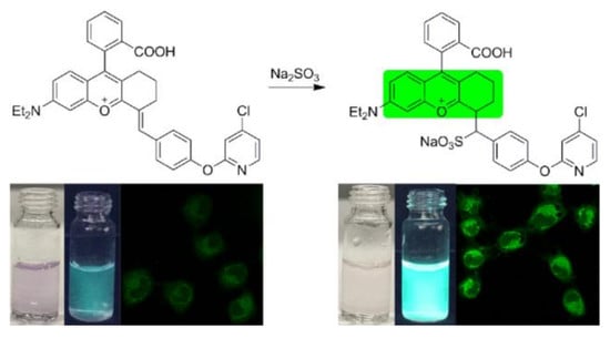
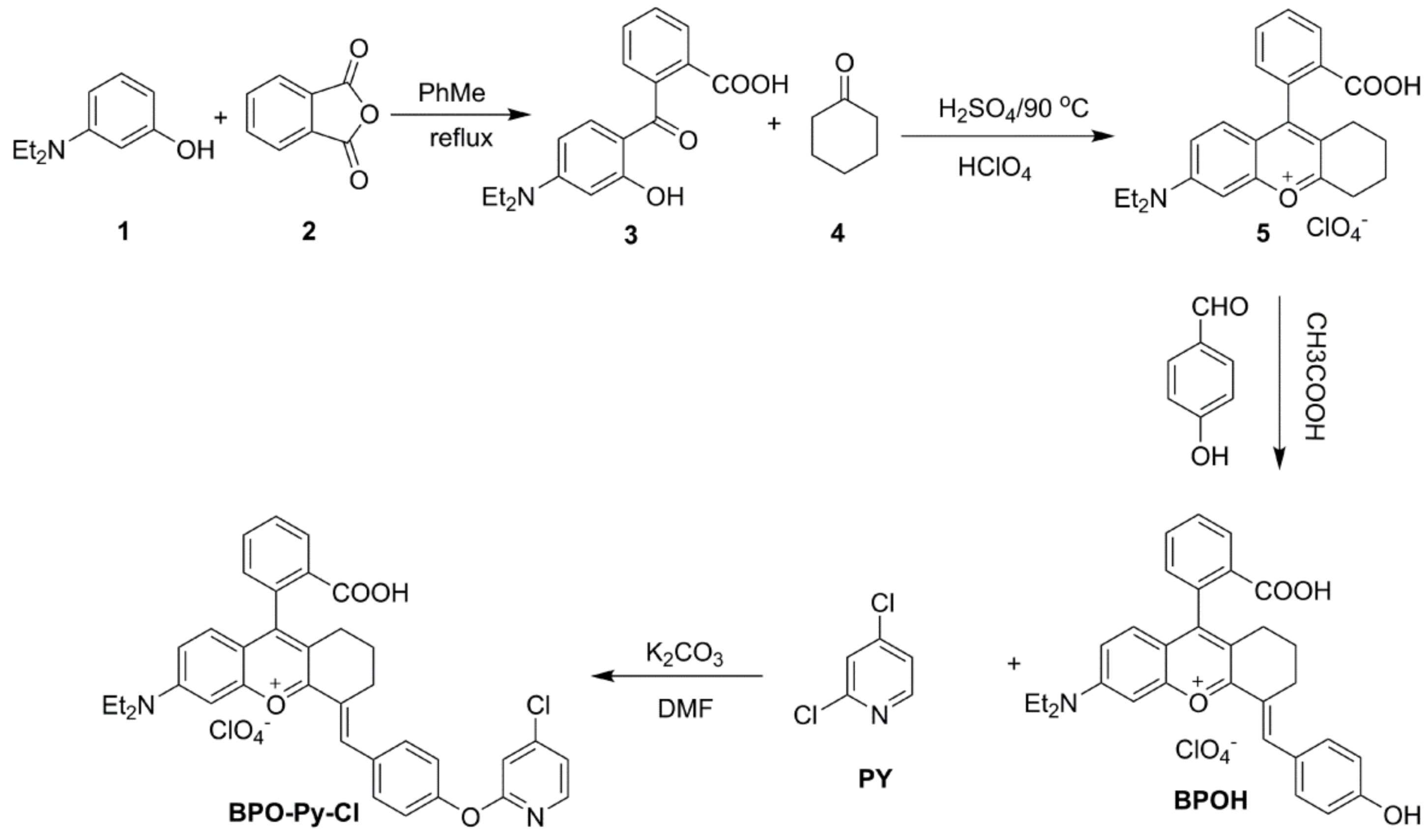
2.1. Synthesis and Characterization of BPO-Py-Cl

The synthesis route of the target molecule is illustrated in Scheme 2. It takes four steps to complete its composition, as follows: BPOH was synthesized according to the literature [40,42]. First, the Friedel-Crafts acylation reaction of 3-(diethylamino)phenol with phthalic anhydride can obtain compound 3 which could further react with cyclohexanone by a two-step cascade reaction to acquire oxonium 5. Then, a condensation reaction between 4-hydroxybenzaldehyde with compound 5 will produce BPOH. Finally, BPO-Py-Cl could be obtained by a nucleophilic substitution reaction of BPOH with the 2-chloropyridine derivative. The probe has been characterized by 1H-NMR, 13C-NMR, and HRMS. Detailed synthetic procedures and structure characterizations were given in the experimental section and supporting information.

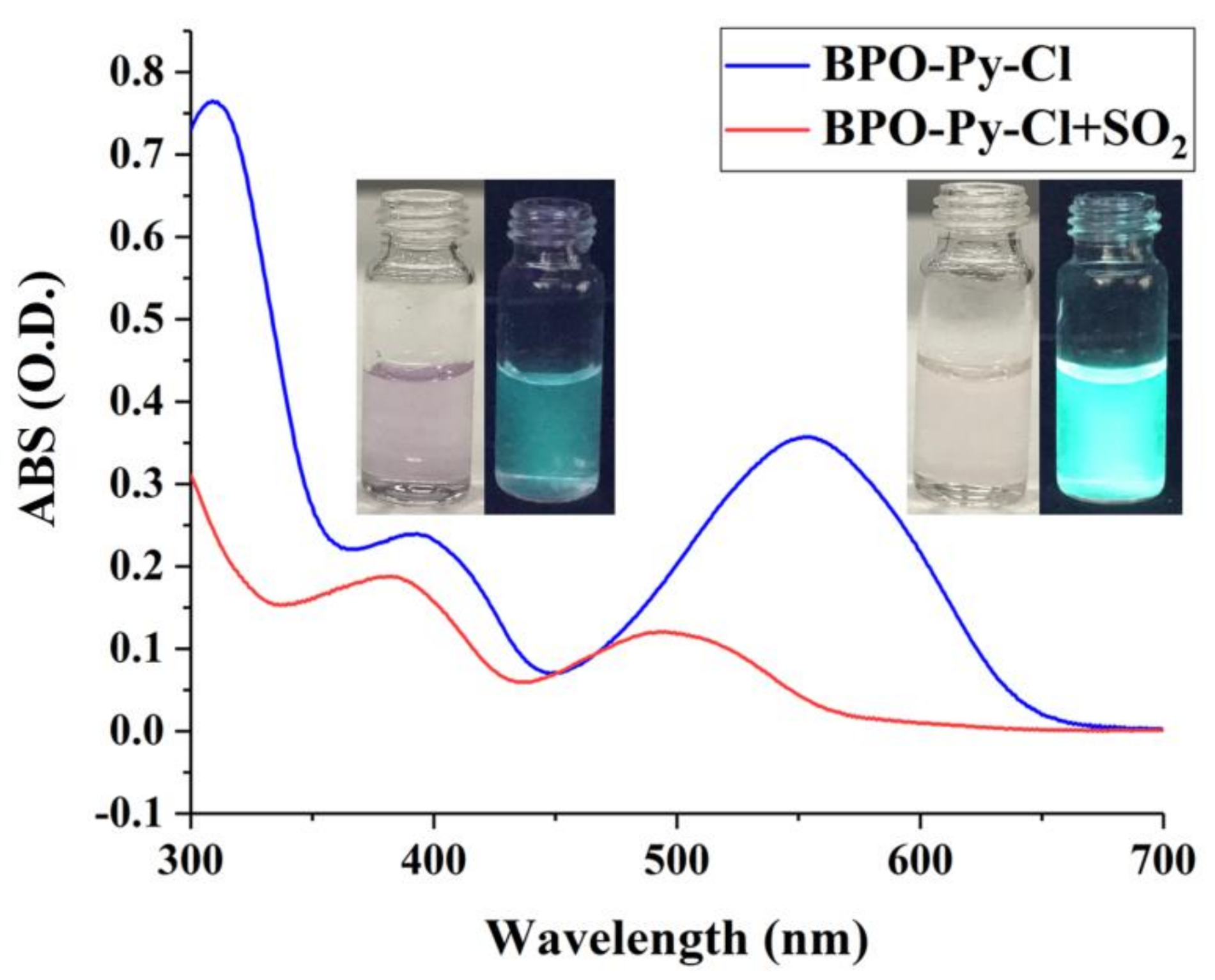
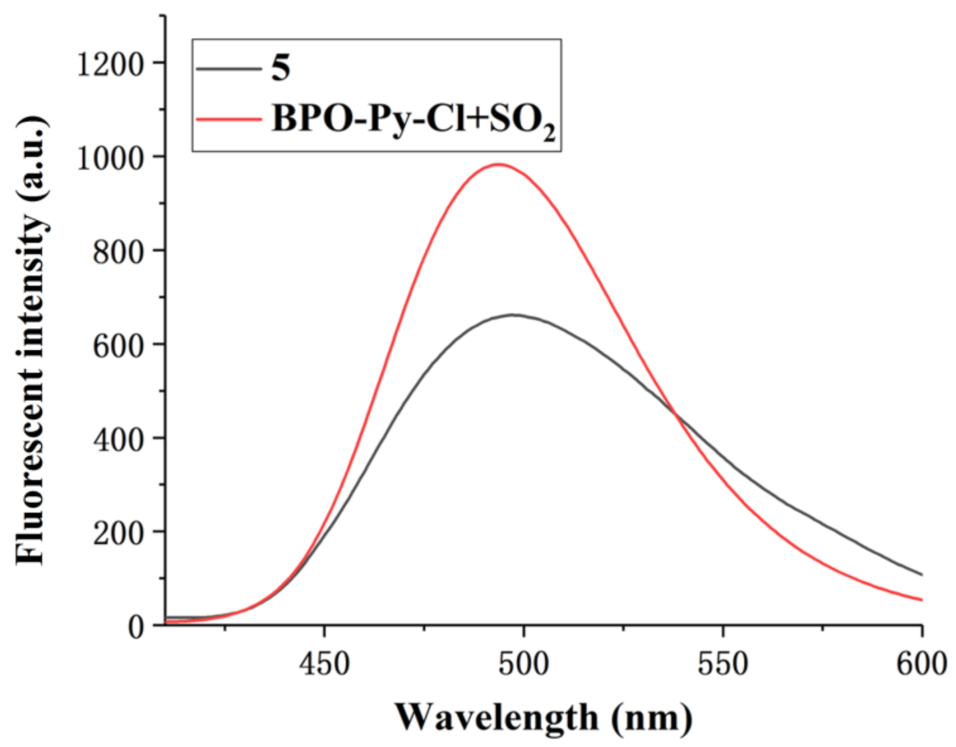
2.2. Optical Response of BPO-Py-Cl toward SO2

The optical property of BPO-Py-Cl (10 μM) was explored and the maxima absorption at 552 and 391 nm were observed in 10 mmol PBS (pH 7.4) buffer solution containing 20% DMSO as co-solvent (Figure 1). After interacting with 500 μM sulphite, the colour of the solution changed from purple to pale yellow under visible light and green fluorescence increased prominently under a 365 nm ultraviolet lamp. The absorption peaks of BPO-Py-Cl were blue-shifted to 493 nm and 379 nm, respectively. These results demonstrated that BPO-Py-Cl could detect SO2 efficiently.

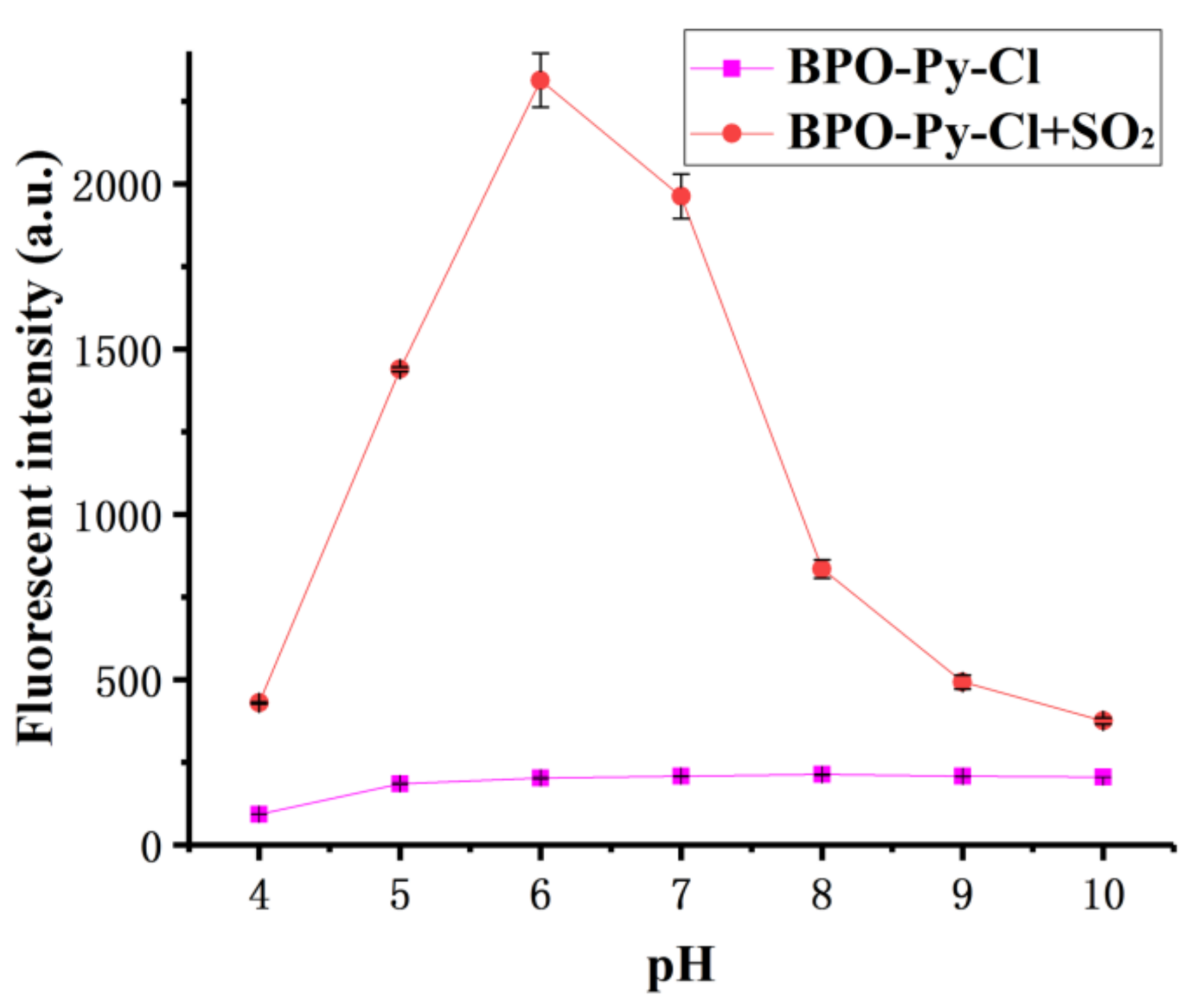
2.3. pH-Dependent Fluorescence Response of the Probe toward SO2

The effect of pH on the fluorescent properties was examined. As shown in Figure 2, the fluorescent intensity of the BPO-Py-Cl had almost no remarkable changes in 10 mmol PBS buffer solution containing 20% DMSO when the pH increased from 4.0 to 10.0. However, upon addition of 500 μM Na2SO3, the fluorescent intensity at 495 nm exhibited conspicuous enhancement when pH increased from 4.0 to 6.0. When the pH continuously increased to 10.0, the fluorescent intensity decreased dramatically. Therefore, pH 6.0 was chosen as the optimal condition for BPO-Py-Cl.

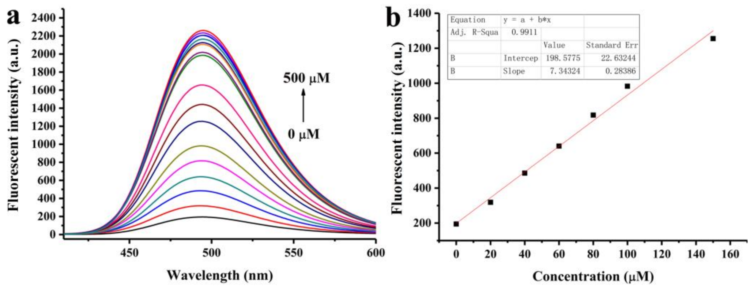
2.4. Quantitative Determination of SO2

The fluorescent titrations were conducted in 10 mmol pH 6.0 PBS buffer containing 20% DMSO as a co-solution. As expected, the fluorescence intensity of BPO-Py-Cl at 495 nm increased gradually along with the increment of Na2SO3. To our delight, 400 μM Na2SO3 can lead to an intensity increase as much as 11.7 times greater (Figure 3a and Figure S1). The quantum yields increased from 0.043 to 0.673 after interacting with 400 μM Na2SO3. In the given concentration rage from 0 to 160.0 μM, the fluorescent signal intensity was linearly related to the concentration of Na2SO3 (Figure 3b), with the detection limit of 99.27 nM.

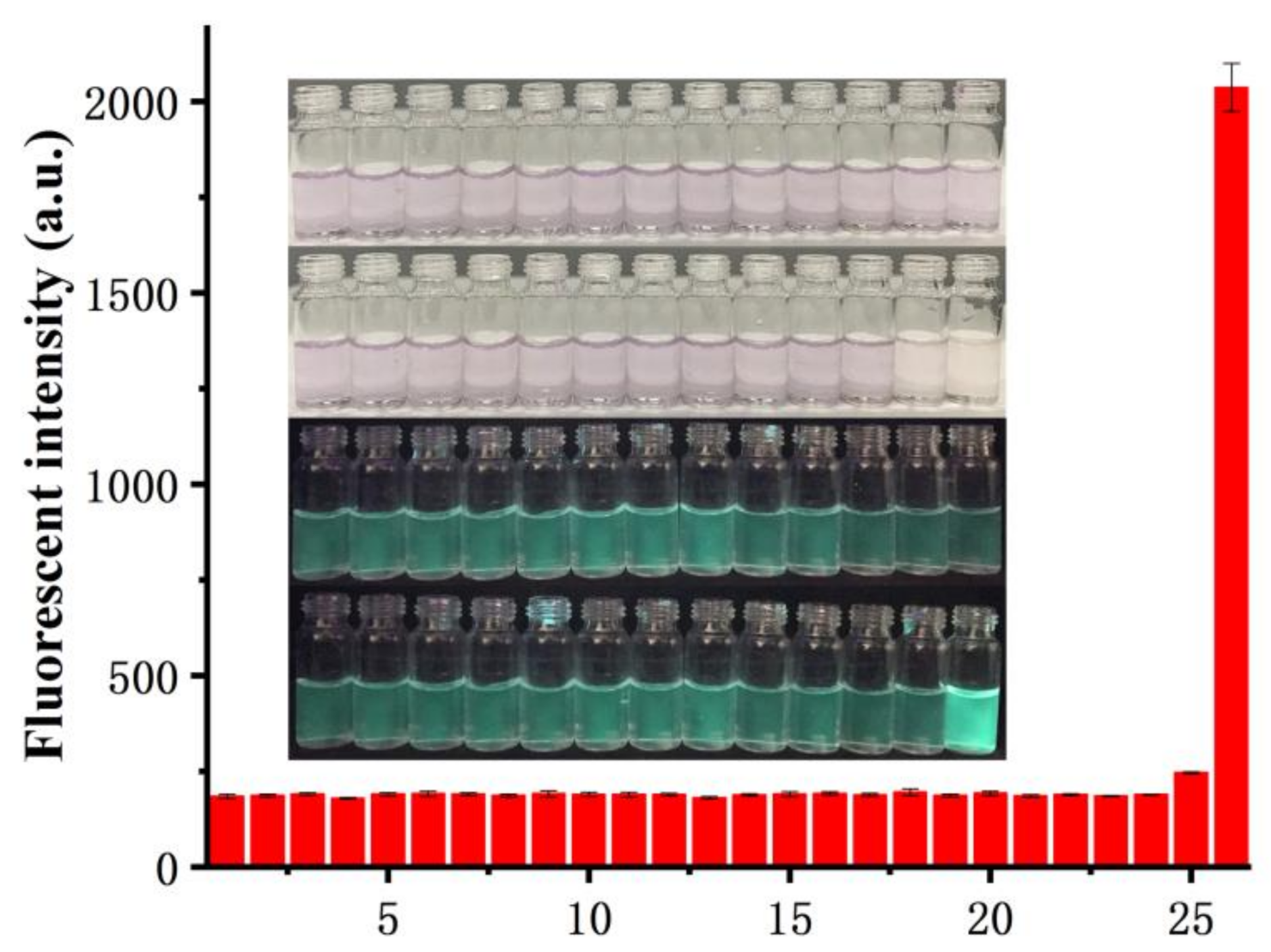
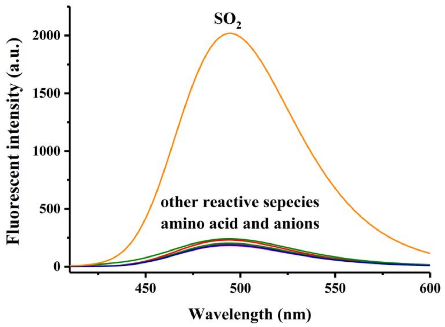
2.5. Selectivity Experiments

Based on the excellent fluorescent properties observed with Na2SO3, the selectivity of the probe for SO2 over other reactive species and amino acids were examined. As shown in Figure 4 and Figure S2, 500 μM sulphite could lead to noteworthy fluorescent spectrum changes of BPO-Py-Cl. Meanwhile, 500 μM of representative anions (F, Cl, Br, I, CO32−, AcO, PO43−, SCN, S2O32−), biologically-abundant metal ions (Li+, Na+, K+, Mg2+, Ca2+, Cu2+, Zn2+, Ni2+, Fe2+, Fe3+, Pb2+), reactive oxygen species (NaClO, H2O2, TBHP), reactive nitrogen species (NO2 and NO3), other reactive sulphur species (SO42−, H2S, Cys, HCy, and GSH) and amino acids (Ala, Arg, Asp, Glu, Gly, His, Ile, Leu, Lys, Met, Phe, Pro, Ser, Val) hardly led to any change. Figure 5 shows the fluorescent intensity and colour changes of the BPO-Py-Cl before and after the addition of sulphite or other species under visible light and 365 nm UV lamp. Obviously, only sulphite caused a remarkable colour change under the UV lamp.

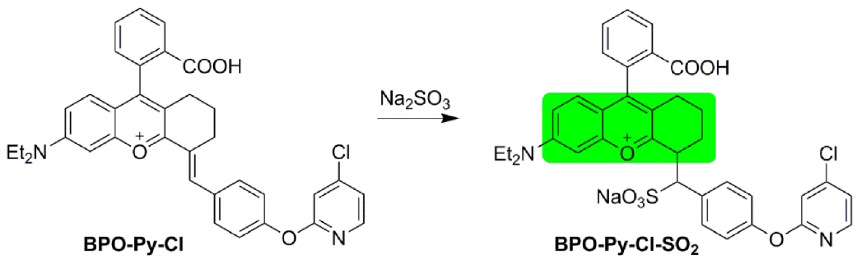
2.6. Possible Mechanism

As reported by Lin, the carbon–carbon double bond linked with oxonium can provide the latent nucleophilic addition site for SO2 [37]. From the absorption spectrum (Figure 1), it can be speculated that Na2SO3 broke the conjugate system of BPO-Py-Cl. To verify the nucleophilic addition reaction of sulphite to the carbon–carbon double bond, the spectrum of compound 5 was investigated in pH 6.0 PBS buffer solution containing 20% DMSO. As we can see in Figure 6 and Figure S3, compared with the BPO-Py-Cl interacting with SO2, compound 5 had a similar excitation and emission wavelength, which demonstrated that both of them have similar conjugated systems. Therefore, nucleophilic addition reactions of sulphite to the carbon–carbon double bond was considered as the possible mechanism.

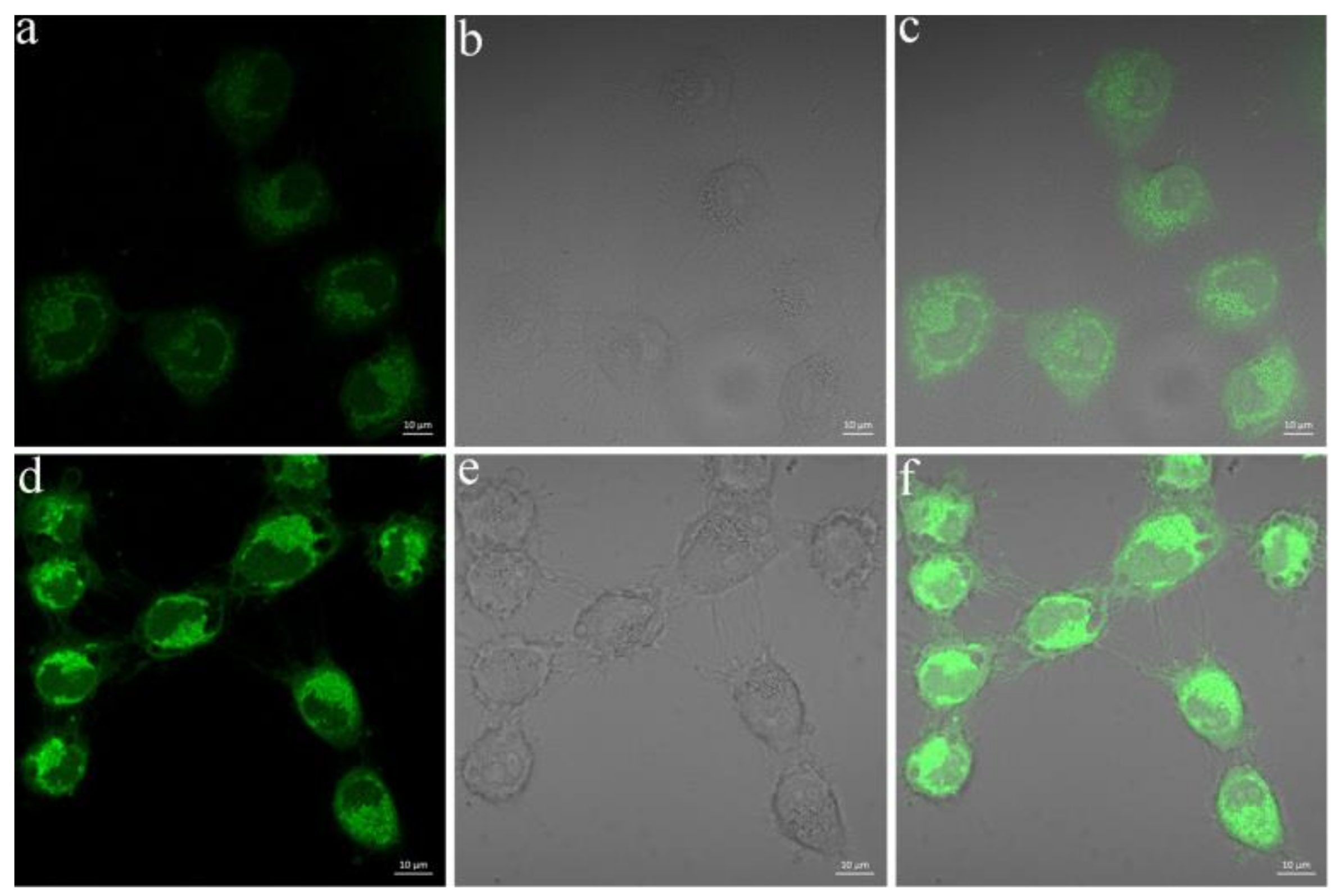
2.7. Living Cell Imaging

Encouraged by the aforementioned results, we further performed the capacity of probe for the fluorescent imaging in living cells. First, a standard CCK-8 assay in HeLa cells was investigated at different concentrations of BPO-Py-Cl. Figure 7 shows the changes of the cell survival rate with the concentration of BPO-Py-Cl increasing, and the cell survival rate was about 85% after incubation with 2 μM BPO-Py-Cl for 24 h. With 10 μM BPO-Py-Cl, it decreased to 40%.
Finally, the utility of BPO-Py-Cl to image SO2 was next carried out for living cells (Figure 8). The HeLa cells were incubated with BPO-Py-Cl (10 μM) in culture medium for 30 min at 37 °C, and exhibited weak fluorescence in the green channel. In a control experiment, the cells were pre-treated with BPO-Py-Cl for 30 min and further incubated with Na2SO3 (500 μM) for another 90 min. Obvious fluorescence increases in the green channel were observed compared with BPO-Py-Cl alone. These results demonstrated that BPO-Py-Cl could be used for the detection and imaging of SO2 in living cells.

3. Materials and Methods

3.1. Materials and General Instruments

All chemicals and reagents were purchased from commercial suppliers Aladdin-Reagent (Shanghai, China), Sigma-Aldrich (St. Louis, MO, USA), and TCI (Shanghai, China) and used without any further purification if not declared. All the solvents for optical spectroscopic studies were either HPLC or spectroscopic grade. TLC analyses were performed on GF 254silica gel. Silica gel (HG/T2354-92, 200–300 mesh) used for column chromatographic purifications were obtained from Qingdao Haiyang chemical Co., Ltd (Shandong, China). High-performance mass spectra were performed on a Bruker Daltonics Bio TOF (Karlsruhe, Germany) mass spectrometer in electrospray ionization mode. NMR spectra were recorded on a Bruker AMX-400 instrument using tetramethyl silane as the internal reference for 1H-NMR (400 MHz) or CDCl3 as the internal standard for 13C-NMR (100 MHz). UV–VIS absorption spectra and fluorescent spectra were recorded by a UV-1900 UV–VIS spectrophotometer and F4600 spectrofluorimeter with a 10 mm quartz cuvette from Hitachi PharmaSpec (Tokyo, Japan).

3.2. Synthesis and Characterization

2-(4-(Diethylamino)-2-hydroxybenzoyl)benzoic acid (3): 16.5 g (100 mmol) 3-diethylamino phenol, 19.0 g (128 mmol) phthalic anhydride, and 70 mL toluene were added to a 250-mL round round-bottom flask which was heated to 80 °C for 10 h, 90 °C for 5 h, 100 °C for 2 h, and 110 °C for 1 h, successively under N2. After cooling to RT, the precipitate was filtered off and washed with PhMe to obtain 28 g crude purple solid (compound 3) (89.5% yield) which was used for the next reaction without purification.
9-(2-Carboxyphenyl)-6-(diethylamino)-1,2,3,4-tetrahydroxanthylium perchlorate (4): concentrated H2SO4 (70 mL) was cooled to 0 °C. Then, 6.6 mL of cyclohexanone (63.7 mmol) and 9.5 g of compound 3 (32 mmol) were added dropwise to the cooled H2SO4, respectively, with vigorous stirring. The mixture was further stirred at 90 °C for 1.5 h, cooled down to room temperature, and poured onto ice (300 g). The resulting precipitate was filtered off and washed with cold water (100 mL) after 7 mL 70% perchloric acid was added to the ice mixture. The precipitate was further purified by silica gel column chromatography with DCM to DCM/MeOH (10/1, v/v). About 9.4 g of purple solid (compound 4) was obtained (62% yield).
(E)-9-(2-Carboxyphenyl)-6-(diethylamino)-4-(4-hydroxybenzylidene)-1,2,3,4-tetrahydroxanthylium perchlorate (BPOH): 4-hydroxybenzaldehyde (0.53 g, 4.32 mmol) and 1.72 g (3.60 mmol) of compound 5 were mixed in 40 mL AcOH. Then the mixture was heated to 90 °C overnight under N2. Subsequently, the solvent was removed under reduced pressure. After that, the crude product was dissolved in 50 mL dichloromethane, washed with water (50 mL) three times, and dried by sodium sulphate. The crude product was concentrated and purified with chromatography on a silica gel column with DCM to DCM/MeOH (10/1, v/v) to get 1.35 g BPOH as dark purple solid (65% yield).
(E)-9-(2-Carboxyphenyl)-4-(4-((4-chloropyridin-2-yl)oxy)benzylidene)-6-(diethylamino)-1,2,3,4-tetrahydroxanthylium (BPO-Py-Cl): About 174.0 mg (0.3 mmol) of BPOH was dissolved in 10 mL of DMF. Then, 441.0 mg (3.0 mmol) of 2,4-dichloropyridine and 207 mg (1.5 mmol) of K2CO3 were added into the above solution. After reacting for 3–5 h at 50 °C under supervision by TLC, the solvent was removed under reduced pressure to obtain the crude product which as purified on a silica gel column chromatography with DCM to DCM/MeOH (20/1, v/v) to obtain an aubergine solid of 70 mg BPO-Py-Cl (33.3% yield). 1H-NMR (400 MHz, CDCl3), 8.28 (d, 1H, J = 5.6 Hz), 7.99 (d, 1H, J = 7.6 Hz), 7.68 (t, 1H, J = 7.6 Hz), 7.58 (t, 1H, J = 7.2 Hz), 7.49 (d, 2H, J = 8.4 Hz), 7.41 (s, 1H), 7.27 (d, 1H, J = 7.6 Hz), 7.13 (d, 2H, J = 8.4 Hz), 6.89–6.85 (m, 2H), 6.53 (d, 1H, J = 8.8 Hz), 6.45 (d, 1H, J = 2.4 Hz), 6.39 (dd, 1H, J = 2.8 Hz, J = 8.8 Hz), 3.39 (q, 4H, J = 6.8 Hz), 2.86–2.82 (m, 1H), 2.69–2.65 (m, 1H), 2.11–2.07 (m, 1H), 1.75–1.63 (m, 3H), 1.20 (t, 4H, J = 6.8 Hz). 13C-NMR (100 MHz, CDCl3), 170.1, 166.4, 152.8,152.3, 152.2, 150.8, 149.4, 146.8, 134.5, 131.5, 131.2, 129.3, 128.6, 123.5, 120.6, 112.0, 111.5, 108.9, 108.5, 104.8, 97.3, 44.5, 27.2, 23.1, 22.4, 12.6. HRMS (ESI): m/z [M]+ calcd. for C36H32ClN2O4: 591.2045; found 591.2043.

3.3. Preparation of the Test Solutions

Na2SO3 was used as the source of SO2. A probe stock solution of the BPO-Py-Cl (2.0 mM) was prepared in DMSO. Other stock solutions of representative anions (F, Cl, Br, I, CO32−, AcO, PO43−, SCN, S2O32−), biologically-abundant metal ions (Li+, Na+, K+, Mg2+, Ca2+, Cu2+, Zn2+, Ni2+, Fe2+, Fe3+, Pb2+), reactive oxygen species (NaClO, H2O2, TBHP), reactive nitrogen species (NO2 and NO3), other reactive sulphur species (SO42−, H2S, Cys, HCy, and GSH), and amino acids (Lys, Glu, Leu, Met, Arg, His, Phe, Ser, Ile, Val, Ala, Asp, Pro,) were prepared in deionized water at a concentration of 50 mM. All the test solutions were prepared by mixing 75 μL of each species stock solution and 15 μL of the probe stock solution in 3.0 mL with PBS buffer/DMSO (8:2, v/v) solution. The absorption and emission spectra were recorded after the test solution was incubated at room temperature for 2 h.

3.4. Quantum Yields

The fluorescence quantum yield (Φ) was calculated using the following formula:
Φx = Φs × (As/Ax) × (Dx/Ds) × (nx2/ns2)
where the subscripts s and x refer to the standard material and test sample, respectively. Φs and Φx represent the fluorescence quantum yield of the standard and test sample; ns and nx represent the refractive index of the solvents used; As and Ax represent the absorption intensity at the excitation wavelength of the standard and test sample; Ds and Dx represent the integral of the fluorescence intensity of the standard and test sample. Fluorescein in 0.1 M NaOH (Φs = 0.85) was used as the standard [43].

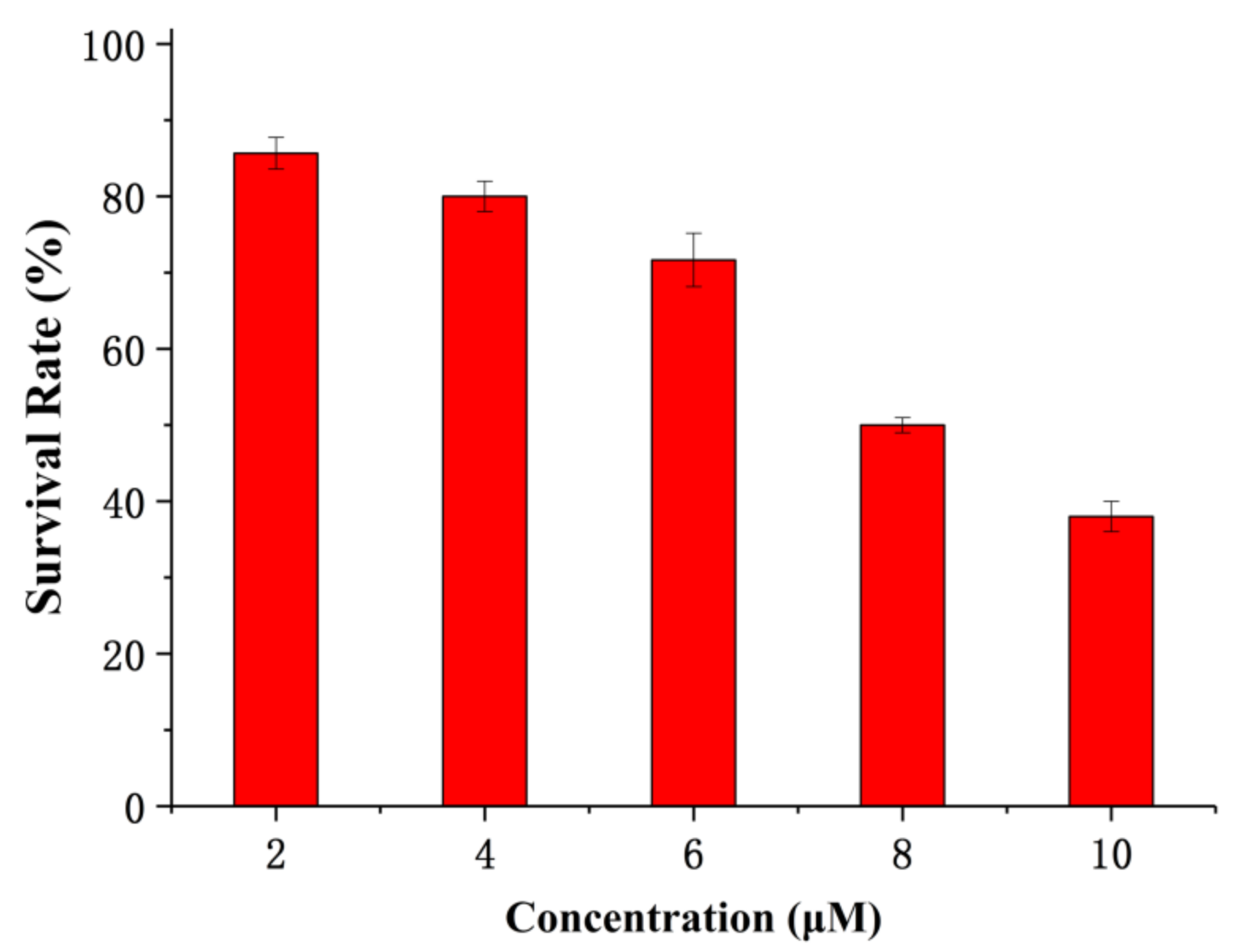
3.5. CCK-8 Assay for the Cell Cytotoxicity

The cytotoxicity of the probe was determined by CCK-8 assays. HeLa cells were first seeded in 96-well plates (about 7000 cells per well) and cultured overnight for 70–80% cell confluence. After the cells were incubated with BPO-Py-Cl at different concentrations (2, 4, 6, 8, 10 μM in DMSO/cell culture medium = 1:49) for twenty-four hours, 10 μL of CCK-8 mixed in 90 μL of PBS was added to each well of the 96-well assay plate for additional 1 h incubation at 37 °C. The absorbance was measured at a wavelength of 450 nm using an ELISA plate reader (Model 550, BioRad, Hercules, CA, USA). All the samples were repeated three times.

3.6. Determination of the Detection Limit

The detection limit was calculated based on the fluorescence titration [44] with the following equation:
detection limit = 3σbi/m
where σbi is the standard deviation of blank measurements, m is the slope between intensity versus sample concentration (signal-to-noise ratio of 3:1). The standard deviation of blank measurements was determined when the emission intensity of probe (10 μM) without Na2SO3 was measured 10 times. Then, fluorescent titrations were conducted under the present conditions. A good linear relationship between the fluorescence intensity and the concentration of Na2SO3 was obtained, the slope between intensity versus Na2SO3 concentration was m in the above-mentioned formula.

3.7. Fluorescence Imaging of SO2 in Living Cells

HeLa cells were incubated in DMEM medium (which contained 10% foetal bovine serum and 1% antibiotic-antimycotic) at 37 °C in an incubator (5% CO2/95% air). Then cells were seeded in confocal dishes (4 × 10³ per well) and incubated for 24 h. Before staining, the cells were washed three times with physiological PBS and incubated with BPO-Py-Cl (10 μM) for 30 min at 37 °C. After being washed with physiological PBS three times to remove free probes, cell imaging was carried out after the cells were further incubated with Na2SO3 (500 μM) for another 90 min. Meanwhile, the control experiment was performed. The cells were incubated with BPO-Py-Cl (10 μM) for 30 min and then washed with PBS three times. Then, cell imaging was performed. The confocal fluorescent images were recorded for the green channel, corresponding to the wavelength range 460–530 nm, which was monitored for excitation at 405 nm.

4. Conclusions

In summary, a novel chromenylium derivative bearing the electron-withdrawing group BPO-Py-Cl was synthesized, and the probe exhibited a remarkable turn-on fluorescence response toward SO2 with a 105 nm Stock shift. It exhibited high sensitivity and selectivity with a low detection limit (99.27 nM) for SO2. In addition, the probe was cell membrane permeable and could be successfully applied to the imaging of SO2 in living cells.

Supplementary Materials

The supplementary materials are available online.

Acknowledgments

This work was supported by the National Natural Science Foundation of China (no. 21708030) and the Fundamental Research Funds for the Central University (no. 2682016CX102). We also thank the Comprehensive Training Platform of Specialized Laboratory, College of Chemistry, Sichuan University for laser scanning confocal imaging.

Author Contributions

M.-Y.W. and X.-Q.Y. conceived and designed the work. M.-Y.W. carried out the synthetic work and wrote the paper. J.W., Y.W., and Y.-H.L. performed the fluorescence properties assay. All authors read and approved the final manuscript.

Conflicts of Interest

The authors declare no conflict of interest.

References

  1. Chen, T.-M.; Kuschner, W.G.; Gokhale, J.; Shofer, S. Outdoor air pollution: Nitrogen dioxide, sulfur dioxide, and carbon monoxide health effects. Am. J. Med. Sci. 2007, 333, 249–256. [Google Scholar] [CrossRef] [PubMed]
  2. Li, X.; Bazer, F.W.; Gao, H.; Jobgen, W.; Johnson, G.A.; Li, P.; McKnight, J.R.; Satterfield, M.C.; Spencer, T.E.; Wu, G. Amino acids and gaseous signalling. Amino Acids 2009, 37, 65–78. [Google Scholar] [CrossRef] [PubMed]
  3. Griffith, O.W. Mammalian sulfur amino acid metabolism: An overview. Methods Enzymol. 1987, 143, 366–376. [Google Scholar] [PubMed]
  4. Li, J.; Meng, Z. The role of sulfur dioxide as an endogenous gaseous vasoactive factor in synergy with nitric oxide. Nitric Oxide 2009, 20, 166–174. [Google Scholar] [CrossRef] [PubMed]
  5. Li, J.; Li, R.; Meng, Z. Sulfur dioxide upregulates the aortic nitric oxide pathway in rats. Eur. J. Pharmacol. 2010, 645, 143–150. [Google Scholar] [CrossRef] [PubMed]
  6. Song, A.; Liao, Q.; Li, J.; Lin, F.; Liu, E.; Jiang, X.; Deng, L. Chronic exposure to sulfur dioxide enhances airway hyperresponsiveness only in ovalbumin-sensitized rats. Toxicol. Lett. 2012, 214, 320–327. [Google Scholar] [CrossRef] [PubMed]
  7. Jin, H.; Wang, Y.; Wang, X.; Sun, Y.; Tang, C.; Du, J. Sulfur dioxide preconditioning increases antioxidative capacity in rat with myocardial ischemia reperfusion (I/R) injury. Nitric Oxide 2013, 32, 56–61. [Google Scholar] [CrossRef] [PubMed]
  8. Li, W.; Tang, C.; Jin, H.; Du, J. Regulatory effects of sulfur dioxide on the development of atherosclerotic lesions and vascular hydrogen sulfide in atherosclerotic rats. Atherosclerosis 2011, 215, 323–330. [Google Scholar] [CrossRef] [PubMed]
  9. Sun, Y.; Tian, Y.; Prabha, M.; Liu, D.; Chen, S.; Zhang, R.; Liu, X.; Tang, C.; Tang, X.; Jin, H.; et al. Effects of sulfur dioxide on hypoxic pulmonary vascular structural remodelling. Lab. Investig. 2010, 90, 68–82. [Google Scholar] [CrossRef] [PubMed]
  10. Liang, Y.; Liu, D.; Ochs, T.; Tang, C.; Chen, S.; Zhang, S.; Geng, B.; Jin, H.; Du, J. Endogenous sulfur dioxide protects against isoproterenol-induced myocardial injury and increases myocardial antioxidant capacity in rats. Lab. Investig. 2011, 91, 12–23. [Google Scholar] [CrossRef] [PubMed]
  11. Li, G.K.; Sang, N. Delayed rectifier potassium channels are involved in SO2 derivative-induced hippocampal neuronal injury. Ecotoxicol. Environ. Saf. 2009, 72, 236–241. [Google Scholar] [CrossRef] [PubMed]
  12. Migliore, L.; Coppedè, F. Environmental-induced oxidative stress in neurodegenerative disorders and aging. Mutat. Res. 2009, 674, 73–84. [Google Scholar] [CrossRef] [PubMed]
  13. Sang, N.; Yun, Y.; Yao, G.; Li, H.; Guo, L.; Li, G. SO2-induced neurotoxicity is mediated by cyclooxygenases-2-derived prostaglandin E2 and its downstream signaling pathway in rat hippocampal neurons. Toxicol. Sci. 2011, 124, 400–413. [Google Scholar] [CrossRef] [PubMed]
  14. Liu, D.; Huang, Y.; Bu, D.; Liu, A.D.; Holmberg, L.; Jia, Y.; Tang, C.; Du, J.; Jin, H. Sulfur dioxide inhibits vascular smooth muscle cell proliferation via suppressing the Erk/MAP kinase pathway mediated by cAMP/PKA signaling. Cell Death Dis. 2014, 5, e1251. [Google Scholar] [CrossRef] [PubMed]
  15. Yu, F.; Han, X.; Chen, L. Fluorescent probes for hydrogen sulfide detection and bioimaging. Chem. Commun. 2014, 50, 12234–12249. [Google Scholar] [CrossRef] [PubMed]
  16. Lin, V.S.; Chen, W.; Xian, M.; Chang, C.J. Chemical probes for molecular imaging and detection of hydrogen sulfide and reactive sulfur species in biological systems. Chem. Soc. Rev. 2015, 44, 4596–4618. [Google Scholar] [CrossRef] [PubMed]
  17. Jiao, X.; Li, Y.; Niu, J.; Xie, X.; Wang, X.; Tang, B. Small-molecule fluorescent probes for imaging and detection of reactive oxygen, nitrogen, and sulfur species in biological systems. Anal. Chem. 2018, 90, 533–555. [Google Scholar] [CrossRef] [PubMed]
  18. Sun, W.; Guo, S.; Hu, C.; Fan, J.; Peng, X. Recent development of chemosensors based on cyanine platforms. Chem. Rev. 2016, 116, 7768–7817. [Google Scholar] [CrossRef] [PubMed]
  19. Hou, J.-T.; Ren, W.X.; Li, K.; Seo, J.; Sharma, A.; Yu, X.-Q.; Kim, J.S. Fluorescent bioimaging of pH: From design to applications. Chem. Soc. Rev. 2017, 46, 2076–2090. [Google Scholar] [CrossRef] [PubMed]
  20. Xu, W.; Zeng, Z.; Jiang, J.-H.; Chang, Y.-T.; Yuan, L. Discerning the chemistry in individual organelles with small-molecule fluorescent probes. Angew. Chem. Int. Ed. 2016, 55, 13658–13699. [Google Scholar] [CrossRef] [PubMed]
  21. Wu, M.-Y.; He, T.; Li, K.; Wu, M.-B.; Huang, Z.; Yu, X.-Q. A real-time colorimetric and ratiometric fluorescent probe for sulfite. Analyst 2013, 138, 3018–3025. [Google Scholar] [CrossRef] [PubMed]
  22. Wu, M.-Y.; Li, K.; Li, C.-Y.; Hou, J.-T.; Yu, X.-Q. A water-soluble near-infrared probe for colorimetric and ratiometric sensing of SO2 derivatives in living cells. Chem. Commun. 2014, 50, 183–185. [Google Scholar] [CrossRef] [PubMed]
  23. Liu, Y.; Li, K.; Xie, K.-X.; Li, L.-L.; Yu, K.-K.; Wang, X.; Yu, X.-Q. A water-soluble and fast-response mitochondria-targeted fluorescent probe for colorimetric and ratiometric sensing of endogenously generated SO2 derivatives in living cells. Chem. Commun. 2016, 52, 3430–3433. [Google Scholar] [CrossRef] [PubMed]
  24. Liu, Y.; Li, K.; Wu, M.-Y.; Liu, Y.-H.; Xie, Y.-M.; Yu, X.-Q. A mitochondria-targeted colorimetric and ratiometric fluorescent probe for biological SO2 derivatives in living cells. Chem. Commun. 2015, 51, 10236–10239. [Google Scholar] [CrossRef] [PubMed]
  25. Yang, J.; Li, K.; Hou, J.-T.; Li, L.-L.; Lu, C.-Y.; Xie, Y.-M.; Wang, X.; Yu, X.-Q. Novel tumor-specific and mitochondria-targeted near-infrared-emission fluorescent probe for SO2 derivatives in living cells. ACS Sens. 2016, 1, 166–172. [Google Scholar] [CrossRef]
  26. Xu, W.; Teoh, C.L.; Peng, J.; Su, D.; Yuan, L.; Chang, Y.-T. A mitochondria-targeted ratiometric fluorescent probe to monitor endogenously generated sulfur dioxide derivatives in living cells. Biomaterials 2015, 56, 1–9. [Google Scholar] [CrossRef] [PubMed]
  27. Sun, Y.-Q.; Liu, J.; Zhang, J.; Yang, T.; Guo, W. Fluorescent probe for biological gas SO2 derivatives bisulfite and sulfite. Chem. Commun. 2013, 49, 2637–2639. [Google Scholar] [CrossRef] [PubMed]
  28. Wu, W.-L.; Wang, Z.-Y.; Dai, X.; Miao, J.-Y.; Zhao, B.-X. An effective colorimetric and ratiometric fluorescent probe based FRET with a large Stokes shift for bisulfite. Sci. Rep. 2016, 6, 25315. [Google Scholar] [CrossRef] [PubMed]
  29. Li, D.-P.; Wang, Z.-Y.; Cao, X.-J.; Cui, J.; Wang, X.; Cui, H.-Z.; Miao, J.-Y.; Zhao, B.-X. A mitochondria-targeted fluorescent probe for ratiometric detection of endogenous sulfur dioxide derivatives in cancer cells. Chem. Commun. 2016, 52, 2760–2763. [Google Scholar] [CrossRef] [PubMed]
  30. Li, G.; Chen, Y.; Wang, J.; Wu, J.; Gasser, G.; Ji, L.; Chao, H. Direct imaging of biological sulfur dioxide derivatives in vivo using a two-photon phosphorescent probe. Biomaterials 2015, 63, 128–136. [Google Scholar] [CrossRef] [PubMed]
  31. Ma, Y.; Tang, Y.; Zhao, Y.; Gao, S.; Lin, W. Two-photon and deep-red emission ratiometric fluorescent probe with a large emission shift and signal ratios for sulfur dioxide: Ultrafast response and applications in living cells, brain tissues, and zebrafishes. Anal. Chem. 2017, 89, 9388–9393. [Google Scholar] [CrossRef] [PubMed]
  32. Zhang, W.; Liu, T.; Huo, F.; Ning, P.; Meng, X.; Yin, C. Reversible ratiometric fluorescent probe for sensing bisulfate/H2O2 and Its application in zebrafish. Anal. Chem. 2017, 89, 8079–8083. [Google Scholar] [CrossRef] [PubMed]
  33. Dou, K.; Fu, Q.; Chen, G.; Yu, F.; Liu, Y.; Cao, Z.; Li, G.; Zhao, X.; Xia, L.; Chen, L.; et al. A novel dual-ratiometric-response fluorescent probe for SO2/ClO detection in cells and in vivo and its application in exploring the dichotomous role of SO2 under the ClO induced oxidative stress. Biomaterials 2017, 133, 82–93. [Google Scholar] [CrossRef] [PubMed]
  34. Zhu, Y.; Du, W.; Zhang, M.; Xu, Y.; Song, L.; Zhang, Q.; Tian, X.; Zhou, H.; Wu, J.; Tian, Y. A series of water-soluble A–π–A′ typological indolium derivatives with two-photon properties for rapidly detecting HSO3/SO32− in living cells. J. Mater. Chem. B 2017, 5, 3862–3869. [Google Scholar] [CrossRef]
  35. Che, S.; Dao, R.; Zhang, W.; Lv, X.; Li, H.; Wang, C. Designing an anion-functionalized fluorescent ionic liquid as an efficient and reversible turn-off sensor for detecting SO2. Chem. Commun. 2017, 53, 3862–3865. [Google Scholar] [CrossRef] [PubMed]
  36. Dou, K.; Chen, G.; Yu, F.; Sun, Z.; Li, G.; Zhao, X.; Chen, L.; You, J. A two-photon ratiometric fluorescent probe for the synergistic detection of the mitochondrial SO2/HClO crosstalk in cells and in vivo. J. Mater. Chem. B 2017, 5, 8389–8398. [Google Scholar] [CrossRef]
  37. Shang, H.; Liu, K.; Lin, W. Construction of a novel ratiometric near-infrared fluorescent probe for SO2 derivatives and its application for biological imaging. Anal. Methods 2017, 9, 3790–3794. [Google Scholar] [CrossRef]
  38. Chen, H.; Dong, B.; Tang, Y.; Lin, W. A unique “integration” strategy for the rational design of optically tunable near-infrared fluorophores. Acc. Chem. Res. 2017, 50, 1410–1422. [Google Scholar] [CrossRef] [PubMed]
  39. Dong, B.; Song, X.; Kong, X.; Wang, C.; Tang, Y.; Liu, Y.; Lin, W. Simultaneous near-infrared and two-photon in vivo imaging of H2O2 using a ratiometric fluorescent probe based on the unique oxidative rearrangement of oxonium. Adv. Mater. 2016, 28, 8755–8759. [Google Scholar] [CrossRef] [PubMed]
  40. Yuan, L.; Lin, W.; Yang, Y.; Chen, H. A unique class of near-infrared functional fluorescent dyes with carboxylic-acid-modulated fluorescence ON/OFF switching: Rational design, synthesis, optical properties, theoretical calculations, and applications for fluorescence imaging in living animals. J. Am. Chem. Soc. 2012, 134, 1200–1211. [Google Scholar] [CrossRef] [PubMed]
  41. Wei, Y.; Cheng, D.; Ren, T.; Li, Y.; Zeng, Z.; Yuan, L. Design of NIR chromenylium-cyanine fluorophore library for “switch-ON” and ratiometric detection of bio-active species in vivo. Anal. Chem. 2016, 88, 1842–1849. [Google Scholar] [CrossRef] [PubMed]
  42. Yuan, L.; Lin, W.; Chen, H. Analogs of Changsha near-infrared dyes with large Stokes Shifts for bioimaging. Biomaterials 2013, 34, 9566–9571. [Google Scholar] [CrossRef] [PubMed]
  43. Parker, B.C.A.; Rees, W.T. Correction of fluorescence spectra and measurement of fluorescence quantum efficiency. Analyst 1960, 85, 587–600. [Google Scholar] [CrossRef]
  44. Joshi, B.P.; Park, J.; Lee, W.I.; Lee, K. Ratiometric and turn-on monitoring for heavy and transition metal ions in aqueous solution with a fluorescent peptide sensor. Talanta 2009, 78, 903–909. [Google Scholar] [CrossRef] [PubMed]
Sample Availability: Samples of the compounds BPOH and BPO-Py-Cl are available from the authors.
Scheme 1. The detection process of BPO-Py-Cl with SO2.
Molecules 23 00871 sch001
Scheme 2. Synthesis route of fluorescent probe BPO-Py-Cl.
Molecules 23 00871 sch002
Figure 1. The absorption spectra and colorimetric changes of BPO-Py-Cl (10 μM) interacting with SO2 (500 μM) under visible and 365 nm ultraviolet lamps.
Molecules 23 00871 g001
Figure 2. The pH dependent fluorescent changes of BPO-Py-Cl (10 μM) in PBS or after addition of SO2 (500 μM). The experiment was repeated three times (±S.D.).
Molecules 23 00871 g002
Figure 3. (a) Fluorescence titration of BPO-Py-Cl (10 µM) with SO32− (0, 20, 40, 60, 80, 100, 150, 200, 250, 300, 350, 400, 450, 500 µM) (λex = 390 nm, slit: 2.5 nm/5 nm) in a 10 mM PBS:DMSO = 8:2 pH 6.0 buffer solution; and (b) linear fit of fluorescence intensity changes with Na2SO3. λex = 390 nm. Slit: 2.5 nm/5 nm.
Molecules 23 00871 g003
Figure 4. Fluorescence spectrum changes of BPO-Py-Cl (10 μM) interacting with 500 μM amino acids (Arg, Met, Ser, Asp, Gly, Ala, His, Val, Lys, Leu, Glu, Pro, Ile, Phe), reactive oxygen species (H2O2, NaClO, TBHP), reactive nitrogen species (NO3, NO2), and reactive sulphur species (SO42−, Cys, Hcy, GSH, H2S, SO2) in 10 mM pH 6.0 PBS buffer solution containing 20% DMSO, λex = 390 nm. Slit: 2.5 nm/5 nm.
Molecules 23 00871 g004
Figure 5. Fluorescence intensity changes of BPO-Py-Cl with SO2 and other species. Inset: fluorescence changes of BPO-Py-Cl upon addition of 500 μM SO2 and other species under visible light and UV light (Ex = 365 nm). 1. BPO-Py-Cl, 2. Ala, 3. Arg. 4. Asp, 5. Glu, 6. Gly, 7. His, 8. Ile, 9. Leu, 10. Lys, 11. Met, 12. Phe, 13. Pro, 14, Ser, 15. Val, 16. H2O2, 17, NaClO, 18. TBHP, 19. NO3, 20. NO2, 21. SO42−, 22. Cys, 23. Hcy, 24. GSH, 25. H2S, 26. SO2. The experiment was repeated three times (±S.D.).
Molecules 23 00871 g005
Figure 6. The fluorescent spectrum of compound 5 and BPO-Py-Cl interacting with SO2.
Molecules 23 00871 g006
Figure 7. Cell viability by a standard CCK-8 assay. The experiment was repeated three times (±S.D.).
Molecules 23 00871 g007
Figure 8. Cell image experiments of BPO-Py-Cl. HeLa cells were incubated with 10 μM BPO-Py-Cl for 30 min, (ac) green channel, bright field, and the merged channel of green and bright field channels. HeLa cells were incubated with 10 μM BPO-Py-Cl for 30 min, then washed with PBS and incubated with SO32− (500 μM) for another 90 min; (df) are the green channel, bright field, and the merged channel of green and bright field channels.
Molecules 23 00871 g008

