Potential Anti-Inflammatory and Anti-Cancer Properties of Farnesol


Abstract
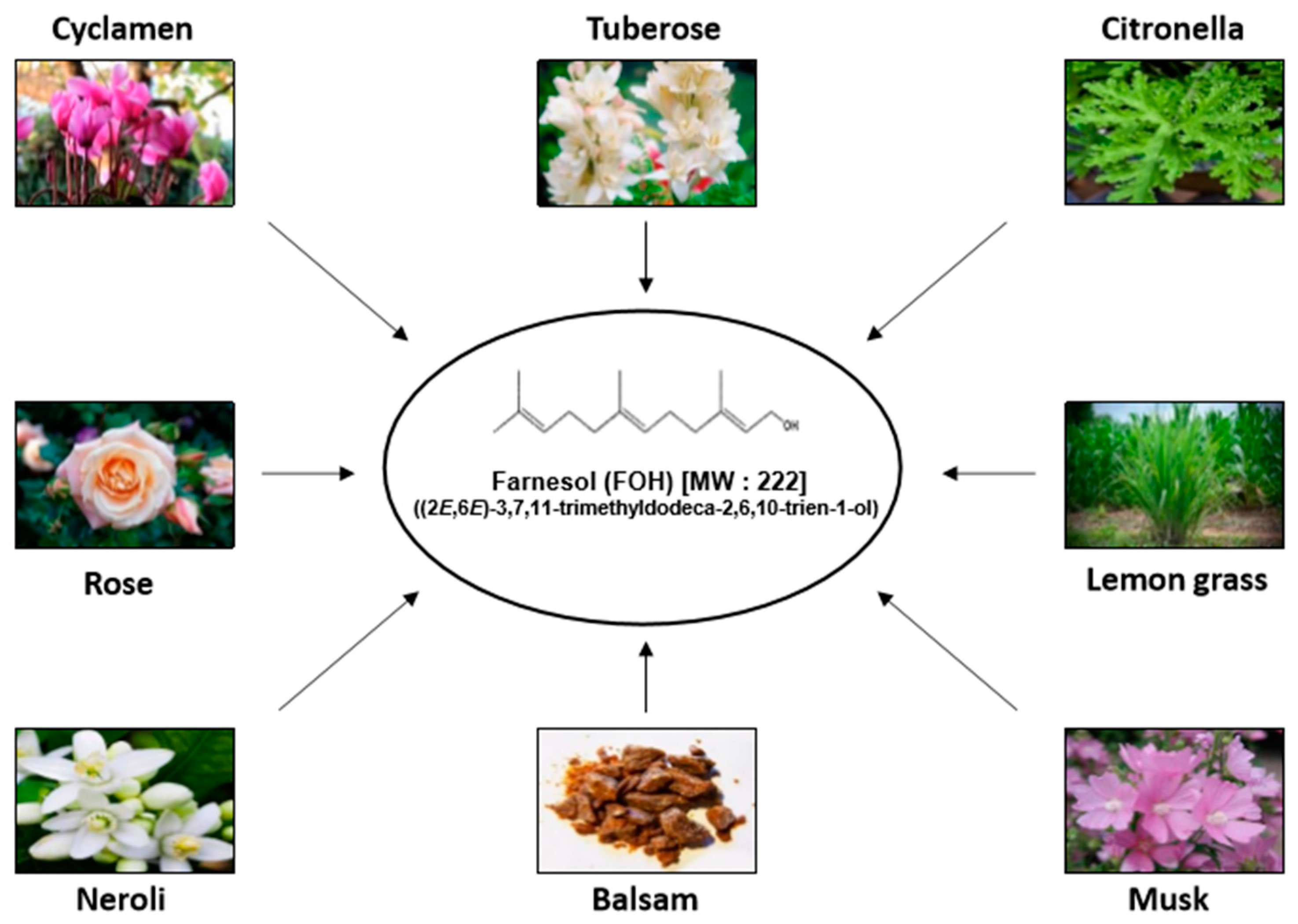
1. Introduction
2. In Vitro Inflammatory-Modulatory and Anti-Tumor Effects of Farnesol
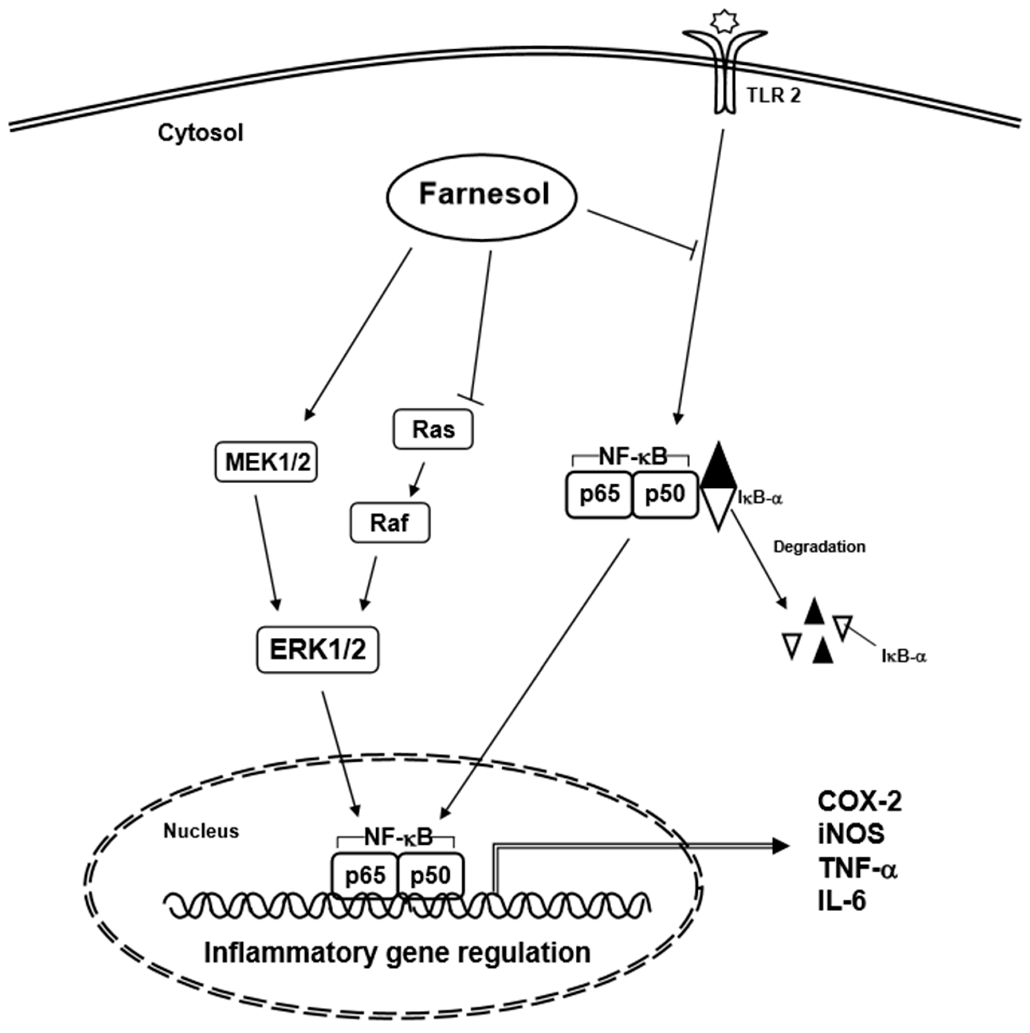
2.1. In Vitro Pro-Inflammatory Effects of Farnesol
2.2. In Vitro Anti-Inflammatory Effects of Farnesol
2.3. In Vitro Anti-Tumor Effects of Farnesol
2.4. Prostate Cancer
2.5. Breast Cancer
2.6. Lung Cancer
2.7. Pancreatic Cancer
2.8. Cervical Cancer
2.9. Oral Squamous Cell Carcinoma (OSCC)
2.10. Meningioma
2.11. Multiple Myeloma
2.12. T Lymphoblastic Leukemia
3. In Vivo Anti-Inflammatory and Anti-Cancer Effects of Farnesol
3.1. Anti-Inflammatory
3.2. Anti-Cancer
3.2.1. Pancreatic Tumor
3.2.2. Prostate Tumor
3.2.3. Multiple Myeloma
4. Conclusions and Future Perspectives
Author Contributions
Funding
Conflicts of Interest
Abbreviations
| Akt | Serine/threonine kinase |
| ATF | Activating transcription factor |
| CDK2 | Cyclin dependent kinase 2 |
| CHAC1 | Cation transport regulator-like protein 1 |
| COX-2 | Cyclooxygenase-2 |
| CPT | Choline phosphate transferase |
| DAG | Diacylglycerol |
| DDIT3 | DNA damage-inducible transcript 3 |
| ER | Endoplasmic reticulum |
| IL | Interleukin |
| iNOS | Inducible nitric oxide synthase |
| IRE1 | Inositol requiring protein 1 |
| MAPK | Mitogen-activated protein kinase |
| MMP | Mitochondrial membrane potential |
| NF-κB | Nuclear factor kappa-light-chain-enhancer of activated B cells |
| OSCC | Oral squamous cell carcinoma |
| PARP | Poly (ADP-ribose) polymerase |
| PC | Phosphatidylcholine |
| PERK | PKR-like ER kinase |
| PI3K | Phosphatidylinositol-3-kinase |
| PPAR | Peroxisome proliferator activated receptor |
| STAT3 | Signal transducer and activator of transcription 3 |
| THR | Thyroid hormone receptor |
| TNFα | Tumor necrosis factor alpha |
| UPR | Unfolded protein response |
| XBP1 | X-box binding protein 1 |
| ↓ | Downregulation |
| ↑ | Upregulation |
| ED50 | Effective dose 50 |
References
- Jung, S.; Spiegelman, D.; Baglietto, L.; Bernstein, L.; Boggs, D.A.; van den Brandt, P.A.; Buring, J.E.; Cerhan, J.R.; Gaudet, M.M.; Giles, G.G.; et al. Fruit and vegetable intake and risk of breast cancer by hormone receptor status. J. Natl. Cancer. Inst. 2013, 105, 219–236. [Google Scholar] [CrossRef] [PubMed]
- Park, S.Y.; Ollberding, N.J.; Woolcott, C.G.; Wilkens, L.R.; Henderson, B.E.; Kolonel, L.N. Fruit and vegetable intakes are associated with lower risk of bladder cancer among women in the Multiethnic Cohort Study. J. Nutr. 2013, 143, 1283–1292. [Google Scholar] [CrossRef] [PubMed]
- Chen, G.C.; Lv, D.B.; Pang, Z.; Liu, Q.F. Fruits and vegetables consumption and risk of non-Hodgkin’s lymphoma: A meta-analysis of observational studies. Int. J. Cancer 2013, 133, 190–200. [Google Scholar] [CrossRef] [PubMed]
- Koushik, A.; Spiegelman, D.; Albanes, D.; Anderson, K.E.; Bernstein, L.; van den Brandt, P.A.; Bergkvist, L.; English, D.R.; Freudenheim, J.L.; Fuchs, C.S.; et al. Intake of fruits and vegetables and risk of pancreatic cancer in a pooled analysis of 14 cohort studies. Am. J. Epidemiol. 2012, 176, 373–386. [Google Scholar] [CrossRef] [PubMed]
- Goossens, A.; Merckx, L. Allergic contact dermatitis from farnesol in a deodorant. Contact Dermat. 1997, 37, 179–180. [Google Scholar] [CrossRef]
- Haustein, U.F.; Herrmann, J.; Hoppe, U.; Engel, W.; Sauermann, G. Growth-inhibition of coryneform bacteria by a mixture of 3 natural-products—Farnesol, glyceryl monolaurate, and phenoxyethanol: Hgq. J. Soc. Cosmet. Chem. 1993, 44, 211–220. [Google Scholar]
- Ishizaka, H.; Yamada, H.; Sasaki, K. Volatile compounds in the flowers of Cyclamen persicum, C-purpurascens and their hybrids. Sci. Hortic. 2002, 94, 125–135. [Google Scholar] [CrossRef]
- Krupcik, J.; Gorovenko, R.; Spanik, I.; Sandra, P.; Armstrong, D.W. Enantioselective comprehensive two-dimensional gas chromatography. A route to elucidate the authenticity and origin of Rosa damascena Miller essential oils. J. Sep. Sci. 2015, 38, 3397–3403. [Google Scholar] [CrossRef] [PubMed]
- Azanchi, T.; Shafaroodi, H.; Asgarpanah, J. Anticonvulsant activity of Citrus aurantium blossom essential oil (neroli): Involvment of the GABAergic system. Nat. Prod. Commun. 2014, 9, 1615–1618. [Google Scholar] [PubMed]
- Joo, J.H.; Jetten, A.M. Molecular mechanisms involved in farnesol-induced apoptosis. Cancer Lett. 2010, 287, 123–135. [Google Scholar] [CrossRef] [PubMed]
- Duncan, R.E.; Archer, M.C. Farnesol decreases serum triglycerides in rats: Identification of mechanisms including up-regulation of PPARalpha and down-regulation of fatty acid synthase in hepatocytes. Lipids 2008, 43, 619–627. [Google Scholar] [CrossRef] [PubMed]
- Takahashi, N.; Kawada, T.; Goto, T.; Yamamoto, T.; Taimatsu, A.; Matsui, N.; Kimura, K.; Saito, M.; Hosokawa, M.; Miyashita, K.; et al. Dual action of isoprenols from herbal medicines on both PPARgamma and PPARalpha in 3T3-L1 adipocytes and HepG2 hepatocytes. FEBS Lett. 2002, 514, 315–322. [Google Scholar] [CrossRef]
- Goto, T.; Kim, Y.I.; Funakoshi, K.; Teraminami, A.; Uemura, T.; Hirai, S.; Lee, J.Y.; Makishima, M.; Nakata, R.; Inoue, H.; et al. Farnesol, an isoprenoid, improves metabolic abnormalities in mice via both PPARalpha-dependent and -independent pathways. Am. J. Physiol. Endocrinol. Metab. 2011, 301, E1022–E1032. [Google Scholar] [CrossRef] [PubMed]
- Ku, C.M.; Lin, J.Y. Farnesol, a sesquiterpene alcohol in herbal plants, exerts anti-inflammatory and antiallergic effects on ovalbumin-sensitized and -challenged asthmatic mice. Evid. Based Complement Alternat. Med. 2015, 2015, 387357. [Google Scholar] [CrossRef] [PubMed]
- Ghosh, S.; Howe, N.; Volk, K.; Tati, S.; Nickerson, K.W.; Petro, T.M. Candida albicans cell wall components and farnesol stimulate the expression of both inflammatory and regulatory cytokines in the murine RAW264.7 macrophage cell line. FEMS Immunol. Med. Microbiol. 2010, 60, 63–73. [Google Scholar] [CrossRef] [PubMed]
- Joo, J.H.; Jetten, A.M. NF-κB-dependent transcriptional activation in lung carcinoma cells by farnesol involves p65/RelA(Ser276) phosphorylation via the MEK-MSK1 signaling pathway. J. Biol. Chem. 2008, 283, 16391–16399. [Google Scholar] [CrossRef] [PubMed]
- Qamar, W.; Sultana, S. Farnesol ameliorates massive inflammation, oxidative stress and lung injury induced by intratracheal instillation of cigarette smoke extract in rats: an initial step in lung chemoprevention. Chem. Biol. Interact. 2008, 176, 79–87. [Google Scholar] [CrossRef] [PubMed]
- Chaudhary, S.C.; Alam, M.S.; Siddiqui, M.S.; Athar, M. Chemopreventive effect of farnesol on DMBA/TPA-induced skin tumorigenesis: involvement of inflammation, Ras-ERK pathway and apoptosis. Life Sci. 2009, 85, 196–205. [Google Scholar] [CrossRef] [PubMed]
- Khan, R.; Sultana, S. Farnesol attenuates 1,2-dimethylhydrazine induced oxidative stress, inflammation and apoptotic responses in the colon of Wistar rats. Chem. Biol. Interact. 2011, 192, 193–200. [Google Scholar] [CrossRef] [PubMed]
- Qamar, W.; Khan, A.Q.; Khan, R.; Lateef, A.; Tahir, M.; Rehman, M.U.; Ali, F.; Sultana, S. Benzo(a)pyrene-induced pulmonary inflammation, edema, surfactant dysfunction and injuries in rats: Alleviation by farnesol. Exp. Lung Res. 2012, 38, 19–27. [Google Scholar] [CrossRef] [PubMed]
- Santhanasabapathy, R.; Vasudevan, S.; Anupriya, K.; Pabitha, R.; Sudhandiran, G. Farnesol quells oxidative stress, reactive gliosis and inflammation during acrylamide-induced neurotoxicity: Behavioral and biochemical evidence. Neuroscience 2015, 308, 212–227. [Google Scholar] [CrossRef] [PubMed]
- Park, J.S.; Kwon, J.K.; Kim, H.R.; Kim, H.J.; Kim, B.S.; Jung, J.Y. Farnesol induces apoptosis of DU145 prostate cancer cells through the PI3K/Akt and MAPK pathways. Int. J. Mol. Med. 2014, 33, 1169–1176. [Google Scholar] [CrossRef] [PubMed]
- Epplen, R.; Stockle, M.; Engelmann, U.; Heidenreich, A.; Ohlmann, C.H. Differential effects of ibandronate, docetaxel and farnesol treatment alone and in combination on the growth of prostate cancer cell lines. Acta Oncol. 2011, 50, 127–133. [Google Scholar] [CrossRef] [PubMed]
- Duncan, R.E.; Archer, M.C. Farnesol induces thyroid hormone receptor (THR) beta1 but inhibits THR-mediated signaling in MCF-7 human breast cancer cells. Biochem. Biophys. Res. Commun. 2006, 343, 239–243. [Google Scholar] [CrossRef] [PubMed]
- Wang, Z.; Chen, H.T.; Roa, W.; Finlay, W. Farnesol for aerosol inhalation: Nebulization and activity against human lung cancer cells. J. Pharm. Pharm. Sci. 2003, 6, 95–100. [Google Scholar] [PubMed]
- Miquel, K.; Pradines, A.; Tercé, F.; Selmi, S.; Favre, G. Competitive inhibition of choline phosphotransferase by geranylgeraniol and farnesol inhibits phosphatidylcholine synthesis and induces apoptosis in human lung adenocarcinoma A549 Cells. J. Bio. Chem. 1998, 273, 26179–26186. [Google Scholar] [CrossRef]
- Joo, J.H.; Liao, G.; Collins, J.B.; Grissom, S.F.; Jetten, A.M. Farnesol-induced apoptosis in human lung carcinoma cells is coupled to the endoplasmic reticulum stress response. Cancer Res. 2007, 67, 7929–7936. [Google Scholar] [CrossRef] [PubMed]
- Burke, Y.D.; Ayoubi, A.S.; Werner, S.R.; McFarland, B.C.; Heilman, D.K.; Ruggeri, B.A.; Crowell, P.L. Effects of the isoprenoids perillyl alcohol and farnesol on apoptosis biomarkers in pancreatic cancer chemoprevention. Anticancer Res. 2002, 22, 3127–3134. [Google Scholar] [PubMed]
- Burke, Y.D.; Stark, M.J.; Roach, S.L.; Sen, S.E.; Crowell, P.L. Inhibition of pancreatic cancer growth by the dietary isoprenoids farnesol and geraniol. Lipids 1997, 32, 151–156. [Google Scholar] [CrossRef] [PubMed]
- Wiseman, D.A.; Werner, S.R.; Crowell, P.L. Cell cycle arrest by the isoprenoids perillyl alcohol, geraniol, and farnesol is mediated by p21(Cip1) and p27(Kip1) in human pancreatic adenocarcinoma cells. J. Pharmacol. Exp. Ther. 2007, 320, 1163–1170. [Google Scholar] [CrossRef] [PubMed]
- Wang, Y.L.; Liu, H.F.; Shi, X.J.; Wang, Y. Antiproliferative activity of farnesol in HeLa cervical cancer cells is mediated via apoptosis induction, loss of mitochondrial membrane potential (LambdaPsim) and PI3K/Akt signalling pathway. J. BUON 2018, 23, 752–757. [Google Scholar] [PubMed]
- Scheper, M.A.; Shirtliff, M.E.; Meiller, T.F.; Peters, B.M.; Jabra-Rizk, M.A. Farnesol, a fungal quorum-sensing molecule triggers apoptosis in human oral squamous carcinoma cells. Neoplasia 2008, 10, 954–963. [Google Scholar] [CrossRef] [PubMed]
- Pfister, C.; Pfrommer, H.; Tatagiba, M.S.; Roser, F. Detection and quantification of farnesol-induced apoptosis in difficult primary cell cultures by TaqMan protein assay. Apoptosis 2013, 18, 452–466. [Google Scholar] [CrossRef] [PubMed]
- Lee, J.H.; Kim, C.; Kim, S.H.; Sethi, G.; Ahn, K.S. Farnesol inhibits tumor growth and enhances the anticancer effects of bortezomib in multiple myeloma xenograft mouse model through the modulation of STAT3 signaling pathway. Cancer Lett. 2015, 360, 280–293. [Google Scholar] [CrossRef] [PubMed]
- Joo, J.H.; Ueda, E.; Bortner, C.D.; Yang, X.P.; Liao, G.; Jetten, A.M. Farnesol activates the intrinsic pathway of apoptosis and the ATF4-ATF3-CHOP cascade of ER stress in human T lymphoblastic leukemia Molt4 cells. Biochem. Pharmacol. 2015, 97, 256–268. [Google Scholar] [CrossRef] [PubMed]
- Katsuyama, M.; Kobayashi, Y.; Ichikawa, H.; Mizuno, A.; Miyachi, Y.; Matsunaga, K.; Kawashima, M. A novel method to control the balance of skin microflora Part 2. A study to assess the effect of a cream containing farnesol and xylitol on atopic dry skin. J. Dermatol. Sci. 2005, 38, 207–213. [Google Scholar] [PubMed]
- Schnuch, A.; Uter, W.; Geier, J.; Lessmann, H.; Frosch, P.J. Sensitization to 26 fragrances to be labelled according to current European regulation. Results of the IVDK and review of the literature. Contact Dermat. 2007, 57, 1–10. [Google Scholar] [CrossRef] [PubMed]
- Gilpin, S.; Maibach, H. Allergic contact dermatitis caused by farnesol: Clinical relevance. Cutan. Ocul. Toxicol. 2010, 29, 278–287. [Google Scholar] [CrossRef] [PubMed]
- Schnuch, A.; Uter, W.; Geier, J.; Lessmann, H.; Frosch, P.J. Contact allergy to farnesol in 2021 consecutively patch tested patients. Results of the IVDK. Contact Dermat. 2004, 50, 117–121. [Google Scholar] [CrossRef] [PubMed]
- Chai, E.Z.; Siveen, K.S.; Shanmugam, M.K.; Arfuso, F.; Sethi, G. Analysis of the intricate relationship between chronic inflammation and cancer. Biochem. J. 2015, 468, 1–15. [Google Scholar] [CrossRef] [PubMed]
- Sethi, G.; Shanmugam, M.K.; Ramachandran, L.; Kumar, A.P.; Tergaonkar, V. Multifaceted link between cancer and inflammation. Biosci. Rep. 2012, 32, 1–15. [Google Scholar] [CrossRef] [PubMed]
- Li, F.; Zhang, J.; Arfuso, F.; Chinnathambi, A.; Zayed, M.E.; Alharbi, S.A.; Kumar, A.P.; Ahn, K.S.; Sethi, G. NF-κB in cancer therapy. Arch. Toxicol. 2015, 89, 711–731. [Google Scholar] [CrossRef] [PubMed]
- Cutolo, M.; Soldano, S.; Contini, P.; Sulli, A.; Seriolo, B.; Montagna, P.; Brizzolara, R. Intracellular NF-kB-decrease and IKBalpha increase in human macrophages following CTLA4-Ig treatment. Clin. Exp. Rheumatol. 2013, 31, 943–946. [Google Scholar] [PubMed]
- Ku, C.M.; Lin, J.Y. Anti-inflammatory effects of 27 selected terpenoid compounds tested through modulating Th1/Th2 cytokine secretion profiles using murine primary splenocytes. Food Chem. 2013, 141, 1104–1113. [Google Scholar] [CrossRef] [PubMed]
- Navarathna, D.H.; Nickerson, K.W.; Duhamel, G.E.; Jerrels, T.R.; Petro, T.M. Exogenous farnesol interferes with the normal progression of cytokine expression during candidiasis in a mouse model. Infect. Immun. 2007, 75, 4006–4011. [Google Scholar] [CrossRef] [PubMed]
- Juvekar, A.; Hu, H.; Yadegarynia, S.; Lyssiotis, C.A.; Ullas, S.; Lien, E.C.; Bellinger, G.; Son, J.; Hok, R.C.; Seth, P.; et al. Phosphoinositide 3-kinase inhibitors induce DNA damage through nucleoside depletion. Proc. Natl. Acad. Sci. USA 2016, 113, E4338–E4347. [Google Scholar] [CrossRef] [PubMed]
- Luo, J.; Manning, B.D.; Cantley, L.C. Targeting the PI3K-Akt pathway in human cancer: Rationale and promise. Cancer Cell 2003, 4, 257–262. [Google Scholar] [CrossRef]
- Carver, B.S.; Chapinski, C.; Wongvipat, J.; Hieronymus, H.; Chen, Y.; Chandarlapaty, S.; Arora, V.K.; Le, C.; Koutcher, J.; Scher, H.; et al. Reciprocal feedback regulation of PI3K and androgen receptor signaling in PTEN-deficient prostate cancer. Cancer Cell 2011, 19, 575–586. [Google Scholar] [CrossRef] [PubMed]
- Ng, S.S.; Tsao, M.S.; Nicklee, T.; Hedley, D.W. Wortmannin inhibits pkb/akt phosphorylation and promotes gemcitabine antitumor activity in orthotopic human pancreatic cancer xenografts in immunodeficient mice. Clin. Cancer Res. 2001, 7, 3269–3275. [Google Scholar] [PubMed]
- Bondar, V.M.; Sweeney-Gotsch, B.; Andreeff, M.; Mills, G.B.; McConkey, D.J. Inhibition of the phosphatidylinositol 3′-kinase-AKT pathway induces apoptosis in pancreatic carcinoma cells in vitro and in vivo. Mol. Cancer Ther. 2002, 1, 989–997. [Google Scholar] [PubMed]
- Fahy, B.N.; Schlieman, M.; Virudachalam, S.; Bold, R.J. AKT inhibition is associated with chemosensitisation in the pancreatic cancer cell line MIA-PaCa-2. Br. J. Cancer 2003, 89, 391–397. [Google Scholar] [CrossRef] [PubMed]
- McCubrey, J.A.; Lahair, M.M.; Franklin, R.A. Reactive oxygen species-induced activation of the MAP kinase signaling pathways. Antioxid Redox Signal 2006, 8, 1775–1789. [Google Scholar] [CrossRef] [PubMed]
- Liu, X.; Li, Q.; Dowdell, K.; Fischer, E.R.; Cohen, J.I. Varicella-Zoster virus ORF12 protein triggers phosphorylation of ERK1/2 and inhibits apoptosis. J. Virol. 2012, 86, 3143–3151. [Google Scholar] [CrossRef] [PubMed]
- Forman, B.M.; Goode, E.; Chen, J.; Oro, A.E.; Bradley, D.J.; Perlmann, T.; Noonan, D.J.; Burka, L.T.; McMorris, T.; Lamph, W.W.; et al. Identification of a nuclear receptor that is activated by farnesol metabolites. Cell 1995, 81, 687–693. [Google Scholar] [CrossRef]
- Aranda, A.; Pascual, A. Nuclear hormone receptors and gene expression. Physiol. Rev. 2001, 81, 1269–1304. [Google Scholar] [CrossRef] [PubMed]
- Ahn, K.S.; Sethi, G.; Sung, B.; Goel, A.; Ralhan, R.; Aggarwal, B.B. Guggulsterone, a farnesoid X receptor antagonist, inhibits constitutive and inducible STAT3 activation through induction of a protein tyrosine phosphatase SHP-1. Cancer Res. 2008, 68, 4406–4415. [Google Scholar] [CrossRef] [PubMed]
- Buettner, R.; Mora, L.B.; Jove, R. Activated STAT signaling in human tumors provides novel molecular targets for therapeutic intervention. Clin. Cancer Res. 2002, 8, 945–954. [Google Scholar] [PubMed]
- Miquel, K.; Pradines, A.; Favre, G. Farnesol and geranylgeraniol induce actin cytoskeleton disorganization and apoptosis in A549 lung adenocarcinoma cells. Biochem. Biophys. Res. Commun. 1996, 225, 869–876. [Google Scholar] [CrossRef] [PubMed]
- Park, J.I. Growth arrest signaling of the Raf/MEK/ERK pathway in cancer. Front. Biol. 2014, 9, 95–103. [Google Scholar] [CrossRef] [PubMed]
- Lateef, A.; Rehman, M.U.; Tahir, M.; Khan, R.; Khan, A.Q.; Qamar, W.; Sultana, S. Farnesol protects against intratracheally instilled cigarette smoke extract-induced histological alterations and oxidative stress in prostate of wistar rats. Toxicol. Int. 2013, 20, 35–42. [Google Scholar] [PubMed]


| Plants | Scientific Name | Usages | Ref. |
|---|---|---|---|
| Citronella | Cymbopogon nardus | Source of soap, candles and incense, perfume, cosmetics, and flavoring | [5] |
| Lemon Grass | Cymbopogon citratus | Sterilization Relieves sweating, fever, abdominal pain, and controls skin secretions | [5] |
| Tuberose | Polianthes tuberosa L. | Source of perfume | [5,6] |
| Cyclamen | Cyclamen persicum | [7] | |
| Rose | Rosa hybrida | Source of perfume | [8] |
| Neroli | Citrus aurantium subsp. amara | Source of perfume | [9] |
| Balsam | Source of medicinal products, fragrances for varnishes and lacquers, air freshener perfume, and natural remedy for skin rashes | [5] | |
| Musk | Abelmoschus moschatus | Source of perfume | [6] |
| Origin | Cell Lines | Molecular Targets | Mechanism of Actions | ED50 (μM) | Ref. |
|---|---|---|---|---|---|
| Prostate | DU145 | PI3K/Akt, MAPK ↓ | apoptosis ↑ | 30 and 60 | [22] |
| LNCaP | Ras | apoptosis ↑, cell proliferation ↓ | 75 | [23] | |
| Breast | MCF-7 | Farnesoid X receptor | anti-estrogens ↑, cell growth ↓ | 10 and 20 | [24] |
| Lung | A549 | CPT activity ↓ PC biosynthesis ↓ | cell growth ↓ apoptosis ↑, G0/G1 phase arrest ↑ | 4.5 or 80 | [25,26] |
| H460 | NF-κB ↑ MEK1/2, ERK1/2, UPR ↑ | Immune response ↑ cell growth ↓, apoptosis ↑ | 4.5 or 250 | [16,25,27] | |
| Pancreatic | BxPC-3 | Bak ↑; p21, p27 ↑, Cyclin A, cyclin B1 ↓ CDK2 ↓ | apoptosis ↑ G0/G1 phase arrest ↑ | 30, 60 and 90 | [28,30] |
| PC-1 | Bak ↑ | apoptosis ↑ | 30, 60 and 90 | [29] | |
| MIA PaCa2 | Bak ↑; p21, p27 ↑, Cyclin A, cyclin B1 ↓, Cyclin D1, CDK2 ↓ | apoptosis ↑ G0/G1 phase arrest ↑ | 25, 30, 60 and 90 | [29,30] | |
| Cervical | HeLa | PI3K, p-Akt ↓ | apoptosis ↑ | 33.5, 23.8 and 17.6 | [31] |
| Oral squamous cell | OSCC9 | Cleaved caspase-9 & -3 ↑ | cell growth ↓, apoptosis ↑ | 30 and 60 | [32] |
| OSCC25 | Cleaved caspase-9 & -3 ↑ | cell growth ↓, apoptosis ↑ | 30 and 60 | [32] | |
| Meningioma | Primary meningioma cells | Cleaved caspase-3 ↑ | apoptosis ↑ | 0.4 and 8 | [33] |
| IOMM-Lee | Cleaved caspase-3 ↑ Cleaved PARP ↑ | apoptosis ↑ | 0.4 and 8 | [33] | |
| Multiple myeloma | U266 | STAT3, JAK ↓ | apoptosis ↑, cell proliferation ↓ | 100 | [34] |
| MM.1S | STAT3, JAK ↓ | apoptosis ↑, cell proliferation ↓ | 100 | [34] | |
| T lymphoblastic leukemia | Molt4-hyg | p-eI2Fα, ATF3, ATF4, CHOP, CHAC1 ↑ (UPR ↑) GRP78 ↓ | ER stress ↑ apoptosis ↑ | 75 | [35] |
| Macrophage | RAW264.7 | TNF-α, IL-6 ↑ | Immune response ↑ | 5 | [15] |
| Primary murine peritoneal macrophage | IL-12 ↓ | Inflammation ↓ | 100 | [45] |
| Disease | Animal | Molecular Targets | Mechanism(s) of Action | Dose and Day of Administration | Ref. |
|---|---|---|---|---|---|
| Allergic asthmatic | Mouse | TNF-α ↓ IL-6, IL-10 ↓ | Inflammation ↓ | 5, 25 and 100 mg/kg/day for 5 weeks | [14] |
| Lung chemoprevention | Rat | Lung prevention ↑ | 50 and 100 mg/kg/day for 7 days | [17] | |
| Skin tumorigenesis | Mouse | Ras, Raf, ERK1/2 ↓ Bax/Bcl-2 ↑ | Inflammation ↓ Apoptosis ↑ | 25, 50, and 100 mg/kg for 3 days | [18] |
| Colon carcinogenesis | Rat | Apoptosis ↑ | 50 and 100 mg/kg for 7 days | [19] | |
| Lung inflammation, Edema | Rat | B(a)P ↓ | Lung protection ↑ | 100 and 200 mg/kg/day for 14 days | [20] |
| Gliosis | Mouse | iNOS ↓ TNF-α ↓ | Inflammation ↓ | 100 mg/kg for 4 weeks | [21] |
| Pancreatic cancer | Hamster | Tumor growth ↓ | 20 g/kg diet for 20 days | [29] | |
| Prostate cancer | Mouse | PI3K/Akt ↓ | Apoptosis ↑ | 50 mg/kg daily for 5 weeks | [22] |
| Multiple myeloma | Mouse | p-STAT3 ↓ | Cell proliferation ↓ Apoptosis ↑ | 60 mg/kg 3 times/week for 3 weeks | [34] |
| Oxidative damage to prostate gland | Rat | Glutathione ↓, Antioxidant, enzymes ↑ | Xanthine oxidase activity ↓, Lipid hydroperoxide ↓ | 50 or 100 mg/kg for 7 days | [60] |
© 2018 by the authors. Licensee MDPI, Basel, Switzerland. This article is an open access article distributed under the terms and conditions of the Creative Commons Attribution (CC BY) license (http://creativecommons.org/licenses/by/4.0/).
Share and Cite
Jung, Y.Y.; Hwang, S.T.; Sethi, G.; Fan, L.; Arfuso, F.; Ahn, K.S. Potential Anti-Inflammatory and Anti-Cancer Properties of Farnesol. Molecules 2018, 23, 2827. https://doi.org/10.3390/molecules23112827
Jung YY, Hwang ST, Sethi G, Fan L, Arfuso F, Ahn KS. Potential Anti-Inflammatory and Anti-Cancer Properties of Farnesol. Molecules. 2018; 23(11):2827. https://doi.org/10.3390/molecules23112827
Chicago/Turabian StyleJung, Young Yun, Sun Tae Hwang, Gautam Sethi, Lu Fan, Frank Arfuso, and Kwang Seok Ahn. 2018. "Potential Anti-Inflammatory and Anti-Cancer Properties of Farnesol" Molecules 23, no. 11: 2827. https://doi.org/10.3390/molecules23112827
APA StyleJung, Y. Y., Hwang, S. T., Sethi, G., Fan, L., Arfuso, F., & Ahn, K. S. (2018). Potential Anti-Inflammatory and Anti-Cancer Properties of Farnesol. Molecules, 23(11), 2827. https://doi.org/10.3390/molecules23112827