
Biotransformation of Bicyclic Halolactones with a Methyl Group in the Cyclohexane Ring into Hydroxylactones and Their Biological Activity


 , ,
, ,  and
and
Abstract
:
1. Introduction
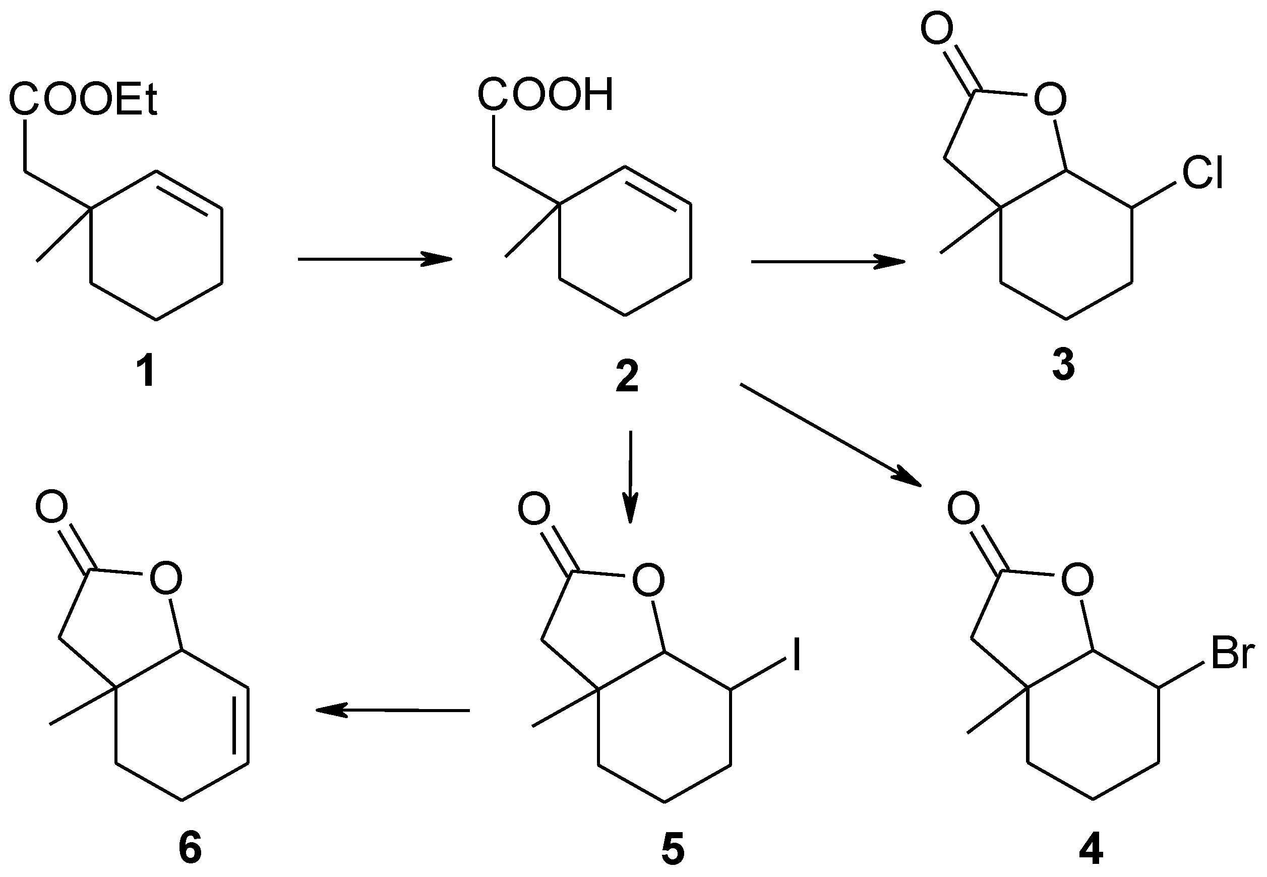
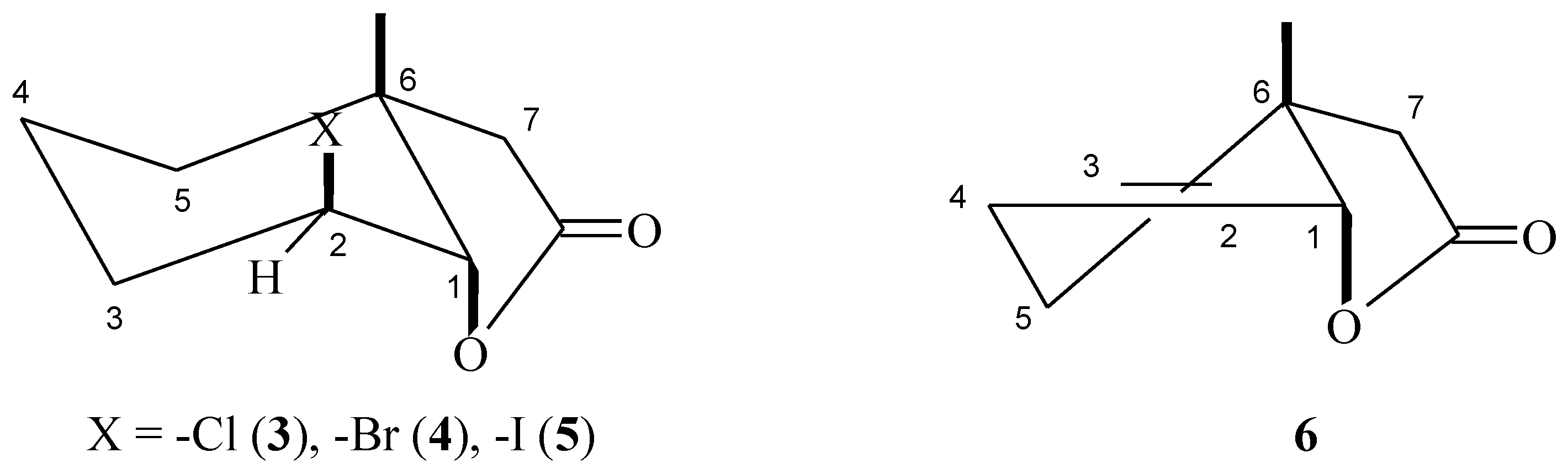
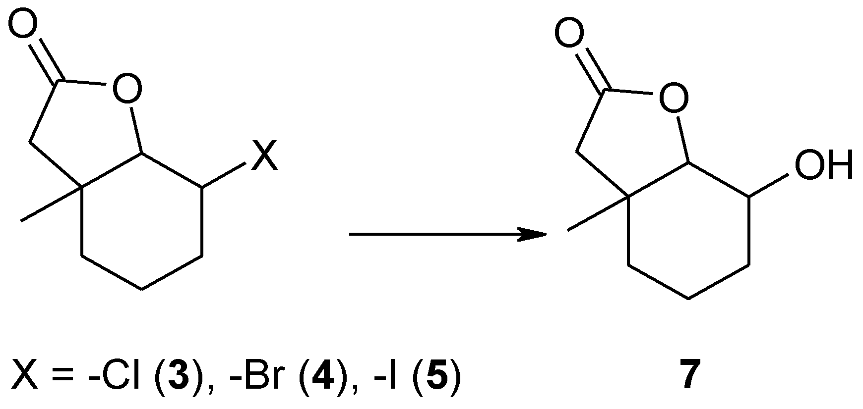
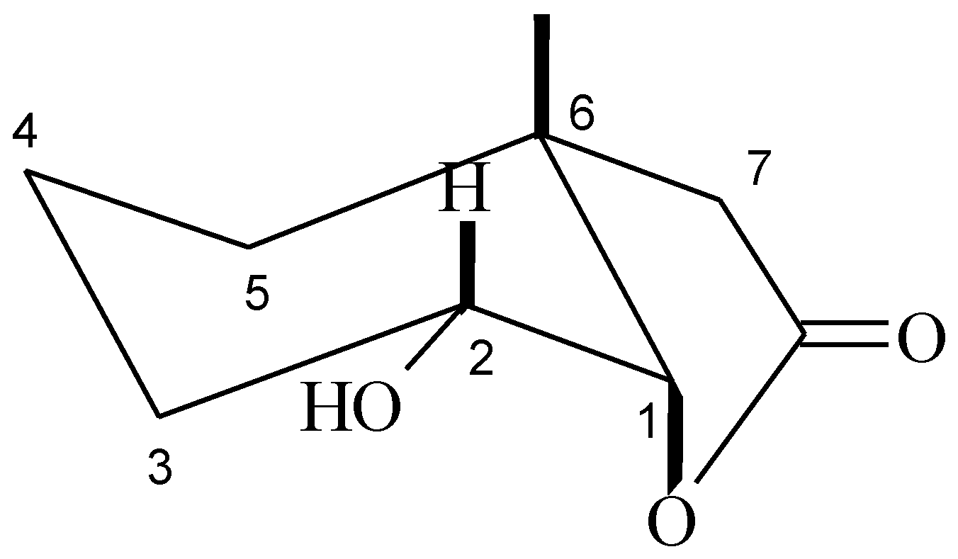
2. Results and Discussion
3. Experimental Section
3.1. General Information
3.2. Synthesis of Substrates
3.3. Microorganisms
3.3.1. Screening Procedure
3.3.2. Preparative Biotransformation
3.4. Bioassay Tests on Microorganisms
3.5. Aphid Settling Bioassays
4. Conclusions
Supplementary Materials
Acknowledgments
Author Contributions
Conflicts of Interest
References
- Scher, R.K.; Nakamura, N.; Tavakkol, A. Luliconazole: A review of a new antifungal agent for the topical treatment of onychomycosis. Mycoses 2014, 57, 389–393. [Google Scholar] [CrossRef] [PubMed]
- Moussa, Z.; El-Sharief, M.A.; El-Sharief, A.M. Synthesis and characterization of new types of halogenated and alkylated imidazolidineiminothiones and a comparative study of their antitumor, antibacterial, and antifungal activities. Eur. J. Med. Chem. 2011, 46, 2280–2289. [Google Scholar] [CrossRef] [PubMed]
- Vairappan, C.S.; Kawamoto, T.; Miwa, H.; Suzuki, M. Potent antibacterial activity of halogenated compounds against antibiotic-resistant bacteria. Planta Med. 2004, 70, 1087–1090. [Google Scholar] [CrossRef] [PubMed]
- Lakshmanan, K.; Sekar, K.G.; Sathiyendiran, V.; Muthuvel, I. Insect antifeedant potent quinoxalines. Int. Lett. Chem. Phys. Astron. 2015, 2, 82–87. [Google Scholar] [CrossRef]
- Boulogne, I.; Petit, P.; Ozier-Lafontaine, H.; Desfontaines, L.; Loranger-Merciris, G. Insecticidal and antifungal chemicals produced by plants: A review. Environ. Chem. Lett. 2012, 10, 325–347. [Google Scholar] [CrossRef]
- Zhenchuk, A.; Lotfi, K.; Juliusson, G.; Albertioni, F. Mechanisms of anti-cancer action and pharmacology of clofarabine. Biochem. Pharmacol. 2009, 78, 1351–1359. [Google Scholar] [CrossRef] [PubMed]
- Bossert, I.D.; Häggblom, M.M.; Young, L.Y. Microbial ecology of dehalogenation. In Dehalogenation: Microbial Processes and Environmental Applications; Häggblom, M.M., Bossert, I.D., Eds.; Kluwer Academic Publishers: Boston, MA, USA, 2003; pp. 33–52. [Google Scholar]
- Vairappan, C.S. Potent antibacterial activity of halogenated metabolites from Malaysian red algae, Laurencia majuscula (Rhodomelaceae, Ceramiales). Biomol. Eng. 2003, 20, 255–259. [Google Scholar] [CrossRef]
- Ren, D.; Wood, T.K. (5Z)-4-bromo-5-(bromomethylene)-3-butyl-2(5H)-furanone reduces corrosion from Desulfotomaculum orientis. Environ. Microbiol. 2004, 6, 535–540. [Google Scholar] [CrossRef] [PubMed]
- Vairappan, C.S.; Ishii, T.; Lee, T.K.; Suzuki, M.; Zhaoqi, Z. Antibacterial activities of a new brominated diterpene from Borneon Laurencia spp. Mar. Drugs 2010, 8, 1743–1749. [Google Scholar] [CrossRef] [PubMed]
- Jensen, S.R.; Gotfredsen, C.H.; Harput, U.S.; Saracoglu, I. Chlorinated iridoid glucosides from Veronica longifolia and their antioxidant activity. J. Nat. Prod. 2010, 73, 1593–1596. [Google Scholar] [CrossRef] [PubMed]
- Nenkep, V.; Yun, K.; Zhang, D.; Choi, H.D.; Kang, J.S.; Son, B.W. Induced production of bromomethylchlamydosporols A and B from the marine-derived fungus Fusarium tricinctum. J. Nat. Prod. 2010, 73, 2061–2063. [Google Scholar] [CrossRef] [PubMed]
- Yin, S.; Boyle, G.M.; Carroll, A.R.; Kotiw, M.; Dearnaley, J.; Quinn, R.J.; Davis, R.A. Caelestines A−D, Brominated quinolinecarboxylic acids from the Australian ascidian Aplidium caelestis. J. Nat. Prod. 2010, 73, 1586–1589. [Google Scholar] [CrossRef] [PubMed]
- Yang, X.; Davis, R.A.; Buchanan, M.S.; Duffy, S.; Avery, V.M.; Camp, D.; Quinn, R.J. Antimalarial bromotyrosine derivatives from the Australian marine sponge Hyattella sp. J. Nat. Prod. 2010, 73, 985–987. [Google Scholar] [CrossRef] [PubMed]
- Sobczak, A. Czynniki chemiczne w środowisku zagrażające zdrowiu ludzi. Environ. Med. 2012, 15, 7–17. [Google Scholar]
- Grabarczyk, M.; Mączka, W.; Wińska, K.; Żarowska, B.; Anioł, M. Antimicrobial activity of hydroxylactone obtained by biotransformation of bromo- and iodolactone with gem-dimethylcyclohexane ring. J. Braz. Chem. Soc. 2013, 24, 1913–1919. [Google Scholar] [CrossRef]
- Grabarczyk, M.; Wińska, K.; Mączka, W.; Żarowska, B.; Maciejewska, G.; Dancewicz, K.; Gabryś, B.; Anioł, M. Synthesis, biotransformation and biological activity of halolactones obtained from β-ionone. Tetrahedron 2016, 72, 637–644. [Google Scholar] [CrossRef]
- Grabarczyk, M.; Wińska, K.; Mączka, W.; Żołnierczyk, A.K.; Żarowska, B.; Anioł, M. Lactones with methylcyclohexane system obtained by chemical and microbiological methods and their antimicrobial activity. Molecules 2015, 20, 3335–3353. [Google Scholar] [CrossRef] [PubMed]
- Monod, J. The growth of bacterial cultures. Annu. Rev. Microbiol. 1949, 3, 371–394. [Google Scholar] [CrossRef]
- Naranga, A.; Pilyugin, S.S. Bacterial gene regulation in diauxic and non-diauxic growth. J. Theor. Biol. 2007, 244, 326–348. [Google Scholar] [CrossRef] [PubMed]
- Loomis, W.F., Jr.; Magasanik, B. Glucose-Lactose Diauxie in Escherichia coli. J. Bacteriol. 1967, 93, 1397–1401. [Google Scholar] [PubMed]
- Grudniewska, A.; Dancewicz, K.; Białońska, A.; Ciunik, Z.; Gabryś, B.; Wawrzeńczyk, C. Synthesis of piperitone-derived halogenated lactones and their effect on aphid probing, feeding, and settling behavior. RSC Adv. 2011, 1, 498–510. [Google Scholar] [CrossRef]
- Grudniewska, A.; Dancewicz, K.; Białońska, A.; Wawrzeńczyk, C.; Gabryś, B. Piperitone-derived saturated lactones: Synthesis and aphid behavior-modifying activity. J. Agric. Food Chem. 2013, 61, 3364–3372. [Google Scholar] [CrossRef] [PubMed]
- Grudniewska, A.; Kłobucki, M.; Dancewicz, K.; Szczepanik, M.; Gabryś, B.; Wawrzeńczyk, C. Synthesis and antifeedant activity of racemic and optically active hydroxy lactones with the p-menthane system. PLoS ONE 2015, 10, 1–17. [Google Scholar] [CrossRef] [PubMed]
- Burgstahler, A.W.; Nordin, I.C. Specyfic angular alkylation. A new application of the Claisen rearrangement. J. Am. Chem. Soc. 1961, 83, 198–206. [Google Scholar] [CrossRef]
- Grabarczyk, M.; Białońska, A. Biotransformations of chloro-, bromo- and iodolactone with trimethylcyclohexane system using fungal strains. Biocatal. Biotransform. 2010, 5–6, 408–414. [Google Scholar] [CrossRef]
- Grabarczyk, M.; Szumny, A.; Gładkowski, W.; Białońska, A.; Ciunik, Z.; Wawrzeńczyk, C. Lactones 18. Synthesis of bicyclic lactones with methyl-, di- and trimethyl substituted cyclohexane system. Polish J. Chem. 2005, 79, 1763–1771. [Google Scholar] [CrossRef]
- Sample Availability: Samples of the compounds 1–10 are available from the authors.


| Entry | Microorganism | Screening Biotransformation/% | |||||
|---|---|---|---|---|---|---|---|
| 3 | 7 | 4 | 7 | 5 | 7 | ||
| 1 | Fusarium culmorum AM10 | 100 | - | 100 | - | 100 | - |
| 2 | Fusarium avenaceum AM11 | 100 | - | 100 | - | 100 | - |
| 3 | Fusarium oxysporum AM13 | 100 | - | 100 | - | 100 | - |
| 4 | Fusarium tricinctum AM16 | 100 | - | 100 | - | 100 | - |
| 5 | Fusarium semitectum AM20 | 100 | - | 100 | - | 100 | - |
| 6 | Fusarium oxysporum AM21 | 100 | - | 100 | - | 100 | - |
| 7 | Fusarium equiseti AM22 | 65.4 | 34.6 | 3.3 | 96.7 | 40.0 | 60.0 |
| 8 | Fusarium solani AM203 | 86.1 | 13.9 | 76.6 | 23.4 | 74.7 | 25.3 |
| 9 | Syncephalastrum racemosum AM105 | 100 | - | 74.0 | 26.0 | 69.9 | 30.1 |
| 10 | Absidia coerulea AM93 | 100 | - | 100 | - | 100 | - |
| 11 | Absidia cylindrospora AM336 | 100 | - | 75.0 | 25.0 | 86.0 | 14.0 |
| 12 | Penicillium camembertii AM83 | 85.3 | 14.7 | 100 | - | 100 | - |
| 13 | Penicillium chermesinum AM113 | 100 | - | 100 | - | 100 | - |
| 14 | Penicillium frequentans AM351 | 100 | - | 100 | - | 100 | - |
| 13 | Aspergillus wenthi AM413 | 100 | - | 100 | - | 100 | - |
| 14 | Aspergillus ochraceus AM456 | 100 | - | 100 | - | 100 | - |
| 15 | Pleurotus ostreatus AM482 | 100 | - | 100 | - | 100 | - |
| 16 | Pleurotus ostreatus AM600 | 100 | - | 100 | - | 100 | - |
| 17 | Mucor hiemalis AM450 | 100 | - | 100 | - | 100 | - |
| 18 | Yarrowia lipolytica AM71 | 75.5 | 27.4 | 13.3 | 86.7 | 40.7 | 59.3 |
| 19 | Rhodotorula marina AM77 | 100 | - | 100 | - | 100 | - |
| Entry | Microorganism | Screening Biotransformation/% | |||||
|---|---|---|---|---|---|---|---|
| 3 | 8 | 4 | 9 | 5 | 10 | ||
| 1 | Penicillium vermiculatum AM30 | 69.7 | 30.3 | 79.1 | 20.9 | 65.4 | 34.6 |
| Substrate | Strain | Product | Isolated Yield g/% | Enatiomeric Excess (%) | Optical Rotation | Concentration in CH3Cl (g/100 mL) |
|---|---|---|---|---|---|---|
| 3 | P. vermiculatum AM30 | 8 | 0.0098/9.0 | 7.6 | +5.404 | 0.48 |
| 4 | F. equiseti AM22 | 7 | 0.028/38.4 | 10.7 | −6.762 | 0.56 |
| 4 | Y. lipolytica AM71 | 7 | 0.021/28.8 | 5.8 | −6.244 | 0.62 |
| 4 | P. vermiculatum AM30 | 9 | 0.0114/10.7 | 1.6 | +0.645 | 0.78 |
| 5 | F. equiseti AM22 | 7 | 0.016/26.3 | 27.0 | −17.886 | 0.44 |
| 5 | Y. lipolytica AM71 | 7 | 0.024/40.2 | 52.9 | −31.214 | 0.51 |
| 5 | P. vermiculatum AM30 | 10 | 0.015/14.2 | 5.5 | +4.108 | 0.67 |
| Strain | E. coli | S. aureus | P. fluorescens | C. albicans | F. avenaceum | A. niger | |
|---|---|---|---|---|---|---|---|
| Control | Lag-phase [h] | 5 | 16 | 8 | 14.5 | 15.5 | 7.5 |
| ΔOD ± SD | 1.29 ± 0.07 | 1.09 ± 0.05 | 1.36 ± 0.07 | 1.14 ± 0.05 | 1.18 ± 0.04 | 1.94 ± 00.6 | |
| 3 | Lag-phase [h] | 9 | - | 14.5 | 21 | 35 | 13 |
| ΔOD ± SD | 0.29 ± 0.04 | 0 | 1.05 ± 0.06 | 1.11 ± 0.04 | 1.40 ± 0.05 | 1.48 ± 0.05 | |
| 4 | Lag-phase [h] | 18 | - | 16 | 29 | - | 13 |
| ΔOD ± SD | 0.43 ± 0.05 | 0 | 1.08 ± 0.04 | 1.18 ± 0.04 | 0 | 1.02 ± 0.04 | |
| 5 | Lag-phase [h] | 9 | 23 | 13 | 15 | 38 | 9 |
| ΔOD ± SD | 0.32 ± 0.03 | 0.37 ± 0.04 | 0.95 ± 0.03 | 0.95 ± 0.03 | 0.71 ± 0.04 | 1.97 ± 0.05 | |
| 6 | Lag-phase [h] | 9 | - | 14 | 21 | 32.5 | 11.5 |
| ΔOD ± SD | 0.27 ± 0.03 | 0 | 1.32 ± 0.02 | 0.77 ± 0.02 | 1.80 ± 0.06 | 1.33 ± 0.07 | |
| 7 | Lag-phase [h] | 5 | 36 | 12 | 19.5 | 14.5 | 7.5 |
| ΔOD ± SD | 0.57 ± 0.05 | 0.62 ± 0.04 | 1.15 ± 0.04 | 1.99 ± 0.06 | 1.87 ± 0.07 | 1.94 ± 0.06 | |
| 1 h | 2 h | 24 h | |||||||
|---|---|---|---|---|---|---|---|---|---|
| C | T | p | C | T | p | C | T | p | |
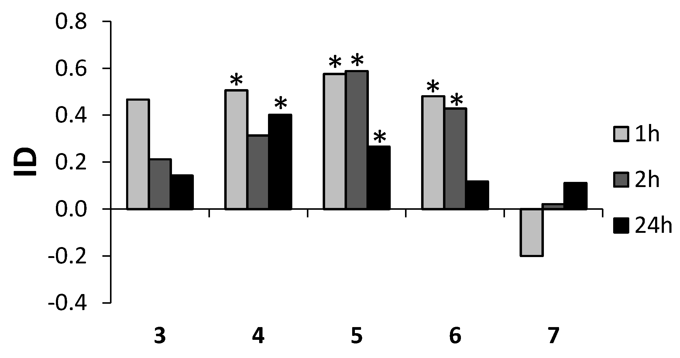
| 3 | 2.8 (±0.8) | 1.0 (±0.3) | 0.0615 | 2.5 (±0.6) | 1.6 (±0.3) | 0.2189 | 6.0 (±1.0) | 4.5 (±1.5) | 0.4168 |
| 4 | 7.6 (±1.1) | 2.5 (±0.5) | 0.0007 | 5.5 (±1.7) | 2.9 (±0.7) | 0.1791 | 8.5 (±1.5) | 3.6 (±0.8) | 0.0116 |
| 5 | 3.3 (±0.8) | 0.9 (±0.3) | 0.0106 | 3.4 (±0.5) | 0.9 (±0.5) | 0.0028 | 6.3 (±0.9) | 3.6 (±0.8) | 0.0470 |
| 6 | 9.6 (±1.3) | 3.4 (±1.3) | 0.0035 | 10.6 (±1.5) | 4.3 (±1.3) | 0.0067 | 7.8 (±1.8) | 6.1 (±1.5) | 0.4972 |
| 7 | 1.8 (±0.7) | 2.6 (±1.0) | 0.4951 | 3.0 (±1.0) | 2.9 (±0.9) | 0.9285 | 5.6 (±0.8) | 4.5 (±1.0) | 0.3996 |
© 2016 by the authors. Licensee MDPI, Basel, Switzerland. This article is an open access article distributed under the terms and conditions of the Creative Commons Attribution (CC-BY) license ( http://creativecommons.org/licenses/by/4.0/).
Share and Cite
Wińska, K.; Grabarczyk, M.; Mączka, W.; Żarowska, B.; Maciejewska, G.; Dancewicz, K.; Gabryś, B.; Szumny, A.; Anioł, M. Biotransformation of Bicyclic Halolactones with a Methyl Group in the Cyclohexane Ring into Hydroxylactones and Their Biological Activity. Molecules 2016, 21, 1453. https://doi.org/10.3390/molecules21111453
Wińska K, Grabarczyk M, Mączka W, Żarowska B, Maciejewska G, Dancewicz K, Gabryś B, Szumny A, Anioł M. Biotransformation of Bicyclic Halolactones with a Methyl Group in the Cyclohexane Ring into Hydroxylactones and Their Biological Activity. Molecules. 2016; 21(11):1453. https://doi.org/10.3390/molecules21111453
Chicago/Turabian StyleWińska, Katarzyna, Małgorzata Grabarczyk, Wanda Mączka, Barbara Żarowska, Gabriela Maciejewska, Katarzyna Dancewicz, Beata Gabryś, Antoni Szumny, and Mirosław Anioł. 2016. "Biotransformation of Bicyclic Halolactones with a Methyl Group in the Cyclohexane Ring into Hydroxylactones and Their Biological Activity" Molecules 21, no. 11: 1453. https://doi.org/10.3390/molecules21111453
APA StyleWińska, K., Grabarczyk, M., Mączka, W., Żarowska, B., Maciejewska, G., Dancewicz, K., Gabryś, B., Szumny, A., & Anioł, M. (2016). Biotransformation of Bicyclic Halolactones with a Methyl Group in the Cyclohexane Ring into Hydroxylactones and Their Biological Activity. Molecules, 21(11), 1453. https://doi.org/10.3390/molecules21111453