Review

Small-Molecule Inhibitors of the Type III Secretion System

School of Pharmaceutical Engineering and Life Science, Changzhou University, Changzhou 213164, China
*
Author to whom correspondence should be addressed.
Molecules 2015, 20(9), 17659-17674; https://doi.org/10.3390/molecules200917659
Submission received: 16 July 2015 / Revised: 17 September 2015 / Accepted: 18 September 2015 / Published: 23 September 2015

Abstract

:
Drug-resistant pathogens have presented increasing challenges to the discovery and development of new antibacterial agents. The type III secretion system (T3SS), existing in bacterial chromosomes or plasmids, is one of the most complicated protein secretion systems. T3SSs of animal and plant pathogens possess many highly conserved main structural components comprised of about 20 proteins. Many Gram-negative bacteria carry T3SS as a major virulence determinant, and using the T3SS, the bacteria secrete and inject effector proteins into target host cells, triggering disease symptoms. Therefore, T3SS has emerged as an attractive target for antimicrobial therapeutics. In recent years, many T3SS-targeting small-molecule inhibitors have been discovered; these inhibitors prevent the bacteria from injecting effector proteins and from causing pathophysiology in host cells. Targeting the virulence of Gram-negative pathogens, rather than their survival, is an innovative and promising approach that may greatly reduce selection pressures on pathogens to develop drug-resistant mutations. This article summarizes recent progress in the search for promising small-molecule T3SS inhibitors that target the secretion and translocation of bacterial effector proteins.

Graphical Abstract

1. Introduction

Although antibiotic therapy is the most commonly-used strategy to control infectious pathogens, most antibiotics affect cellular processes of microorganisms and, therefore, kill them, which becomes a strong selective pressure to develop resistance against antibiotics [1]. In order to contain the risk of increasing antibiotic resistance, targeting bacterial virulence factors instead of bacterial survivability provides a novel alternative approach for the development of new antimicrobials, because virulence-specific therapeutics create less selection pressure for antibiotic-resistant mutations [2,3]. The discovery of bacterial secretion systems is an important milestone in studies on the mechanisms of bacterial pathogenesis. Most enterobacterial pathogens, such as animal pathogens Pseudomonas aeruginosa, Salmonella Typhimurium and Yersinia pestis, and plant pathogens Dickeya dadantii, Erwinia amylovora and Pseudomonas syringae, possess at least one type III secretion system (T3SS) as a major virulence determinant [4,5]. Through this protein secretion/injection system, pathogens translocate their virulence factors (effectors) directly into host cells, enabling infection by subverting cells’ defense mechanisms [6,7]. T3SS represents a particularly appealing target for antimicrobial agents, because the antimicrobial therapies using T3SS-specific inhibitors would affect the virulence rather than the viability of pathogens, creating low selective pressure for developing drug resistance [2,3].

2. The Components of T3SS

As one of the most complicated currently known protein secretion systems, the T3SS is usually encoded by a 30–40-kbp gene and exists in the bacterial chromosome or plasmid in the form of a pathogenicity island [8]. T3SSs of animal or plant pathogens possess many highly conserved main structural components, which are comprised of more than 20 proteins. Various pathogenic T3SSs are very similar in their structures, and their syringe-like appearance in transmission electron microscope images has led them to be called needle complexes (NCs) [9]. The core of the T3SS injection device is a needle-shaped complex that consists of a multi-ring base and a needle protrusion. The two parts are connected by an external needle, which protrudes from the bacterial surface [10,11,12] (Figure 1). The multi-ring base is composed of a pair of inner and outer rings that span the bacterial inner membrane (IM) and outer membrane (OM), and the two rings are connected by a rod-shaped structure passing through the IM and OM [13,14]. Needle protrusions have a straight hollow cylindrical structure containing the narrow center channel (~28 Å in diameter), specialized in transporting secretory proteins. The center channel stretches from the bottom of the ring structure to the top of the needle tip [15].
Although the three-dimensional structure of the T3SS needle is currently unknown, due to its inherent non-crystallinity (for X-ray crystallography) and insolubility (for solution NMR analysis), a higher-resolution atomic model of the Salmonella needle has been reported by American and German researchers [16]. They used solid-state NMR, electron microscopy and Rosetta model techniques to reveal the key features of the Salmonella needle: (1) the needle is composed of PrgI protomers with an α-helical hairpin head structure; (2) the needle has an 80 Å outer diameter with a 25 Å lumen; (3) the 80 residue subunits form a right-handed helical assembly with roughly 11 subunits per two turn; and (4) the N-terminus of PrgI is positioned on the surface of the needle, while the highly conserved C-terminus points towards the interior [16].
Figure 1. Schematic diagram of the T3SS in which the needle apparatus is contacting the host cell. OM, outer membrane; IM, inner membrane.
Molecules 20 17659 g001
A key component of all T3SSs is the translocon, a proteinaceous channel (20–30 Å) that is inserted into the target membrane and that allows the passage of effectors into the host cell [12]. Another essential component of T3SS is a family of customized chaperones that specifically bind a ~100-amino acid domain at the N-terminus of the cognate secretion substrates [17]. Chaperones belong to three different classes: Class I chaperones recognize effectors; class II chaperones interact with translocators; and class III chaperones sequester the needle-forming proteins [18]. The chaperones of the translocon proteins are class II chaperones, and they are responsible for partitioning the intracellular pools of the translocon proteins in order to prevent their premature degradation [12]. Effector proteins travel in an unfolded or partially-folded conformation through the type III secretion channels, and the chaperones may keep the proteins in a secretion-competent conformation, probably by preventing them from folding. For example, Feldman et al. have confirmed that Yop effectors are never folded on their way to the Y. enterocolitica secretion system. Instead, SycE (the cognate chaperone of YopE) binds to the proteins and keeps the proteins in a partially-folded or unfolded conformation [19].

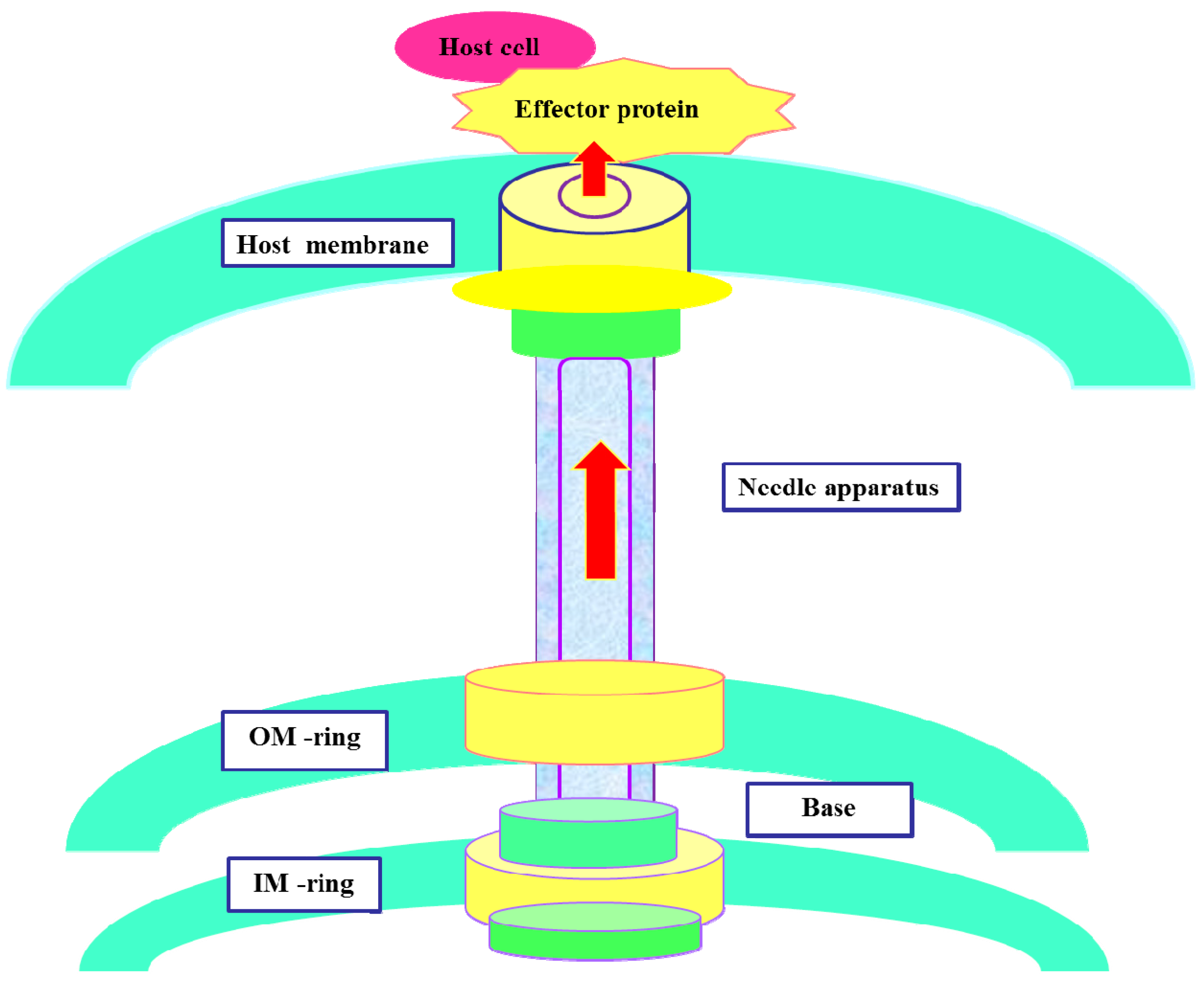
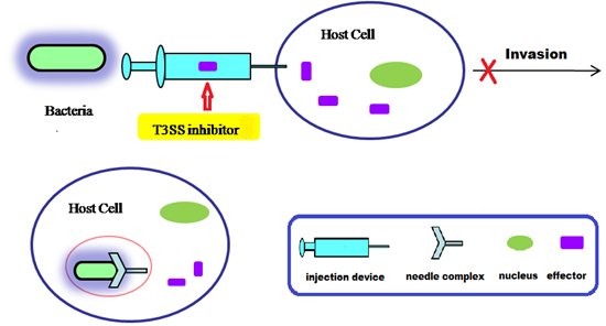
3. Action Mechanism of T3SS

Highly conserved T3SS is a secretion system consisting of a variety of components, and the secretion follows a single-step, sec-independent model [18]. The secretory signal of T3SS secretory protein is not dependent on the signal peptide, but occurs through a 15–20-amino acid domain at the N-terminus of the secretory protein. The secreted proteins are not cleaved in the cytoplasm, but are transported to the cell surface from the cytoplasm [15]. The T3SS injection device forms a temporary structure when bacteria interact with the host cell. The injection device is activated for assembly under physiological conditions [15]; However, under low calcium conditions, the injection device can also be activated. The needle complex of T3SS secretes and translocates protein effectors into the host cytoplasm, interfering with the function of the host cell (Figure 1) [20,21,22,23]. Hundreds of effector proteins have been identified as potential T3SS substrates from diverse bacterial pathogens. Pathogenic T3SS effector proteins can alter signal transduction pathways in order to evade the host defense system. Some bacterial effectors can enter into host cells and destroy their immune system, ultimately inducing host cell death (Figure 2) [24]. Although the detailed molecular mechanisms of T3SS are still unclear, current research has shown that effector translocation by T3SS is a key way that Gram-negative bacteria can infect host cells [23,24]. T3SS has emerged as an attractive target for developing novel anti-virulence agents. In terms of therapeutic strategies, T3SS inhibitors might be used to prevent effector proteins from invading the host cell through active or passive immunization, so that the pathogens lose the ability to infect host cells (Figure 2).
Figure 2. Schematic diagram of anti-virulence strategies by using T3SS inhibitors in Gram-negative bacterial pathogens.
Molecules 20 17659 g002
In 2014, American pharmaceutical company Kalobios reported antibody KB001-A as a P. aeruginosa T3SS inhibitor, and it is currently in phase II clinical trials for treating patients who have respiratory tract inflammation and cystic fibrosis (CF) caused by chronic P. aeruginosa infection [25]. To the best of our knowledge, this is the first T3SS inhibitor drug used in humans, suggesting an alternative to traditional antibiotics for treating bacterial infection. The action mechanism of T3SS inhibitors is very different from that of conventional antibiotics, because these inhibitors specifically target the virulence of bacteria, rather than their viability, making selective pressure on bacteria low and reducing the likelihood of bacterial resistance. T3SS inhibitors, especially small-molecule inhibitors, have attracted much attention in recent years, and this review summarizes several classes of small-molecule T3SS inhibitors reported in the literature between 2003 and 2015.

4. Small Molecule Inhibitors of T3SS

4.1. Salicylidene Acyl Hydrazides as T3SS Inhibitors

Salicylidene acyl hydrazides were reported as the first class of small-molecule T3SS inhibitors that can block one or more effector proteins. Using a luciferase reporter gene assay [26] in viable Yersinia pseudotuberculosis, Kauppi et al. [27] screened a chemical library of 9400 compounds and identified several salicylidene acyl hydrazide derivatives, such as INP0007 (1) and INP0010 (2) (Figure 3). These derivatives dramatically suppressed the reporter gene signal expressed by the yopE promoter, as well as effector protein secretion at low micromolar concentrations without affecting bacterial growth. INP0007 (1) blocked Yersinia T3SS secretion and prevented bacteria from invading HeLa cells. When the uninfected HeLa cells were incubated in the presence of INP0007 (50 μM), INP0007 strongly reduced the YopE-induced cytotoxic reaction in the cells [28]. This result implies that INP0007 can curb the translocation of several Yop effectors proteins, such as YopE. INP0010 (2) blocked microinjection of Yops into the cytosol of the target cell and inhibited replication of Chlamydia in Hep-2 cells [29].
Another salicylidene acyl hydrazide, INP0400 (3) (Figure 3), inhibited the intracellular replication and infectivity of Chlamydia trachomatis. Interestingly, the inhibitor has different effects at different stages of the infectious cycle of C. trachomatis [30]: when given at the time of infection, INP0400 partially blocked the entry of elementary bodies (EBs) into McCoy cells (host cells). In the mid-cycle, the inhibitor suppressed secretion of the effector IncA and homotypic vesicular fusions mediated by this protein. In the late phase, treatment resulted in detachment of reticulate bodies (RBs) from the inclusion membrane, ultimately preventing the conversion of RB to EB, thereby significantly reducing the infectivity of C. trachomatis.
More Gram-negative pathogens have been investigated using salicylidene acyl hydrazide inhibitors [31,32]. Hudson et al. found that INP0007 and INP0400 inhibited T3SS-mediated secretion of proteins without affecting the growth of bacteria in Salmonella [32]. Subsequently, they confirmed that the two inhibitors were capable of inhibiting secreted proteins via T3SS-mediated and inflammatory response by Salmonella in vitro. These results have demonstrated that salicylidene acyl hydrazides have the capacity to inhibit T3SSs in multiple species of pathogens and could serve as promising starting points for drug development [29].
Figure 3. Structures of INP0007 (1), INP0010 (2) and INP0400 (3).
Molecules 20 17659 g003

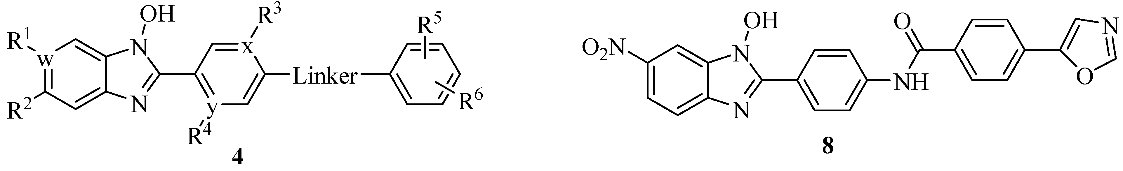
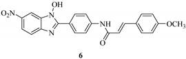
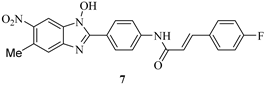
4.2. N-Hydroxybenzimidazoles as T3SS Inhibitors

LcrF is a member of the AraC family of transcription activators [33] and is a multiple adaptational response (MAR) transcription factor associated with virulence in Yersinia pestis and Y. pseudotuberculosis [34]. Upon contacting host cells or changing temperature, LcrF is expressed and activates the expression of Yersinia cytotoxic Yop proteins, such as YopH, YopJ and YopE [35,36]. Yops are secreted into host cells via T3SS and cause cellular apoptosis [37,38]. Y. pestis is the causative agent of plague and raises serious concerns regarding its potential use as a biological weapon by terrorists [39].
Kim et al. synthesized a series of N-hydroxybenzotriazole derivatives [40] (general structure shown as 4 in Figure 4) and identified some derivatives as potent LcrF inhibitors using a primary cell-free LcrF-DNA binding assay, as well as a T3SS-dependent whole cell assay (T3SS-dependent Y. pseudotuberculosis cytotoxicity assay) [34]. Lead Compounds 5, 6 and 7 exhibited very similar inhibitory activity against both LcrF and ExsA (Table 1); ExsA is an MAR transcription factor found in P. aeruginosa and has 85% identity and 92% similarity with LcrF. However, these compounds have very weak inhibitory activity against SlyA, a member of the MarR family of transcription factors in Salmonella spp. The DNA binding motif of SlyA is different from that of MAR proteins [41,42]. These results indicated that the N-hydroxybenzimidazole inhibitors are non-DNA binding and specifically target MAR proteins. In a murine model of Y. pseudotuberculosis pneumonia, Compound 5 and another N-hydroxybenzimidazole derivative, 8 (Figure 4), significantly reduced the bacterial burden in the lungs, offering a dramatic survival advantage [43]. The inhibitors attenuated the T3SS-mediated virulence without affecting the normal bacterial growth in Y. pseudotuberculosis and did not demonstrate toxicity against mammalian cells [43]. Since the members of the MAR family of transcription factors play central roles in pathogenesis across bacterial genera, the N-hydroxybenzimidazole inhibitors could have broad applicability as, for example, lead compounds for preclinical studies of preventing Y. spp. infection.
Figure 4. Structure of N-hydroxybenzimidazoles.
Molecules 20 17659 g004

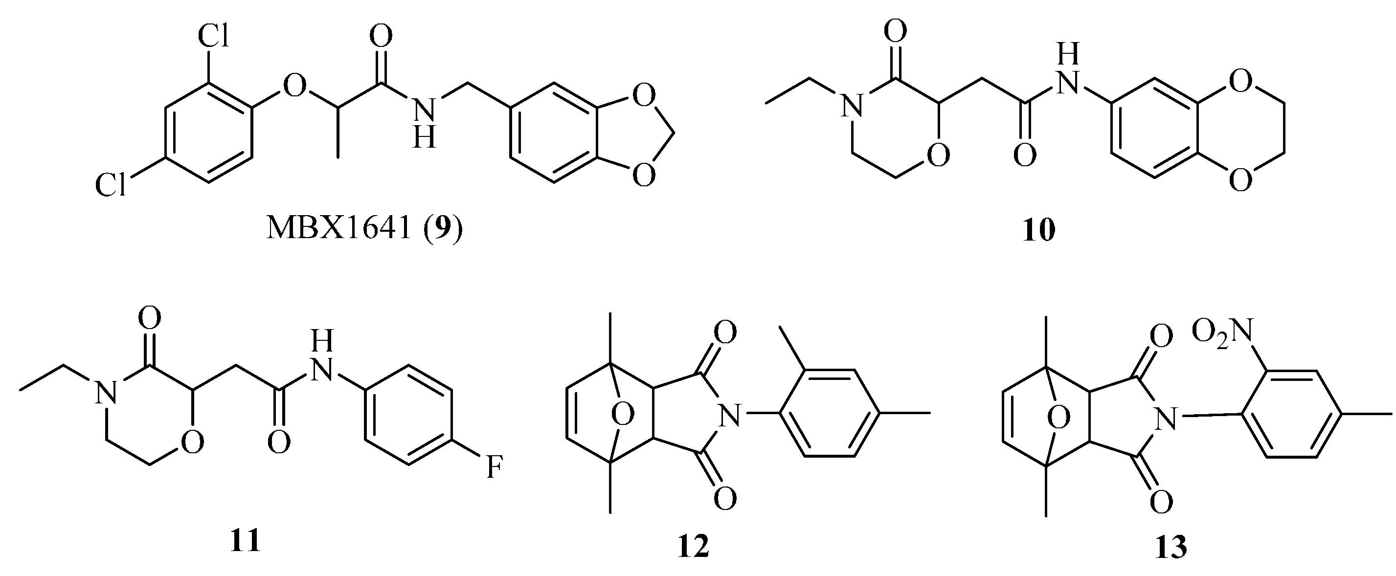
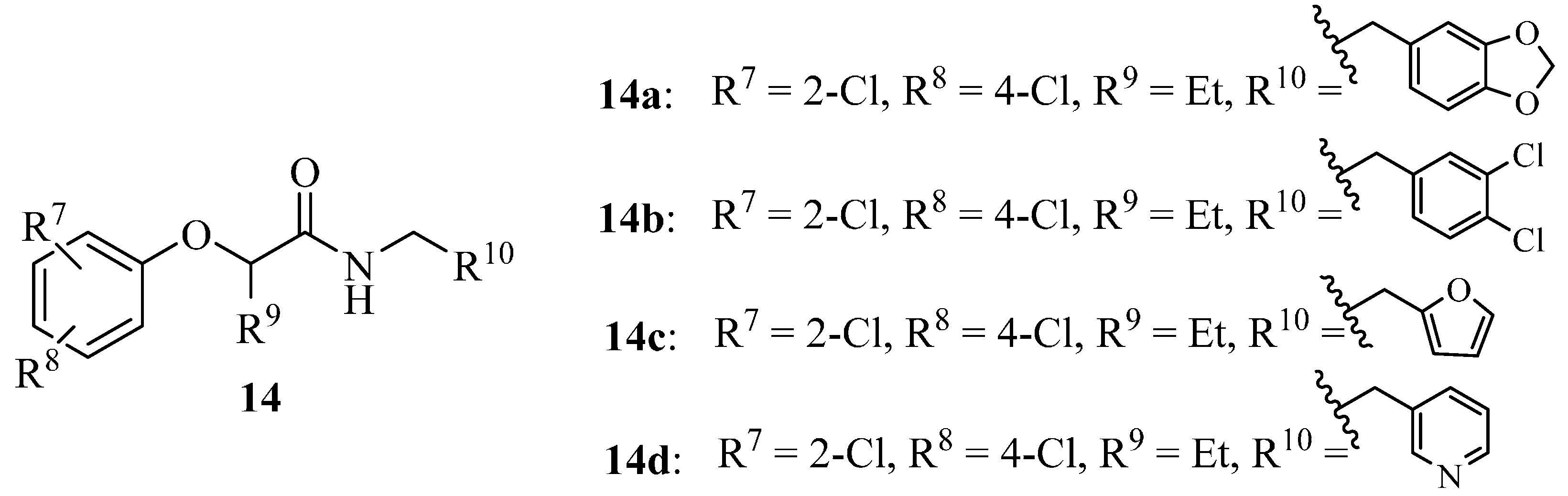
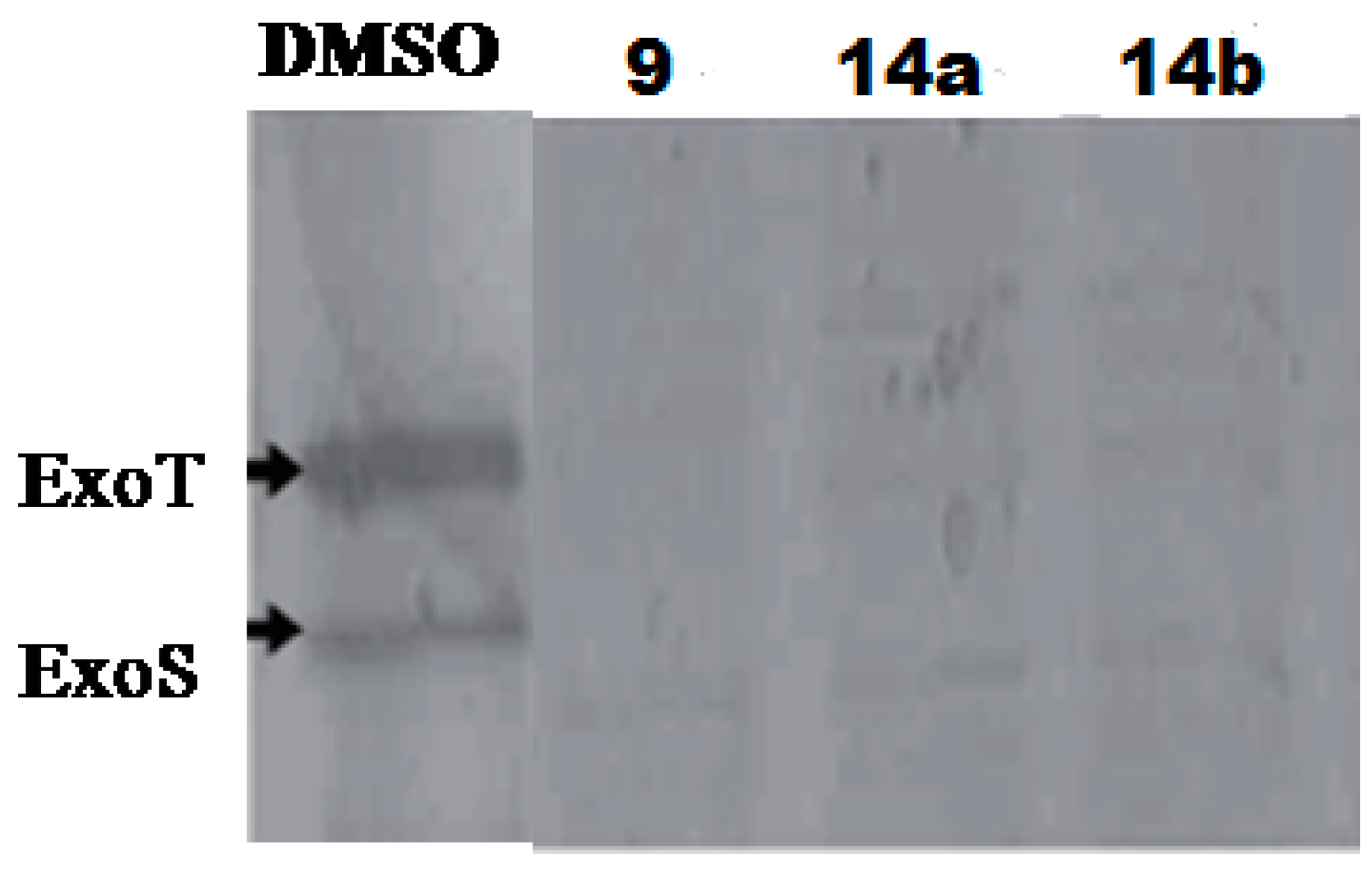
4.3. Phenoxyacetamides as T3SS Inhibitors

The opportunistic pathogen P. aeruginosa is the leading cause of hospital-acquired infections by Gram-negative bacteria, and T3SS is the major virulence factor contributing to infections [44]. There are four T3SS effectors in P. aeruginosa strains: ExoS, ExoT, ExoY and ExoU. ExoU and ExoS contributed significantly to persistence, dissemination and mortality, and ExoT produced minor effects on virulence in a mouse lung infection model, but ExoY did not play a major role in P. aeruginosa pathogenesis [44]. Aiello et al. developed two cellular reporter assays and screened a library of 80,000 compounds to search for inhibitors of P. aeruginosa type III secretion [45]. The primary assay consisted of a transcriptional fusion of the Photorhabdus luminescens luxCDABE operon to the P. aeruginosa exoT effector gene, and the secondary assay included direct measurements of T3SS-mediated secretion of a P. aeruginosa ExoS effector-β-lactamase fusion protein, as well as detecting the secretion of native ExoS. Five compounds (Figure 5) were identified and demonstrated inhibitory activity against P. aeruginosa type III secretion without affecting bacterial growth. These inhibitors also blocked the T3SS-mediated secretion of a YopE effector-β-lactamase fusion protein from an attenuated Y. pestis strain. The best inhibitor identified is phenoxyacetamide MBX1641 (9), which effectively inhibits the secretion of T3SS effector protein ExoS in P. aeruginosa.
Table 1. Target specificity and in vitro antibacterial activity of three lead N-hydroxybenzimidazoles.
CompoundIC50 (μM) a
LcrFExsA bSlyA c
Molecules 20 17659 i0013.93.5>53.8
Molecules 20 17659 i0028.05.9>55.2
Molecules 20 17659 i0037.66.7>52
a IC50 was determined using a dose-response analysis with the maximum concentration of 25 μg/mL; b MAR transcription factor in P. aeruginosa; c MarR family transcription factor in Salmonella spp.
Figure 5. Structures of 913.
Molecules 20 17659 g005
In order to search for more potent T3SS inhibitors against P. aeruginosa, our lab studied the structure-activity relationship (SAR) of MBX1641 in collaboration with Yang’s group at the University of Wisconsin at Milwaukee. We designed and synthesized a series of new α-phenoxyacetamide derivatives by modifying the 2,4-dichlorophenoxy group, the length of the amide side chain and the 3,4-(methylenedioxy)benzyl group [46]. P. aeruginosa PAO1 was chosen as the target strain, and a promoter-probe vector pPROBE-AT containing the promoterless green fluorescent protein (gfp) gene was used to construct an exoS promoter-gfp plasmid reporter pATexoS. PAO1 containing pATexoS was cultured in T3SS-inducing medium containing DMSO or supplemented with 250 μM of each synthesized compound, and promoter activity was assessed at 6 h by measuring GFP intensity using flow cytometry. Four new derivatives (14a14d; Figure 6) have shown a strong inhibitory effect against exoS gene expression in P. aeruginosa. Among them, 14d not only exhibits stronger potency against PAO1 T3SS than MBX1641, but also has better solubility in aqueous solution. In addition, we selected a number of compounds to determine the expression level of ExoS and ExoT proteins via Western hybridization. MBX1641, 14a and 14b significantly inhibited the expression of ExoS and ExoT proteins (Figure 7). The results have demonstrated that the inhibitors can effectively suppress the expression of PAO1 T3SS effectors ExoS and ExoT.
Figure 6. Structures of Compounds 14a14d.
Molecules 20 17659 g006
Figure 7. Alteration of the production of T3SS effectors ExoS and ExoT by T3SS inhibitors. P. aeruginosa PAO1 cells were grown in LB broth supplemented with 10 mM NTA (nitrilotriacetic acid) and 250 μM inhibitors. The same volume of DMSO was added to the culture as a negative control. The Western blot was performed using an anti-ExoS antibody.
Molecules 20 17659 g007

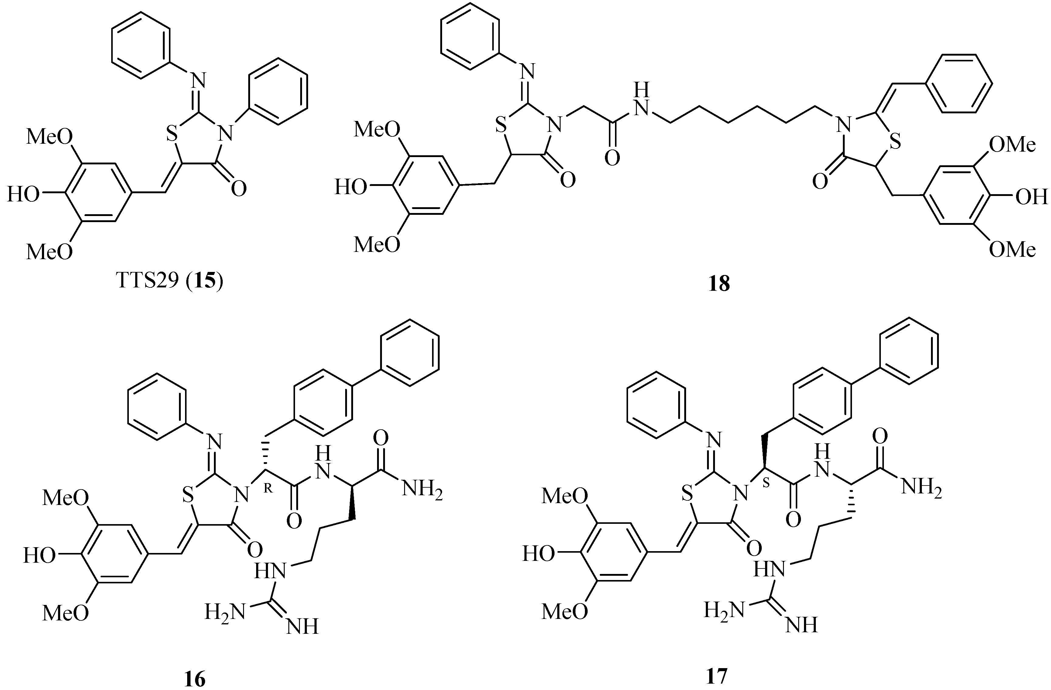
4.4. 2-Imino-5-arylidenethiazolidinones as T3SS Inhibitors

In search of S. typhimurium T3SS inhibitors, Felise et al. developed and performed a high-throughput screening for compound libraries of 92,000 small molecules and discovered that 2-imino-5-arylidene thiazolidinone (TTS29; Figure 8) inhibited T3SS secretion or assembly without affecting bacterial growth [47]. They purified the needle complexes of S. typhimurium and found that TTS29-treated samples had lower levels of needle components, whereas the levels of whole-cell proteins were unchanged. These results indicated that TTS29 disrupts the needle assembly. This inhibitor can block the secretion and virulence functions of a wide array of animal and plant Gram-negative bacterial pathogens, such as Y. spp., P. aeruginosa and Francisella novicida. However, the major disadvantage for TTS29 is its moderate potency (only 83 μM). Therefore, the researchers synthesized a series of new derivatives of TTS29, and the structure-activity relationship results showed that imino nitrogen, the aryl group and the substitution pattern on the arylidene ring are critical for inhibitory activity, whereas the amido nitrogen is tolerated for modification. Dipeptide analogs 16 and 17 (Figure 8) have much better potency than TTS29 at low micromolar IC50 values (8 μM and 3 μM, respectively) [48]. The new analogs were synthesized through a solid-phase approach. Some tethered thiazolidinone dimers had also been synthesized, and many of these dimers inhibited the T3SS-dependent secretion of a virulence protein at a concentrations lower than that of TTS29 [49]. For example, the IC50 value of dimer 18 is 5 μM.
Figure 8. Structures of Compounds 1518.
Molecules 20 17659 g008

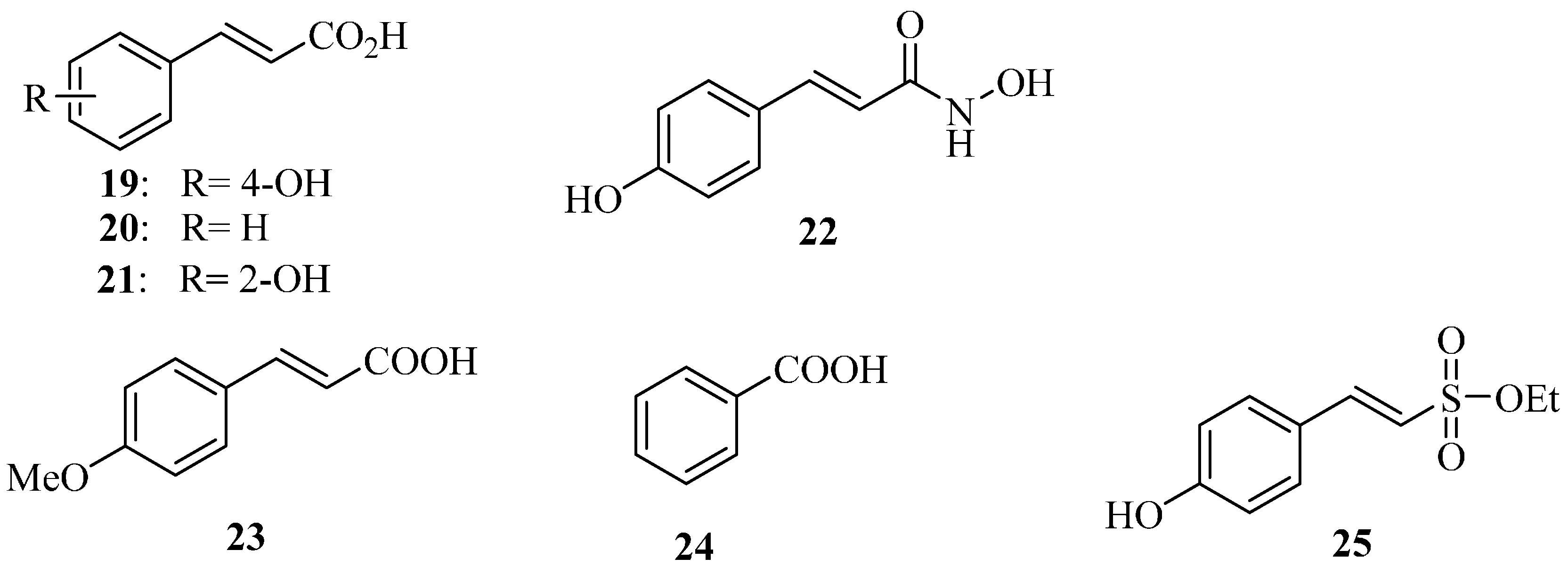
4.5. Plant Phenolic Compounds as T3SS Inhibitors

Natural plant phenolic compounds are secondary metabolites synthesized by plants. Apart from their role in growth, pigmentation and reproduction, they also play an important role in disease resistance. Plants have evolved a systemic acquired resistance mechanism to protect themselves from pathogen invasion [50]. Inspired by this self-protection mechanism found in nature, we explored inhibitors of D. dadantii T3SS hrpA genes. After systematically screening a series of natural plant phenolic compounds and their derivatives, we found that p-coumaric acid (19) (Figure 9) significantly repressed the expression of T3SS regulatory genes through the HrpS-Hrp two-component system [51]. On the other hand, trans-cinnamic acid (20) and o-coumaric acid (21) induced the expression of D. dadantii T3SS genes hrpA via the RsmB-RsmA pathway.
Very recently, we converted 19 into the corresponding hydroxamic acid and found that the resulting trans-4-hydroxycinnamohydroxamic acid (22) has an eight-fold higher inhibitory potency than 19 [52]. 22 inhibits the T3SS of D. dadantii HrpY via HrpX/HrpY two-component signal transduction and Rsm systems. To the best of our knowledge, this is the first inhibitor to affect the T3SS of D. dadantii through both the transcriptional and post-transcriptional pathways. In addition, our previous studies show that 22 reduces the biofilm formation of P. aeruginosa PAO1 and affects the T3SS of P. aeruginosa via the GacSA-RsmYZ-RsmA-ExsA regulatory pathway [53].
Figure 9. Structures of Compounds 1925.
Molecules 20 17659 g009
E. amylovora, of the family Enterobacteriaceae, is a Gram-negative bacterial plant pathogen. It is the causal agent of fire blight in apple, pear, raspberry and other rosaceous plant species, and it infects leaves, blossoms, succulent shoots and immature fruits. Annual losses to fire blight and costs of control in the USA are estimated at over $100 million. Losses and costs in many other countries are also huge [54]. Currently, the approach for controlling fire blight pathogen employs antibiotics and copper spray. The most commonly-used antibiotics for treating E. amylovora are streptomycin and oxytetracycline: streptomycin kills E. amylovora, whereas oxytetracycline merely inhibits its growth. In the 1990s, 30%–40% of planted pear trees in the USA were treated with streptomycin or oxytetracycline or both, and 19% of planted apple trees were treated with streptomycin each year [55]. However, the streptomycin-resistant strains of E. amylovora are now widespread and common in apple and pear orchards of the western United States and British Columbia in Canada and have also been reported in New Zealand, Israel and Lebanon. Resistance in the fire blight pathogen has had widespread economic and political implications [55]. The U.S. EPA strictly regulates the use of existing antibiotics (e.g., streptomycin and oxytetracycline) on plants in the USA and has established a higher standard for approval of new antibiotics due to antibiotic resistance concerns.
We have found that some plant phenolic compounds can inhibit the expression of E. amylovora T3SS [56]. Using a green fluorescent protein (GFP) reporter combined with a high-throughput flow cytometry assay for measuring the expression of E. amylovora T3SS, 4-methoxycinnamic acid (23) and benzoic acid (24) (Figure 9) were identified as E. amylovora T3SS inhibitors. 23 and 24 altered the expression of E. amylovora T3SS via the HrpS-HrpL pathway. 23 inhibited the T3SS gene through the RsmB-RsmA system. We observed that the two inhibitors weakened the hypersensitive response (HR) in tobacco leaves by suppressing the T3SS of E. amylovora. Additionally, the sulfonate ester 25 was identified as the E. amylovora T3SS inducer [56]. The phenolic compounds (23, 24) and their derivatives, which specifically targeted the E. amylovora T3SS, may provide an alternative strategy to antibiotics in fire blight control.

4.6. Polyol Products as T3SS Inhibitors

Although the majority of Escherichia coli strains are benign for humans, several strains are pathogenic. For example, enteropathogenic E. coli (EPEC) infects the human intestinal epithelium and is a major cause of infantile diarrhea in developing countries. EPEC is an extracellular bacterial pathogen and uses a T3SS to deliver bacterial effectors into host cells [57]. Linington et al. isolated a complex glycolipid, caminoside A (26; Figure 10), from the marine sponge Caminus sphaeroconia [58]. Caminoside A effectively blocked the pathogenicity of EPEC by inhibiting the T3SS expression with an IC50 value of 20 μM and without killing the bacteria. Caminosides B, C and D were isolated from the marine sponge C. sphaeroconia, and all of the glycolipids showed inhibitory activity against the EPEC T3SS (IC50 = 20 μM) [59].
Six guadinomines were isolated from the culture broth of Streptomyces sp. K01-0509 as inhibitors of EPEC T3SS-induced hemolysis by Iwatsuki et al. [60,61]. Guadinomine B (27; Figure 10) showed the most potent inhibition with an IC50 value of 7 ng/mL.
A linear polyketide, aurodox (28; Figure 10), was isolated from the culture broth of S. sp. by using a screening system for the T3SS-mediated hemolysis of EPEC [62]. Aurodox strongly inhibited the T3SS-mediated hemolysis with an IC50 value of 1.5 μg/mL without affecting bacterial growth, and it specifically blocked the secretion of T3SS proteins, such as EspB, EspF and Map. In an in vivo infection study, after aurodox was administered, mice survived a lethal dose of Citrobacter rodentium, a model bacterium for human pathogens, such as EPEC. This study suggests that certain microbes might produce small molecules capable of counteracting bacterial infection [62].
Figure 10. Structures of caminoside A, guadinomine B and aurodox.
Molecules 20 17659 g010

5. Conclusions and Prospects

In the past few years, extraordinary efforts have been made by a number of laboratories to discover new T3SS inhibitors as potential anti-infective agents. However, some challenges still need to be addressed before these inhibitors are proven to be effective therapeutics. None of the small-molecule T3SS inhibitors described in this review have advanced into the clinic. One major issue is that the precise mechanisms of action and effective targets remain unclear. Another is that the current small-molecule T3SS inhibitors have limited potency, with IC50 values in the single digits of micromolar concentrations.
Clinical development of the antibody KB001-A against P. aeruginosa infection has clearly demonstrated the therapeutic potential of T3SS inhibitors. Since the atomic structure of the needle body of S. typhimurium has been successfully revealed, it is hoped that the next major breakthroughs will be the complete structural elucidation of all of the T3SS structural proteins, effector proteins and chaperones. If this can be achieved, then clear targets for T3SS inhibitors will be within reach. The design and synthesis of small-molecule T3SS inhibitors will be more specific and potent, and researchers will more easily discover and develop such inhibitors for treating human bacterial infections.

Acknowledgments

We are grateful for the financial support from the National Science Foundation of China (Grant 21272029), the “333 High-level Talent Training Program” of Jiangsu Province of China, the Priority Academic Program Development (PAPD) of Jiangsu Higher Education Institutions and the International Collaboration Program of Changzhou City (CZ20130024).

Author Contributions

Lingling Gu, Shanshan Zhou, Lanping Zhu, Cuirong Liang and Xin Chen have equally contributed in writing the material of the manuscript, and Xin Chen had also the general supervision.

Conflicts of Interest

The authors confirm that this article content has no conflicts of interest.

References

  1. Cegelski, L.; Marshall, G.R.; Eldridge, G.R.; Hultgren, S.J. The biology and future prospects of antivirulence therapies. Nat. Rev. Microbiol. 2008, 6, 17–27. [Google Scholar] [CrossRef] [PubMed]
  2. Escaich, S. Antivirulene as a new antibacterial approach for chemotherapy. Curr. Opin. Chem. Biol. 2008, 12, 400–408. [Google Scholar] [CrossRef] [PubMed]
  3. Rasko, D.A.; Sperandio, V. Anti-virulene strategies to combat bacteria-mediated disease. Nat. Rev. Drug Discov. 2010, 9, 117–128. [Google Scholar] [CrossRef] [PubMed]
  4. Desvaux, M.; Hebraud, M.; Henderson, I.R.; Pallen, M.J. Type III secretion: What’s in a name? Trends Microbiol. 2006, 14, 157–160. [Google Scholar] [CrossRef] [PubMed]
  5. Coburn, B.; Sekirov, I.; Finlay, B.B. Type III secretion systems and disease. Clin. Microbiol. Rev. 2007, 20, 535–549. [Google Scholar] [CrossRef] [PubMed]
  6. Tang, X.Y.; Xiao, Y.M.; Zhou, J.M. Regulation of the type III secretion system in phytopathogenic bacteria. Mol. Plant Microbe Interact. 2006, 19, 1159–1166. [Google Scholar] [CrossRef] [PubMed]
  7. Waterman, S.R.; Holden, D.W. Functions and effectors of the Salmonella pathogenicity island 2 type III secretion system. Cell. Microbiol. 2003, 5, 501–511. [Google Scholar] [CrossRef] [PubMed]
  8. Yip, C.K.; Strynadka, N.C. New structural insights into the bacterial type III secretion system. Trends Biochem. Sci. 2006, 31, 223–230. [Google Scholar] [CrossRef] [PubMed]
  9. Schraidt, O.; Marlovits, T.C. Three-dimensional model of Salmonella’s needle complex at subnamometer resolution. Science 2011, 331, 1192–1195. [Google Scholar] [CrossRef] [PubMed]
  10. Okada, N.; Iida, T.; Park, K.S.; Goto, N.; Yasunaga, T.; Hiyoshi, H.; Matsuda, S.; Kodama, T.; Honda, T. Identification and characterization of a novel type III secretion system in trh-positive vibrio parahaemolyticus strain TH3996 reveal genetic lineage and diversity of pathogenic machinery beyond the species level. Infect. Immun. 2009, 77, 904–913. [Google Scholar] [CrossRef] [PubMed]
  11. Cornelis, G.R. The type III secretion injectisome, a complex nanomachine for intracellular “toxin” delivery. Biol. Chem. 2010, 391, 745–751. [Google Scholar] [CrossRef] [PubMed] [Green Version]
  12. Chatterjee, S.; Chaudhury, S.; McShan, A.C.; Kaur, K.; Guzman, N.D. Structure and biophysics of type III secretion in bacteria. Biochemistry 2013, 52, 2508–2517. [Google Scholar] [CrossRef] [PubMed]
  13. Tamano, K.; Aizawa, S.I.; Katayama, E.; Nonaka, T.; Imajoh-Ohmi, S.; Kuwae, A.; Nagai, S.; Sasakawa, C. Supramolecular structure of the Shigella type III secretion machinery: The needle part is changeable in length and essential for delivery of effectors. EMBO J. 2000, 19, 3876–3887. [Google Scholar] [CrossRef] [PubMed]
  14. Spreter, T.; Yip, C.K.; Sanowar, S.; André, I.; Kimbrough, T.G.; Vuckovic, M.; Pfuetzner, R.A.; Deng, W.; Yu, A.C.; Finlay, B.B.; et al. A conserved structural motif mediates formation of the periplasmic rings in the type III secretion system. Nat. Struct. Mol. Biol. 2009, 16, 468–476. [Google Scholar] [CrossRef] [PubMed]
  15. Galan, J.E.; Wolf-Watz, H. Protein delivery into eukaryotic cells by type III secretion machines. Nature 2006, 444, 567–573. [Google Scholar] [CrossRef] [PubMed]
  16. Loquet, A.; Sgourakis, N.G.; Gupta, R.; Giller, K.; Riedel, D.; Goosmann, C.; Griesinger, C.; Kolbe, M.; Baker, D.; Becker, S.; et al. Atomic model of the type III secretion system needle. Nature 2012, 486, 276–279. [Google Scholar] [CrossRef] [PubMed]
  17. Akeda, Y.; Galan, J.E. Chaperone release and unfolding of substrates in type III secretion. Nature 2005, 437, 911–915. [Google Scholar] [CrossRef] [PubMed]
  18. Izore, T.; Job, V.; Dessen, A. Biogenesis, regulation, and targeting of the type III secretion system. Structure 2011, 19, 603–612. [Google Scholar] [CrossRef] [PubMed]
  19. Feldman, M.F.; Muller, S.; Wuest, E.; Cornelis, G.R. SycE allows secretion of YopE-DHFR hybrids by the Yersinia enterocolitica type III Ysc system. Mol. Micrbiol. 2002, 46, 1183–1197. [Google Scholar] [CrossRef]
  20. Schraidt, O.; Lefebre, M.D.; Brunner, M.J.; Schmied, W.H.; Schmidt, A.; Radics, J.; Mechtler, K.; Galan, J.E.; Marlovits, T.C. Topology and organization of the Salmonella typhimurium type III secretion needle complex components. PLoS Pathog. 2010, 6, e1000824. [Google Scholar] [CrossRef] [PubMed]
  21. Marlovits, T.C.; Kubori, T.; Sukhan, A.; Thomas, D.R.; Galan, J.E.; Unger, V.M. Structural insights into the assembly of the type III secretion needle complex. Science 2004, 306, 1040–1042. [Google Scholar] [CrossRef] [PubMed]
  22. Marlovits, T.C.; Kubori, T.; Lara-Tejero, M.; Thomas, D.; Unger, V.M.; Galan, J.E. Assembly of the inner rod determines needle length in the type III secretion injectisome. Nature 2006, 441, 637–640. [Google Scholar] [CrossRef] [PubMed]
  23. Galan, J.E. Salmonella interactions with host cells: Type III secretion at work. Annu. Rev. Cell Dev. Biol. 2001, 17, 53–86. [Google Scholar] [CrossRef] [PubMed]
  24. Tsou, L.K.; Dossa, P.D.; Hang, H.C. Small molecules aimed at type III secretion systems to inhibit bacterial virulence. Med. Chem. Commun. 2013, 4, 68–79. [Google Scholar] [CrossRef] [PubMed]
  25. Milla, C.E.; Chmiel, J.F.; Accurso, F.J.; van Devanter, D.R.; Konstan, M.W.; Yarranton, G.; Geller, D.E. Anti-PcrV antibody in cystic fibrosis: A novel approach targeting Pseudomonas aeruginosa airway infection. Pediatr. Pulmonol. 2014, 49, 650–658. [Google Scholar] [CrossRef] [PubMed]
  26. Pettersson, J.; Nordfelth, R.; Dubinina, E.; Bergman, T.; Gustafsson, M.; Magnusson, K.E.; Wolf-Watz, H. Modulation of virulence factor expression by pathogen target cell contact. Science 1996, 273, 1231–1233. [Google Scholar] [CrossRef] [PubMed]
  27. Kauppi, A.M.; Nordfelth, R.; Uvell, H.; Wolf-Watz, H.; Elofsson, M. Targeting bacterial virulence: Inhibitors of type III secretion in Yersinia. Chem. Biol. 2003, 10, 241–249. [Google Scholar] [CrossRef]
  28. Nordfelth, R.; Kauppi, A.M.; Norberg, H.A.; Wolf-Watz, H.; Elofsson, M. Small-molecule inhibitors specifically targeting type III secretion. Infect. Immun. 2005, 73, 3104–3114. [Google Scholar] [CrossRef] [PubMed]
  29. Keyser, P.; Elofsson, M.; Rosell, S.; Wolf-Watz, H. Virulence blockers as alternatives to antibiotics: Type III secretion inhibitors against Gram-negative bacteria. J. Intern. Med. 2008, 264, 17–29. [Google Scholar] [CrossRef] [PubMed]
  30. Muschiol, S.; Bailey, L.; Gylfe, A.; Sundin, C.; Hultenby, K.; Bergstro, S.; Elofsson, M.; Wolf-Watz, H.; Normark, S.; Normark, H.B. A small-molecule inhibitor of type III secretion inhibits different stages of the infectious cycle of Chlamydia trachomatis. Proc. Natl. Acad. Sci. USA 2006, 103, 14566–14571. [Google Scholar] [CrossRef]
  31. Gauthier, A.; Robertson, M.L.; Lowden, M.; Ibarra, J.A.; Puente, J.L.; Finlay, B.B. Transcriptional inhibitor of virulence factors in enteropathogenic Escherichia coli. Antimicrob. Agents Chemother. 2005, 49, 4101–4109. [Google Scholar] [CrossRef] [PubMed]
  32. Hudson, D.L.; Layton, A.N.; Field, T.R.; Bowen, A.J.; Wolf-Watz, H.; Elofsson, M.; Stevens, M.P.; Galyov, E.E. Inhibition of type III secretion in Salmonella enterica serovar typhimurium by small-molecule inhibitors. Antimicrob. Agents Chemother. 2007, 51, 2631–2635. [Google Scholar] [CrossRef] [PubMed]
  33. Hoe, N.P.; Minion, F.C.; Goguen, J.D. Temperature sensing in Yersinia pestis: Regulation of yopE transcription by lcrF. J. Bacteriol. 1992, 174, 4275–4286. [Google Scholar] [PubMed]
  34. Kim, O.K.; Garrity-Ryan, L.K.; Bartlett, V.J.; Grier, M.C.; Verma, A.K.; Medjanis, G.; Donatelli, J.E.; Macone, A.B.; Tanaka, S.K.; Levy, S.B.; et al. N-Hydroxybenzimidazole inhibitors of the transcription factor LcrF in Yersinia: Novel antivirulence agents. J. Med. Chem. 2009, 52, 5626–5634. [Google Scholar] [CrossRef] [PubMed]
  35. Cornelis, G.R.; Wolf-Watz, H. The Yersinia Yop virulon: A bacterial system for subverting eukaryotic cells. Mol. Microbiol. 1997, 23, 861–867. [Google Scholar] [CrossRef] [PubMed]
  36. Logsdon, L.K.; Mecsas, J. Requirement of the Yersinia pseudotuberculosis effectors YopH and YopE in colonization and persistence in intestinal and lymph tissues. Infect. Immun. 2003, 71, 4595–4607. [Google Scholar] [CrossRef] [PubMed]
  37. Mota, L.J.; Cornelis, G.R. The bacterial injection kit: Type III secretion systems. Ann. Med. 2005, 37, 234–249. [Google Scholar] [CrossRef] [PubMed]
  38. Monack, D.M.; Mecsas, J.; Bouley, D.; Falkow, S. Yersinia-induced apoptosis in vivo aids in the establishment of a systemic infection of mice. J. Exp. Med. 1998, 188, 2127–2137. [Google Scholar] [CrossRef] [PubMed]
  39. Hawley, R.J.; Eitzen, E.M. Biological weapons-a primer for microbiologists. Annu. Rev. Microbiol. 2001, 55, 235–253. [Google Scholar] [CrossRef] [PubMed]
  40. Gardiner, J.M.; Procter, J. Synthesis of N-alkoxybenzimidazoles with differentiated C2 and O-substituents. Tetrahedron Lett. 2001, 42, 5109–5111. [Google Scholar] [CrossRef]
  41. Ellison, D.W.; Miller, V.L. Regulation of virulence by members of the MarR/SlyA family. Curr. Opin. Microbiol. 2006, 9, 153–159. [Google Scholar] [PubMed]
  42. Hong, M.; Fuangthong, M.; Helmann, J.D.; Brennan, R.G. Structure of an OhrR-ohrA operator complex reveals the DNA binding mechanism of the MarR family. Mol. Cell. 2005, 20, 131–141. [Google Scholar] [CrossRef] [PubMed]
  43. Garrity-Ryan, L.K.; Kim, O.K.; Balada-Llasat, J.M.; Bartlett, V.J.; Verma, A.K.; Fisher, M.L.; Castillo, C.; Songsungthong, W.; Tanaka, S.K.; Levy, S.B.; et al. Small molecule inhibitors of LcrF, a Yersinia pseudotuberculosis transcription factor, attenuate virulence and limit infection in a murine pneumonia model. Infect. Immun. 2010, 78, 4683–4690. [Google Scholar] [CrossRef] [PubMed]
  44. Shaver, C.M.; Hauser, A.R. Relative contributions of Pseudomonas aeruginosa ExoU, ExoS, and ExoT to virulence in the lung. Infect. Immun. 2004, 72, 6969–6977. [Google Scholar] [CrossRef] [PubMed]
  45. Aiello, D.; Williams, J.D.; Majgier-Baranowska, H.; Patel, I.; Peet, N.P.; Huang, J.; Lory, S.; Bowlin, T.L.; Moir, D.T. Discovery and characterization of inhibitors of Pseudomonas aeruginosa type III secretion. Antimicrob. Agents Chemother. 2010, 54, 1988–1999. [Google Scholar] [CrossRef] [PubMed]
  46. Zhang, C.F.; Wu, X.G.; Li, Yan; Liang, C.R.; Che, Y.Z.; Gu, L.L.; Ren, J.; Hu, K.; Sun, X.Q.; Yang, C.H.; et al. Synthesis and bioactivity of novel inhibitors for type III secretion system of Pseudomonas aeruginosa PAO1. Chin. J. Org. Chem. 2013, 33, 1309–1318. [Google Scholar] [CrossRef]
  47. Felise, H.B.; Nguyen, H.V.; Pfuetzner, R.A.; Barry, K.C.; Jackson, S.R.; Blanc, M.P.; Bronstein, P.A.; Kline, T.; Miller, S.I. An inhibitor of Gram-negative bacterial virulence protein secretion. Cell Host Microbe 2008, 4, 325–336. [Google Scholar] [CrossRef] [PubMed]
  48. Kline, T.; Felise, H.B.; Barry, K.C.; Jackson, S.R.; Nguyen, H.V.; Miller, S.I. Substituted 2-imino-5-arylidenethiazolidin-4-one inhibitors of bacterial type III secretion. J. Med. Chem. 2008, 51, 7065–7074. [Google Scholar] [CrossRef] [PubMed]
  49. Kline, T.; Barry, K.C.; Jackson, S.R.; Felise, H.B.; Nguyen, H.V.; Miller, S.I. Tethered thiazolidinone dimers as inhibitors of the bacterial type III secretion system. Bioorg. Med. Chem. Lett. 2009, 19, 1340–1343. [Google Scholar] [CrossRef] [PubMed]
  50. Aoki, T.; Akashi, T.; Ayabe, S.I. Flavonoids of leguminous plants: Structure, biological activity, and biosynthesis. J. Plant Res. 2000, 113, 475–488. [Google Scholar] [CrossRef]
  51. Li, Y.; Peng, Q.; Selimi, D.; Wang, Q.; Charkowski, A.O.; Chen, X.; Yang, C.H. The plant phenolic compound p-coumaric acid represses gene expression in the Dickeya dadantii type III secretion system. Appl. Enviro. Microbiol. 2009, 75, 1223–1228. [Google Scholar] [CrossRef] [PubMed]
  52. Li, Y.; Hutchins, W.C.; Wu, X.; Liang, C.; Zhang, C.; Yuan, X.; Khokhani, D.; Chen, X.; Che, Y.; Wang, Q.; et al. Derivatives of plant phenolic compound inhibits the type III secretion system of Dickeya dadantii via HrpX/HrpY two-component signal transduction and Rsm system. Mol. Plant Pathol. 2015, 16, 150–163. [Google Scholar] [CrossRef] [PubMed]
  53. Yamazaki, A.; Li, J.; Zeng, Q.; Khokhani, D.; Hutchins, W.C.; Yost, A.C.; Biddle, E.; Toone, E.J.; Chen, X.; Yang, C.H. Derivatives of plant phenolic compound affect the type III secretion system of Pseudomonas aeruginosa via a GacS/GacA two component signal transduction system. Antimicrob. Agents Chemother. 2012, 56, 36–43. [Google Scholar] [CrossRef] [PubMed]
  54. Norelli, J.L.; Jones, A.L.; Aldwinckle, H.S. Fire blight management in the twenty-first century: Using new technologies that enhance host resistance in apple. Plant Dis. 2003, 87, 756–765. [Google Scholar] [CrossRef]
  55. McManus, P.S.; Stockwell, V.O.; Sundin, G.W.; Jones, A.L. Antibiotic use in plant agriculture. Annu. Rev. Phytopathol. 2002, 40, 443–463. [Google Scholar] [CrossRef] [PubMed]
  56. Khokhani, D.; Zhang, C.F.; Li, Y.; Wang, Q.; Zeng, Q.; Yamazaki, A.; Hutchins, W.; Zhou, S.S.; Chen, X.; Yang, C.H. Discovery of plant phenolic compounds that act as type III secretion system inhibitors or inducers of the fire blight pathogen, Erwinia amylovora. Appl. Environ. Microbiol. 2013, 79, 5424–5436. [Google Scholar] [CrossRef] [PubMed]
  57. Grado, M.; Rosenberger, C.M.; Gauthier, A.; Vallance, B.A.; Finlay, B.B. Enteropathogenic Escherichia coli infection induces expression of the early growth response factor by activating mitogen-activated protein kinase cascades in epithelial cells. Infect. Immun. 2001, 69, 6217–6224. [Google Scholar] [CrossRef] [PubMed]
  58. Linington, R.G.; Robertson, M.; Gauthier, A.; Finlay, B.B.; van Soest, R.; Andersen, R.J. Caminoside A, an antimicrobial glycolipid isolated from the marine sponge Caminus sphaeroconia. Org. Lett. 2002, 4, 4089–4092. [Google Scholar] [CrossRef] [PubMed]
  59. Linington, R.G.; Robertson, M.; Gauthier, A.; Finlay, B.B.; MacMillan, J.B.; Molinski, T.F.; van Soest, R.; Andersen, R.J. Caminosides B-D, antimicrobial glycolipids isolated from the marine sponge Caminus sphaeroconia. J. Nat. Prod. 2006, 69, 173–177. [Google Scholar] [CrossRef] [PubMed]
  60. Iwatsuki, M.; Uchida, R.; Yoshijima, H.; Ui, H.; Shiomi, K.; Matsumoto, A.; Takahashi, Y.; Abe, A.; Tomoda, H.; Omura, S. Guadinomines, type III secretion system inhibitors, produced by Streptomyces sp. K01-0509 I. Taxonomy, fermentation, isolation and biological properties. J. Antibiot. 2008, 61, 222–229. [Google Scholar] [CrossRef] [PubMed]
  61. Iwatsuki, M.; Uchida, R.; Yoshijima, H.; Ui, H.; Shiomi, K.; Kim, Y.P.; Hirose, T.; Sunazuka, T.; Abe, A.; Tomoda, H.; et al. Guadinomines, type III secretion system inhibitors, produced by Streptomyces sp. K01-0509 II. Physico-chemical properties and structure elucidation. J. Antibiot. 2008, 61, 230–236. [Google Scholar] [CrossRef] [PubMed]
  62. Kimura, K.; Iwatsuki, M.; Nagai, T.; Matsumoto, A.; Takahashi, Y.; Shiomi, K.; Omura, S.; Abe, A. A small-molecule inhibitor of the bacterial type III secretion system protects against in vivo infection with Citrobacter rodentium. J. Antibiot. 2011, 64, 197–203. [Google Scholar] [CrossRef] [PubMed]

