Article

Four New Triterpenoids from Callicarpa kwangtungensis

1
Jiangxi Provincial Institute for Drug and Food Control, Nanchang 330029, China
2
State Key Laboratory of Bioactive Substance and Function of Natural Medicines, Institute of Materia Medica, Chinese Academy of Medical Sciences & Peking Union Medical College, Beijing 100050, China
3
Pharmaceutical Department of Nanchang University, Nanchang 330006, China
*
Authors to whom correspondence should be addressed.
Molecules 2015, 20(5), 9071-9083; https://doi.org/10.3390/molecules20059071
Submission received: 1 March 2015 / Revised: 8 May 2015 / Accepted: 11 May 2015 / Published: 19 May 2015
(This article belongs to the Section Natural Products Chemistry)

Abstract

:
Four new triterpenoids which were identifed as 2α,3β,6β,19α-tetrahydroxy- oleanolic acid 28-O-β-d-glucopyranoside (1), 2-O-β-d-glucopyranosyloxy-3α,19α-di-hydroxyoleanolic acid (2), 2-O-β-d-glucopyranosyloxy-3α,19α-dihydroxyursolic acid (3), 2α,3α,6β,19α-tetrahydroxyursolic acid 28-O-β-d-glucopyranoside (4), were isolated from the aerial parts of Callicarpa kwangtungensis together with three known triterpenoids identified as 2α,3β,21β-trihydroxyursolic acid 28-O-β-d-glucopyranoside (5), 2α,3α,19α,23-tetrahydroxyoleanolic acid 28-O-β-d-glucopyranoside (6), 2α,3α,19α,23-tetrahydroxyursolic acid 28-O-β-d-glucopyranoside (7). Their structures were elucidated by the combination of mass spectrometry (MS), one and two-dimensional NMR experiments.

1. Introduction

Callicarpa kwangtungensis Chun, belonging to the family Verbenaceae, is distributed widely in the Guangdong, Guangxi, and Jiangxi provinces of China [1]. The aerial parts of C. kwangtungensis are used in Chinese herbal medicine for the treatment of bleeding wounds and hematemesis [1]. Previous phytochemical studies of the genus Callicarpa led to the isolation of flavonoids, triterpenoids, and phenylpropanoid glycosides. Some sesquiterpenoids such as callicarpenal, intermedeol, α-humulane were isolated from Callicarpa americana, C. japonica and C. pedunculata; They also include a few diterpenoids such as 16,17-dihydroxy-3-oxophyllocladane, 16-hydroxy-17-acetoxy-3-oxophyllocladane, isopropylidenocalliterpenone, and calliphyllin from C. acuminata, C. formosana, C. macrophylla, C. maingayi and C. pentandra; and many triterpenoids such as 2a,3a,24-trihydroxyoleanolic acid, ursolic acid and β-amyrin from C. formosana; oleanolic acid, betulin and α-amyrin from C. macrophylla; maslinic acid, bauerenol, 2α,3β-dihydroxyursolic acid from C. bodinieri; several phenylpropanoid glycosides from C. pentandra, C. kwangtungensis and C. furfuraceae [2,3,4]. In our previous investigations, a novel phenylpropanoid glycoside was isolated from the aerial parts of C. kwangtungensis [5,6].
Figure 1. Structures of compounds 17.
Molecules 20 09071 g001
As part of a study of the chemical constituents of C. kwangtungensis, a 95% EtOH extract of the aerial parts of C. kwangtungensis has now been investigated. Four new triterpenoids have been isolated and identified as 2α,3β,6β,19α-tetrahydroxyoleanolic acid 28-O-β-d-glucopyranoside (1), 2-O-β-d-glucopyranosyloxy-3α,19α-dihydroxyoleanolic acid (2), 2-O-β-d-glucopyranosyloxy-3α,19α-dihydroxyursolic acid (3), 2α,3α,6β,19α-tetrahydroxyursolic acid 28-O-β-d-glucopyranoside (4), In addition three known triterpenoids identified as 2α,3β,21β-trihydroxyursolic acid 28-O-β-d-glucopyranoside (5), 2α,3α,19α,23-tetrahydroxyoleanolic acid 28-O-β-d-glucopyranoside (6) and 2α,3α,19α,23-tetrahydroxyursolic acid 28-O-β-d-glucopyranoside (7) have also been isolated (Figure 1). We report herein the isolation and structure elucidation of these compounds.

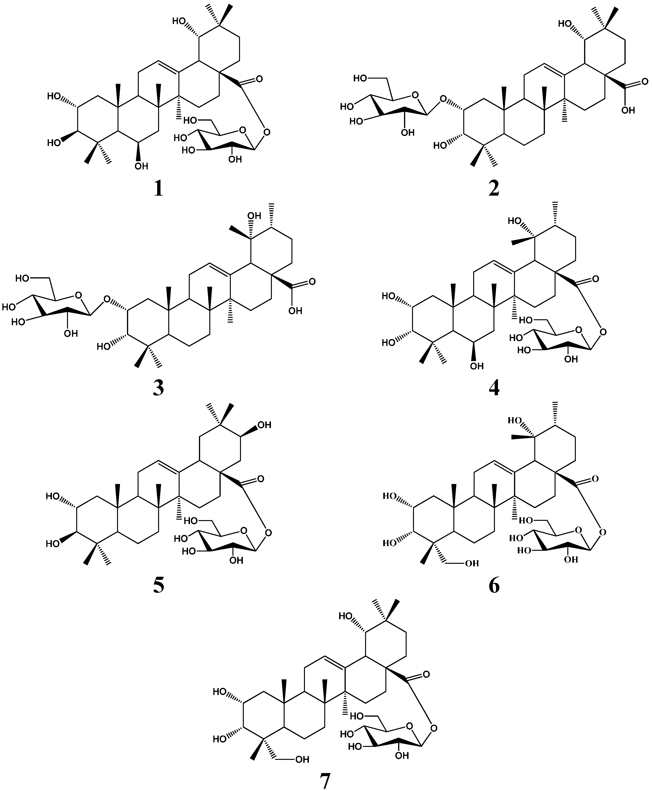
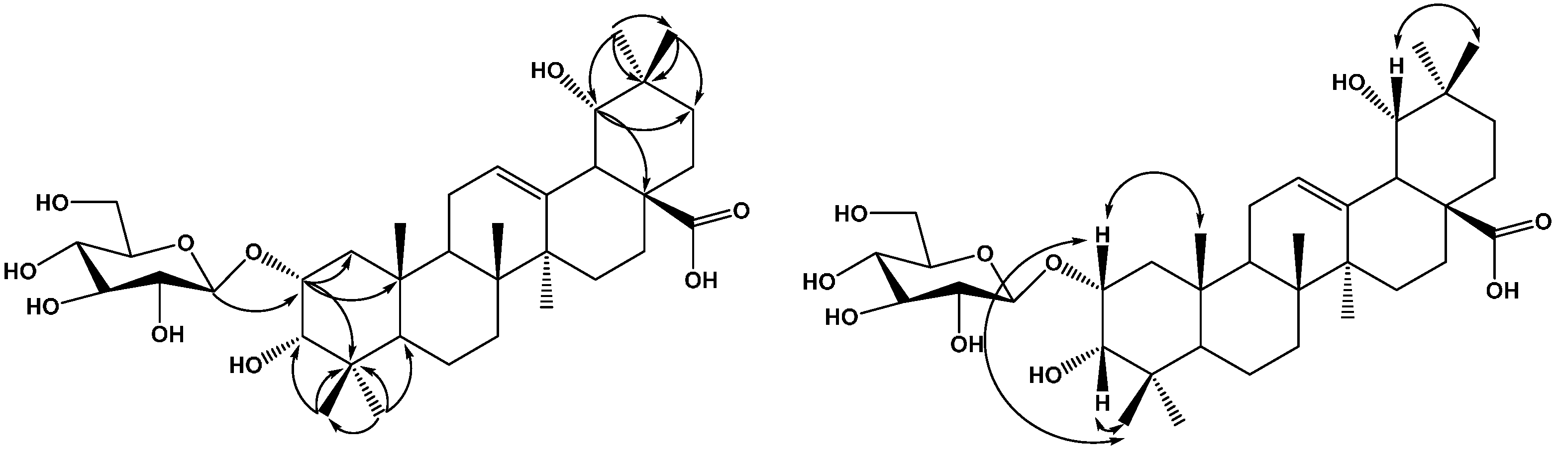
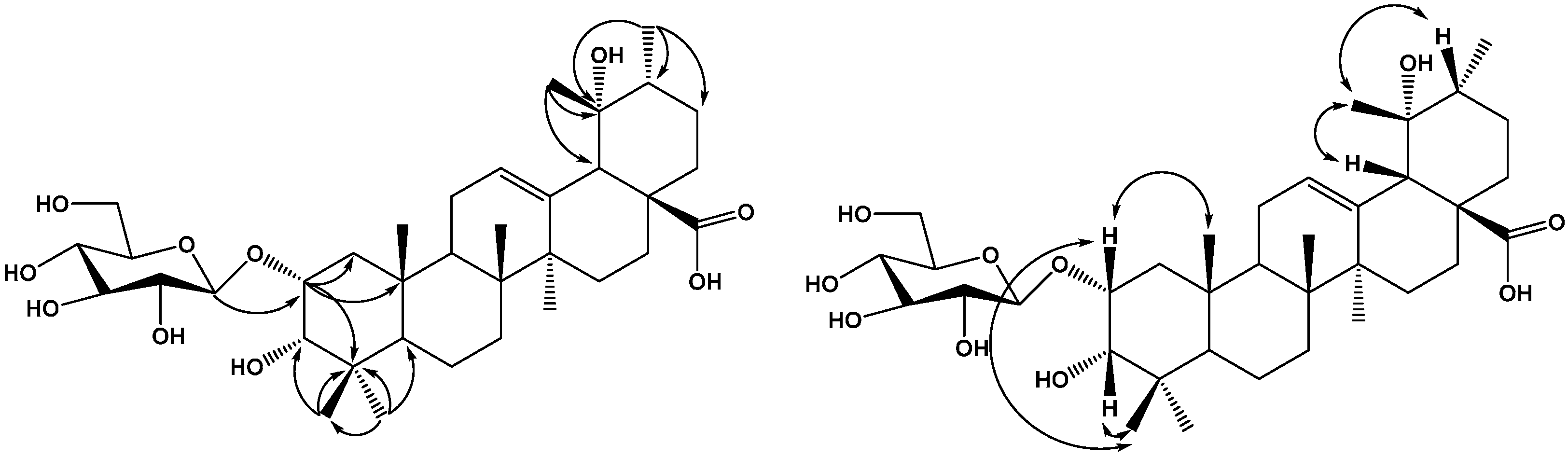
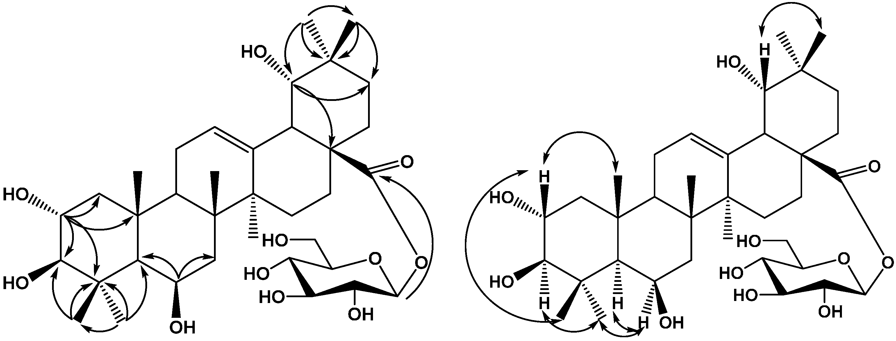
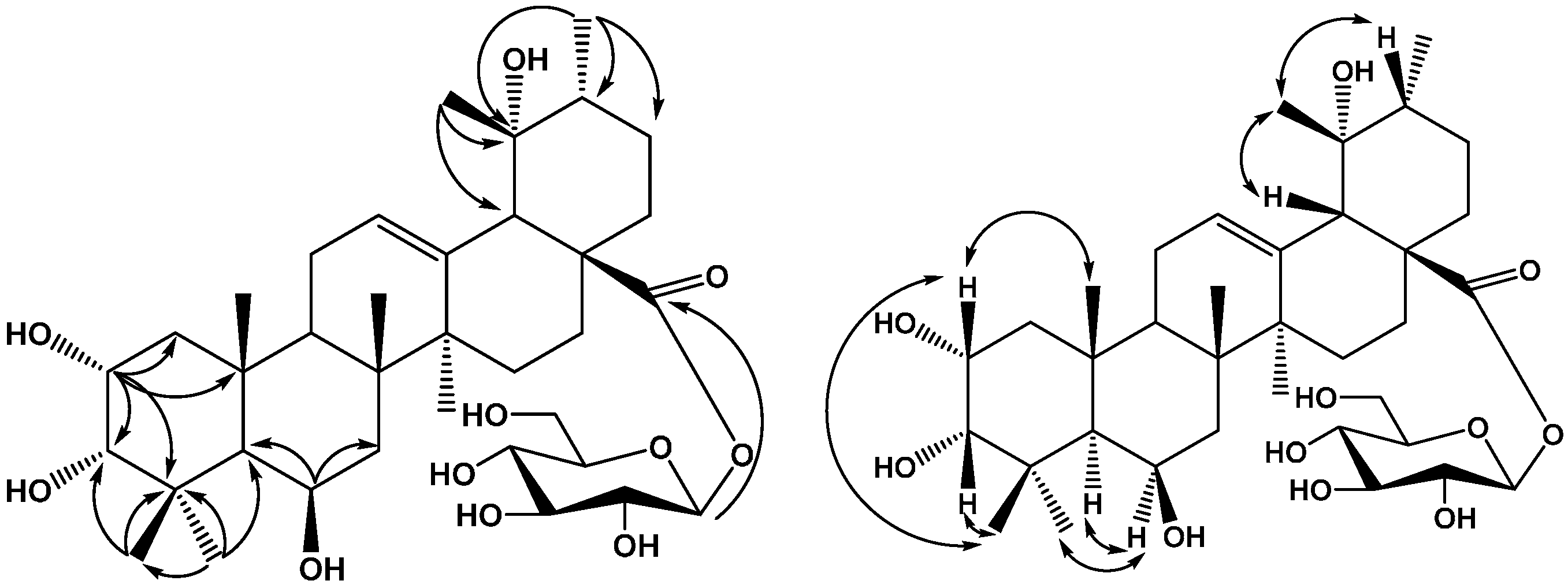
2. Results and Discussion

The EtOH extract of the aerial parts of Callicarpa kwangtungensis was successively partitioned with petroleum ether, EtOAc, and n-BuOH. The EtOAc-soluble portion was separated by a combination of silica gel, ODS column chromatography, and preparative HPLC to afford four new triterpenoids: 2α,3β,6β,19α-tetrahydroxyoleanolic acid 28-O-β-d-glucopyranoside (1), 2-O-β-d-glucopyranosyloxy-3α,19α-dihydroxyoleanolic acid (2), 2-O-β-d-glucopyranosyloxy-3α,19α-dihydroxyursolic acid (3), 2α,3α,6β,19α-tetrahydroxyursolic acid 28-O-β-d-glucopyranoside (4), together with three known triterpenoids 2α,3β,21β-trihydroxyursolic acid 28-O-β-d-glucopyranoside (5), 2α,3α,19α,23-tetrahydroxyoleanolic acid 28-O-β-d-glucopyranoside (6) and 2α,3α,19α,23-tetra-hydroxyursolic acid 28-O-β-d-glucopyranoside (7). Their structures were elucidated by extensive NMR techniques mainly including 1D NMR (1H- and 13C-NMR), 2D NMR (COSY, NOESY, HSQC, and HMBC), and ESI-MS.
Compound 1 was obtained as a white amorphous powder, which gave a positive result in the Liebermann-Burchard test. Acid hydrolysis of compound 1 with 2 mol/L HCl/1,4-dioxane (1:1, v/v) furnished glucose, identified by TLC by comparison with an authentic sample. The positive optical rotation ( [ α ] D 20 +45.1, c 0.03, H2O) indicated the d-configuration of glucose, The sugar identity was further confirmed by the chemical shifts and coupling constants in the 1H- and 13C-NMR spectra. The HR-ESI-MS of 1 showed a quasi-molecular ion peak at m/z 665.3901 [M−H], indicating a molecular formula of C36H58O10 (calcd. for C36H57O10, 665.3909, Δamu 2.6 ppm). The 1H- and 13C-NMR spectra of 1 in pyridine-d5 showed typical signals for an oleanane pentacyclic triterpenoid skeleton including seven tertiary methyl groups [δH 0.99,1.17, 1.49, 1.66, 1.79, 1.79, 1.83, (each 3H, s)], as well as one olefinic proton at δH 5.61 (1H, br s), two olefinic carbons (δC 124.5 and 144.2) and an ester carbonyl at δC 177.17. The 1H-NMR spectra of 1 exhibited four oxymethine protons at δH 4.87 (1H, s), 4.30 (1H, m), 3.63 (1H, s), and 3.43 (1H, d, J = 9.0 Hz). The data thus suggested that 1 is an oleanane-type triterpene with four hydroxy groups, a trisubstituted double bond, and a carboxyl. Comparison of the NMR spectroscopic data of 1 with those of arjunetin [7] demonstrated that the two compounds were almost identical, except for an additional hydroxyl group at C-6 (δC 68.3). These data suggested that 1 is a 6-oxygenated derivative of arjunetin, which was further confirmed by HMBC and NOESY experiments on 1. The existence of four hydroxy groups at C-2, C-3, C-6 and C-19 was supported by the HMBC spectrum. HMBC correlations (Figure 2) were observed between H-1 (δH 2.35 and δH 1.40) and C-25 (δC 18.9), C-4 (δC 40.2), C-2 (δC 69.3), C-3 (δC 84.4); between H-2 (δH 4.30) and C-3 (δC 84.4); H-3 (δH 3.43) and C-2 (δC 69.3), C-4 (δC 40.2), C-24 (δC 19.0); between H-19 (δH 3.63) and C-21 (δC 29.5), C-17 (δC 47.0); between H-5 (δH 1.21), H-7 (δH 2.00) and C-6 (δC 68.3). The orientations of the hydroxyls at C-2, C-3, C-6 and C-19 were determined using NOESY correlations. The NOESY correlation of H-3 (δH 3.43) with H-23 (δH 1.43) indicated that the hydroxyl at C-3 should be β-oriented; the NOESY correlations of H-2 (δH 4.30) with H-24 (δH 1.79) and H-25 (δH 1.79) implied that 2-OH group had an α-orientation; the NOESY correlations of H-6 (δH 4.87) with H-5 (δH 1.21) and H-23 (δH 1.49) implied that the 6-OH group had a β-orientation; the NOESY correlations of H-19 (δH 3.63) with H-30 (δH 1.66) implied that the 19-OH group had an α-orientation; Therefore, the aglycon moiety of 1 was identified as 2α,3β,6β,19α-tetrahydroxyoleanolic acid. In the 1H-NMR spectrum of 1, the relatively large 3JH-1,H-2 coupling constant of the anomeric proton at δH 6.36 of the d-glucopyranosyl moiety (J = 7.8 Hz) indicated a β-configuration for d-glucose. HMBC correlations between the anomeric proton at δH 6.36 (1H, d, J = 7.8 Hz) and the carbon signal at C-28 (δC 177.7) indicated that a β-d-glucopyranosyl moiety was attached to the C-28 position of the aglycone. On the basis of the foregoing evidence, the structure of 1 was determined as 2α,3β,6β,19α-tetrahydroxyoleanolic acid 28-O-β-d-glucopyranoside.
Figure 2. Key HMBC and NOESY correlations of compound 1.
Molecules 20 09071 g002
Compound 2 was obtained as a white amorphous powder, which gave a positive result in the Liebermann-Burchard test. Acid hydrolysis of compound 2 with 2 mol/L HCl/1,4-dioxane (1:1, v/v) furnished glucose, identified by TLC comparison with an authentic sample. The positive optical rotation ( [ α ] D 20 +46.2, c 0.03, H2O) indicated the d-configuration of glucose. In the (−) and (+)-ESI-MS of 2, quasimolecular ion peaks were observed at m/z: 649 [M−H] and 673 [M+Na]+, respectively. The HR-ESI-MS (m/z 649.3962 [M−H]) analysis revealed the molecular formula of 2 to be C36H58O10 (calcd. for C36H57O10, 649.3968, Δamu 0.9 ppm). The 1H- and 13C-NMR spectra of 2 in pyridine-d5 showed typical signals for an oleanane pentacyclic triterpenoid skeleton including seven tertiary methyl groups [δH 0.88, 0.97, 1.06, 1.11, 1.19, 1.23, 1.54, (each 3H, s)], as well as one olefinic proton at δH 5.57 (1H, br s), a pair of olefinic carbons at δC 124.3 and 145.3, typical for a double bond at C-12 (13) in an oleanane pentacyclic triterpenoid skeleton [6,7] and a carboxyl carbon at δC 181.3. The 1H-NMR spectra of 2 exhibited three oxymethine protons at δH 4.46 (1H, m), 4.05 (1H, d, J = 2.4 Hz), 3.63 (1H, s), The data thus suggested that the aglycon moiety of 2 is an oleanane-type triterpene with three hydroxy groups, a trisubstituted double bond, and a carboxyl. Comparison of the NMR spectroscopic data of 2 with those of 2α,3α,19α-dihydroxyoleanolic acid 28-O-β-d-glucopyranoside [7] demonstrated that the aglycon moiety of the two compounds were almost identical. These data suggesting that 2 has the same aglycon moiety as 2α,3α,19α-dihydroxyoleanolic acid 28-O-β-d-glucopyranoside were further confirmed by HMBC and NOESY experiments on 2. The existence of three hydroxy groups at C-2, C-3 and C-19 was supported by the HMBC spectrum, HMBC correlations (Figure 3) were observed between H-1 (δH 2.00 and δH 1.86) and C-25 (δC 16.9), C-4 (δC 39.2), C-2 (δC 76.9); between H-2 (δH 4.46) and C-3 (δC 79.0); between H-3 (δH 4.05) and C-2 (δC 76.9), C-5 (δC 49.5); between H-19 (δH 3.63) and C-21 (δC 29.6), C-17 (δC 44.6). The configuration of the hydroxyls at C-2, C-3 and C-19 were determined using NOESY correlations. The NOESY correlation of H-2 (δH 4.46) with H-24 (δH 0.88) and H-25 (δH 0.97) indicated that the hydroxyl at C-2 should be in an α-orientation; the NOESY correlations of H-3 (δH 4.05) with H-24 (δH 0.88) implied that 3-OH group had an α-orientation; the NOESY correlations of H-19 (δH 3.63) with H-30 (δH 1.54) implied that 19-OH group had an α-orientation. Therefore, the aglycon moiety of 2 was identified as 2α,3α,19α-trihydroxyoleanolic acid. In the 1H-NMR spectrum of 2, the relatively large 3JH-1,H-2 coupling constant of the anomeric proton at δH 5.16 of the d-glucopyranosyl moiety (J = 7.8 Hz) indicated a β-configuration for d-Glc. HMBC correlations between the anomeric proton at δH 6.36 (1H, d, J = 7.8 Hz) and the carbon signal at C-2 (δC 76.9) indicated that a β-d-glucopyranosyl moiety was attached to the C-2 position of the aglycone. On the basis of the foregoing evidence, the structure of 2 was determined as 2-O-β-d-glucopyranosyloxy-3α,19α-dihydroxyoleanolic acid.
Figure 3. Key HMBC and NOESY correlations of compound 2.
Molecules 20 09071 g003
Compound 3 was obtained as a white amorphous powder, which gave a positive result in the Liebermann-Burchard test. Acid hydrolysis of compound 3 with 2 mol/L HCl/1,4-dioxane (1:1, v/v) furnished glucose, identified by TLC comparison with an authentic sample. The positive optical rotation ( [ α ] D 20 +45.7, c 0.03, H2O) indicated the d-configuration of glucose. In the (−) and (+)-ESI-MS of 3, quasimolecular ion peaks observed at m/z: 649 [M−H] and 673 [M+Na]+ indicated the molecular weight of 3 is 650. The HR-ESI-MS of 3 showed a quasi-molecular ion peak at m/z 649.3952 [M−H], indicating a molecular formula of C36H58O10 (calcd. for C36H57O10, 649.3958, Δamu 0.41 ppm). The 1H and 13C-NMR spectra of 3 in pyridine-d5 showed typical signals for an ursane pentacyclic triterpenoid skeleton, including six tertiary methyl groups [δH 0.86, 0.96, 1.09, 1.22, 1.44, 1.62 (each 3H, s)] and one secondary methyl signal at δH 1.14 (3H, d, J = 6.6 Hz), as well as one olefinic proton at δH 5.56 (1H, br s), two olefinic carbons (δC 128.4 and 140.4) and a carboxyl carbon at δC 181.1. The 1H and 13C-NMR spectra of 3 exhibited two oxymethine protons at δH 4.46 (1H, m), 4.03 (1H, d, J = 2.4 Hz) and one hydroxy group attached to a tertiary carbon. The data thus suggested that 3 is an ursane-type triterpene with three hydroxy groups, a trisubstituted double bond, and a carboxyl. Comparison of the NMR spectroscopic data of 3 with those of 2α,3β,19α-trihydroxyurs-12-en-28-O-β-d-glucopyranoside. [7] demonstrated that the two compounds have the same aglycon moiety, only differing in the orientation of the hydroxy group at C-3. The existence of three hydroxy groups at C-2, C-3 and C-19 was supported by the HMBC spectrum, HMBC correlations (Figure 4) were observed between H-1 (δH 1.80 and δH 1.94) and C-25 (δC 17.0), C-4 (δC 39.0), C-2 (δC 76.6), C-3 (δC 79.1); between H-2 (δH 4.46) and C-3 (δC 79.1); H-3 (δH 4.03) and C-2 (δC 76.6), C-4 (δC 39.0), C-24 (δC 22.8); between H-18 (δH 3.05), H-30 (δH 1.14), H-29 (δH 1.44) and C-19 (δC 73.1). The configuration of the hydroxyls at C-2, C-3 and C-19 were determined using NOESY correlations. The NOESY correlation of H-3 (δH 4.03) with H-24 (δH 0.86) indicated that the hydroxyl at C-3 should be α-oriented; the NOESY correlations of H-2 (δH 4.46) with H-24 (δH 0.86) and H-25 (δH 0.96) implied that the 2-OH group had an α-orientation; the NOESY correlations of H-29 (δH 1.44) with H-18 (δH 3.05) and H-20 (δH 1.50) implied that 19-OH group had an α-orientation; Therefore, the aglycon moiety of 3 was identified as 2α,3α,19α-trihydroxyoursolic acid. In the 1H-NMR spectrum of 3, the relatively large 3JH-1,H-2 coupling constant of the anomeric proton at δH 5.14 of d-glucopyranosyl moiety (J = 7.8 Hz) indicated a β-configuration for d-Glc. HMBC correlations between the anomeric proton at δH 5.14 (1H,d, J = 7.8 Hz) and the carbon signal at C-2 (δC 76.6) indicated that a β-d-glucopyranosyl moiety was attached to the C-2 position of the aglycone. On the basis of the foregoing evidence, the structure of 3 was determined as 2-O-β-d-glucopyranosyloxy-3α,19α-dihydroxyursolic acid.
Figure 4. Key HMBC and NOESY correlations of compound 3.
Molecules 20 09071 g004
Compound 4 was obtained as a white amorphous powder, which gave a positive result in the Liebermann-Burchard test. Acid hydrolysis of compound 4 with 2 mol/L HCl/1,4-dioxane (1:1, v/v) furnished glucose, identified by TLC comparison with an authentic sample. The positive optical rotation ( [ α ] D 20 +45.7, c 0.03, H2O) indicated the d-configuration of glucose. The HR-ESI-MS of 4 showed a quasi-molecular ion peak at m/z 665.3901 [M−H], indicating a molecular formula of C36H58O10 (calcd. for C36H57O10, 665.3909, Δamu 1.2 ppm). The 1H and 13C-NMR spectra of 4 in pyridine-d5 showed typical signals for an ursane pentacyclic triterpenoid skeleton, including six tertiary methyl groups [δH 1.04, 1.07, 1.22, 1.25, 1.33, 1.38 (each 3H, s)] and one secondary methyl signals at δH 0.94 (3H, d, J = 6.6 Hz), as well as one olefinic proton at δH 5.36 (1H, br s), two olefinic carbons (δC 129.9 and 139.9) and an ester carbonyl carbon at δC 178.5. The 1H and 13C-NMR spectra of 4 exhibited three oxymethine protons at δH 4.38 (1H, s), 4.02 (1H, m), 3.29 (1H, d, J = 2.4 Hz) and one hydroxy group attached to a tertiary carbon. The data thus suggested that 4 is an ursane-type triterpene with four hydroxy groups, a trisubstituted double bond, and a carboxyl. Comparison of the NMR spectroscopic data of 4 with those of 2α,3β,19α-trihydroxyurs-12-en-28-O-β-d-glucopyranoside [7] demonstrated that two compounds were almost identical, except for an additional hydroxyl group at C-6 (δC 69.2). This was further confirmed by HMBC and NOESY experiments on 4. The existence of four hydroxy groups at C-2, C-3, C-6 and C-19 was supported by the HMBC spectrum, HMBC correlations (Figure 5) were observed between H-1 (δH 1.54 and δH 1.28) and C-25 (δC 18.5), C-4 (δC 40.1), C-2 (δC 67.1), C-3 (δC 81.5); between H-2 (δH 4.02) and C-3 (δC 81.5); H-3 (δH 3.29) and C-2 (δC 67.1), C-4 (δC 40.1), C-24 (δC 24.4); between H-19 (δH 3.63) and C-21 (δC 29.5), C-17 (δC 47.0); between H-5 (δH 1.28), H-7 (δH 1.53) and C-6 (δC 69.2). The configuration of the hydroxyls at C-2, C-3, C-6 and C-19 were determined using NOESY correlations. The NOESY correlation of H-3 (δH 3.29) with H-24 (δH 1.25) indicated that the hydroxyl at C-3 should be in an α-orientation; the NOESY correlations of H-2 (δH 4.02) with H-24 (δH 1.25) and H-25 (δH 1.38) implied that the 2-OH group had an α-orientation; the NOESY correlations of H-6 (δH 4.38) with H-5 (δH 1.28) and H-23 (δH 1.07) implied that the 6-OH group had a β-orientation; the NOESY correlations of H-19 (δH 3.63) with H-30 (δH 1.66) the NOESY correlations of H-29 (δH 1.22) with H-18 (δH 2.54) and H-20 (δH 1.36) implied that the 19-OH group had an α-orientation. Therefore, the aglycon moiety of 4 was identified as 2α,3α,6β,19α-tetrahydroxyursolic acid. In the 1H-NMR spectrum of 4, the relatively large 3JH-1,H-2 coupling constant of the anomeric proton at δH 5.32 of the d-glucopyranosyl moiety (J = 8.4 Hz) indicated a β-configuration for d-Glc. HMBC correlations between the anomeric proton at δH 5.32 (1H, d, J = 8.4 Hz) and the carbon signal at C-28 (δC 178.5) indicated that a β-d-glucopyranosyl moiety was attached to the C-28 position of the aglycone. On the basis of the foregoing evidence, the structure of 4 was determined as 2α,3α,6β,19α-tetrahydroxyursolic acid 28-O-β-d-glucopyranoside.
Figure 5. Key HMBC and NOESY correlations of compound 4.
Molecules 20 09071 g005
The structures of the three known triterpenoids 2α,3β,21β-trihydroxy ursolic acid 28-O-β-d-glucopyranoside (5) [8], 2α,3α,19α,23-tetrahydroxyoleanolic acid 28-O-β-d-glucopyranoside (6) [9] and 2α,3α,19α,23-tetrahydroxyursolic acid 28-O-β-d-glucopyranoside (7) [10] were determined by comparison of their NMR spectral data with those reported in the literature.

3. Experimental Section

3.1. General Experimental Procedures

Optical rotations were measured on an Autopol IV-T/V (Rudolph Research Analytical, Hackettstown, NJ, USA). UV spectra were recorded in MeOH on a Jasco V650 spectrophotometer (JASCO, Inc., Easton, MD, USA). The 1H (600 MHz), 13C- (150 MHz), and 2D-NMR spectra were recorded on a Bruker AVANCE III 600 instrument using TMS (tetramethylsilane) as an internal reference (Bruker Company, Billerica, MA, USA). HRTOFMS data were obtained on an Agilent 7890–7000 A mass spectrometer (Agilent Technologies, Santa Clara, CA, USA). Preparative HPLC (high-performance liquid chromatography) was conducted with an Agilent Technologies 1200 series instrument with an multiple wavelength detector using a YMC-pack ODS (Octadecylsilyl)-A column (5 µm, 250 × 20 mm). Column chromatography was performed with silica gel (200–300 mesh, Qingdao Marine Chemical Ltd., Qingdao, China), Develosil ODS (50 µm, Nomura Chemical Co. Ltd., Osaka, Japan), and Sephadex LH-20 GE Healthcare Bio-Sciences AB, Uppsala, Sweden). TLC (thin layer chromatography) was carried out with glass precoated with silica gel GF254. Spots were visualized under UV light or by spraying with 10% sulfuric acid in EtOH followed by heating. All reagents and solvents are analystical grade.

3.2. Plant Material

The aerial parts of C. kwangtungensis Chun were collected from Pingxiang, Jiangxi province of China, in July 2012. The plant was identified by Guiping Yuan at Jiangxi Provincial Institute for Drug and Food Control, China. A voucher specimen (No. 20120715) is deposited in the Herbarium of Jiangxi Provincial Institute for Drug and Food Control.

3.3. Extraction and Isolation

The aerial parts of C. kwangtungensis (10.5 kg) were extracted three times with 95% EtOH under reflux (2 h each). The extracted solution was evaporated under reduced pressure to yield a dark-brown residue (1.2 kg). The residue was suspended in water (20 L) and then successively partitioned with petroleum ether (3 × 20 L), EtOAc (3 × 20 L), and n-BuOH (3 × 20 L). After removing the solvent, the EtOAc-soluble portion (130 g) was fractionated via silica gel column chromatography (CC), eluting with CHCl3/MeOH (5:1, v/v), to give 10 major fractions A1-A10 on the basis of TLC analysis. Fraction A2 (7.8 g) was subjected to silica gel CC and eluted with CHCl3/MeOH (30:1–1:1, v/v) to afford nine fractions (A2-1-A2-9). Fraction A2-4 (2.5 g) was separated by ODS CC (50 µm, 20%–100%, MeOH/H2O) to give four subfractions (A2-4-1-A2-4-4). Subfraction A2-4-3 (1.1 g) was separated by Sephadex LH-20 CC using MeOH to afford five fractions (A2-4-3-1–A2-4-3-5) on the basis of TLC analysis. Fraction A2-4-3-3 (108 mg) was further separated by preparative HPLC (YMC-ODS-A, 5 µm, 250 mm × 20 mm, detection at 210 nm) using 23% CH3CN–H2O (7 mL/min) as mobile phase to yield 1 (20.2 mg) and 2 (4.0 mg). A2-4-2 (1.1 g) was subjected to silica gel CC and eluted with CHCl3/MeOH (12:1–4:1) to afford three fractions (A2-4-2-1-A2-4-2-3). Subfraction A2-4-2-2 (108 mg) was separated by preparative HPLC (YMC-ODS-A, 5 µm, 250 mm × 20 mm, detection at 210 nm) using 23% CH3CN–H2O (7 mL/min) to yield 3 (4.5 mg), 4 (21.3 mg) and 5 (5.8 mg). Fraction 5 (8.9 g) was subjected to silica gel CC and eluted with CHCl3/MeOH (30:1–1:1, v/v) to afford five fractions (A5-1-A5-5). A5-4 (2.7 g) was subjected to ODS CC (50 µm, 20%–100%, MeOH-H2O) to afford four subfractions (A5-4-1-A5-4-4). A5-4-3 (1.31 g) was subjected to silica gel CC and eluted with CHCl3/MeOH (30:1–1:1, v/v) to afford three fractions (A5-4-3-1~A5-4-3-3), A5-4-3-1 (217 mg) separated by preparative HPLC (YMC-ODS-A, 5 µm, 250 mm × 20 mm, detection at 210 nm) using 23% CH3CN–H2O (7 mL/min) to yield 6 (5.3 mg)and 7 (16.8 mg).

3.4. Acid Hydrolysis of Compounds 14

Compounds 1 (1.0 mg), 2 (1.0 mg), 3 (1.0 mg) and 4 (1.0 mg) were heated in an ampule with aqueous 2 mol/L HCl/1,4-dioxane (1:1, 2 mL) at 80 °C for 6 h. The aglycone was extracted with chloroform (3 × 3 mL). The aqueous layer was evaporated under reduced pressure and subjected to the column chromatography over Sephadex LH-20, eluting with CH3CN/H2O (8:1) to yield the sugar residue. Compound 1, 2, 3 and 4 gave d-glucose which was identified by TLC comparison with a standard sample (CH3CN/H2O (6:1); Rf = 0.35 and its positive optical rotation.

3.5. The Physicochemical Data of Compounds 17

2α,3β,6β,19α-Tetrahydroxyoleanolic Acid 28-O-β-d-Glucopyranoside (1). White amorphous powder; [ α ] D 20 –12.5 (c 0.12, MeOH); UV (MeOH) λmax (logε): 207.6 (3.33) nm; for 1H-NMR (600 MHz, C5D5N) and 13C-NMR (150 MHz, C5D5N) spectral data, see Table 1; HR-ESI-MS m/z 665.3901 [M−H], (calcd for C36H57O10, 665.3909, Δamu 2.6 ppm).
2-O-β-d-Glucopyranosyloxy-3α,19α-dihydroxyoleanolic Acid (2). White amorphous powder; [ α ] D 20 −23.3 (c 0.03, MeOH); UV (MeOH) λmax (logε): 206.2 (3.22) nm; for 1H-NMR (600 MHz, C5D5N) and 13C-NMR (150 MHz, C5D5N) spectral data, see Table 1; HR-ESI-MS m/z 649.3962 [M−H] (calcd for C36H57O10, 649.3968, Δamu 0.9 ppm).
Table 1. 1H-NMR (600 MHz) and 13C-NMR (150 MHz) spectral data of 14 (δ in ppm, J in Hz, in pyridine-d5).
No.1234
CHCHCHCH
150.41.40, 1H, br t, J = 12.0 Hz39.81.86, 1H, br t, J = 12.0 Hz39.91.80, 1H, br t, J = 12.0 Hz44.91.28, 1H, s
2.35, 1H, m 2.00, 1H, m 1.94, 1H, m 1.54, 1H, m
269.34.30, 1H, m76.94.46, 1H, m76.64.46, 1H, m67.14.02, 1H, m
384.43.43, 1H, d, J = 9.0 Hz794.05,1H, d, J = 2.4 Hz79.14.03, 1H, d, J = 2.4 Hz81.53.29, 1H, d, J = 2.4 Hz
440.2 39.2 39.0 40.1
557.41.21, 1H, s49.51.68, 1H, m48.81.66, 1H, m49.71.28, 1H, s
668.34.87, 1H, s19.11.34, 1H, m19.01.33, 1H, m69.24.38, 1H, s
1.49 m 1.47 m
741.32.00, 2H, m34.11.36, 1H, m33.91.35, 1H, m40.61.53, 2H, m
1.58, 1H, m 1.65, 1H, m
842.1 40.7 41.0 41.7
949.52.14, 1H, m48.72.08, 1H, m48.12.03, 1H, m49.51.90, 1H, m
1039.0 39.2 39.0 38.3
1124.82.33, 1H, m24.82.01, 1H, m24.62.00, 1H, s24.42.09, 2H, m
12124.55.61, 1H, s124.35.57, 1H, s128.45.56, 1H, s129.95.36, 1H, s
13144.2 145.3 140.4 139.9
1443.3 42.7 42.7 43.2
1529.61.32, 1H, m29.71.33, 1H, m29.71.25, 1H, m29.31.03, 1H, m
2.08, 1H, m 2.12, 1H, m 2.33, 1H, m 1.91, 1H, m
1628.72.17, 1H, m28.92.16, 1H, m27.41.33, 1H, m27.11.26, 1H, m
2.83, 1H, m 2.80, 1H, m 2.08, 1H, m 1.77, 1H, m
1747.0 46.6 49.3 49.6
1845.13.58, 1H, s45.33.60, 1H, m55.03.05, 1H, t55.02.54, 1H, s
1981.63.63, 1H, s81.83.63, 1H, t73.2 73.7
2036.0 36.2 42.81.50, 1H, m43.01.36, 1H, m
2129.51.07, 1H, m29.61.25, 1H, m26.92.04, 1H, m26.61.64, 1H, m
2.48, 1H, t J = 6.0 Hz 1.35, 1H, m 3.11, 1H, m 2.62, 1H, m
2233.42.00, 1H, m33.82.04, 1H, m39.22.08, 1H, m38.81.66, 1H, m
2.08, 1H, m 2.17, 1H, m 2.16, 1H, m 1.79, 1H, m
2329.61.49, 3H, s29.91.23, 3H, s29.81.22, 3H, s29.61.07, 3H, s
2419.01.79, 3H, s22.80.88, 3H, s22.80.86, 3H, s24.41.25, 3H, s
2518.91.79, 3H, s17.00.97, 3H, s17.00.96, 3H, s18.51.38, 3H, s
2619.81.83, 3H, s18.11.06, 3H, s17.71.09, 3H, s18.71.04, 3H, s
2725.30.99, 3H, s25.31.54, 3H, s25.11.62, 3H, s24.71.33, 3H, s
28177.7 181.3 181.1 178.5
2929.21.17, 3H, s29.31.19, 3H, s27.51.44, 3H, s27.21.22, 3H, s
3025.31.66, 3H, s25.31.54, 3H, s17.31.14, 3H, d, J = 6.6 Hz16.60.94, 3H, d, J = 6.6 Hz
Glc
196.46.36, 1H, d, J = 7.8 Hz104.25.16, 1H, d, J = 7.2 Hz104.15.14, 1H, d, J = 7.8 Hz95.95.32, 1H, d, J = 7.8 Hz
274.64.24, 1H, t, J = 7.8 Hz75.84.07,1H, t, J = 7.2 Hz75.94.07, 1H, t, J = 7.8 Hz73.93.35, 1H, t, J = 7.8 Hz
379.74.02, 1H, d, J = 9.0 Hz 78.34.28,1H, d, J = 9.0 Hz78.34.29, 1H, d, J = 8.4 Hz78.33.35, 1H, d, J = 8.4 Hz
471.74.39, 1H, t, J = 9.0 Hz72.34.30,1H, t, J = 9.0 Hz72.24.31, 1H, t, J = 8.4 Hz71.33.40, 1H, t, J = 8.4 Hz
579.34.30, 1H, t, J = 9.0 Hz78.84.32, 1H, t, J = 9.0 Hz78.94.33, 1H, t, J = 8.4 Hz78.64.03, 1H, dd, J = 8.4 Hz J = 4.2 Hz
662.64.41, 1H, t, J = 9.6 Hz63.34.37, 1H, dd, J = 4.8 Hz J = 12.0 Hz63.24.37, 1H, dd, J = 8.4 Hz J = 11.4 Hz62.43.70, 1H, dd, J = 4.2 Hz J = 12.0 Hz
4.44, 1H, m 4.54, 1H, dd, J = 2.4 Hz J = 12.0 Hz 4.54, 1H, dd, J = 2.4 Hz J = 11.4 Hz 3.81, 1H, dd, J = 1.8 Hz J = 12.0 Hz
2-O-β-d-Glucopyranosyloxy-3α,19α-dihydroxyursolic Acid (3). White amorphous powder; [ α ] D 20 −13.0 (c 0.1, MeOH); UV (MeOH) λmax (logε): 208.6 (3.16) nm; for 1H-NMR (600 MHz, C5D5N) and 13C-NMR (150 MHz, C5D5N) spectral data, see Table 1; HR-ESI-MS m/z 649.3952 [M−H] (calcd for C36H57O10, 649.3958, Δamu 0.41 ppm).
2α,3α,6β,19α-Tetrahydroxyursolic Acid 28-O-β-d-Glucopyranoside (4). White amorphous powder; [ α ] D 20 −15.0 (c 0.08, MeOH); UV (MeOH) λmax (logε): 210.1 (3.55) nm; for 1H-NMR (600 MHz, C5D5N) and 13C-NMR (150 MHz, C5D5N) spectral data, see Table 1; HR-ESI-MS m/z 665.3901 [M–H] (calcd for C36H57O10, 665.3909, Δamu 1.2 ppm).
2α,3β,21β-Trihydroxyursolic acid 28-O-β-d-glucopyranoside (5), 2α,3α,19α,23-tetrahydroxyoleanolic acid 28-O-β-d-glucopyranoside (6), and 2α,3α,19α,23-tetrahydroxyursolic acid 28-O-β-d-glucopyranoside (7), for 1H-NMR (600 MHz, C5D5N) and 13C-NMR (150 MHz, C5D5N) spectral data, see Table 2.
Table 2. 1H-NMR (600 MHz) and 13C-NMR (150 MHz) spectral data of 57 (δ in ppm, J in Hz, in pyridine-d5).
Position567
146.70.95 (1H, m)42.71.90 (1H, m)481.23 (1H, m)
2.25 (1H, m) 2.00 (1H, m) 2.08 (1H, m)
267.23.31 (1H, m)66.74.08 (1H, m)69.44.10 (1H, m)
383.12.95 (1H, d, J = 9.6 Hz)79.93.78 (1H, d, J = 2.4 Hz)78.83.76 (1H, d, J = 3.6 Hz)
439 42.6 44.2
555.60.93 (1H, m)441.41 (1H, s)48.51.33 (1H, m)
1.23 (1H, m)191.21 (1H, m)
618.51.38 (1H, m)1.34 (1H, m)19.31.48 (2H, m)
1.25 (1H, m)33.71.25 (1H, m)
732.81.42(1H, m)1.40 (1H, m)33.91.70 (2H, m)
839 41.2 40.8
947.81.93 (1H, m)48.32.06 (1H, m)492.03 (1H, m)
1037.7 38.2 39.1
1123.32.16 (1H, m)252.00 (1H, m)24.82.01 (1H, m)
12122.34.76 (1H, d, J = 4.5 Hz)128.75.56 (1H, br s)1283.65.54 (1H, br s)
13143.7 139.6 144.8
1441.4 42.7 42.7
1528.50.98 (1H, m)29.71.34 (1H, m)29.71.31 (1H, m)
1.83 (1H, m) 2.10 (1H, m) 2.04 (1H, m)
1624.31.06 (1H, m)27.12.14 (1H, m)26.72.08 (1H, m)
1.68 (1H, m) 2.62 (1H, m) 2.74 (1H, m)
1747 49.1 47
1841.82.52 (1H, s)54.92.52 (1H, s)45.13.52 (1H, s)
1946.61.06 (1H, m)73.1 81.53.57 (1H, s)
2.16 (1H, m)
2036 42.11.43 (1H, m)36
2171.43.53 (1H, m)26.62.00 (1H, m)29.51.88 (2H, m)
3.13 (1H, m)
2241.51.91 (1H, m)38.92.04 (1H, m)33.52.04 (1H, m)
2.22 (1H, m) 2.14 (1H, m) 2.10 (1H, m)
2329.50.84 (3H, s)713.75 (1H, d, 10.8 Hz)673.57 (1H, d, 10.2 Hz)
3.92 (1H, d, 10.8 Hz) 3.73 (1H, d, 10.2 Hz)
Table 2. Cont. 2417.31.06 (3H, s)18.20.90 (3H, s)14.70.99 (3H, s)
2516.71.00 (3H, s)17.61.09 (3H, s)17,81.22 (3H, s)
26170.91 (3H, s)181.26 (3H, s)18.21.10 (3H, s)
2725.81.41 (3H, s)27.51.65 (3H, s, )25.91.56 (3H, s)
28176.6 177.9 177.8
2929.71.12 (3H, s)24.71.39 (3H, s)29.41.15 (3H, s)
3020.11.09 (3H, s)17.21.07 (3H, d, J = 6.6 Hz)25.11.17 (3H, s)
Glc
1′96.65.44 (1H, d, J = 7.8 Hz)96.46.30 (1H, d, J = 7.8 Hz)96.46.30 (1H, d, J = 7.8 Hz)
2′743.33 (1H, m)74.64.22 (1H, m)74.64.21 (1H, m)
3′78.93.43 (1H, m)79.84.02 (1H, m)79.93.98 (1H, m)
4′71.33.3 (1H, m)71.74.33 (1H, m)71.64.32 (1H, m)
5′79.43.31 (1H, m)79.54.27 (1H, m)79.84.25 (1H, m)
6′61.93.64 (1H, m)62.74.35 (1H, m)62.74.37 (1H, m)
3.85 (1H, m) 4.37 (1H, m) 4.40 (1H, m)

4. Conclusions

Four new triterpenoids which were identifed as 2α,3β,6β,19α-tetrahydroxyoleanolic acid 28-O-β-d-glucopyranoside (1), 2-O-β-d-glucopyranosyloxy-3α,19α-dihydroxyoleanolic acid (2), 2-O-β-d-glucopyranosyloxy-3α,19α-dihydroxyursolic acid (3) and 2α,3α,6β,19α-tetrahydroxyursolic acid 28-O-β-d-glucopyranoside (4), were isolated together with three known triterpenoids identified as 2α,3β,21β-trihydroxyursolic acid 28-O-β-d-glucopyranoside (5), 2α,3α,19α,23-tetrahydroxyoleanolic acid 28-O-β-d-glucopyranoside (6), and 2α,3α,19α,23-tetrahydroxyursolic acid 28-O-β-d-glucopyranoside (7) from the aerial parts of Callicarpa kwangtungensis. This finding represents an addition to the ongoing research on the pharmacological activity of this plant, which may be helpful to understand the use of Callicarpa kwangtungensis in traditional medicine and should continue to clarify its actual health benefits.

Supplementary Materials

Supplementary materials can be accessed at: https://www.mdpi.com/1420-3049/20/05/9071/s1.

Acknowledgments

This study was supported financially by the National Natural Science Foundation of China (NSFC, Grant No. 81460589) and Natural Science Foundation of Jiangxi, China (No. 20114BAB205070 and No. 20122BAB215062). The authors thank A.H.L. at the Center of Analysis and Testing Nanchang University for NMR measurements.

Author Contributions

Tengfei Ji conceived and designed the experiments; Guo-Ping Zhou, Yan Yu performed the experiments and analyzed the data; Ming-Ming Yuan, Tengfei Ji and Hui-Zheng Fu contributed materials and analysis tools; Guo-Ping Zhou, Tengfei Ji and Rui-Jian Zhong wrote the paper.

Conflicts of Interest

The authors declare no conflict of interest.

References

  1. Chinese Academy of Sciences, Flora of China Editorial Committee. Flora of China, 1st ed.; Science Press: Beijing, China, 1982; Volume 65, p. 25. [Google Scholar]
  2. Zhou, B.T.; Li, X.Z.; Xu, P.S.; Ye, W.C.; Wang, M.S. Chemical constituents from the upground of Callicarpa kwangtungensis Chun (I). Cent. South Pharm. 2004, 2, 238–239. [Google Scholar]
  3. Zhou, B.T.; Li, X.Z.; Xu, P.S.; Ouyang, J.D.; Zhang, Y. Chemical constituents from the upground of Callicarpa kwangtungensis Chun (II). J. TCM Univ. Hunan 2005, 25, 20–22. [Google Scholar]
  4. Wang, Y.J.; Yang, Y.F.; Gao, D. Advances in studies on chemical constituents in plants of Callicarpa Linn and their bioactivities. Chin. Tradit. Herb. Drugs 2008, 33, 133–138. [Google Scholar]
  5. Xie, E.L.; Zhou, G.P.; Ji, T.F.; Wu, J.; Yuan, G.P. A Novel Phenylpropanoid Glycoside from Callicarpa kwangtungensis Chun. Chin. Chem. Lett. 2009, 20, 833–835. [Google Scholar] [CrossRef]
  6. Yuan, M.M.; Zhong, R.J.; Chen, G.; Zhou, G.P.; Fu, H.Z.; Yan, Q.W. Two new triterpenoids from Callicarpa kwangtungensis. J. Asian Nat. Prod. Res. 2015, 17, 138–142. [Google Scholar] [CrossRef] [PubMed]
  7. Wang, Y.; Ye, W.C.; Yin, Z.Q. Triterpene saponins from Adinandra nitida. Acta Pharm. Sin. 2008, 43, 504–508. [Google Scholar]
  8. Tabopda, T.K.; Ngoupayo, J.; Tanoli, S.A.K. Antimicrobial Pentacyclic Triterpenoids from Terminalia superba. Planta Med. 2009, 75, 522–527. [Google Scholar] [CrossRef] [PubMed]
  9. Ngounou, F.N.; Choudhary, M.I.; Malik, S.; Zareen, S.; Ali, R.; Lontsi, D.; Sondengam, B.L. Two saponins from Pteleopsis hylodendron. Phytochemistry 1999, 52, 917–921. [Google Scholar] [CrossRef]
  10. Zhang, J.; Li, B.Q.; Feng, F. Chemical constituents from Callicarpa nudiflora and their hemostatic acitivity. Chin. Tradit. Herb. Drugs 2010, 35, 3297–3301. [Google Scholar]
  • Sample Availability: Samples of the compounds 17 are available from the authors.

