
Secondary Metabolite Localization by Autofluorescence in Living Plant Cells

Abstract
:
1. Introduction
2. Results and Discussion
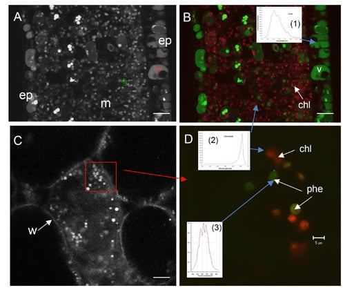
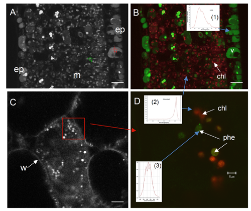
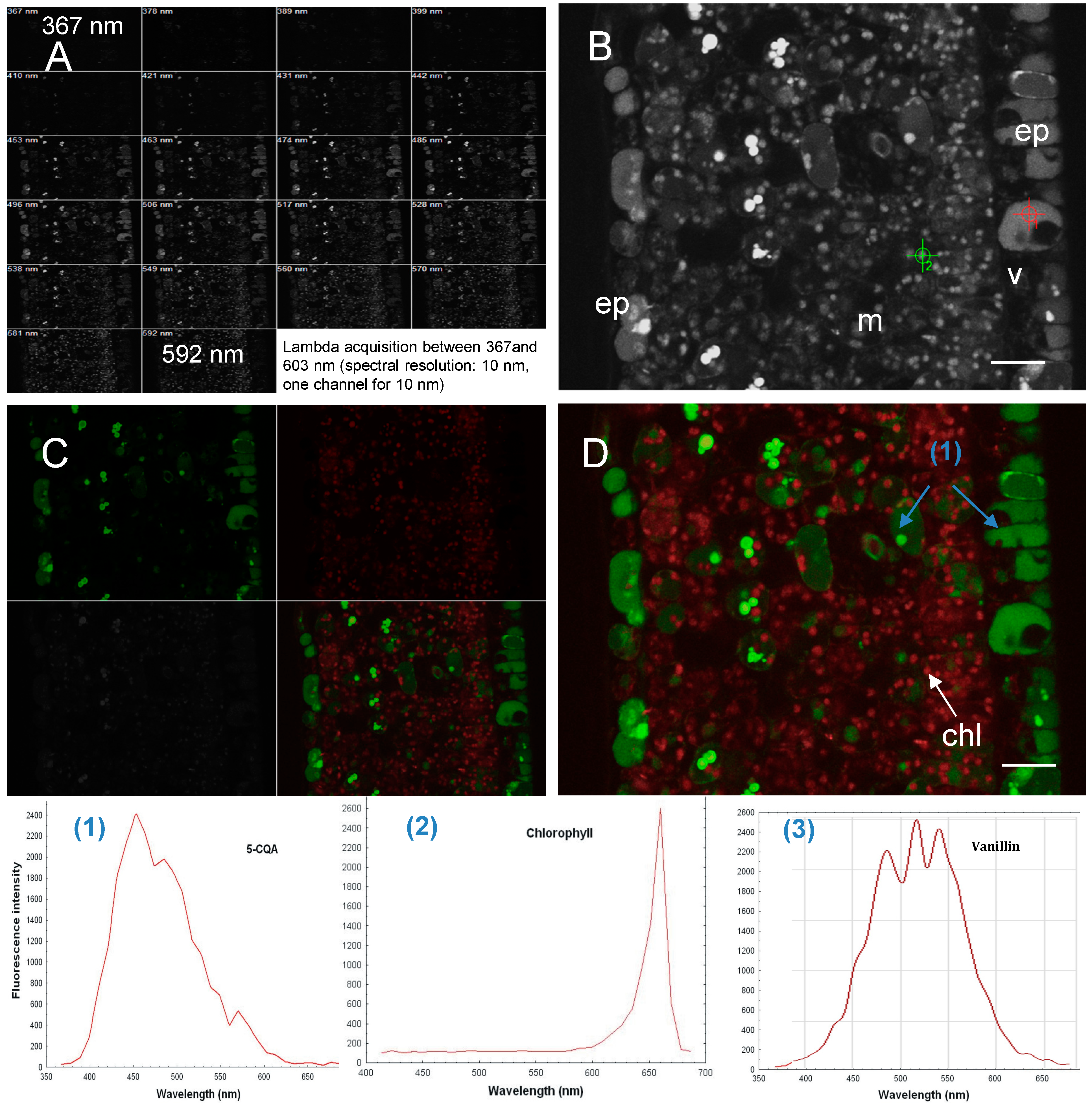
2.1. Autofluorescence of Living Plant Cells and Tissues

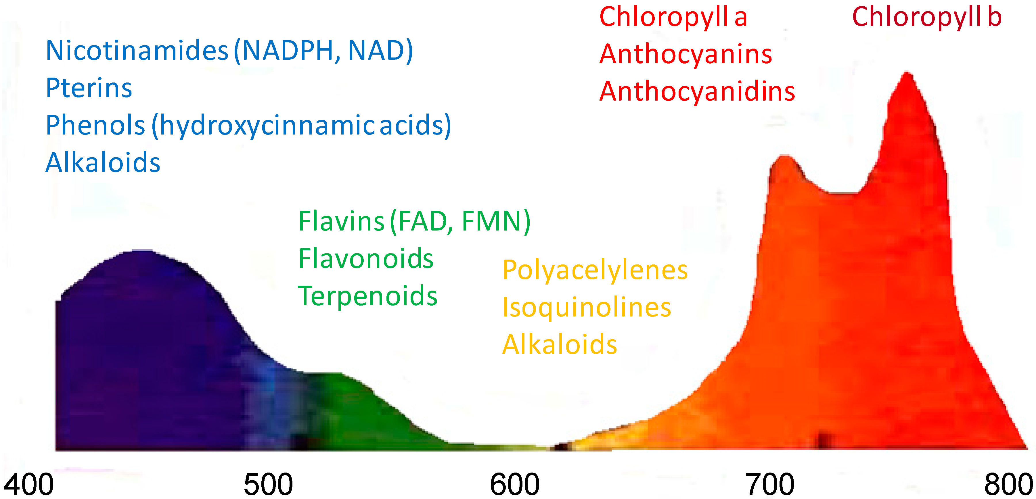
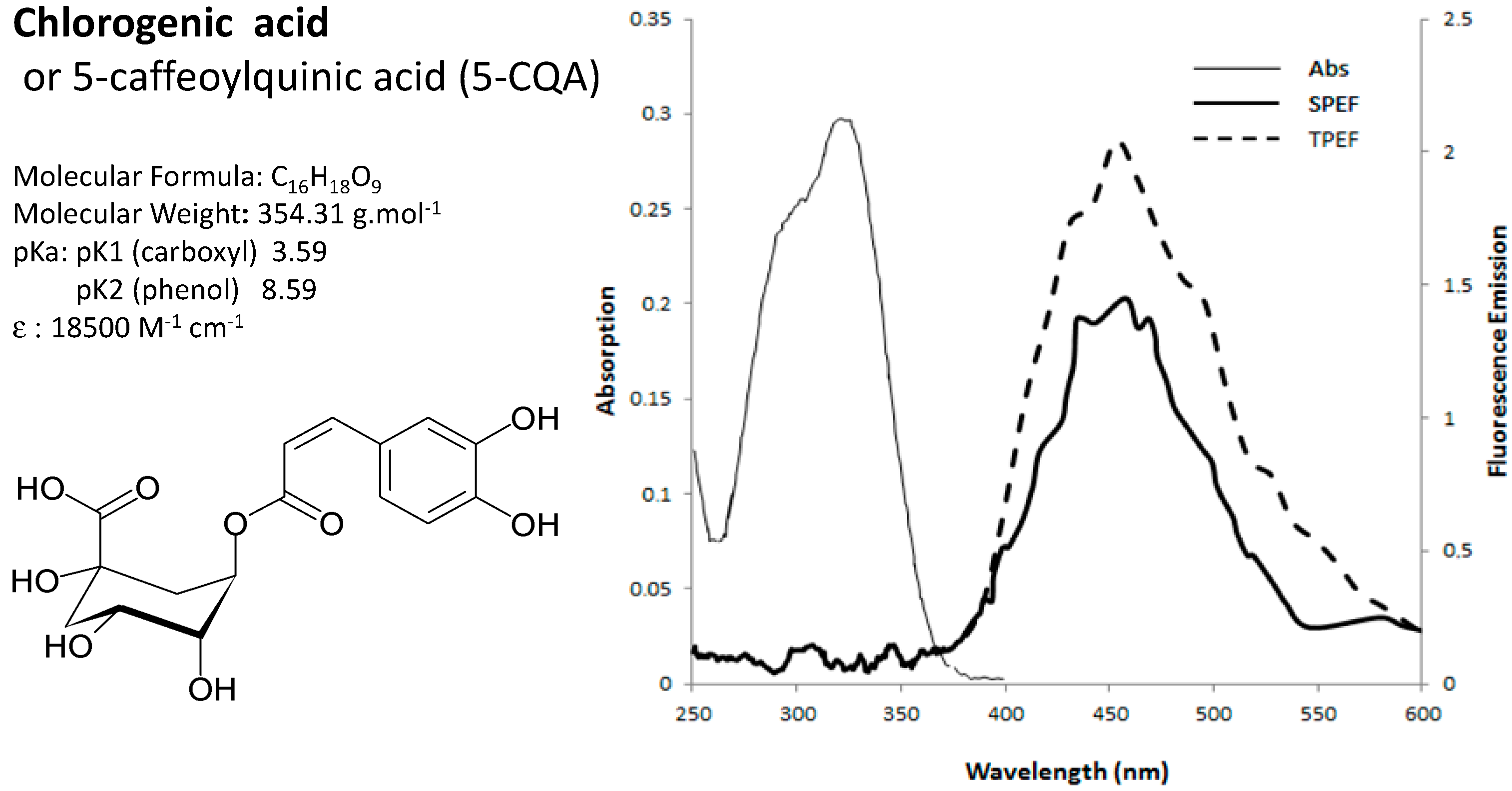
2.2. Spectral Signatures of Endogenous Fluorophores

| Metabolite | Conventional Spectrometer | Multiphoton Laser | |
|---|---|---|---|
| λabsorption | λemission | λemission | |
| Maxima (nm) | Maxima/Bandwidth (nm) * | Maxima/Bandwidth (nm) * | |
| Chlorophyll a | 430 | 685/22 | 660/35 |
| Chlorogenic acid | 324 | 455/90 | 453/105 |
| Methoxybenzaldehyde | 318 | 424/90 | 515/115 |
| Mangiferin | 318, 366 | nd | 570/80 |
| Caffeine | 272 | 397/80 | 405/60 |
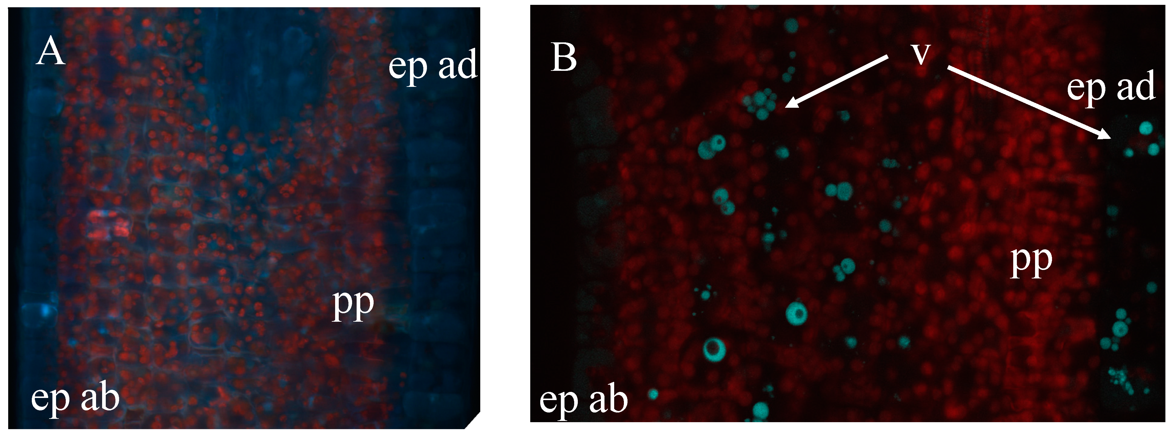
2.3. Phenolic Compounds Localization in Coffee Leaf


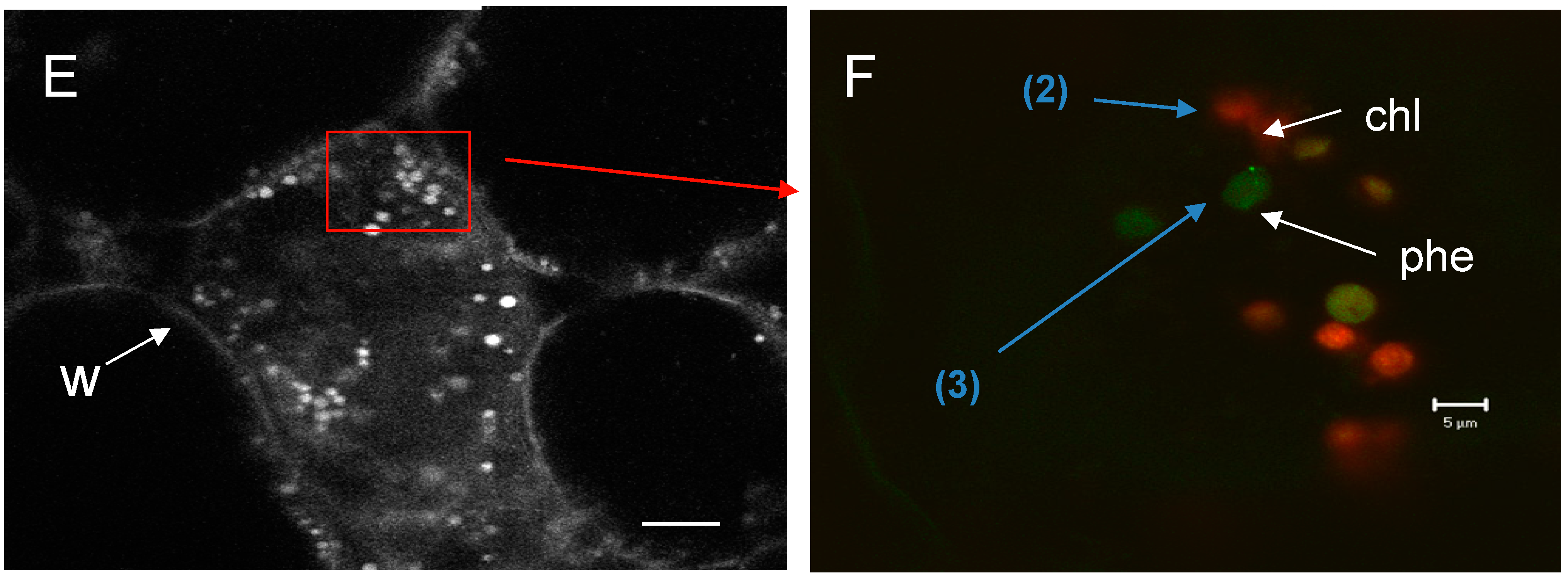
2.4. Phenolic Compound in Vanilla Fruit
3. Experimental Section
3.1. General Experimental Procedures
3.2. Processing Spectral Images
Or more simply:
I(λ) = ∑i Ci × Ri(λ)
3.3. Plant Material
3.4. Leaf and Fruit Cross-Sections
4. Conclusions
Acknowledgments
Author Contributions
Conflicts of Interest
References
- Zimmerman, T.; Rietdorf, J.; Pepperkok, R. Spectral imaging and its application in live cell microscopy. FEBS Lett. 2003, 546, 87–92. [Google Scholar] [CrossRef] [PubMed]
- Berg, R.H. Evaluation of spectral imaging for plant cell analysis. J. Microsc. 2004, 214, 174–181. [Google Scholar] [CrossRef] [PubMed]
- Chappelle, E.W.; Wood, F.M.; McMurtrey, J.E.L.; Newcomb, W.W. Laser induced fluorescence of green plants. 1: A technique for the remote detection of plant stress and species differentiation. Appl. Opt. 1984, 23, 134–138. [Google Scholar] [CrossRef] [PubMed]
- Tsurui, H.; Nishimura, H.; Hattori, S.; Hirose, S.; Okumura, K.; Shirai, T. Seven-color fluorescence imaging of tissue samples based on Fourier spectroscopy and singular value decomposition. J. Histochem. Cytochem. 2000, 48, 653–662. [Google Scholar] [CrossRef] [PubMed]
- Mylle, E.; Codreanu, M.C.; Boruc, J.; Russinova, E. Emission spectra profiling of fluorescent proteins in living plant cells. Plant Methods 2013, 9, 1–8. [Google Scholar] [CrossRef] [PubMed]
- Buschmann, C.; Langsdorf, G.; Lichtenthaler, H.K. Imaging of blue, green and red fluorescence emission of plants: On overview. Photosynthetica 2000, 38, 483–491. [Google Scholar] [CrossRef]
- Meyer, G.E.; Cartelat, A.; Moya, L.; Cerovic, Z.G. UV-induced blue-green and farred fluorescence along wheat leaves: A potential signature of leaf ageing. J. Exp. Bot. 2003, 54, 757–769. [Google Scholar] [CrossRef] [PubMed]
- Cerovic, Z.G.; Samson, G.; Morales, F.; Trembaly, N.; Moya, I. Ultraviolet-induced fluorescence for plant monitoring present state and prospects. Agronomie 1999, 19, 543–578. [Google Scholar] [CrossRef]
- Monici, M. Cell and tissue autofluorescence research and diagnostic applications. Biotechnol. Annu. Rev. 2005, 11, 1387–2656. [Google Scholar]
- Chappelle, E.W.; Wood, F.M.; McMurtray, J.E.I.; Newcomb, W.W. Laser induced fluorescence of green plants. 3: LIF spectral signatures of five major plant types. Appl. Opt. 1985, 24, 74–80. [Google Scholar] [CrossRef] [PubMed]
- Haris, P.J.; Hartley, R.D. Detection of bound ferulic acid in the cell walls of the Gramineae by ultra-violet fluorescence microscopy. Nature 1980, 259, 508–510. [Google Scholar] [CrossRef]
- Ounis, A.; Cerovic, Z.G.; Briantais, J.M.; Moya, I. Dual-excitation FLIDAR for the estimation of epidermal UV absorption in leaves and canopies. Remote Sens. Environ. 2001, 76, 33–48. [Google Scholar] [CrossRef]
- Hartmann, T. From waste products to ecochemicals: Fifty years research of plant secondary metabolism. Phytochemistry 2007, 68, 2831–2846. [Google Scholar]
- Buschmann, C.; Lichtenthaler, H.K. Principles and characteristics of multicolour fluorescence imaging of plants. J. Plant Physiol. 1998, 152, 297–314. [Google Scholar] [CrossRef]
- Wolfbeis, O.S. The fluorescence of organic natural products, chap 3. In Molecular Luminescence Spectroscopy: Methods and Applications; Schulman, S.G., Ed.; John Wiley & Sons: New York, NY, USA, 1985; Volume 77, pp. 167–370. [Google Scholar]
- Roshchina, V.V. Fluorescing World of Plant Secreting Cells; BIOS Scientific Publishers Limited: Milton Park, UK, 2007. [Google Scholar]
- Roshchina, V.V. Vital Autofluorescence: Application to the study of plant living cells. Int. J. Spectrosc. 2012, 2012. [Google Scholar] [CrossRef]
- Agati, G. Response of the in vivo chlorophyll fluorescence spectrum to environmental factors and laser excitation wavelength. Pure Appl. Opt. 1998, 7, 797–807. [Google Scholar] [CrossRef]
- Agati, G.; Galardi, C.; Gravano, E.; Romani, A.; Tattini, M. Flavonoid Distribution in Tissues of Phillyrea latifolia L. Leaves as Estimated by Microspectrofluorometry and Multispectral Fluorescence Microimaging. Photochem. Photobiol. 2002, 76, 350–360. [Google Scholar] [CrossRef] [PubMed]
- Tattini, M.; Galardi, C.; Pinelli, P.; Massai, R.; Remorini, D.; Agati, G. Differential accumulation of flavonoids and hydroxycinnamates in leaves of Ligustrum vulgare under excess light and dought stress. New Phytol. 2004, 163, 547–561. [Google Scholar] [CrossRef]
- Hutzler, P.; Fischbach, R.; Heller, W. Tissue localization of phenolic compounds in plants by confocal laser scanning microscopy. J. Exp. Bot. 1998, 49, 953–965. [Google Scholar] [CrossRef]
- Denk, W.; Strickler, J.; Webb, W. Two-photon laser scanning fluorescence microscopy. Science 1990, 248, 73–76. [Google Scholar] [CrossRef] [PubMed]
- Rost, F.W.D. Autofluorecence in plants, fungi and bacteria. In Fluorescence Microscopy; Rost, F.W.D., Ed.; Cambridge University Press: Cambridge, UK, 1995; Volume 2, pp. 16–36. [Google Scholar]
- Mondolot, L.; la Fisca, P.; Buatois, B.; Talansier, E.; de Kochko, A.; Campa, C. Evolution in caffeoylquinic acid content and histolocalization during Coffea canephora leaf development. Ann. Bot. 2006, 98, 33–40. [Google Scholar] [CrossRef] [PubMed]
- Campa, C.; Mondolot, L.; Rakotondravao, A.; Bidel, L.P.; Gargadennec, A.; Couturon, E.; la Fisca, P.; Rakotomalala, J.J.; Jay-Allemand, C.; Davis, A.P. A survey of mangiferin and hydroxycinnamic acid ester accumulation in coffee (Coffea) leaves: Biological implications and uses. Ann Bot. 2012, 110, 595–613. [Google Scholar] [CrossRef] [PubMed]
- Conejero, G.; Talamond, P.; Verdeil, J.L. A new approach to visualize secondary metabolites in plants. In Proceedings of the FESPB, XVII Congress of Federation of European Societies of Plant Biology, Valencia, Spain, 4–9 July 2010.
- Conejero, G.; Noirot, M.; Talamond, P.; Verdeil, J.L. Spectral analysis combined with advanced linear unmixing allows for histolocalization of phenolics in leaves of coffee trees. Front. Plant Sci. 2014, 5, 39–46. [Google Scholar] [CrossRef] [PubMed]
- Rubart, M. Two-Photon Microscopy of Cells and Tissue. Circ. Res. 2004, 95, 1154–1165. [Google Scholar] [CrossRef] [PubMed]
- Xu, C.; Zipfel, W.; Shear, J.B.; Williams, R.M.; Webb, W.W. Multiphoton fluorescence excitation: New spectral windows for biological nonlinear microscopy. Proc. Natl. Acad. Sci. USA 1996, 93, 10763–10768. [Google Scholar] [CrossRef] [PubMed]
- Patterson, G.H.; Piston, D.W. Photobleaching in Two-Photon Excitation Microscopy. Biophys. J. 2000, 78, 2159–2162. [Google Scholar] [CrossRef] [PubMed]
- Smith, W.L. Handbook of Laser Science and Technology; supplement 2, Weber, M.J., Ed.; CRC Press (Chemical Rubber Comapgny): Baca Raton, FL, USA, 1986; pp. 229–258. [Google Scholar]
- Xu, C.; Webb, W.W. Measurement of two-photon excitation cross sections of molecular fluorophores with data from to 1050 nm. J. Opt. Soc. Am. B 1996, 13, 481–491. [Google Scholar] [CrossRef]
- Parma, L.; Omenetto, N. Fluorescence behavior of 7-hydroxycoumarine excited by one-photon and two-photon absorption by means of a tunable dye laser. Chem. Phys. Lett. 1978, 54, 544–546. [Google Scholar] [CrossRef]
- Bestvater, F.; Spiess, E.; Stobrawa, G.; Hacker, M.; Feurer, T.; Porwol, T.; Berchner-Pfannschmidt, U.; Wotzlaw, C.; Acher, H. Two-photon fluorescence absorption and emission spectra of dyes relavant for cell imaging. J. Microsc. 2002, 208, 108–115. [Google Scholar] [CrossRef] [PubMed]
- Garini, Y.; Young, I.T.; McNamara, G. Spectral imaging: Principles and Applications. Cytometry Part A 2006, 69, 735–747. [Google Scholar] [CrossRef]
- Neu, R. A new reagent for differentiating and determining flavones on paper chromatograms. Naturwissenschaften 1957, 43, 82. [Google Scholar] [CrossRef]
- Dai, G.H.; Andary, C.; Mondolot-Cosson, L.; Boubals, D. Histochemical responses of leaves of in vitro plantlets of Vitis spp. to infection with Plasmopara viticola. Phytopathology 1995, 85, 149–154. [Google Scholar] [CrossRef]
- Talamond, P.; Mondolot, L.; Gardennec, A.; de Kochko, A.; Hamon, S.; Fruchier, A.; Campa, C. First report on mangiferin (C-glucosyl-xanthone) isolated from leaves of a wild coffee plant, Coffea pseudozanguebariae (Rubiaceae). Acta Bot. Gallica 2008, 155, 513–519. [Google Scholar] [CrossRef]
- Talamond, P.; Conéjéro, G.; Verdeil, J.L.; Poëssel, J.L. Isolation of C-glycosyl Xanthones from Coffea pseudozanguebariae and their Location. Nat. Prod. Commun. 2011, 6, 1885–1888. [Google Scholar] [PubMed]
- Brillouet, J.M.; Verdeil, J.L.; Odoux, E.; Lartaud, M.; Grisoni, M.; Conéjéro, G. Phenol homeostasis is ensured in vanilla fruit by storage under solid form in a new chloroplast-derived organelle, the phenyloplast. J. Exp. Bot. 2014, 65, 2427–2435. [Google Scholar] [CrossRef] [PubMed]
- Brillouet, J.M.; Romieu, C.; Schoefs, B.; Solymosi, K.; Cheynier, V.; Fulcrand, H.; Verdeil, J.L.; Conéjéro, G. The tannosome is an organelle forming condensed tannins in the chlorophyllous organs of Tracheophyta. Ann. Bot. 2013, 112, 1003–1014. [Google Scholar] [CrossRef] [PubMed]
- Littlejohn, G.R.; Meckel, T.; Scharzländer, M.; Costa, A. Functional imaging in living plants–cell biology meets physiology. Front. Plant Sci. 2014, 5, 1–3. [Google Scholar]
- Sample Availability: Samples of the compounds are not available from the authors.
© 2015 by the authors. Licensee MDPI, Basel, Switzerland. This article is an open access article distributed under the terms and conditions of the Creative Commons Attribution license ( http://creativecommons.org/licenses/by/4.0/).
Share and Cite
Talamond, P.; Verdeil, J.-L.; Conéjéro, G. Secondary Metabolite Localization by Autofluorescence in Living Plant Cells. Molecules 2015, 20, 5024-5037. https://doi.org/10.3390/molecules20035024
Talamond P, Verdeil J-L, Conéjéro G. Secondary Metabolite Localization by Autofluorescence in Living Plant Cells. Molecules. 2015; 20(3):5024-5037. https://doi.org/10.3390/molecules20035024
Chicago/Turabian StyleTalamond, Pascale, Jean-Luc Verdeil, and Geneviève Conéjéro. 2015. "Secondary Metabolite Localization by Autofluorescence in Living Plant Cells" Molecules 20, no. 3: 5024-5037. https://doi.org/10.3390/molecules20035024
APA StyleTalamond, P., Verdeil, J.-L., & Conéjéro, G. (2015). Secondary Metabolite Localization by Autofluorescence in Living Plant Cells. Molecules, 20(3), 5024-5037. https://doi.org/10.3390/molecules20035024