Article

Isoeugenin, a Novel Nitric Oxide Synthase Inhibitor Isolated from the Rhizomes of Imperata cylindrica

1
Department of Pharmacology, College of Korean Medicine, Sangji University, Wonju 220-702, Korea
2
Department of Agro-Industrial Technology, Faculty of Agriculture, Lambung Mangkurat University, Indonesia 70712, Indonesia
3
Department of Forest Science, College of Life Science and Natural Resources, Sangji University, Wonju 220-702, Korea
4
Department of Pharmaceutical Engineering, College of Health Science, Sangji University, Wonju 220-702, Korea
*
Author to whom correspondence should be addressed.
Molecules 2015, 20(12), 21336-21345; https://doi.org/10.3390/molecules201219767
Submission received: 28 October 2015 / Revised: 20 November 2015 / Accepted: 24 November 2015 / Published: 1 December 2015
(This article belongs to the Section Natural Products Chemistry)

Abstract

:
Phytochemical studies on the constituents of the rhizomes of Imperata cylindrica (Gramineae) were performed using high-performance liquid chromatography (HPLC). We also aimed to search for any biologically active substance capable of inhibiting nitric oxide (NO) formation in lipopolysaccharide (LPS)-activated macrophage 264.7 cells, by testing four compounds isolated from this plant. Four compounds, including a new chromone, isoeugenin, along with ferulic acid, p-coumaric acid, and caffeic acid were isolated and identified by NMR spectroscopy. The structure of isoeugenin was determined as 7-hydroxy-5-methoxy-2-methylchromone by the 2D-NMR technique. Among the four compounds, isoeugenin has the lowest IC50 value on the inhibition of NO production in LPS-activated macrophage RAW264.7 cells (IC50, 9.33 μg/mL). In addition, isoeugenin significantly suppressed the LPS-induced expressions of inducible nitric oxide synthase (iNOS), cyclooxygenase-2 (COX-2), and proinflammatory cytokines mRNA levels. Taken together, these results suggest that the anti-inflammatory activity of isoeugenin is associated with the down-regulation of iNOS, COX-2, and pro-inflammatory cytokines in RAW264.7 cells. Accordingly, our results suggest that the new chromone isoegenin should be considered a potential treatment for inflammatory disease.

Graphical Abstract

1. Introduction

Imperata cylindrica, also called cogongrass, a traditional medicinal plant in Asia, is a widespread grass and is among the top 10 worst invasive weeds in the world. Traditionally, I. cylindrica is an Asian herb used to stop bleeding, as an expectorant, and as an anti-fever and anti-inflammatory agent. Recently, several compounds isolated from cogongrass exhibited medicinal properties, including anticancer properties, platelet aggregation inhibition, and hepatoprotective activities [1].
The major phytochemical constituents identified in I. cylindrica are phenylpropanoids [2], lignan glycosides [3], biphenyl ethers [4], sesquiterpenoids [5], phenolic compounds [6], lignans [5], and megastigmatrienone [7].
Several biologically active substances have been also reported to be present in I. cylindrica. These include the 5-lipoxygenase inhibitor cylindol A, vasodilator cylindrene, platelet aggregation inhibitor imperanene, and neuroprotectives 5-hydroxy-2(2-phenylethyl) chromone and 5-hydroxy-2(2-(2-hydroxyphenyl)ethyl) chromone. However, anti-inflammatory effect of I. cylindrica and its components have not been investigated.
Inflammation is a response to injury caused by physical or chemical noxious stimuli or microbiological toxins, and occurs in multiple pathologies, such as, arthritis, asthma, multiple sclerosis, inflammatory bowel diseases, and atherosclerosis [8]. In the inflammatory state, activated immune cells, such as macrophages secrete large amounts of proinflammatory cytokines and nitric oxide (NO). High levels of proinflammatory cytokines and NO in chronic inflammatory states can result in various pathological conditions [9]. In macrophages, lipopolysaccharide (LPS), a well-known endotoxin, induces the production of the inflammatory cytokines such as tumor necrosis factor-α (TNF-α), interleukin (IL)-6, and IL-1β, as well as inflammatory mediators, including NO and prostaglandin E2 (PGE2), that are synthesized by inducible NO synthase (iNOS) and cyclooxygenase-2 (COX-2), respectively [8,10].
Accordingly, control of the production of proinflammatory cytokines and NO in macrophages are current research topics for the development of new anti-inflammatory agents. As a part of our on-going screening project to evaluate the anti-inflammatory potentials of natural compounds, we isolated four compounds from the rhizomes of I. cylindrica and investigated their anti-inflammatory effects on LPS-stimulated RAW264.7 macrophages.

2. Results and Discussion

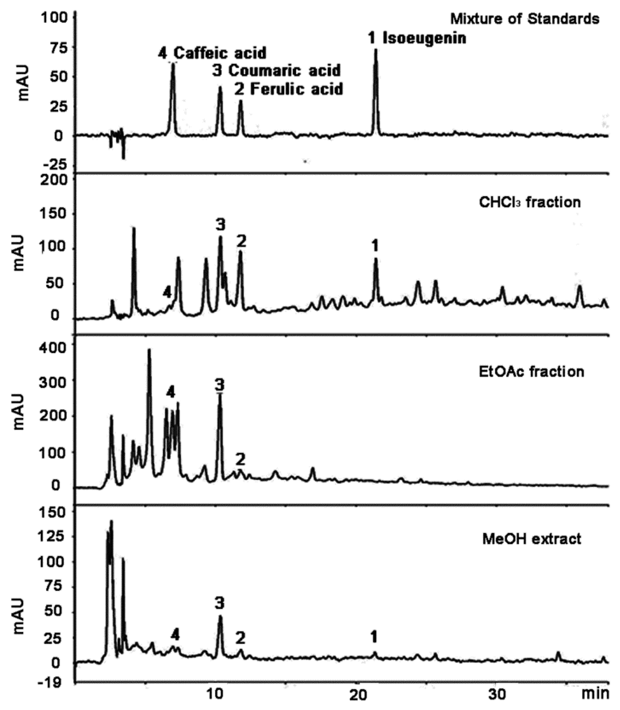
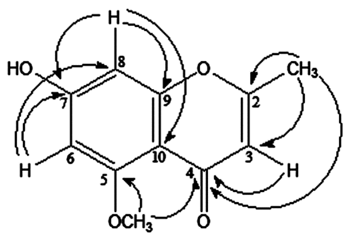
2.1. Isolation and Characterization of Compounds

The CHCl3 fraction fractionated from the MeOH extract of I. cylindrica was subjected to column chromatography to afford four compounds. The 1H-NMR spectrum of compound 1 measured in DMSO-d6 exhibited a vinylic methyl signal at δ 2.52. A singlet peak of H-3 at δ 5.60, the two doublet peaks of H-6 and 8 appearing as meta-coupled peaks (J = 1.8 Hz), together with a peak attributed to a OCH3 group at δ 3.93 (3H, s) suggests that compound 1 belongs to the chromone-type family of compounds.
The representative chromone-type compounds include eugenin, noreugenin, eugenitin, isoeugenitin, and isoeugenitol [11]. All of these have two CH3 groups, but some of them do not have a OCH3. The HR-ESI-MS showed that compound 1 exhibited an m/z of 206.0579 corresponding to a [C11H10O4]+ molecular ion. Although the molecular formula of compound 1 is the same as that of eugenin, its NMR data was different from other chromone-type compounds, therefore, compound 1 is different from known chromone-type compounds. In particular, the literature [11] indicates that eugenin possessing a 5-OH exhibits a peak of δC 182.7 (in CDCl3); therefore, it is suggested that compound 1 has a 5-OCH3 instead of a 5-OH according to its δ 169.9 value (in DMSO).
The peak at δ 2.52 (3H, s) is attributable to 2-CH3, commonly observed in this type of compounds [12] because the heteronuclear multiple-bond correlation spectroscopy (HMBC) spectrum showed its long-range coupling with δC 138.7, 87.2, and 169.9 as shown in Figure 1. A singlet peak (δH 5.60) attributable to H-3 overlapped with the peak at δC 169.9 of carbonyl. The position of 5-OCH3 assignable to δH 3.93 can be determined since the OCH3 peak is correlated with δC 169.9 (C-4). Therefore, the structure of compound 1 was determined as 7-hydroxy-5-methoxy-2-methyl-chromone, called isoeugenin. Compounds 24 were identified as ferulic acid [13], p-coumaric acid [14], and caffeic acid [15], respectively, after comparing our 1H- and 13C-NMR data with the literature data. Furthermore, Compounds 24 were identical to each corresponding standard compound on thin layer chromatography (TLC) and high performance liquid chromatography (HPLC).
The four phenolic substances were analyzed using HPLC, and the chromatograms are shown in Figure 2. Regression equations were established as shown in Table 1, by estimating peak areas measured at six concentrations (12,5, 25, 50, 100, 200, and 1000 μg/mL). Each equation was verified for linearity at R2 > 0.999.
Figure 1. Structure of isoeugenin (1) isolated from I. cylindrica var. major and heteronuclear multiple-bond correlation spectroscopy (HMBC) correlation.
Molecules 20 19767 g001
Figure 2. HPLC chromatograms of the MeOH extract of I. cylindrica and its fractions.
Molecules 20 19767 g002
Table 1. Linearity of standard curves and detection/quantification limits for the standard compounds.
Standard CompoundstR (min)Calibration Equation (Linear Model) aLinear Range (μg/mL)R2 bLOD c (μg/mL)LOQ d (μg/mL)
Caffeic acid6.92y = 747.05x + 83.3312.50–200.00.99980.160.52
p-Coumaric acid10.30y = 245.73x + 95.8312.50–200.00.99980.421.41
Ferulic acid11.75y = 276.37x + 41.6612.50–200.00.99930.571.91
Isoeugenin21.42y = 40.468x + 30.2625.00–1000.00.99984.1913.97
a y, peak area at 254 nm; x, concentration of the standard (μg/mL); b R2, correlation coefficient for 5 data points in the calibration curves (n = 4); c LOD, limit of detection (S/N = 3); d LOQ, limit of quantification (S/N = 10).
The contents of the four compounds were evaluated and revealed to be in the following order of abundance: isoeugenin (approximately 0.268 mg/g dry weight) > ferulic acid (approximately 0.042 mg/g) > p-coumaric acid (approximately 0.182 mg/g) > caffeic acid (approximately 0.020 mg/g). These four compounds were distributed mainly in the CHCl3 and EtOAc fractions as shown in Table 2. However, these substances are present in minute quantities in the hexane and BuOH fractions and isoeugenin was not detected in the EtOAc fraction.
Table 2. Content of analytes in the extract and fractions of I. cylidrica.
AnalyteMeOH ExtractFractions (mg/g Extract)
(mg/g Dry Weight)(mg/g Extract)CHCl3EtOAc
Caffeic acid0.0200.150.324.59
p-Coumaric acid0.1821.323.7316.18
Ferulic acid0.0420.304.892.97
Isoeugenin0.2681.9428.09n.d.
Total0.5123.7137.0323.74

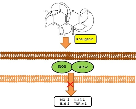
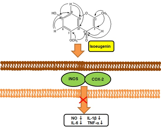
2.2. Inhibitory Effect of Compounds on LPS-Induced NO Production

When the cytotoxicity of the MeOH extract and its fractions were evaluated in macrophage 264.7 cells, only the CHCl3 fraction exhibited cytotoxicity at more than 100 μg/mL. The MeOH extract and other fractions displayed almost no cytotoxicity below 100 μg/mL concentration. When the cytotoxicity of the isolated compounds was evaluated in the cells, isoeugenin has a little cytotoxicity at 100 μg/mL and no cytotoxicity below 50 μg/mL. Ferulic acid, coumaric acid, and caffeic acid have no cytotoxicity at 12.5, 25, and 50 μg/mL (data not shown).
The inhibitory effects of the isolated compounds on NO formation in the LPS-activated macrophage 264.7 cells are shown in Table 3. In this experiment, concentrations less than those showing cytotoxicity were chosen for the nitrite assay. Isoeugenin considerably reduced NO formation in LPS-activated cells, although other compounds exhibited very low activities. The IC50 value of isoeugenin on the inhibition of NO formation was 9.33 μg/mL.
Table 3. Inhibitory effect of components of I. cylidrica on the LPS-induced NO production.
GroupConcentrationNO Production (μM)Inhibition (%)
NOR-11.90 ± 0.65-
LPS-77.18 ± 0.82 #-
l-NIL (μM)2036.12 ± 1.56 ***53.21 ± 2.02
Isoeugenin (μg/mL)12.532.96 ± 0.47 ***57.29 ± 0.61
2510.36 ± 0.16 ***86.58 ± 0.20
506.12 ± 0.16 ***92.07 ± 0.20
Ferulic Acid (μg/mL)2579.05 ± 3.08−2.42 ± 3.99
5079.67 ± 3.16−3.22 ± 4.09
10072.56 ± 0.71 **5.99 ± 0.91
Coumaric acid (μg/mL)2577.89 ± 1.49−0.92 ± 1.92
5077.00 ± 2.460.23 ± 3.19
10072.83 ± 1.67 **5.64 ± 2.16
Caffeic acid (μg/mL)2576.38 ± 1.491.04 ± 1.06
5074.87 ± 0.672.99 ± 0.87
10066.60 ± 0.41 ***13.70 ± 0.53
Cells were pretreated with different concentrations (12.5, 25, 50, 100 μg/mL) of samples for 1 h, then with LPS (1 μg/mL), and incubated for 24 h. Normal (NOR) values were obtained in the absence of LPS and samples. l-NIL was used as a positive control at a concentration of 20 μM. # p < 0.05 vs. the normal controls; ** p < 0.01; *** p < 0.001 vs. 1 μg/mL LPS-treated cells; the significances of differences between treated groups were determined using ANOVA and Dunnett’s post hoc test.

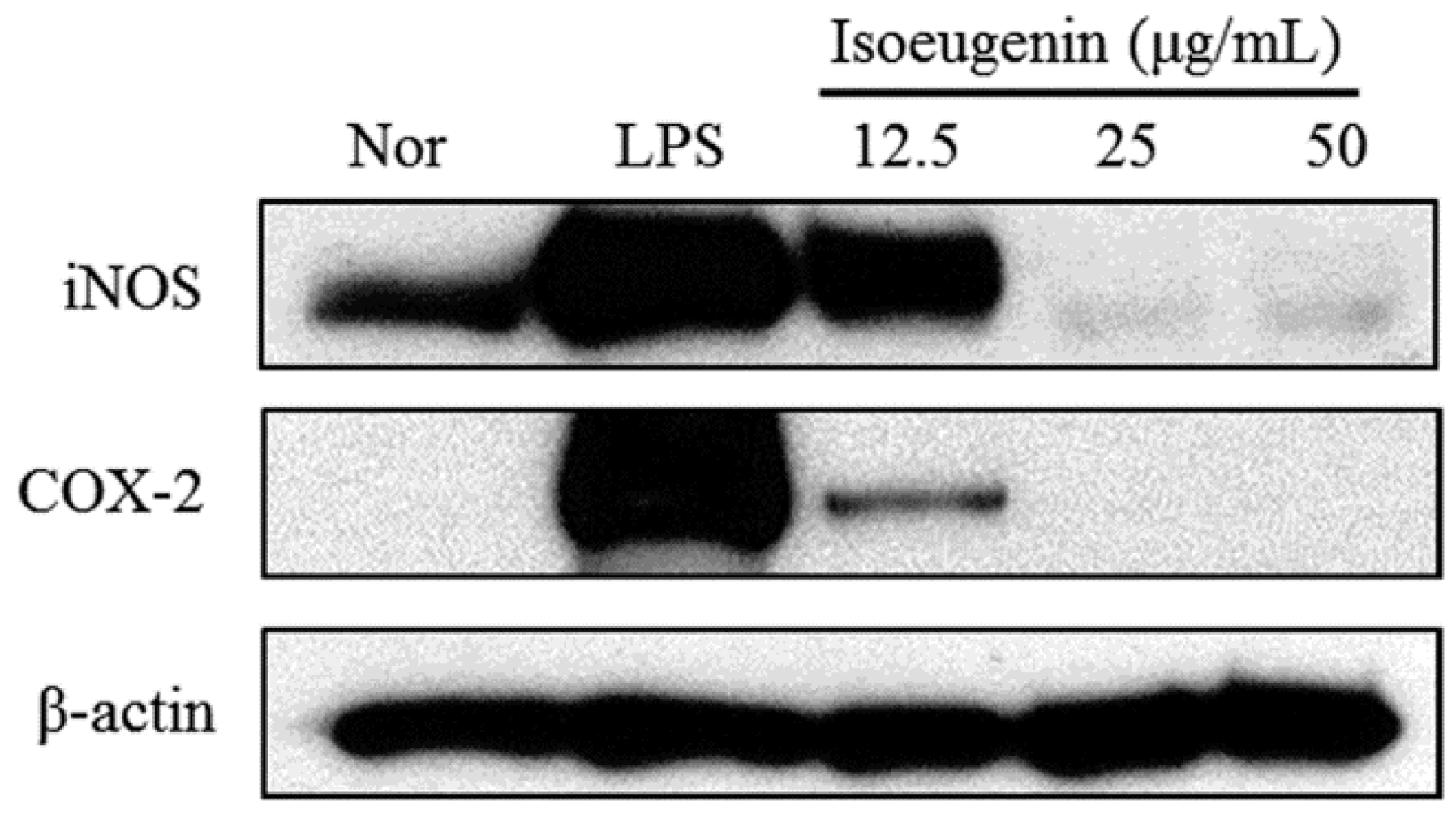
2.3. Inhibitory Effect of Isoeugenin on the LPS-Induced iNOS and COX-2 Expressions

To determine whether the inhibitory effects of isoeugenin on NO productions are related to the modulation of iNOS and COX-2 enzymes, we examined their expression levels by western blotting. In unstimulated RAW264.7 cells, iNOS and COX-2 protein levels were undetectable. However, in response to LPS, the expression levels of iNOS and COX-2 were markedly upregulated, and isoeugenin significantly inhibited the LPS-stimulated iNOS and COX-2 expressions in a dose-dependent manner (Figure 3).
Figure 3. Inhibitory effects of isoeugenin on LPS-induced iNOS and COX-2 protein expressions in RAW264.7 cells. Cells were pretreated with different concentrations (12.5, 25, 50 μg/mL) of isoeugenin for 1 h, then with LPS (1 μg/mL), and incubated for 24 h. Total cellular proteins (30 μg) were resolved by SDS-PAGE, transferred to nitrocellulose membranes, and detected with specific antibodies, as described in the Experimental Section. NOR values were obtained in the absence of LPS and samples. LPS values were obtained in only LPS treatment. A representative immunoblot of three separate experiments is shown.
Molecules 20 19767 g003

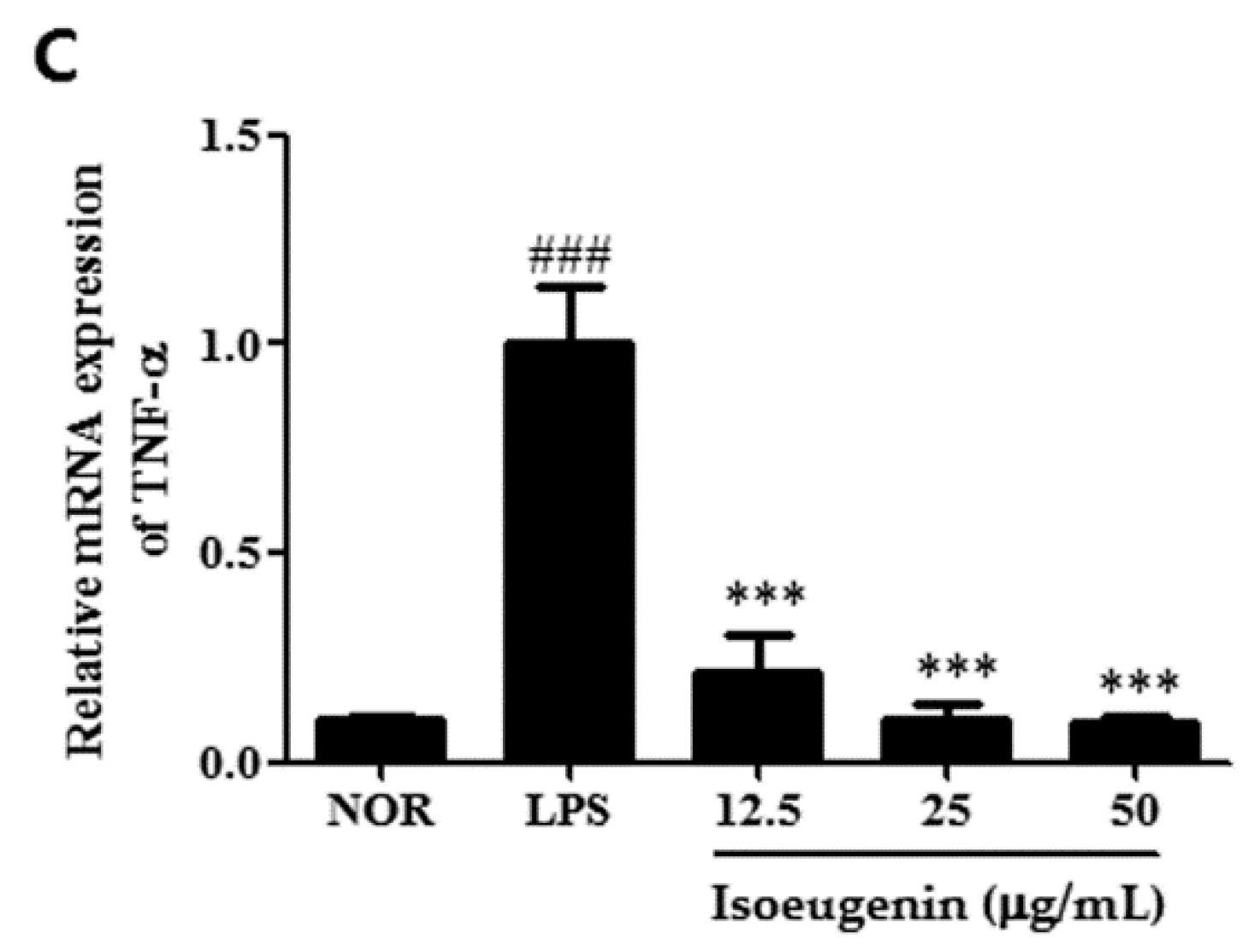
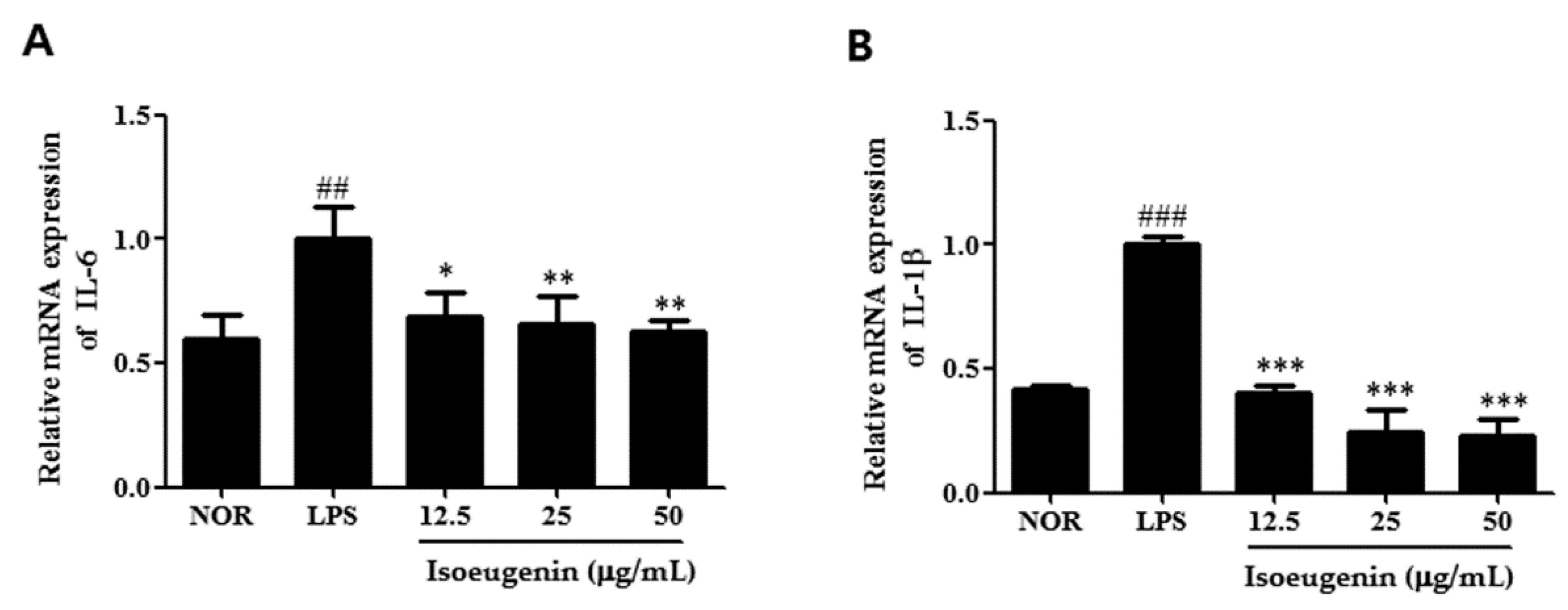
2.4. Inhibitory Effect of Isoeugenin on the LPS-Induced mRNA Levels of Cytokines

To evaluate the effect of isoeugenin on the pro-inflammatory cytokines, we further examined the expressions of TNF-α, IL-6, and IL-1β in LPS-stimulated macrophages pretreated with isoeugenin by quantitative Real-time PCR. Pretreatment with isoeugenin was found to reduce LPS-induced TNF-α, IL-6, and IL-1β mRNA expressions in a concentration-dependent manner (Figure 4). These three cytokines are known to act as pro-inflammatory mediators in vitro and in vivo. TNF-α exhibits its pro-inflammatory activity by regulating several intercellular and vascular cell adhesion molecules, which results in the recruitment of leukocytes to sites of inflammation [16]. IL-6 is a cytokine released by LPS-activated monocytes and plays a crucial role in immune response [17]. For example, the overexpression of IL-6 is involved in pathological conditions, such as, rheumatoid arthritis [18]. IL-1β is normally produced in response to infection, injury, or immunologic challenge; at minimal concentrations, it causes fever, hypotension, and production of additional proinflammatory cytokines, such as IL-6 [19]. Collectively, isoeugenin has the potential for inhibition not only iNOS and COX-2 expression but also a wide range of the pro-inflammatory genes regulated.
Figure 4. Inhibitory effect of isoeugenin on LPS-induced TNF-α (A); IL-6 (B); IL-1β (C) mRNA expression in RAW264.7 cells. Total RNA was prepared for the Real Time-PCR analysis of TNF-α, IL-6, IL-1β gene expression from RAW264.7 macrophage cells pretreated with different concentrations (12.5, 25, 50 μg/mL) of isoeugenin for 1 h followed by LPS (1 μg/mL) for 24 h. NOR values were obtained in the absence of LPS and samples. The experiment was repeated three times and similar results were obtained. Values represent means ± S.D. of three independent experiments. ## p < 0.01, ### p < 0.001 vs. the control group; * p < 0.05, ** p < 0.01, *** p < 0.001 vs. 1 μg/mL LPS-treated group; significant differences between treated groups were determined using ANOVA and Dunnett’s post hoc test.
Molecules 20 19767 g004aMolecules 20 19767 g004b

3. Experimental Section

3.1. General Information

Chromatographic isolation was performed on the open columns using normal or reverse stationary phases. Spectra of the isolated compounds were measured using a NMR spectrometer (Bruker Co., Rheinstetten, Germany). A quantitative analysis was performed on the HPLC system (Varian Co., Palo Alto, CA, USA) consisting of two Prostar 210 pumps, a Prostar 325 UV-vis detector and a Shiseido Capcell PAK C18 column (5 μm, 4.6 mm × 250 mm, Japan). Dulbecco’s modified Eagle’s minimum essential medium (DMEM), fetal bovine serum (FBS), penicillin, and streptomycin were obtained from Life Technologies Inc. (Grand Island, NY, USA). iNOS, COX-2, and β-actin monoclonal antibodies and the peroxidase-conjugated secondary antibody were purchased from Santa Cruz Biotechnology, Inc. (Santa Cruz, CA, USA).

3.2. Plant Material

The rhizome of I. cylindrica was purchased from the Chun-Il medicinal herb store in Wonju City, Korea. This medicinal herb was identified by Prof. Byong-Min Song (Department of Forestry Science, Sangji University, Wonju City, Korea). The voucher specimen (natchem #68) was deposited in the laboratory of Natural Product Chemistry, Department of Pharmaceutical Engineering, Sangji University.

3.3. Extraction and Fractionation

The plant material (3 kg) consisting of the rhizome of I. cylindrica was extracted with MeOH (4 L) under reflux for 5 h and repeated three times. The extracted solution was filtered and evaporated under reduced pressure to give a MeOH extract (501.7 g). This extract was suspended in H2O and fractionated in a separating funnel with hexane (0.8 L) three times. The hexane-soluble portion was concentrated to dryness in vacuo to give a hexane fraction (24.40 g). Similarly, the remaining H2O layer was further successively fractionated with CHCl3, EtOAc, and BuOH, and then concentrated. The yields of the CHCl3, EtOAc, and BuOH fractions were 14.56 g, 12.58 g, and 136.1 g, respectively. The concentration of the final aqueous phase gave an H2O fraction (348.8 g).

3.4. Isolation of the Componds

The CHCl3 fraction (10 g) was chromatographed on a silica gel column (280 g, 5 cm × 35 cm) using CHCl3–MeOH-H2O (10:2:2, lower layer). Forty-five fractions (fr.4–fr.5; 60 mL (each)) were then grouped into 10 subfractions (Gr.1–Gr.10) according to their band profiles in TLC.
To obtain the main component from Gr.2, silica gel (55 g, 3 cm × 30 cm) column chromatography was performed using CHCl3–MeOH–H2O (10:2:2, lower layer) to yield compound 1. Further, to obtain the main component from Gr.6, silica gel (60 g, 3 cm × 30 cm) column chromatography was undertaken using CHCl3–MeOH–H2O (10:2:1, lower layer) to yield compound 2. To obtain the main component from Gr.8, it was chromatographed on silica gel (55 g, 3 cm × 30 cm) with the solvent CHCl3–MeOH–H2O (10:2.2:1, lower layer) to yield compound 4.
Compounds 2, 3, and 4 were identified as ferulic acid, p-coumaric acid, and caffeic acid, respectively, by analysis of their corresponding 1H- and 13C-NMR spectra. The three compounds 24 were the same as standards of each compound by TLC and HPLC. The retention times of p-coumaric acid, ferulic acid and caffeic acid were 10.30 min, 11.75 min, and 6.92 min, respectively, on the HPLC chromatogram. On the other hand, compound 1 with a retention time of 21.42 min on the HPLC chromatogram was structurally determined to be isoeugenin through the interpretation of 2D-NMR spectra including 1H-1H correlation spectroscopy (COSY), 1H-13C COSY, HMBC, and nuclear Overhauser effect spectroscopy NMR spectra.
7-Hydroxy-5-methoxy-2-methylchromone (isoeugenin, 1): Yellow gum; UV λmax MeOH nm (log ε): 209 (4.22), 307 (3.87); 1H-NMR (600 MHz, dimethyl sulfoxide (DMSO-d6): 6.59 (1H, d, J = 1.8 Hz, H-8), 6.55 (1H, d, J = 1.8 Hz, H-6), 5.60 (1H, s, H-3), 3.93 (1H, s, OCH3), 2.52 (3H, s, 2-CH3); 13C-NMR (150 MHz, DMSO-d6): 138.7 (C-2), 87.2 (C-3), 169.9 (C-4), 162.2 (C-5), 101.0 (C-6), 160.9 (C-7), 116.5 (C-8), 156.6 (C-9), 106.6 (C-10), 57.0 (OCH3), 23.5 (CH3); High-resolution electrospray ionization mass spectrometry (HR-ESI-MS) m/z (rel. int.): 206.0605 (100, C11H10O4).
Ferulic Acid (2): Colorless gum; EI-MS (70 eV): m/z 194.1 (M+); 1H-NMR (500 MHz, CD3OD) and 13C-NMR (125.5 MHz, CD3OD) δ: [13].
p-Coumaric Acid (3): Colorless needles; m.p. 210–213 °C; EI-MS (70 eV): m/z 164.1 (M+), 1H-NMR (500 MHz, CD3OD) and 13C-NMR (125.5 MHz, CD3OD): [14].
Caffeic Acid (4): Yellow powder, m.p. 223–225 °C (dec.); EI-MS (70 eV): m/z 180.1 (M+), 163.06, 135.08; 1H-NMR (500 MHz, CD3OD) and 13C-NMR (125.5 MHz, CD3OD) [15].

3.5. HPLC Analysis

The HPLC system used for the present study consists of a Varian Prostar 210 solvent delivery module (Agilent Technologies, Santa Clara, CA, USA), Prostar 325 UV-Vis detector, and 20-μL sample loop. A Shiseido Capcell Pak C18 HPLC column (5 μm, 250 mm × 4.6 mm, Tokyo, Japan) was used. The two solvents used for gradient elution were solvent A 0.5% HOAc aqueous solution, and solvent B 0.5% HOAc-MeOH solution. Regression equations were established as shown in Table 1, by estimating peak areas measured at six concentrations (12, 5, 25, 50, 100, 200, and 1000 μg/mL). Each equation was verified for linearity at R2 > 0.999. Detection was performed at the fixed wavelength of 254 nm.

3.6. Preparation of Standard and Test Solutions

The four compounds, isoeugenin, ferulic acid, p-coumaric acid, and caffeic acid, which were isolated from I. cylindrica were used for preparation of standard solutions. Stock solutions (each 1000 μg/mL) that were made by dissolving each standard compound in MeOH were further diluted to produce working standard solutions. The rhizome of I. cylindrica was pulverized, added to 20 mL MeOH, and then extracted at 60 °C for 6 h using an ultrasonicator. The extracted solution was filtered through a syringe filter and used for sample solution. The other four sample solutions (each 1000 μg/mL) were prepared by dissolving hexane CHCl3, EtOAc, and BuOH fractions in MeOH.

3.7. Cell Culture and Sample Treatment

The RAW264.7 murine macrophage cell line was obtained from the Korea Cell Line Bank (Seoul, Korea). These cells were grown at 37 °C in Dulbecco’s modified Eagle’s medium containing 10% fetal bovine serum, 100 U/mL penicillin, and 100 μg/mL streptomycin in a humidified atmosphere of 5% CO2. Cells were incubated with various concentrations of the MeOH extract, fractions, or with positive controls (l-N6-(1-iminoethyl)lysine (NIL)), and then stimulated with LPS (1 mg/mL) for the indicated time. Various concentrations of the MeOH extract, four fractions, and isolated compounds dissolved in DMSO were added to the medium.

3.8. Measurement of Cell Viability by 3-(4,5-Dimethylthiazol-2-yl)-2,5-diphenyltetrazolium Bromide (MTT) Assay

Cells were incubated after being treated with various concentrations of four compounds for 24 h. This medium was further incubated for 4 h after adding 5 mg/mL MTT solution. After discarding the medium, the formazan crystals formed was dissolved in DMSO and then the absorbance measured at 570 nm using an Epoch microplate spectrometer (Biotek, Winooski, VT, USA).

3.9. Measurement of Nitrite in Culture Media

The concentration of NO produced in RAW264.7 cells was determined by measuring nitrite (NO2) levels in the medium using Griess reagent (1% sulfanilamide in 5% phosphoric acid, 1% α-naphthylamide in H2O) as previous study [20]. After incubating the medium with the supernatant (50 μL) and Griess reagent (50 μL) for 15 min, the absorbance was measured at 540 nm with a microplate reader.

3.10. Western Blot Analysis

The cells were re-suspended in a commercial lysis buffer (PRO-PREP™, Intron Biotechnology, Seoul, Korea) and incubated for 20 min at 4 °C. Cell debris was removed by micro-centrifugation, followed by quick freezing of the supernatants. The protein concentration was determined using the Bio-Rad protein assay reagent according to the manufacturer’s instructions (Bio-Rad, Hercules, CA, USA). Aliquots of each protein sample (30 μg) were separated on a sodium dodecyl sulfate (SDS) polyacrylamide gel and transferred onto a polyvinylidene fluoride (PVDF) membrane. Membranes were incubated for 1 h with 5% skim milk at room temperature, followed by incubation overnight with a primary antibody (iNOS, COX-2 (Santa Cruz Biotechnology)) at 4 °C. Blots were washed three times with Tween 20/Tris-buffered saline (T/TBS) and incubated with a 1:1000 dilution of horseradish peroxidase-conjugated secondary antibody (Jackson Immunoresearch, Inc., Baltmore, MD, USA) for 2 h at room temperature. Blots were again washed three times with T/TBS, and then developed by enhanced chemiluminescence (GE Healthcare, Milwaukee, WI, USA).

3.11. Quantitative Real-Time PCR Analysis

Total RNA was isolated from the cells or liver tissue using a Trizol reagent (Invitrogen, Carlsbad, CA, USA). cDNA was obtained using isolated total RNA (2 μg), d(T)16 primer and AMV reverse transcriptase. Relative gene expression was quantified by use quantitative real-time PCR (Real Time PCR System 7500, Applied Biosystems, Foster, CA, USA) with SYBR Primix Ex Taq. The The oligonucleotide primers used in this study are listed below and were purchased from Bioneer (Seoul, Korea): for TNF-α were ATGAGCACAGAAAGCATGAT (forward) and TACAGGCTTGTCACTCG AAT (reverse); for IL-6 were TTCCATCCAGTTGCCTTCTTG (forward) and GGGAGTGGTATCC TCTGTGAAGTC (reverse); for IL-1β were GATCCACACTCTCCAGCTGCA (forward) and CAAC CAACAAGTGATATTCTCCATG (reverse); for GAPDH GACGGCCGCATCTTCTTGT(forward) and CACACCGACCTTCACCATTTT (reverse:), and the suitable size of synthesized cDNA was 200 bp. The results are expressed as the ratio of optimal density to GAPDH.

4. Conclusions

In the present study, we obtained four compounds from a MeOH extract of I. cylindrica rhizomes and tested them for anti-inflammatory activity. As part of our study, we identified the novel compound isoeugenin for the first time and also we demonstrated the anti-inflammatory effects of isoeugenin on LPS-activated RAW264.7 macrophages. Based on these findings, isoeugenin could be a useful pharmacologic tool for improving our understanding of basic cellular functions and suggest that isoeugenin be considered for evaluation as a potential treatment option for inflammatory diseases.

Acknowledgements

This work (Grants No. 2014C24410) was supported by Business for Cooperative R & D between Industry, Academy, and Research Institute funded Korea Small and Medium Business Administration in 20.

Author Contributions

Hyo-Jin An, Agung Nugroho, Hee-Juhn Park came up with the conception of this experiment. Hyo-Jin An and Hee-Juhn Park wrote the manuscript. Agung Nugroho and Byung-Min Song carried out the experiments and analyzed the spectral data. All authors had read and approved the final manuscript.

Conflicts of Interest

The authors declare no conflict of interest.

References

  1. Kim, B.K.; Lim, J.S.; Kil, K.J. Effects of Imperatae Rhizoma Extract on T helper 2 cell differentiation. Korean J. Herbol. 2014, 29, 27–33. [Google Scholar] [CrossRef]
  2. Yoon, J.S.; Lee, M.K.; Sung, S.H.; Kim, Y.C. Neuroprotective 2-(2-phenylethyl)chromones of Imperata cylindrica. J. Nat. Prod. 2006, 69, 290–291. [Google Scholar] [CrossRef] [PubMed]
  3. Lee, D.Y.; Han, K.M.; Song, M.C.; Lee, D.G.; Rho, Y.D.; Baek, N.I. A new lignan glycoside from the rhizomes of Imperata cylindrica. J. Asian Nat. Prod. Res. 2008, 10, 337–341. [Google Scholar] [CrossRef] [PubMed]
  4. Matsunaga, K.; Ikeda, M.; Shibuya, M.; Ohizumi, Y. Cylindol A, a novel biphenyl ether with 5-lipoxygenase inhibitory activity, and a related compound from Imperata Cylindrica. J. Nat. Prod. 1994, 57, 1290–1293. [Google Scholar] [CrossRef] [PubMed]
  5. Matsunaga, K.; Shibuya, M.; Ohizumi, Y. Cylindrene, a novel sesquiterpenoid from Imperata cylindrica with inhibitory activity on contractions of vascular smooth muscle. J. Nat. Prod. 1994, 57, 1183–1184. [Google Scholar] [CrossRef] [PubMed]
  6. Liu, X.; Zhang, B.F.; Yang, L.; Chou, G.X.; Wang, Z.T. Four new compounds from Imperata cylindrica. J. Nat. Med. 2014, 68, 295–301. [Google Scholar] [CrossRef] [PubMed]
  7. Cerdeira, A.L.; Cantrell, C.L.; Dayan, F.E.; Byrd, J.D. Tabanone, a new phytotoxic constituent of Cogongrass (Imperata cylindrica). Weed Sci. 2012, 60, 212–218. [Google Scholar] [CrossRef]
  8. Kanwar, J.R.; Kanwar, R.K.; Burrow, H.; Baratchi, S. Recent advances on the roles of NO in cancer and chronic inflammatory disorders. Cur. Med. Chem. 2009, 16, 2373–2394. [Google Scholar] [CrossRef]
  9. An, H.J.; Kim, I.T.; Park, H.J.; Kim, H.M.; Choi, J.H.; Lee, K.T. Tormentic acid, a triterpenoid saponin, isolated from Rosa rugosa, inhibited LPS-induced iNOS, COX-2, and TNF-alpha expression through inactivation of the nuclear factor-kappab pathway in RAW264.7 macrophages. Int. Immunopharmacol. 2011, 11, 504–510. [Google Scholar] [CrossRef] [PubMed]
  10. Vuolteenaho, K.; Moilanen, T.; Knowles, R.G.; Moilanen, E. The role of nitric oxide in osteoarthritis. Scand. J. Rheumatol. 2007, 36, 247–258. [Google Scholar] [CrossRef] [PubMed]
  11. Tsui, W.G.; Brown, G.D. Chromones and chromaones from Baeckea frutescens. Phytochemistry 1996, 43, 871–876. [Google Scholar] [CrossRef]
  12. Hauser, D.; Zardin, T. Isolation of 6-hydroxymethyl-eugenin from Chaetomium minutum. Experientia 1972, 28, 1114. [Google Scholar] [CrossRef]
  13. Park, H.J.; Young, H.S.; Kim, J.O.; Rhee, S.H.; Choi, J.S. A Study on the Chemical Constituents of Orostachys japonicus A. Berger. Korean J. Pharmacogn. 1992, 22, 78–84. [Google Scholar]
  14. Yi, B.; Hu, L.; Mei, W.; Zhou, K.; Wang, H.; Luo, Y.; Wei, X.; Dai, H. Antioxidant phenolic compounds of cassava (Manihot esculenta) from Hainan. Molecules 2011, 16, 10157–10167. [Google Scholar] [CrossRef] [PubMed]
  15. Bhatt, B. Chemical constituents of Solanum xanthocarpum. J. Chem. Pharm. Res. 2011, 3, 176–181. [Google Scholar]
  16. Aggarwal, B.B.; Natarajan, K. Tumor necrosis factors: Developments during the last decade. Eur. Cytokine Netw. 1996, 7, 93–124. [Google Scholar] [PubMed]
  17. Kaplanski, G.; Marin, V.; Montero-Julian, F.; Mantovani, A.; Farnarier, C. IL-6: A regulator of the transition from neutrophil to monocyte recruitment during inflammation. Trends Immunol. 2003, 24, 25–29. [Google Scholar] [CrossRef]
  18. Connell, L.; McInnes, I.B. New cytokine targets in inflammatory rheumatic diseases. Best Pract. Res. Clin. Rheumatol. 2006, 20, 865–878. [Google Scholar] [CrossRef] [PubMed]
  19. Dinarello, C.A. Interleukin-1 beta, interleukin-18, and the interleukin-1 beta converting enzyme. Ann. N. Y. Acad. Sci. 1998, 856, 1–11. [Google Scholar] [CrossRef] [PubMed]
  20. Cheon, S.Y.; Chung, K.S.; Jeon, E.; Nugroho, A.; Park, H.J.; An, H.J. Anti-inflammatory Activity of Saxifragin via Inhibition of NF-κB Involves Caspase-1 Activation. J. Nat. Prod. 2015, 78, 1579–1585. [Google Scholar] [CrossRef] [PubMed]
  • Sample Availability: Samples of the isoeugenin, ferulic acid, coumaric acid, caffeic acid are available from Hee-Juhn Park.

