Dot Immunobinding Assay Method with Chlorophyll Removal for the Detection of Southern Rice Black-Streaked Dwarf Virus


Abstract
:1. Introduction
2. Results and Discussion
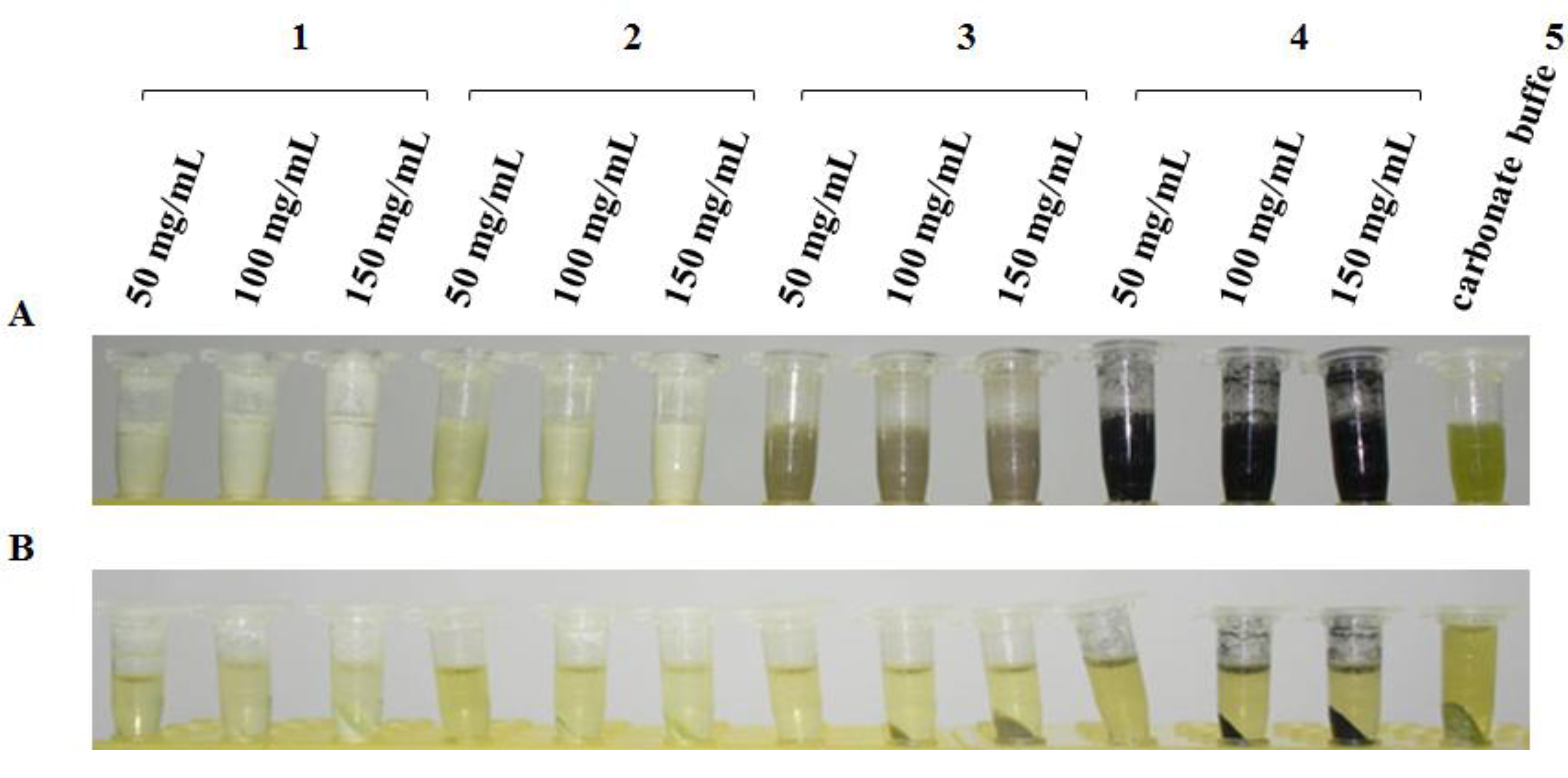
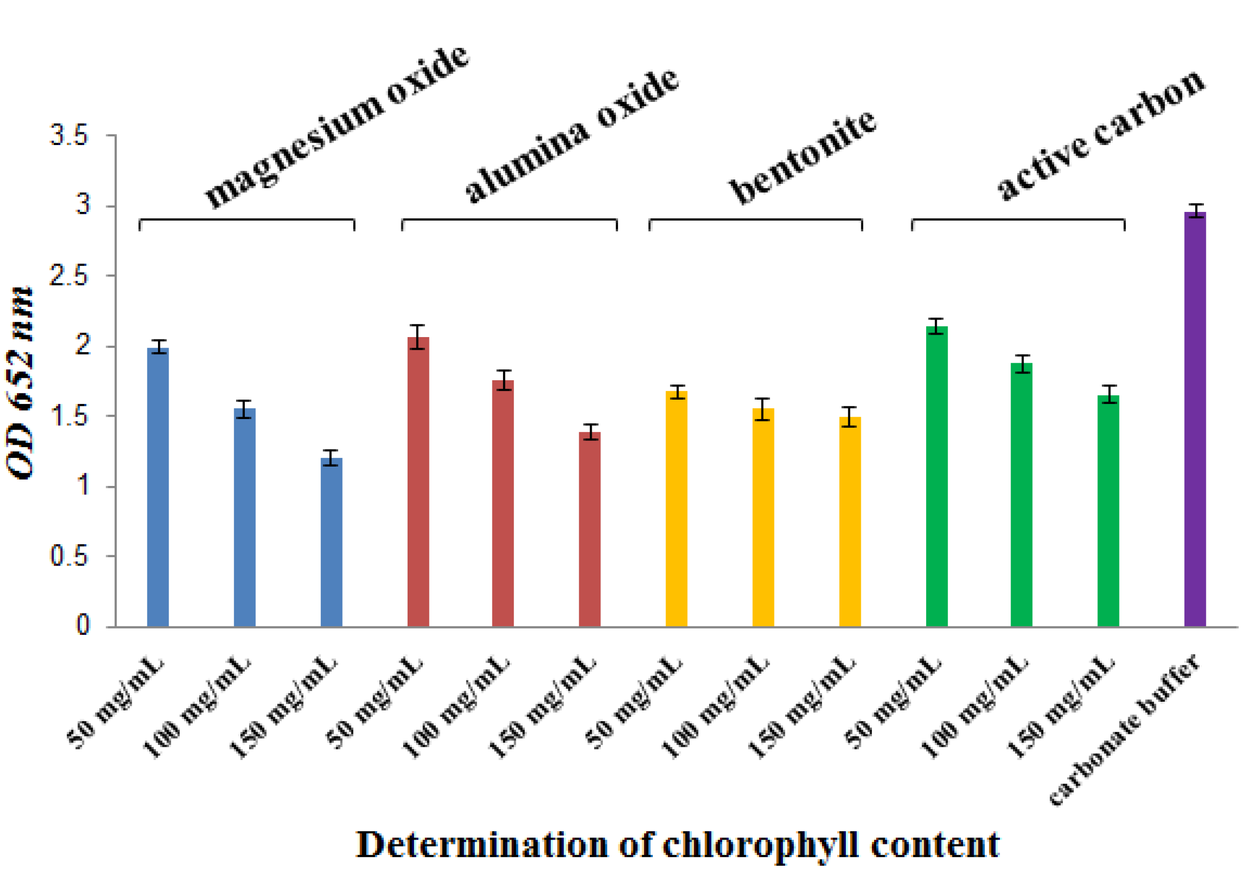
2.1. Chlorophyll Removal from Rice Extracts by Several Reagents


| Reagents | Concentration (mg/mL) | Decreased percent (%) ± SD |
|---|---|---|
| Magnesium oxide | 50 | 67.1 ± 0.044 |
| 100 | 52.3 ± 0.068 | |
| 150 | 40.7 ± 0.051 | |
| Alumina oxide | 50 | 69.8 ± 0.087 |
| 100 | 59.2 ± 0.069 | |
| 150 | 46.8 ± 0.052 | |
| Bentonite | 50 | 56.5 ± 0.041 |
| 100 | 52.2 ± 0.072 | |
| 150 | 50.7 ± 0.069 | |
| Active carbon | 50 | 72.1 ± 0.052 |
| 100 | 63.2 ± 0.064 | |
| 150 | 55.8 ± 0.06 |

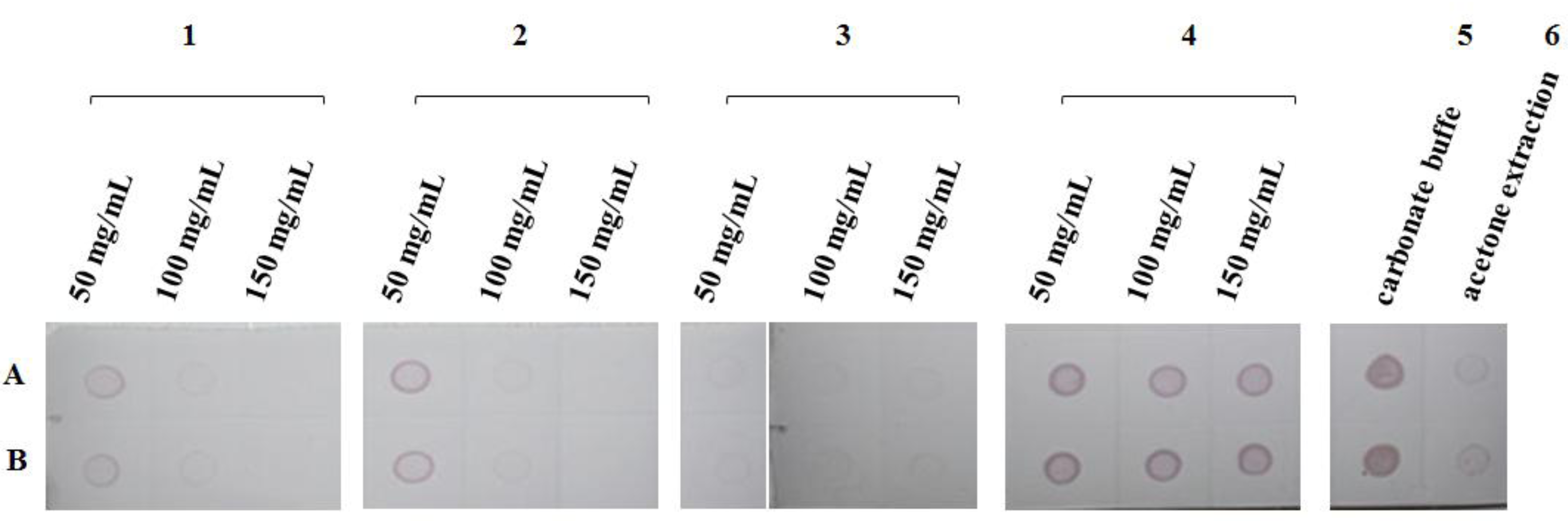
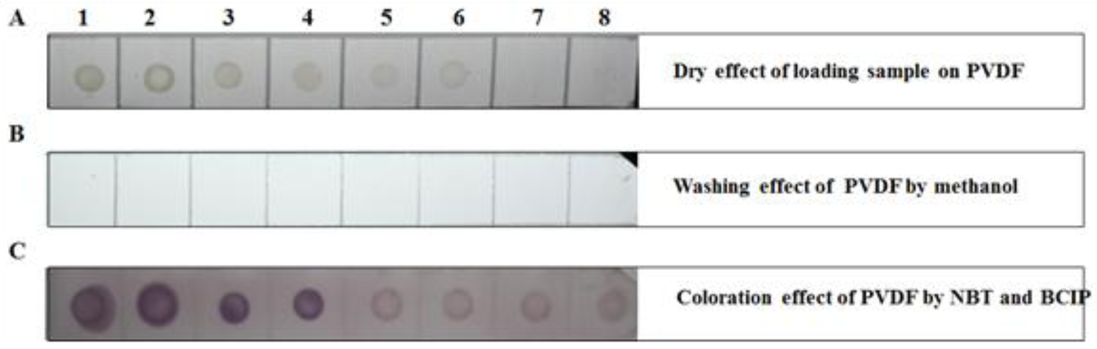
2.2. Results of Chlorophyll Removal from NC Membranes by Washing with Several Organic Solvents

| Organic solvent | Effect | Concentration | |||||
|---|---|---|---|---|---|---|---|
| 5% | 10% | 20% | 30% | 40% | 50% | ||
| Methanol | Chlorophyll removal | Imperceptible | Imperceptible | Imperceptible | Slightly | Slightly | Slightly |
| Membrane destruction | Imperceptible | Imperceptible | Imperceptible | Obvious | Obvious | Obvious | |
| Ethanol | Chlorophyll removal | Imperceptible | Imperceptible | Imperceptible | Slightly | Slightly | Slightly |
| Membrane destruction | Stable | Stable | Stable | Obvious | Obvious | obvious | |
| Acetone | Chlorophyll removal | Imperceptible | Imperceptible | Imperceptible | Imperceptible | Imperceptible | Imperceptible |
| Membrane destruction | Stable | Stable | Stable | Stable | Stable | Stable | |
| Ethyl acetate | Chlorophyll removal | Imperceptible | Imperceptible | Slight | Slight | Slight | Slight |
| Membrane destruction | Slight deformation | Obvious deformation | Slightly dissolved | Medium dissolved | Dissolved | Dissolved | |
| Diethyl ether | Chlorophyll removal | Slight | Slight | Slight | Slightly | Slight | Slight |
| Membrane destruction | Slight deformation | Slight deformation | Slight deformation | Slight deformation | Slight deformation | Slight deformation | |
2.3. Results of Chlorophyll Removal from PVDF Membranes by Washing with Several Organic Solvents
| Organic solvent | Effect | |
|---|---|---|
| Methanol | Chlorophyll removal | Obviously |
| Membrane destruction | Stable | |
| Ethanol | Chlorophyll removal | Obviously |
| Membrane destruction | Stable | |
| Acetone | Chlorophyll removal | Obviously |
| Membrane destruction | Stable | |
| Ethyl acetate | Chlorophyll removal | Obviously |
| Membrane destruction | Stable | |
| Diethyl ether | Chlorophyll removal | Obviously |
| Membrane destruction | Stable | |
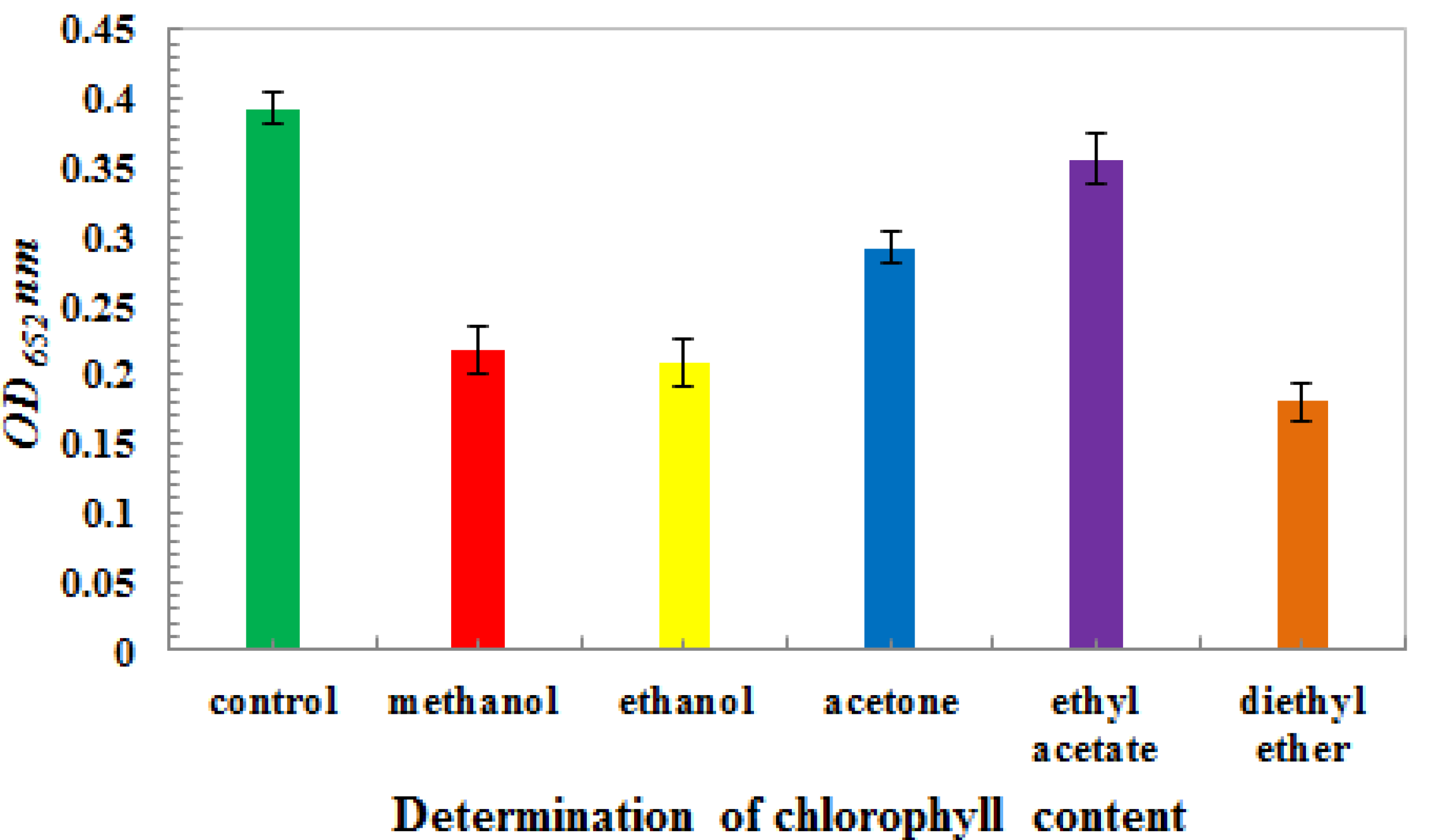
| Concentration (%) | Reduced percent (%) ± SD | |
|---|---|---|
| Methanol | 100 | 54.1 ± 0.017 |
| Ethanol | 100 | 58.2 ± 0.018 |
| Acetone | 100 | 74.3 ± 0.011 |
| Ethyl acetate | 100 | 90.7 ± 0.018 |
| Diethyl ether | 100 | 51.0 ± 0.014 |

3. Experimental
3.1. Materials
| Sample source | Rice cultivars | Sowing date | Year |
|---|---|---|---|
| Indoor growing, artificially infected by Sogatella furcifera | Indica type rice | 2011 | |
| Paddy field, collected from Yuanjiang county, Yunnan province | Indica hybrid rice | Early season rice | 2011 |
| Paddy field, collected from Shidian county, Yunnan province | Indica hybrid rice | Early season rice | 2011 |
| Paddy field, collected from Dushan county, Guizhou province | Indica type rice | Single harvest rice | 2010 |
3.2. Methods
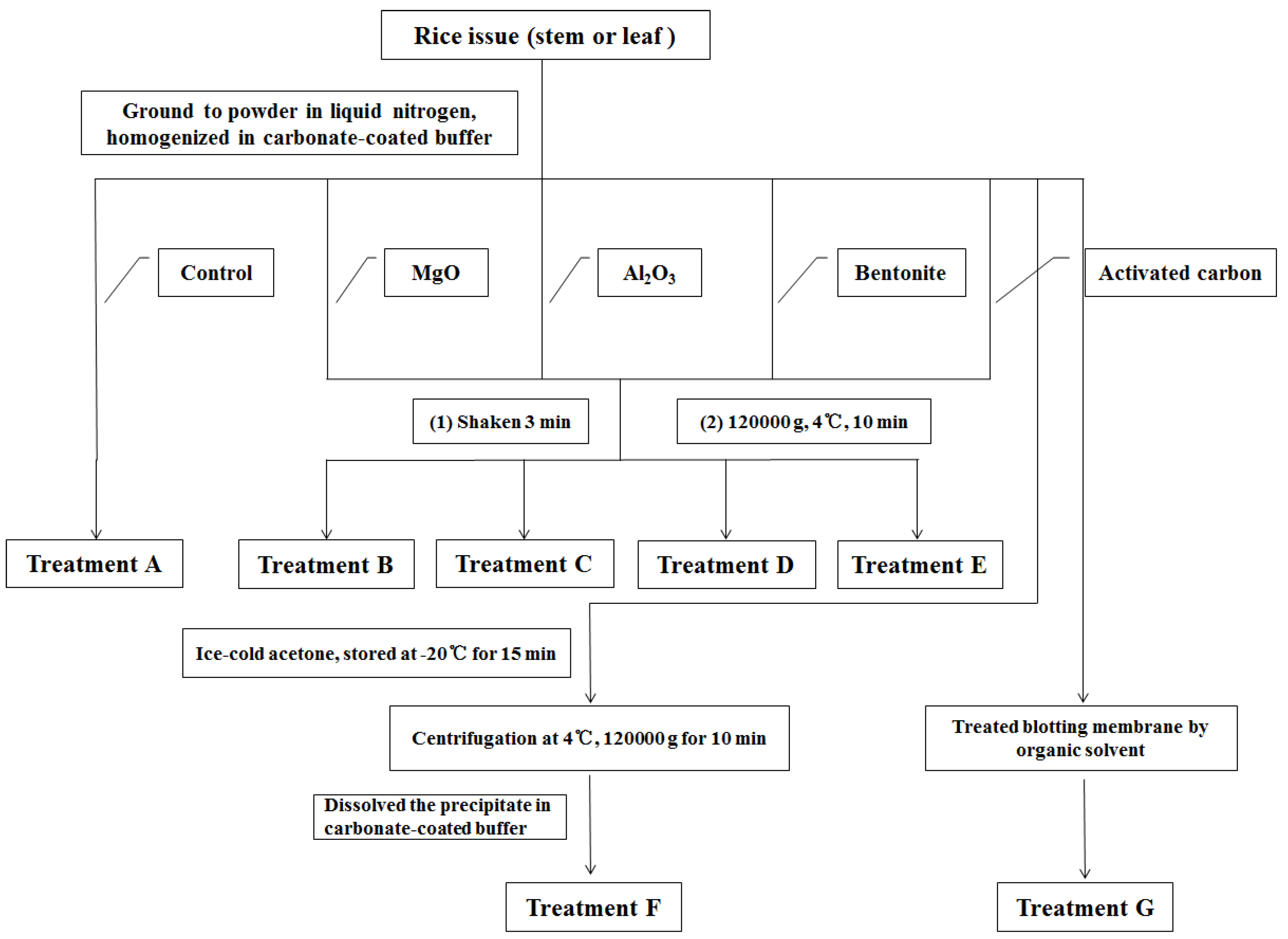
3.2.1. Preparation of Rice Extract
3.2.2. Treatment Methods for Eliminating Chlorophyll
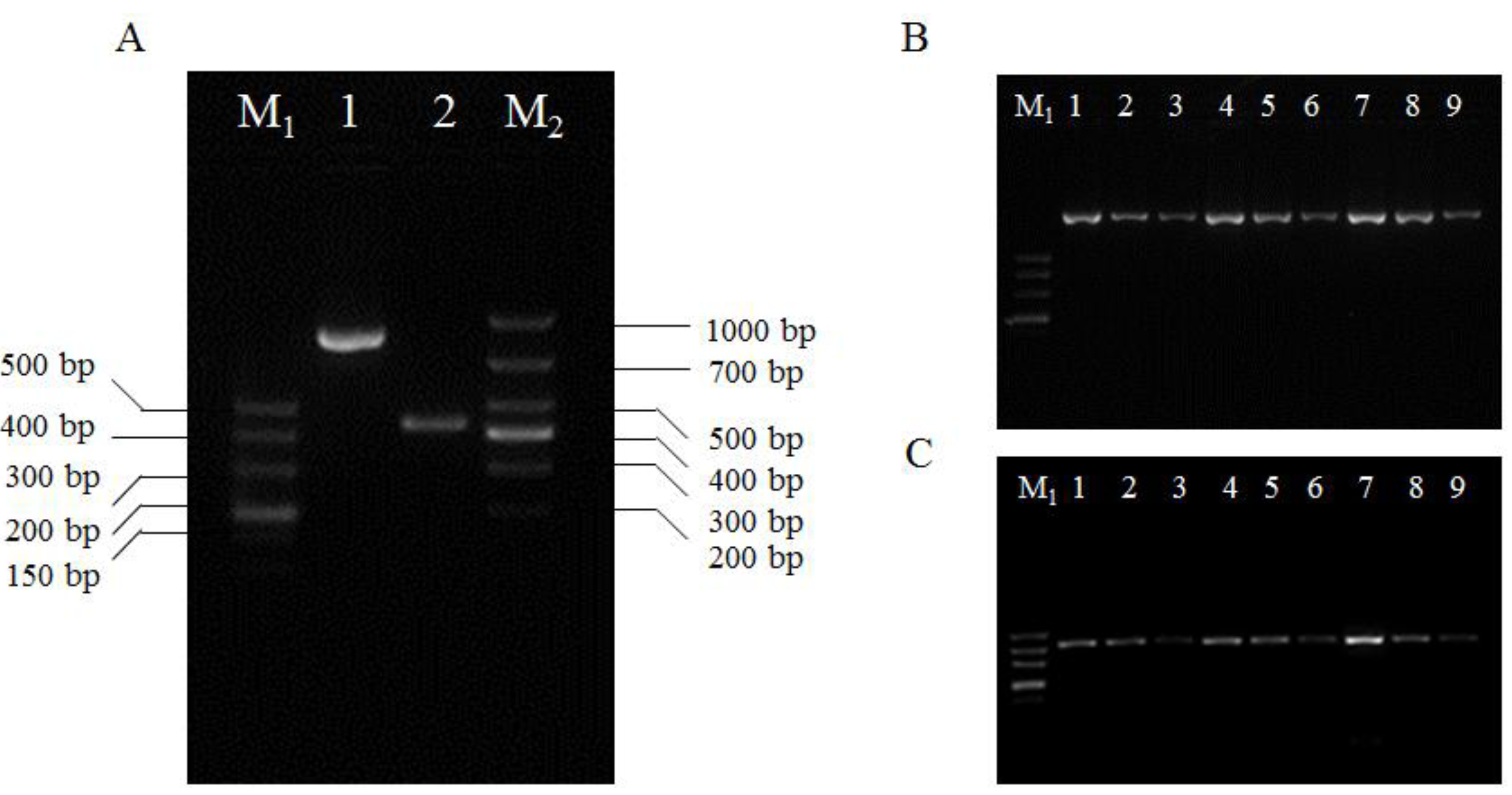
3.2.3. DIBA Test

3.2.4. IC-RT-PCR Demonstration Test
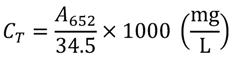
3.2.5. Determination of Chlorophyll Content

3.2.6. Statistical Analysis
4. Conclusions
Acknowledgments
Conflict of Interest
References
- Zhuo, G.H.; Zhang, S.G.; Zou, S.F. The characteristic of the occurrence and the analysis of the trend of the extension of SRBSDV-The new rice disease. Plant Protect. 2010, 36, 235–237. [Google Scholar]
- Zhuo, G.H.; Wen, J.J.; Cai, D.J. A new kind of virus of Feiji genus of Reovirus family: Southern rice black-streaked dwarf virus. Chin. Sci. Ser. B 2008, 53, 2500–2508. [Google Scholar]
- Chen, Z.; Song, B.A. The Technology of Prevention and Control on Southern Rice Black-Streaked Dwarf Virus; Chemical Industrial Press: Beijing, China, 2011; pp. 1–3. [Google Scholar]
- Ha, V.C.; Nguyen, V.H.; Vu, T.M.; Masaru, M. Rice dwarf disease in north Vietnam in 2009 is caused by southern rice black-streaked dwarf virus (SRBSDV). Bull. Inst. Trop. Agric. Kyushu Univ. 2009, 32, 85–92. [Google Scholar]
- Hoang, A.T.; Zhang, H.M.; Yang, J.; Chen, J.P.; Hébrard, E.; Zhou, G.H.; Vinh, V.N.; Cheng, J.A. Identification, characterization, and distribution of southern rice black-streaked dwarf virus in Vietnam. Plant Dis. 2011, 95, 1063–1069. [Google Scholar] [CrossRef]
- Ji, Y.H.; Gao, R.Z.; Zhang, Y.; Chen, Z.B.; Zhou, T.; Fan, Y.J.; Zhou, Y.J. A simplified method for quick detection of rice black streaked dwarf virus and and southern rice black-streaked dwarf virus. Chin. Rice Sci. 2011, 25, 91–94. [Google Scholar]
- Zhang, S.B.; Luo, H.G.; Zhang, Q.D. A dwarf disease on rice in Hubei Province, China is caused by southern rice black-streaked dwarf virus. Chin. Rice Sci. 2011, 25, 223–226. [Google Scholar]
- Wang, Q.; Zhou, G.H.; Zhang, S.G. Detection of Southern rice black-streaked dwarf virus using one-step dual RT-PCR. Acta Phytopathol. Sin. 2012, 42, 84–87. [Google Scholar]
- Zhou, T.; Du, L.; Fan, Y.; Zhou, Y. Reverse transcription loop-mediated isothermal amplification of RNA for sensitive and rapid detection of southern rice black-streaked dwarf virus. J. Virol. Methods 2012, 180, 91–95. [Google Scholar] [CrossRef]
- Hawkes, R.A. Dot-immunobinding assay for monoclonal and other antibodies. Anal. Biochem. 1982, 119, 142–147. [Google Scholar] [CrossRef]
- Pappas, M.G.; Hajkowski, R.; Hockmeyer, W.T. Dot enzyme-linked immunosorbent assay (Dot-ELISA): A micro technique for the rapid diagnosis of visceral leishmaniasis. J. Immunol. Methods 1983, 64, 205–214. [Google Scholar] [CrossRef]
- Bode, L.; Beutin, L.; Kohler, H. Nilrocellulose-enzyme-linked immunosorbent assay (NC-ELISA) -a sensitive technique for the rapid visual detection of both viral antigens and antibodies. J. Virol. Methods 1984, 8, 111–121. [Google Scholar] [CrossRef]
- Hibi, T.; Saito, Y. A dot immunobinding assay for the detection of tobacco mosaic virus in infected tissues. J. Gen. Virol. 1985, 66, 1191–1194. [Google Scholar] [CrossRef]
- Kandana, A.; Ramanathanb, A.; Raguchanderb, T.; Balasubramanianb, P.; Samiyappanb, R. Development and evaluation of an enzyme-linked immunosorbent assay (ELISA) and dot immunobinding assay (DIBA) for the detection of Ganoderma infecting palms. Arch. Phytopathol. Plant Protec. 2010, 43, 1473–1484. [Google Scholar] [CrossRef]
- Sulimenko, T.; Draber, P. A fast and simple dot-immunobinding assay for quantification of mouse immunoglobulins in hybridoma culture supernatants. J. Immunol. Methods 2004, 289, 89–95. [Google Scholar] [CrossRef]
- Salah Zein, H.; Nakazawa, M.; Ueda, M.; Miyatake, K. Development of Serological Procedures for Rapid and Reliable Detection of cucumber mosaic virus with dot-immunobindign assay. World J. Agric. Sci. 2007, 3, 430–439. [Google Scholar]
- Ouyang, Y.L.; Wu, J.X.; Xiong, R.Y. The prokaryotic expression, polyclonal antibody preparation and application of coat protein gene S10 of southern rice black-streaked dwarf virus. Chin. Rice Sci. 2010, 24, 25–30. [Google Scholar]
- Wang, G.Z.; Zhou, Y.J.; Zhou, X.P. Detection of rice stripe virus in Laodelphax striatellus by direct dot immunobinding assay. J. Zhejiang Univ. 2005, 31, 37–40. [Google Scholar]
- Arnon, D.I. Copper enzymes in isolated chloroplasts. Polyphenol oxidase in beta vulgaris. Plant Physiol. 1949, 24, 1–15. [Google Scholar] [CrossRef]
- Gao, J.F. Guide of Plant Physiological Experiment; Higher Education Press: Beijing, China, 2006; pp. 74–77. [Google Scholar]
- Qian, X.H.; Zhou, X.P. The application and technology of dot immunoassay in the study of plant virus. Biotechnology 1994, 4, 39–41. [Google Scholar]
- Rocha-Peña, M.A.; Lee, R.F.; Niblett, C.L. Development of a dot-immunobinding assay for detection of citrus tristeza virus. J. Virol. Methods 1991, 34, 297–309. [Google Scholar] [CrossRef]
- Chen, Z.; Song, B.A. The Technology of Prevention and Control on Southern Rice Black-Streaked Dwarf Virus; Chemical Industrial Press: Beijing, China, 2011; pp. 49–72. [Google Scholar]
- Ma, B.R. SPSS for Windows Ver. 11.5 Application of Statistics in Medicine, 3rd ed; Science Press: Beijing, China, 2005; pp. 180–205. [Google Scholar]
- Porra, R.J. Recent process in porphyrin and chlorophyll biosynthesis. Photochem. Photobiol. 1997, 65, 492–516. [Google Scholar] [CrossRef]
- Khraisheh, M.; Andre, C.; AlGhouti, M. Kinetics of humics removal from water and wastewater using granular activated carbon, iron-coated activated alumina, and beta ferric oxihydroxide. Environ. Eng. Sci. 2010, 27, 387–395. [Google Scholar] [CrossRef]
- Mak, S.M.; Tey, B.T.; Cheah, K.Y.; Siew, W.L.; Tan, K.K. Porosity characteristics and pore developments of various particle sizes palm kernel shells activated carbon (PKSAC) and its potential applications. Adsorption 2009, 15, 507–519. [Google Scholar] [CrossRef]
- Liu, T.; Montastruc, L.; Gancel, F.; Zhao, L.; Nikov, I. Integrated process for production of surfactin-part 1: Adsorption rate of pure surfactin onto activated carbon. Biochem. Eng. J. 2007, 35, 333–340. [Google Scholar] [CrossRef]
- Oubagaranadin, J.U.K.; Sathyamurthy, N.; Murthy, Z.V.P. Evaluation of fuller's earth for the adsorption of mercury from aqueous solutions: A comparative study with activated carbon. J. Hazard. Mater. 2007, 142, 165–174. [Google Scholar] [CrossRef]
- Sample Availability: Samples of the polyclonal antibody against SRBSDV S10 are available from the authors.
© 2012 by the authors; licensee MDPI, Basel, Switzerland. This article is an open-access article distributed under the terms and conditions of the Creative Commons Attribution license (http://creativecommons.org/licenses/by/3.0/).
Share and Cite
Chen, Z.; Liu, J.; Zeng, M.; Wang, Z.; Yu, D.; Yin, C.; Jin, L.; Yang, S.; Song, B. Dot Immunobinding Assay Method with Chlorophyll Removal for the Detection of Southern Rice Black-Streaked Dwarf Virus. Molecules 2012, 17, 6886-6900. https://doi.org/10.3390/molecules17066886
Chen Z, Liu J, Zeng M, Wang Z, Yu D, Yin C, Jin L, Yang S, Song B. Dot Immunobinding Assay Method with Chlorophyll Removal for the Detection of Southern Rice Black-Streaked Dwarf Virus. Molecules. 2012; 17(6):6886-6900. https://doi.org/10.3390/molecules17066886
Chicago/Turabian StyleChen, Zhuo, Jiaju Liu, Mengjiao Zeng, Zhenchao Wang, Dandan Yu, Chengjun Yin, Linhong Jin, Song Yang, and Baoan Song. 2012. "Dot Immunobinding Assay Method with Chlorophyll Removal for the Detection of Southern Rice Black-Streaked Dwarf Virus" Molecules 17, no. 6: 6886-6900. https://doi.org/10.3390/molecules17066886
APA StyleChen, Z., Liu, J., Zeng, M., Wang, Z., Yu, D., Yin, C., Jin, L., Yang, S., & Song, B. (2012). Dot Immunobinding Assay Method with Chlorophyll Removal for the Detection of Southern Rice Black-Streaked Dwarf Virus. Molecules, 17(6), 6886-6900. https://doi.org/10.3390/molecules17066886