Article

t-Bu2SiF-Derivatized D2-Receptor Ligands: The First SiFA-Containing Small Molecule Radiotracers for Target-Specific PET-Imaging

1
Department of Inorganic Chemistry II, Faculty of Chemistry, TU Dortmund, Otto-Hahn-Str. 6, 44221 Dortmund, Germany
2
Department of Nuclear Medicine, Ludwig-Maximilians-University, Marchioninistr. 15, 81377 Munich, Germany
3
Department of Pharmacy, Center for Drug Research, Ludwig-Maximilians-University, Butenandtstr. 7, 81377 Munich, Germany
4
GE Global Research, 85748 Garching, Germany
5
McConnell Brain Imaging Centre, Montreal Neurological Institute, McGill University, 3801 University St., Montreal H3A 2B4, QC, Canada
*
Authors to whom correspondence should be addressed.
Molecules 2011, 16(9), 7458-7479; https://doi.org/10.3390/molecules16097458
Submission received: 28 July 2011 / Revised: 19 August 2011 / Accepted: 31 August 2011 / Published: 2 September 2011
(This article belongs to the Special Issue Fluorine Chemistry 2016)

Abstract

:
The synthesis, radiolabeling and in vitro evaluation of new silicon-fluoride acceptor (SiFA) derivatized D2-receptor ligands is reported. The SiFA-technology simplifies the introduction of fluorine-18 into target specific biomolecules for Positron-Emission-Tomography (PET). However, one of the remaining challenges, especially for small molecules such as receptor-ligands, is the bulkiness of the SiFA-moiety. We therefore synthesized four Fallypride SiFA-conjugates derivatized either directly at the benzoic acid ring system (SiFA-DMFP, SiFA-FP, SiFA-DDMFP) or at the butyl-side chain (SiFA-M-FP) and tested their receptor affinities. We found D2-receptor affinities for all compounds in the nanomolar range (Ki(SiFA-DMFP) = 13.6 nM, Ki(SiFA-FP) = 33.0 nM, Ki(SiFA-DDMFP) = 62.7 nM and Ki(SiFA-M-FP) = 4.21 nM). The radiofluorination showed highest yields when 10 nmol of the precursors were reacted with [18F]fluoride/TBAHCO3 in acetonitrile. After a reversed phased cartridge purification the desired products could be isolated as an injectable solution after only 10 min synthesis time with radiochemical yields (RCY) of more than 40% in the case of SiFA-DMFP resulting in specific activities >41 GBq/µmol (>1,100 Ci/mmol). Furthermore, the radiolabeled products were shown to be stable in the injectable solutions, as well as in human plasma, for at least 90 min.

1. Introduction

Positron-Emission-Tomography (PET) is a non-invasive imaging technique using contrast-agents (radiotracers) labeled with radionuclides such as fluorine-18 which undergo positron emission decay. The resulting positron annihilates with an electron, producing two gamma photons, emitted at a 180° angle, which can be detected in coincidence with high sensitivity, thus yielding a spatial resolution in the mm-range. Although a high number of radiotracers has been developed only a limited number are commonly used as a result of their sometimes cumbersome and difficult synthesis.
The short half-life of many PET-nuclides (18F t½ = 109.7 min) makes it necessary to produce the radiotracer on site, resulting in high investment costs. The effort for a radiotracer synthesis is nearly independent of the number of patient doses produced per synthesis run. Therefore, PET-centers usually focus on the application of well-established radiotracers such as the glucose derivative 18F[FDG](18F-2-fluoro-2-desoxyglucose) and only a few large PET-centers are able to provide a large number of other tracers. To bridge this gap, the development of new labeling techniques for the easy introduction of fluorine-18 into radiotracers without costly equipment would be favorable. A promising approach to simplify the radionuclide introduction significantly is the exploitation of the strong silicon-fluorine (Si–F) bond [1,2]. The silicon fluoride acceptor (SiFA) method, based on the efficient isotopic non-radioactive 19F for radioactive 18F exchange at the silicon atom, was recently developed and applied for simple one- and two-step 18F-fluorinations of peptides [3]. The radiosynthesis of different SiFA-derivatized peptides (RGD-, octreotate-, as well as a bombesin-analogue) resulted in specific activities of up to 680 GBq/µmol (18.4 Ci/µmol) for the final radiotracers, surprisingly high for a carrier added radiosynthesis [4]. This finding can be explained by DFT (density functional theory) model calculations. The most convenient feature of this SiFA labeling technique is that a final HPLC purification of the radiotracer from the precursor is not necessary, since labeling precursor and labeled product are identical. Another approach with Si-18F bearing building blocks was used for the in vivo evaluation of a Si-18F-derivatized bombesin derivative in tumor-bearing rodents [5,6]. However, a heating—as well as an HPLC-purification step—were necessary to obtain the final 18F-labeled compound in high specific activities. Most recently, the implementation of new functionalized SiFAs for the kit-like 18F-labeling of biomolecules was reported [7,8,9,10]. In in vivo studies, the use of SiFA-amounts as low as a few nanomoles resulted in 18F-labeled proteins with specific activities of up to 10–50 GBq/µmol (270–1,350 Ci/mmol), which would be suitable for receptor-imaging with PET. In this particular study, we aimed at evaluating the applicability of the SiFA technique for the derivatization of small molecule radiotracers, such as the D2 receptor ligands fallypride (FP, 1), desmethoxyfallypride (DMFP, 2) and raclopride (3). It was expected that the original SiFA building block, which cannot be extensively modified without losing its stability against hydrolysis, might have a detrimental influence on the binding affinity of the SiFA derivatized D2 receptor ligands. Several new Si-F bearing derivatives derived from basic model compounds were analyzed recently and evaluated as to their stability in aqueous solution with regard to the substitution pattern at the silicon atom [11]. The results are consistent with our previous findings that at least two sterically hindered substituents at the silicon atom are necessary to preserve the stability of the silicon-fluorine bond in vitro [3].
With respect to these steric requirements, SiFA derived model compounds of commonly used PET imaging agents were synthesized to evaluate the potential of the SiFA-concept for the syntheses of SiFA-type small molecule radiotracers. The benzamide derivatives [18F]fallypride (1), [18F]-desmethoxyfallypride (2) and [11C]raclopride (3) radiotracers used for the PET-imaging of the dopaminergic system, were chosen as model compounds for this study [12,13] (Figure 1). All compounds are D2-receptor antagonists, which differ mainly in the receptor affinity, in the nanomolar (desmethoxyfallypride, raclopride) and picomolar range (fallypride), respectively [14]. These imaging agents are used for the diagnosis of different neurological disorders related to the dopaminergic system such as parkinsonism and craving [15,16].
Figure 1. D2-receptor affine benzamide-derivatives used for PET-neuroimaging: [18F]-Fallypride (FP, 1); [18F]-Desmethoxyfallypride (DMFP, 2); [11C]-Raclopride (3).
Molecules 16 07458 g001
Due to the bulkiness of the SiFA-moiety that has to be introduced into the potential radiotracer, the ligand fallypride, having one of the highest affinities to the D2 receptor, was tested first as a scaffold for SiFA derivatization since even a certain loss of target affinity would not necessarily result in an unusable PET radiotracer. The two different strategies applied were: (i) the integration of the SiFA building block into the fallypride/desmethoxyfallypride general structure and (ii) the coupling of an already existing SiFA compound, namely SiFA-maleimide (SiFA-M, [17]), to a fallypride derivatized with an SH moiety at the butyl side chain (Figure 2). Besides the prerequisite of a good binding affinity to the targeted D2-receptor, the radiolabeling has to yield a radiotracer with a sufficiently high specific activity for D2 receptor imaging with PET. In order to simplify the radiosynthesis of the desired 18F fluorinated radiotracer, we only studied one-step radiosyntheses using the non-radioactive standards directly as the labeling precursors (isotopic exchange reaction). Hence, the amount of the precursor used determines the specific activity (in relation to the amount of radioactivity used for labeling).
Figure 2. Synthesized SiFA-conjugated Fallypride-derivatives 4a–c, 5.
Molecules 16 07458 g002
The use of 10 nanomoles precursor would result in specific activities comparable to those of conventionally synthesized 18F radiotracers. If, e.g., 500 MBq (13.5 mCi) 18F are incorporated into 10 nmol of the labeling precursor the resulting specific activity would be as high as 50 GBq/µmol (1,350 Ci/mmol). Consequently, the identification of the lowest limit of the ratio precursor amount/concentration necessary for a sufficient radiochemical yield (introduction of fluorine-18) was the second important focus of this work.

2. Results and Discussion

2.1. Precursor Syntheses

The preparation of the SiFA-modified carboxylic acids 12a and 12b (Scheme 1) followed a general method described in our previous work [17]. Single crystals of compounds 10b, 12a and 12b suitable for X-ray diffraction analysis were obtained as colorless needles by re-crystallization from diethyl ether/hexane. The molecular structures of these compounds are presented in Figure 3, Figure 4, Figure 5, and selected bond distances and bond angles are collected in Table 1.
All compounds crystallized monoclinically with eight (10b) or four (12a, 12b) molecules in the unit cell. There are two crystallographically independent molecules in the unit cell of compound 10b (Figure 3) with one of them being disordered. In Table 1, only the data for the non-disordered molecule are given. The silicon atoms in these compounds are four-coordinate and show each a distorted tetrahedral configuration with average angles of 109.59 (10b), 109.70 (12a) and 109.04 (12b). The largest deviations from the tetrahedral angle are found for C(11)–Si–C(15) (119.40°, 12b) and F–Si–C(11) (104.15°, 12a).
The Si-F distances are similar and fall in the range between 1.6033(11) (12b) and 1.6133(14) (10b) Å. They are slightly longer as compared to t-BuPh2SiF (1.6004(10) Å) but close to the Si–F distances in other SiFA-compounds [17]. Interestingly, intramolecular O(1)–HLO(11A) and O(11)–HLO(1) hydrogen bridges (Table 1) link the molecules of compound 10b to form a one-dimensional polymer chain as presented in Figure 3. A noteworthy feature is the asymmetric intramolecular O(1)–HLO (2A) hydrogen bridge that links two molecules of 12a and 12b, respectively, to give a dimer (Table 2). Such hydrogen bridges are common for solid state structures of p-silylarylcarbonic acids [17].
Scheme 1. Synthesis of the silicon-modified carboxylic acids 12a and 12b.
Molecules 16 07458 g006
Figure 3. Molecular structure of 10b (left) and simplified representation of the intermolecular hydrogen bridges (right). Atomic displacement parameters are drawn at 30% probability level.
Molecules 16 07458 g003
Figure 4. Molecular structure of 12a. Atomic displacement parameters are drawn at 30% probability level.
Molecules 16 07458 g004
Figure 5. Molecular structure of 12b. Atomic displacement parameters are drawn at 30% probability level.
Molecules 16 07458 g005
Table 1. Selected bond lengths [Å] and angles [°] for 10b, 12a, 12b.
Bond lengths [Å]10b12a12b
Si(1)-F(1)1.6133(13)1.6125 (10)1.6033 (11)
Si(1)-C(1)1.8712(19)1.8696 (15)1.8695 (17)
Si(1)-C(11)1.893(2)1.8846 (16)1.872 (2)
Si(1)-C(15)1.888(2)1.8911 (17)1.875 (2)
Bond angles [°]
F(1)-Si(1)-C(1)104.78(8)105.12 (6)104.67 (8)
F(1)-Si(1)-C(11)104.97(9)104.15 (7)105.61 (8)
F(1)-Si(1)-C(15)105.23(9)105.20 (6)104.81 (8)
C(1)-Si(1)-C(11)112.55(9)112.36 (7)109.04 (9)
C(1)-Si(1)-C(15)109.59(9)109.70 (7)111.99 (9)
C(11)-Si(1)-C(15)118.44(10)118.96 (7)119.40 (10)
Table 2. Hydrogen-bonding geometry [Å, °].
CompoundD-H...A d(D-H)d(H...A)d(D...A)<(DHA)
10bO(1)-H(1)...O(11A) 0.82(3)1.87(3)2.682(2)168(2)
10bO(11)-H(11)...O(1)0.83(3)1.86(3)2.695(2)178(3)
12aO(1)-H(1)...O(2A) 0.80(2)1.84(2)2.641(2)173(2)
12bO(1)-H(1)...O(2A) 1.22(4)1.42(4)2.640(2)177(3)
The test reaction for the coupling of carboxylic acids to (S)-(1-allylpyrrolidin-2-yl)methanamine was performed according to a literature procedure [12], using the carboxylic acid 12c [17] (Scheme 2). Dicyclohexylcarbodimide (DCC) was used for the activation of the carboxylic acid and the fallypride-derivative was obtained in poor yield of 11%. In order to achieve a higher yield, the more powerful activating agent N-hydroxysuccinimide (HO-Su) was added to the reaction mixture (Scheme 3). SiFA-DMFP 4a and SiFA-FP 4b were obtained in yields of up to 41% after purification via column chromatography. Both compounds were characterized via two-dimensional NMR spectroscopy and high resolution mass spectrometry. In the latter, the molecular peak was found with high precision.
Scheme 2. Coupling reaction of the SiFA-carboxylic acid 12c to the (S)-(1-allyl-pyrrolidin-2-yl)methyl-amine.
Molecules 16 07458 g007
Scheme 3. Coupling reaction of the SiFA-carboxylic acids to the (S)-(1-allyl-pyrrolidin-2-yl)methyl-amine in the presence of N-hydoxy-succinimide.
Molecules 16 07458 g008
SiFA-M (1-(4-(di-tert-butylfluorosilyl)phenyl)-1H-pyrrole-2,5-dione) was synthesized according to a recently described procedure [17] and reacted with FP-Thiol [(S)-N-((1-allylpyrrolidin-2-yl)methyl)-5-(3-mercaptopropyl)-2,3-dimethoxybenzamide] in a water/acetonitrile mixture at pH 7.2. This Michael-addition was monitored via analytical HPLC and was completed within 10 min at ambient temperature. After semipreparative HPLC purification and lyophilization, the product SiFA-M-FP, 5, was isolated in 49% yield and identified via ESI-MS and NMR spectroscopy.

2.2. In Vitro Evaluation

Compounds 4a–c and 5 (SiFA-DMFP 4a, SiFA-FP 4b, SiFA-DDMFP 4c and SiFA-M-FP 5) were tested for their affinity towards the human D2 receptor (Table 3). All tested compounds showed Ki-values in the nanomolar range. As expected for the derivatization of a small molecule with the bulky SiFA-moiety, the affinities are reduced by factors ranging between 22 [SiFA-DMFP 4acompared to desmethoxyfallypride (2)], 44 [SiFA-M-FP 5 compared to fallypride (1)] and 342 [SiFA-FP 4b compared to fallypride (1)]. Interestingly, SiFA-DMFP (4a) displays a higher affinity to the receptor compared to SiFA-FP (4b) and SiFA-DDMFP (4c). Among the newly developed substances 4a–c and 5, SiFA-M-FP (5) displays the highest binding affinity which might be explained with the higher distance of the derivatization site (SiFA is bound to the butyl side chain) to the receptor binding parts of the molecule. The methoxy moiety, which distinguishes the high affinity tracer fallypride (1) from the medium-affinity tracer desmethoxyfallypride (2) leads to a loss of binding affinity in the pair SiFA-DMFP 4a/SiFA-FP 4b inversely to the situation with fallypride (1)/desmethoxyfallypride (2). We can therefore assume that the benzoic acid ring system still influences the binding affinity but binds most probably in a different conformation to the binding site at the D2-receptor. However, we chose fallypride (1) as a scaffold for this study, because even an unavoidable decrease in binding-affinity towards the D2-receptor might still lead to a medium affinity ligand comparable to raclopride (3—the gold standard for D2-receptor imaging with PET). Therefore, the direct comparison of SiFA-M-FP (5) to [11C]raclopride [11C]-3, which is most frequently used in clinical studies of the dopaminergic system, looked more encouraging: When comparing the medium-affinity D2-receptor radiotracer raclopride (Ki = 1.21 nM) to SiFA-M-FP (5; Ki = 4.21) and SiFA-DMFP (4a; Ki = 13.6 nM) the affinity is only reduced by the factor 3.5 and 11, respectively. Thus, a successful in vivo D2 receptor imaging using radiolabeled SiFA-M-FP (5) or SiFA-DMFP (4a) might be possible.
Table 3. D2-receptor affinities of the developed substances 4a–c, 5.
CompoundKi (nM ± SEM *)
Fallypride0.0965 ± 0.0153
Desmethoxyfallypride0.630 ± 0.089
Raclopride1.21 ± 0.43
SiFA-DMFP, 4a13.6 ± 4.3
SiFA-FP, 4b33.0 ± 7.6
SiFA-DDMFP, 4c62.7 ± 16.9
SiFA-M-FP, 54.21 ± 0.41
* SEM: standard error of the mean.

2.3. Radiolabeling

Radiolabeling reactions based on isotopic exchange usually have the drawback of a limited specific activity (SA), as the precursor cannot be separated from the final product. It is therefore crucial for in vivo applications of the synthesized radiotracer to reduce the precursor amount to the absolute minimum. Our previous studies showed that for radiofluorinations of small molecules such as a SiFA-aldehyde, amounts of 1–5 nmol were sufficient to achieve high radiolabeling yields [4]. For the direct labeling of SiFA-derivatized peptides, precursor amounts of 10–25 nmol were necessary [18]. The highest radioactivity incorporation rates were observed when the SiFA radiolabeling was carried out in polar aprotic solvents such as acetonitrile or DMSO. As the SiFA-moiety gets hydrolyzed very quickly under basic conditions, we tested the following two mild labeling procedures in acetonitrile, DMF or DMSO: (a) Kryptofix2.2.2/potassium oxalate/18F and (b) tetrabutylammonium bicarbonate/18F. Table 4 summarizes the labeling of SiFA-DMFP (4a) under different labeling conditions.
To keep the labeling procedure as simple and convenient as possible, we performed the purification step by using a reversed phase cartridge (SepPak C-18 light) for all tested combinations. We determined the 18F-incroporation of 18F into SiFA-DMFP (4a) after a 5 min reaction time at room temperature with analytical radio-HPLC using samples of the crude reaction mixture. After dilution with 10 mL 1M HEPES buffer at pH = 4.0 (used to prevent hydrolysis) the desired radiotracer was purified using a reversed-phased SepPak-cartridge. The SepPak purification was necessary to remove unreacted 18F, solvent and K2.2.2/potassium oxalate or TBAHCO3 from the product. After washing the cartridge with water, the products could be eluted very efficiently with 1 mL ethanol and were subsequently diluted with isotonic saline. The radiochemical yields (RCY) were calculated to the start of the synthesis and the radiochemical purity (RCP) analyzed by analytical radio-HPLC. Using SiFA-DMFP (4a) as the precursor, we found radioactivity incorporations in acetonitrile using TBAHCO3 of up to 61% in the crude reaction mixture (Table 4) and an overall RCY after purification of up to 42%. Under these conditions, the specific activity was calculated to be in the range of 88 GBq/µmol (2.4 Ci/µmol) using a precursor amount of 5 nmol and at least 41 GBq/µmol (1.1 Ci/µmol) using 10 nmol precursor when starting from ~1GBq (27 mCi) 18F-fluoride. This is in the suitable range for D2-neuroreceptor imaging with PET. The radiochemical purity of all products was >96% after purification. Using DMF or DMSO, the 18F-radioactivity incorporation was drastically reduced to less than 25%. More importantly, the desired product could not be purified via cartridge separation. Likewise, the labeling procedure using K2.2.2 resulted in a lower relative 18F-incorporation as well as a lower RCY compared to the procedure using TBAHCO3.
Table 4. Radiolabeling of 5–10 nmol SiFA-DMFP (4a), 5 min at ambient temperature.
Labeling method (solvent)Amount of precursor18F-incorporation ± SD *RCY ± SD *RCP **
(a) K2.2.2 *** (acetonitrile)10 nmol25.3 ± 2.5%16.7 ± 1.4%>96%
(b) TBAHCO3 (acetonitrile)10 nmol60.7 ± 3.1%41.8 ± 5.7%>96%
(b) TBAHCO3 (acetonitrile)5 nmol57.2 ± 6.2%34.6 ± 5.6%>96%
(b) TBAHCO3 (DMSO)10 nmol10.2 ± 0.3%n.d.<60%
(b) TBAHCO3 (DMF)10 nmol21.4 ± 4.5%n.d.<75%
* SD: standard deviation, all experiments were independently performed at least three times; ** after cartridge purification. *** K2.2.2 = Kryptofix2.2.2.
Using these labeling conditions optimized for [18F]-SiFA-DMFP [18F]-4a, we found comparable radiolabeling yields and purities for [18F]-SiFA-FP ([18F]-4b) and [18F]-SiFA-DDMFP ([18F]-4c), whereas the radiolabeling yields for SiFA-M-FP ([18F]-5) were significantly lower (Table 5).
Table 5. Radiolabeling of 4a–c and 5, 10 nmol, labeling method (b) in acetonitrile.
Compound18F-incorporation ± SD *RCY ± SD *RCP
SiFA-DMFP, 4a60.7 ± 3.1%41.8 ± 5.7%>96%
SiFA-FP, 4b60.8 ± 2.5 %47.3 ± 5.9%>94%
SiFA-DDMFP, 4c54.2 ± 6.0%48.4 ± 0.7%>97%
SiFA-M-FP, 516.6 ± 10.2%n.d.<50%
* SD: standard deviation, all experiments were independently performed at least three times.
Moreover, radiolabeled [18F]-SiFA-M-FP [18F]-5 could not be purified via the SepPak separation method described above. We can only speculate that the chemical design of the phenolic ring system of SiFA-maleimide leads to a significantly different stability of the SiFA-moiety compared to the benzamides 4a–c. This finding is in line with our previous observation that protein labeling using [18F]-SiFA-thiol bound to a maleimide-derivatized protein resulted in higher radiolabeling yields than a vice versa labeling of a thiol-derivatized protein with [18F]-SiFA-maleimide.
The chemical purity was determined for all products by analytical HPLC at 214 nm and showed no side products for all radiolabeled derivative swith a RCP > 94%. The chemical stability of [18F]-SiFA-DMFP ([18F]-4a), SiFA-FP ([18F]-4b) and SiFA-DDMFP ([18F]-4c) was determined over 4 h at room temperature by analytical radio-HPLC. Within this time span, no decomposition was detected. The plasma stability was determined for SiFA-DMFP ([18F]4a) over 90min displaying no degradation of the product.

3. Experimental

3.1. General

All solvents used for the syntheses of 4a–c (SiFA-DMFP, SiFA-FP, SIFA-DDMFP) were purified by distillation from appropriate drying agents under argon atmosphere. Solvents and chemicals used in the synthesis of 5, SiFA-M-FP, were of analytical grade and used without further purification. Chemicals and solvents used for the labeling experiments were purchased in the highest available grade and were used without further purification. The NMR experiments were carried out with Jeol AS500, Bruker DRX 400, Bruker DRX 300, and Varian Mercury 200 spectrometers. Chemical shifts (δ) are given in ppm and are referenced to the solvent peaks, with the usual values calibrated against tetramethylsilane (1H, 13C, 29Si) and CFCl3 (19F). High-resolution mass spectra were obtained by using a Finnigan MAT95Q mass spectrometer and a LTQ Orbitrap mass spectrometer (Thermo Electron) using acetonitrile as the mobile phase. FT infrared spectra were recorded using a Bruker IFS 28 spectrometer. Elemental analyses were performed on a LECO CHNS-932 analyzer. The analytical and semi-preparative HPLC system used was an Agilent 1200 system equipped with a raytest Gabi Star radioactivity detector together with a Chromolith Performance (RP-18e, 100–4.6 mm, Merck, Germany) and a Chromolith (RP-18e, 100–10 mm, Merck, Germany) column, respectively. SiFA-M was synthesized according to a published procedure [17]. The synthesis of the thiol-substituted fallypride will be described elsewhere.

3.2. Crystallography

Crystals of compounds 10b, 12a and 12b suitable for single-crystal X-ray diffraction analyses were grown by re-crystallization from diethyl ether/n-hexane. Crystallographic data are summarized in Table 1. Intensity data were collected with a Nonius KappaCCD diffractometer with graphite-monochromated Mo-Kα radiation. The data collections covered almost the whole sphere of the reciprocal space with 3 (5), 4 (7 and 8b) sets at different κ angles and 227 (5), 339 (7), 494 (8b) frames by ω-rotation (Δ/ω = 1°) at 2 × 160 s (5), 80 s (7), 60 s (8b) per frame. Crystal decay was monitored by repeating the initial frames at the end of the data collection. After analysis of the duplicate reflections, there was no indication of any decay. The structures were solved by direct methods (SHELXS97 [19]). Refinement applied full-matrix least-squares methods (SHELXL97). All H atoms were located in the difference Fourier map and their positions were isotropically refined with Uiso constrained at 1.2 times Ueq of the carrier C atom for non-methyl and 1.5 times Ueq of the carrier C atom for methyl groups. Atomic scattering factors for neutral atoms and real and imaginary dispersion terms were taken from International Tables for X-ray Crystallography [20]. The figures were created by SHELXTL. Crystallographic data are given in Table 6. Crystallographic data for the structures reported in this paper have been deposited with the Cambridge Crystallographic Data Centre as commentary material publication No. CCDC-704771, CCDC-704583 and CCDC-704772. Copies of the data can be obtained free of charge on application to CCDC, 12 Union Road, Cambridge, CB2 1EZ, UK (Fax: +44 1223 336033; Email: [email protected] ).
Table 6. Crystallographic data for 10a, 12a and 12b.
10b12a12b
Empirical formulaC17H29FO3SiC16H25FO3SiC17H27FO4Si
Formula mass [g/mol]328.49312.45342.48
Crystal systemmonoclinicmonoclinicmonoclinic
Crystal size0.44 × 0.28 × 0.100.40 × 0.28 × 0.200.28 × 0.24 × 0.20
Space groupP21/cP21/nP21/c
a [Å]8.0498(3)6.91101(14)12.866(3)
b [Å]35.1976(15)16.975(3)8.1150(16)
c [Å]13.5997(7)14.529(3)19.412(4)
β [°]102.644(2)94.46(3)108.51(3)
V [Å3]3759.8(3)1699.3(6)1921.9(7)
Z844
ρcalcd.[mg/m3]1.1611.2211.184
μ [mm−1]0.1430.1550.146
F(000)1424672736
θ range [°]1.64–27.792.78–27.482.74–27.48
Index ranges−16 ≤ h ≤ 17−8 ≤ h ≤ 8−15 ≤ h ≤ 15
−44 ≤ k ≤ 46−22 ≤ k ≤ 22−10 ≤ k ≤ 10
−8 ≤ l ≤ 10−18 ≤ l ≤ 18−25 ≤ l ≤ 25
No. of reflections collected261131795117285
Completeness of θmax[%]97.399.899.8
No. of independent reflections / Rint.8677/0.0373872/0.0224401/0.022
No. of reflections observed with [I > 2σ(I)]614023891602
No. of refined parameters443200211
GoF(F2)1.0310.9000.729
R1(F) [I > 2σ(I)]0.05170.03620.0364
wR2(F2) (all data)0.14090.09240.0905
(Δ/σ)max0.0010.0000.000
Largest differnce peak/hole [e / Å3]0.448/−0.2510.199/−0.2370.106/−0.171

3.3. Chemistry

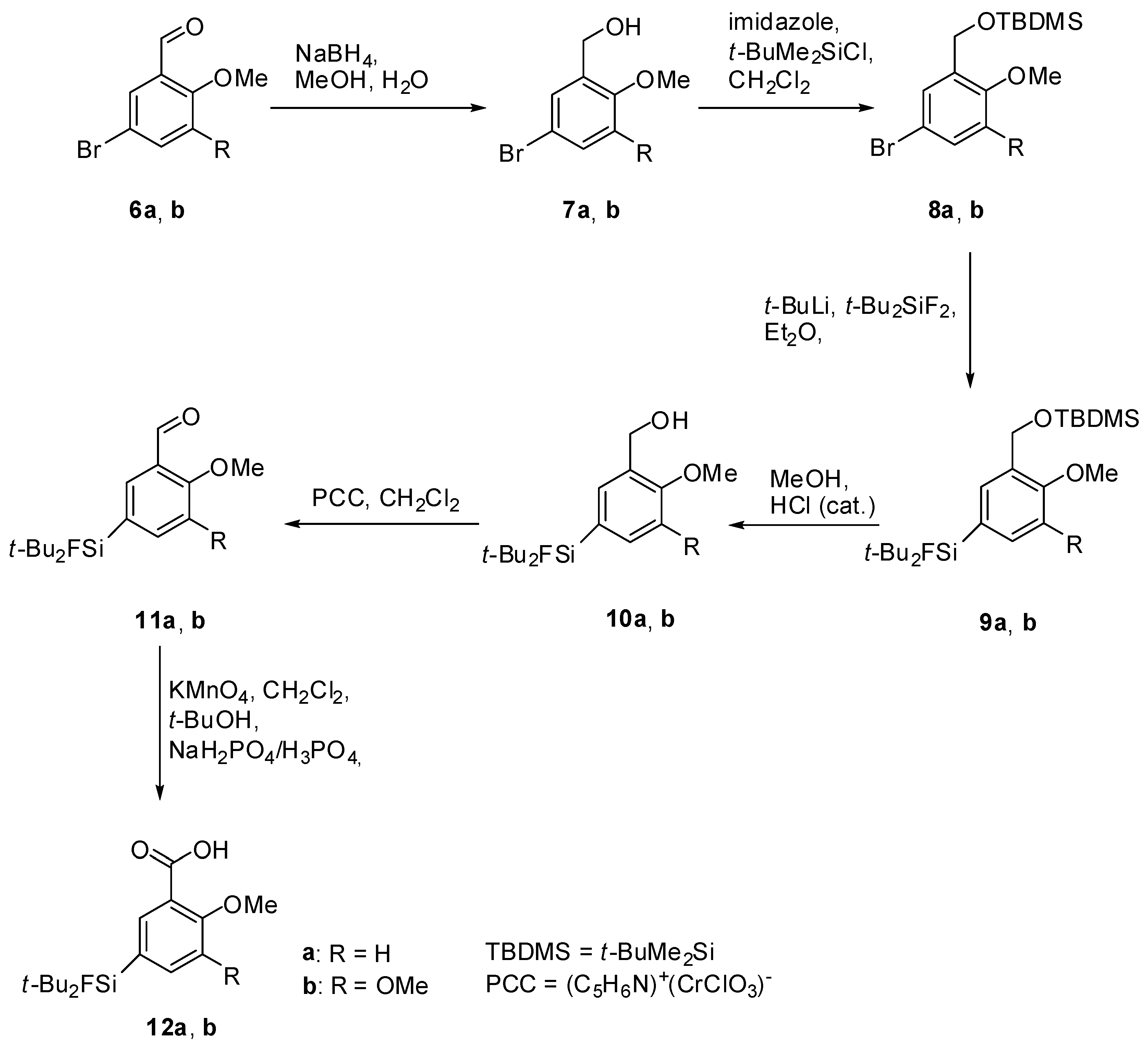
(5-Bromo-2-methoxyphenyl)methanol (7a). A solution of sodium borohydride (0.94 g, 24.8 mmol, 1.0 equiv.) in H2O (30 mL) was added to a solution of the benzaldehyde derivative 6a (5.33 g, 24.8 mmol) in methanol (200 mL). After stirring at room temperature for 24 h, methanol was evaporated and the aqueous residue was diluted with diethyl ether (200 mL). The aqueous phase was extracted with diethyl ether (3 × 100 mL), the combined organic layers were dried with MgSO4, filtered and the filtrate was evaporated to afford 7a (5.06 g, 23.31 mmol, 94%) as colourless oil. 1H-NMR (400.13 MHz, CDCl3): δ (ppm) = 7.39 (d, 4J(1H-1H) = 2.4 Hz, 1H, C(6)H), 7.33 (dd, 3J(1H-1H) = 8.8 Hz, 4J(1H-1H) = 2.4 Hz, 1H, C(4)H), 6.72 (d, 3J(1H-1H) = 8.8 Hz, 1H, C(3)H), 4.61 (s, 2H, CH2), 3.81 (s, 3H, OCH3), 2.35 (s, 1H, OH).
(5-Bromo-2,3-dimethoxyphenyl)methanol (7b). The procedure was analogous to the synthesis of 7a. The benzaldehyde derivative 6b (5.35 g, 21.83 mmol) gave alcohol 7b (5.04 g, 20.40 mmol, 93%) as a colourless oil. 1H-NMR (200.13 MHz, CDCl3): δ (ppm) = 7.05 (d, 4J(1H-1H) = 2.3 Hz, 1H, C(4)H), 6.93 (d, 4J(1H-1H) = 2.3 Hz, 1H, C(6)H), 4.59 (d, 3J(1H-1H) = 5.6 Hz, 2H, CH2), 3.80 (s, 3H, OCH3), 3.78 (s, 3H, OCH3), 2.93 (t, 3J(1H-1H) = 5.6 Hz, 1H, OH).
(5-Bromo-2-methoxybenzyloxy)(tert-butyl)dimethylsilane (8a). To a solution of 7a (5.00 g, 23.00 mmol) in dichloromethane (150 mL) imidazole (2.03 g, 29.89 mmol, 1.3 equiv.) was added and the suspension was stirred at ambient temperature for 10 min. t-Butyldimethylchlorosilane (t-BuMe2SiCl, 4.16 g, 27.59 mmol, 1.2 equiv.) was added and stirring was continued for 20 h. For work-up the mixture was diluted with H2O (40 mL) and the aqueous phase was extracted with dichloromethane (3 × 50 mL). The combined organic layers were dried with MgSO4, filtered and the solvent was evaporated to afford 8a (7.10 g, 21.43 mmol, 93%) as a red oil. 1H-NMR (400.13 MHz, CDCl3): δ (ppm) = 7.57 (d, 4J(1H-1H) = 2.5 Hz, 1H, C(6)H), 7.29 (dd, 4J(1H-1H) = 2.5 Hz, 3J(1H-1H) = 8.6 Hz, 1H, C(4)H), 6.66 (d, 3J(1H-1H) = 8.6 Hz, 1H, C(3)H), 4.71 (s, 2H, CH2), 3.77 (s, 3H, OCH3), 0.97 (s, 9H, CCH3), 0.13 (s, 6H, SiCH3). 13C[1H]-NMR (100.63 MHz, CDCl3): δ (ppm) = 154.9 (s, C(2)), 132.2 (s, C(6)), 130.0 (s, C(4)), 129.5 (s, C(1)), 113.0 (s, C(3)), 111.1 (s, C(5)), 59.7 (s, CH2), 55.3 (s, OCH3), 26.0 (s, CCH3), 18.5 (s, C(CH3)3, −5.3 (s, SiCH3).
(5-Bromo-2,3-dimethoxybenzyloxy)(tert-butyl)dimethylsilane (8b). The procedure was analogous to the synthesis of 8a. The benzyl alcohol derivative 7b (4.93, 19.95 mmol) gave the silylated alcohol 8b (5.88g, 16.27 mmol, 82%) as a slightly yellowish oil. 1H-NMR (400.13 MHz, CDCl3): δ (ppm) = 7.08 (d, 4J(1H-1H) = 2.3 Hz, 1H, C(4)H), 6.82 (d, 4J(1H-1H) = 2.2 Hz, 1H, C(6)H), 4.62 (s, 2 H, CH2), 3.72 (s, 3H, OCH3), 3.67 (s, 3H, OCH3), 0.83 (s, 9H, CCH3), 0.00 (s, 6H, SiCH3).
Di-tert-Butyl(3-((tert-butyldimethylsilyloxy)methyl)-4-methoxyphenyl)fluorosilane (9a). To a stirred solution of 8a (5.00 g, 15.09 mmol) in dry diethyl ether (150 mL), t-BuLi (17.8 mL, 1.7 mol/L, 30.18 mmol, 2.0 equiv.) was added dropwise at −78 °C. After stirring for 10 min t-Bu2SiF2 (3.00 g, 16.60 mmol, 1.1 equiv.) was added and stirring was continued for 19 h while the reaction mixture was allowed to warm to ambient temperature. The mixture was washed with H2O (50 mL) and the aqueous phase was extracted with diethylether (3 × 50 mL). The combined organic layers were dried with MgSO4, filtered and the solvent was evaporated to afford 9a (5.77 g, 13.98 mmol, 93%) as a colourless oil. 1H-NMR (300.13 MHz, CDCl3): δ (ppm) = 7.64 (s, 1H, C(6)H), 7.38 (d, 3J(1H-1H) = 8.1 Hz, 1H, C(4)H), 6.73 (d, 3J(1H-1H) = 8.6 Hz, 1H, C(3)H), 4.69 (s, 2H, CH2), 3.70 (s, 3H, OCH3), 0.96 (s, 18H, CCH3), 0.85 (s, 9H, CCH3), 0.00 (s, 6H, SiCH3). 13C[1H]-NMR (75.48 MHz, CDCl3): δ (ppm) = 157.7 (s, C(2)), 134.4 (d, 3J(13C-19F) = 3.9 Hz, C(6)), 132.4 (d, 3J(13C-19F) = 4.5 Hz, C(4)), 129.3 (s, C(1)), 124.3 (d, 2J(13C-19F) = 13.9 Hz, C(5)), 109.2 (s, C(3)), 60.5 (s, CH2), 55.3 (s, OCH3), 27.8 (s, CCH3), 26.4 (s, CCH3), 20.8 (d, 2J(13C-19F) = 12.5 Hz, C(CH3)3), 18.8 (s, C(CH3)3, −4.9 (s, SiCH3). 19F-NMR (282.38 MHz, CDCl3): δ (ppm) = −189.1 (s, 1J(19F-29Si) = 297 Hz). 29Si-NMR (59.63 MHz, CDCl3): δ (ppm) = 20.6 (s, Si(CH3)2tBu), 14.7 (d, 1J(29Si-19F) = 297 Hz, SiFtBu2). Elemental analysis calculated (%) for C22H41FO2Si2 (412.73 g/mol): C 64.0, H 10.0; found (%): C 63.8, H 9.6. HR-MS (GC-EI): calculated for C22H41O2F28SNa+ 435.2521, found 435.2530 [M+Na+].
Di-tert-Butyl(3-((tert-butyldimethylsilyloxy)methyl)-4,5-dimethoxyphenyl)-fluorosilane (9b). To a stirred solution of 8b (5.70 g, 15.77 mmol) in dry diethylether (150 mL) and dry THF (50 mL) t-BuLi (18.6 mL, 1.7 mol/L, 31.55 mmol, 2.0 equiv.) was added dropwise at −78 °C. After stirring for 5 min t-Bu2SiF2 (3.13 g, 17.35 mmol, 1.1 equiv.) was added and stirring was continued for 19 h while the reaction mixture was allowed to warm to ambient temperature. The mixture was concentrated in vacuo and the aqueous residue dissolved in chloroform. The aqueous phase was extracted with chloroform (4 × 100 mL) and the combined organic layers were concentrated in vacuo, dried with MgSO4 and filtered. The solvent was evaporated and the crude product was purified by column chromatography (hexane/diethylether = 20/1) to afford 9b (4.74 g, 10.71 mmol, 68%) as a colorless oil. 1H-NMR (200.13 MHz, CDCl3): δ (ppm) = 7.33 (s, 1H, C(6)H), 7.07 (s, 1H, C(4)H), 4.82 (s, 2H, CH2), 3.89 (s, 3H, OCH3), 3.87 (s, 3H, OCH3), 1.08 (s, 18H, CCH3), 0.95 (s, 9H, CCH3), 0.11 (s, 6H, SiCH3). 13C[1H]-NMR (100.63 MHz, CDCl3): δ (ppm) = 151.5 (s, C(2)), 147.0 (s, C(3)), 134.4 (s, C(1)), 128.4 (d, 2J(13C-19F) = 13.7 Hz, C(5)), 125.1 (d, 3J(13C-19F) = 4.5 Hz, C(6)), 116.5 (d, 3J(13C-19F) = 3.9 Hz, C(4)), 60.5 (s, CH2 or OCH3), 60.1 (s, CH2 or OCH3), 55.8 (s, OCH3), 27.4 (s, CCH3), 25.9 (s, CCH3), 20.3 (d, 2J(13C-19F) = 12.4 Hz, C(CH3)3), 18.3 (s, C(CH3)3, −5.3 (s, SiCH3). 19F-NMR (282.38 MHz, CDCl3): δ (ppm) = −189.1 (s, 1J(19F-29Si) = 298 Hz). 29Si-NMR (59.63 MHz, CDCl3): δ (ppm) = 20.7 (s, Si(CH3)2tBu), 14.3 (d, 1J(29Si-19F) = 298 Hz, SiFtBu2). Elemental analysis calculated (%) for C23H43FO3Si2 (412.73 g/mol): C 62.4, H 9.8; found (%): C 62.2, H 9.6. HR-MS (GC-EI): calculated for C23H43O3F28SiNa+ 465.2627, found 465.2638 [M+Na+].
(5-(di-tert-Butylfluorosilyl)-2-methoxyphenyl)methanol (10a). To a stirred solution of 9a (4.90 g, 11.88 mmol) in methanol (250 mL) catalytic amounts of concentrated HCl was added. After stirring at room temperature for 19 h, methanol was evaporated and the residue dissolved in H2O and diethyl ether. The aqueous phase was extracted with diethyl ether (3 × 50 mL) and the combined organic layers were washed with saturated NaHCO3-solution (50 mL), dried with MgSO4, and filtrated. The solvent was evaporated to afford 10a (3.05 g, 10.22 mmol, 86%) as a colorless oil. 1H-NMR (400.13 MHz, CDCl3): δ (ppm) = 7.50 (d, 3J(1H-1H) = 8.1 Hz, 1H, C(4)H), 7.47 (s, 1H, C(6)H), 6.89 (d, 3J(1H-1H) = 8.1 Hz, 1H, C(3)H), 4.67 (d, 3J(1H-1H) = 6.2 Hz, 2H, CH2), 3.85 (s, 3H, OCH3), 2.51 (t, 3J(1H-1H) = 6.2 Hz, 1H, OH), 1.03 (s, 18H, CCH3). 13C[1H]-NMR (400.13 MHz, CDCl3): δ (ppm) = 158.7 (s, C(2)), 135.2 (d, 3J(13C-19F) = 4.3 Hz, C(6)), 134.3 (d, 3J(13C-19F) = 4.1 Hz, C(4)), 128.3 (s, C(1)), 124.5 (d, 2J(13C-19F) = 13.9 Hz, C(5)), 109.6 (s, C(3)), 62.2 (s, CH2), 55.0 (s, OCH3), 27.3 (s, CCH3), 20.2 (d, 2J(13C-19F) = 12.4 Hz, C(CH3)3). 19F-NMR (282.38 MHz, CDCl3): δ (ppm) = −189.0 (s, 1J(19F-29Si) = 297 Hz). 29Si-NMR (59.63 MHz, CDCl3): δ (ppm) = 14.6 (d, 1J(29Si-19F) = 297 Hz). Elemental analysis calculated (%) for C16H27FO2Si (298.47 g/mol): C 64.4, H 9.1; found (%): C 64.1, H 9.2. IR (KBr): ν (cm−1) = 3326 (ν(OH)). HR-MS (GC-EI): calculated for C16H27O2F28Si+ 298.1759, found 298.1761 [M+].
(5-(di-tert-Butylfluorosilyl)-2,3-dimethoxyphenyl)methanol (10b). The procedure was analogous to the synthesis of 10a starting from the protected alcohol 9b (4.69, 10.59 mmol). The crude product was purified by column chromatography (hexane/diethylether = 3/1 → hexane/diethylether = 2/1 → hexane/diethylether = 1/1 → hexane/diethylether) to afford 10b (3.13, 9.53 mmol, 90%) as a white crystalline solid of m.p. 80 °C. 1H-NMR (200.13 MHz, CDCl3): δ (ppm) = 7.14 (d, 4J(1H-1H) = 0.9 Hz, 1H, (C4)H), 7.03 (d, 4J(1H-1H) = 0.9 Hz, 1H, (C6)H), 4.61 (s, 2H, CH2), 3.81 (s, 3H, OCH3), 3.80 (s, 3H, OCH3), 3.17 (s, 1H, OH), 1.00 (s, 18H, CCH3). 13C[1H]-NMR (75.48 MHz, CDCl3): δ (ppm) = 152.1 (s, (C2)), 148.6 (s, (C3)), 134.5 (s, (C1)), 129.3 (d, 2J(13C-19F) = 13.6 Hz, (C5)), 126.9 (d, 3J(13C-19F) = 4.1 Hz, (C4)), 117.7 (d, 3J(13C-19F) = 3.9 Hz, (C6)), 61.3 (s, CH2), 61.1 (s, OCH3), 56.1 (s, OCH3), 27.8 (s, CCH3), 20.6 (d, 2J(13C-19F) = 12.4 Hz, C(CH3)3). 19F-NMR (282.38 MHz, CDCl3): δ (ppm) = −188.5 (s, 1J(19F-29Si) = 298 Hz). 29Si-NMR (59.63 MHz, CDCl3): δ (ppm) = 14.1 (d, 1J(29Si-19F) = 298 Hz). Elemental analysis calculated (%) for C17H29FO3Si (328.19 g/mol): C 62.2, H 8.9; found (%): C 62.0, H 8.9. IR (KBr): ν (cm−1) = 3294 (ν(OH)). HR-MS (GC-EI): calculated for C17H29O3F28Si+ 328.1865, found 328.1869 [M+].
(3-(Bromomethyl)-4-methoxyphenyl)di-tert-butylfluorosilane (10c). The procedure was analogous to the synthesis of 10a starting from 9a (2.80 g, 6.78 mmol) and concentrated HBr (50 mL). After crystallisation from diethyl ether/hexane, 10c (1.80 g, 4.98 mmol, 73%) was obtained as white crystalline solid of m.p. 61 °C. 1H-NMR (300.13 MHz, CDCl3): δ (ppm) = 7.54–7.57 (m, 2 H, C(4)H und C(6)H), 6.93 (d, 3J(1H-1H) = 8.7 Hz, 1H, C(3)H), 4.61 (s, 2H, CH2), 3.94 (s, 3H, OCH3), 1.08 (s, 18H, CCH3). 13C[1H]-NMR (75.48 MHz, CDCl3): δ (ppm) = 159.1 (s, C(2)), 137.1 (d, 3J(13C-19F) = 4.1 Hz, C(4)), 136.6 (d, 3J(13C-19F) = 4.2 Hz, C(6)), 126.1 (s, C(1)), 125.2 (d, 2J(13C-19F) = 14.1 Hz, C(5)), 110.8 (s, C(3)), 55.9 (s, OCH3), 29.5 (s, CH2), 27.8 (s, CCH3), 20.7 (d, 2J(13C-19F) = 12.4 Hz, C(CH3)3). 19F-NMR (282.38 MHz, CDCl3): δ (ppm) = −189.0 (s, 1J(19F-29Si) = 297 Hz). 29Si-NMR (59.63 MHz, CDCl3): δ (ppm) = 14.4 (d, 1J(29Si-19F) = 297 Hz). Elemental analysis calculated (%) for C16H26BrFOSi (360.09 g/mol): C 53.2, H 7.3; found (%): C 53.3, H 6.9. HR-MS (LC-ESI): calculated for C16H26O79BrF28Si [M]+ (m/z) 360.0915, found 360.0922; for C16H26O81BrF28Si [M]+ (m/z) 362.0894, found 362.0907.
3-(Chloromethyl)-4-methoxyphenyl)-di-tert-butylfluorosilane (10d). The procedure was analogous to the synthesis of 10a starting from 9a (1.99 g, 4.82 mmol, 1.0 equiv.) and concentrated HCl (50 mL). Crystallisation from diethylether/hexane gave the product 10d (1.16 g, 3.66 mmol, 76%) as white crystalline solid of m.p. 64 °C. 1H-NMR (300.13 MHz, CDCl3): δ (ppm) = 7.54–7.49 (m, 2H, C(4)H und C(6)H), 6.89 (d, 3J(1H-1H) = 7.9 Hz, 1H, C(3)H), 4.64 (s, 2H, CH2), 3.85 (s, 3H, OCH3), 1.03 (s, 18H, CCH3). 13C[1H]-NMR (100.63 MHz, CDCl3): δ (ppm) = 158.5 (s, C(2)), 136.2 (d, 3J(13C-19F) = 4.1 Hz, C(4)), 136.1 (d, 3J(13C-19F) = 4.2 Hz, C(6)), 125.2 (s, C(1)), 124.6 (d, 2J(13C-19F) = 14.1 Hz, C(5)), 110.1 (s, C(3)), 55.3 (s, OCH3), 41.6 (s, CH2), 27.3 (s, CCH3), 20.2 (d, 2J(13C-19F) = 12.5 Hz, C(CH3)3). Elemental analysis calculated (%) for C16H26ClFOSi (316.91 g/mol): C 60.6, H 8.3; found (%): C 60.8. HR-MS (LC-ESI): calculated for calculated for C16H26OF28Si [M−Cl]+ (m/z) 281.1732, found 281.1733.
5-(di-tert-Butylfluorosilyl)-2-methoxybenzaldehye (11a). To an ice-cooled and stirred suspension of pyridinium chlorochromate (2.86 g, 13.27 mmol, 3.0 equiv.) in dry CH2Cl2 (150 mL) was added drop-wise within 10 min a solution of 10a (1.32 g, 4.42 mmol) in dry CH2Cl2 (30 mL). The reaction mixture was stirred at room temperature for 2 h. After the reaction mixture had been diluted with diethyl ether (300 mL), the supernatant solution was decanted and the residue was washed with diethyl ether (2 × 100 mL). Filtration of the combined organic layers over a pad of silica and evaporation of the solvent afforded 11a (1.31 g, 4.42 mmol, quantitative) as a slightly yellowish amorphous solid, m.p. 38 °C. 1H-NMR (400.13 MHz, CDCl3): δ (ppm) = 10.43 (s, 1H, CHO), 8.00 (s, 1H, C(6)H), 7.73 (d, 3J(1H-1H) = 8.3 Hz, 1H, C(4)H), 6.98 (d, 3J(1H-1H) = 8.3 Hz, 1H, C(3)H), 3.88 (s, 3H, OCH3), 0.98 (s, 18H, CCH3). 13C[1H]-NMR (100.63 MHz, CDCl3): δ (ppm) = 189.6 (s, CHO), 162.7 (s, C(2)), 141.6 (d, 3J(13C-19F) = 4.1 Hz, C(4)), 134.1 (d, 3J(13C-19F) = 4.3 Hz, C(6)), 129.9 (s, C(1)), 124.8 (d, 2J(13C-19F) = 14.1 Hz, C(5)), 111.1 (s, C(3)), 55.4 (s, OCH3), 27.2 (s, CCH3), 20.1 (d, 2J(13C-19F) = 12.4 Hz, C(CH3)3). 19F-NMR (282.38 MHz, CDCl3): δ (ppm) = −188.6 (s, 1J(19F-29Si) = 298 Hz). 29Si-NMR (59.63 MHz, CDCl3): δ (ppm) = 14.3 (d, 1J(29Si-19F) = 298 Hz). Elemental analysis calculated (%) for C16H27FO2Si (296.45 g/mol): C 64.8, H 8.5; found (%): C 64.0, H 8.7. HR-MS (GC-EI): calculated for C16H25O2F28Si [M]+ (m/z): 296.1602; found 296.1601.
5-(di-tert-Butylfluorosilyl)-2,3-dimethoxybenzaldehyde (11b). The procedure was analogous to the synthesis of 11a. The alcohol 10b (3.04 g, 9.25 mmol) gave the aldehyde 11b (3.12 g, 9.25 mmol, quantitative) as a slightly yellowish amorphous solid of m.p. 73 °C. 1H-NMR (400.13 MHz, CDCl3): δ (ppm) = 10.36 (s, 1H, CHO), 7.55 (s, 1H, C(6)H), 7.28 (s, 1H, C(4)H), 3.93 (s, 3H, OCH3), 3.84 (s, 3H, OCH3), 0.97 (s, 18H, CCH3). 13C[1H]-NMR (100.63 MHz, CDCl3): δ (ppm) = 189.9 (s, CHO), 153.6.7 (s, C(2)), 152.3 (s, C(3)), 129.2 (d, 2J(13C-19F) = 14.2 Hz, C(5)), 128.9 (s, C(1)), 124.7 (d, 3J(13C-19F) = 4.4 Hz, C(4)), 122.9 (d, 3J(13C-19F) = 4.2 Hz, C(6)), 62.0 (s, OCH3), 55.4 (s, OCH3), 27.1 (s, CCH3), 20.1 (d, 2J(13C-19F) = 12.2 Hz, C(CH3)3). 19F-NMR (282.38 MHz, CDCl3): δ (ppm) = −188.2 (s, 1J(19F-29Si) = 298.9 Hz). 29Si-NMR (59.63 MHz, CDCl3): δ (ppm) = 13.9 (d, 1J(29Si-19F) = 299.0 Hz). Elemental analysis calculated (%) for C17H27FO3Si (326.48 g/mol): C 62.5, H 8.3; found (%): C 62.4, H 8.4. IR (KBr): ν (cm-1) = 1692 (ν (C=O)). HR-MS (GC-EI): calculated for C17H27O3FSi [M]+ (m/z): 326.1708; found 326.1697.
5-(di-tert-Butylfluorosilyl)-2-methoxybenzoic acid (12a). To a stirred solution of 11a (1.31 g, 4.42 mmol) in CH2Cl2 (5 mL) and t-BuOH (30 mL) one after another a buffered solution (30 mL, pH = 3) of NaH2PO4 (1.25 M), concentrated H3PO4 and an aqueous solution of KMnO4 (50 mL, 1 M) were added. Stirring was continued at room temperature for 3 h. The reaction mixture was quenched with saturated Na2SO3-solution (150 mL) and HCl (2 M). The latter reagents were added until the mixture turned colorless. After extraction with diethylether (3 × 200 mL) the combined organic layers were dried with MgSO4, filtered and the solvent was evaporated to afford 12a (1.01 g, 3.23 mmol, 73%) as a white amorphous solid that was re-crystallised from diethyl ether/hexane to give colorless crystals of m.p. 123 °C. 1H-NMR (200 MHz, CDCl3): δ (ppm) = 8.40 (d, 4J(1H-1H) = 1.8 Hz, 1H, C(6)H), 7.81 (dd, 3J(1H-1H) = 8.3 Hz, 4J(1H-1H) = 1.8 Hz, 1H, C(4)H), 7.09 (d, 3J(1H-1H) = 8.3 Hz, 1H, C(3)H), 4.09 (s, 3H, OCH3), 1.04 (s, 18H, CCH3). 13C[1H]-NMR (100.63 MHz, CDCl3): δ (ppm) = 165.5 (s, COOH), 159.8 (s, C(2)), 140.8 (d, 3J(13C-19F) = 4.0 Hz, C(4)), 139.2 (d, 3J(13C-19F) = 4.4 Hz, C(6)), 127.1 (d, 2J(13C-19F) = 14.3 Hz, C(5)), 116.9 (s, C(1)), 111.1 (s, C(3)), 56.5 (s, OCH3), 27.2 (s, CCH3), 20.1 (d, 2J(13C-19F) = 12.2 Hz, C(CH3)3). 19F-NMR (282.38 MHz, CDCl3): δ (ppm) = −188.5 (s, 1J(19F-29Si) = 298 Hz). Elemental analysis calculated (%) for C16H25FO3Si (312.45 g/mol): C 61.5, H 8.1; found (%): C 61.1, H 7.9. HR-MS (GC-EI): calculated for C16H25O3FSi [M]+ (m/z): 312.1552; found 312.1563. IR (KBr): ν (cm−1) = 2989 (ν(OH)), 1701 (ν (C=O)).
5-(di-tert-Butylfluorosilyl)-2,3-dimethoxybenzoic acid (12b). The procedure was analogous to the synthesis of 12a. The aldehyde 11b (2.00 g, 6.13 mmol) gave after re-crystallization from diethylether/hexane the carboxylic acid 12b (2.02 g, 5.90 mmol, 96%) as colorless crystals of m.p. 118 °C. 1H-NMR (400.13 MHz, CDCl3): δ (ppm) = 7.91 (s, 1H, C(6)H), 7.34 (s, 1H, C(4)H), 4.10 (s, 3H, OCH3), 3.92 (s, 3H, OCH3), 1.04 (s, 18H, CCH3). 13C[1H]-NMR (100.63 MHz, C6D6): δ (ppm) = 166.8 (s, COOH), 152.4 (s, C(2)), 150.2 (s, C(3)), 130.1 (d, 2J(13C-19F) = 14.2 Hz, C(5)), 129.3 (d, 3J(13C-19F) = 4.7 Hz, C(4)), 123.3 (s, C(1)), 122.0 (d, 3J(13C-19F) = 3.9 Hz, C(6)), 61.1 (s, OCH3), 55.2 (s, OCH3), 27.1 (s, CCH3), 20.1 (d, 2J(13C-19F) = 12.1 Hz, C(CH3)3). 19F-NMR (282.38 MHz, CDCl3): δ (ppm) = −187.7 (s, 1J(19F-29Si) = 299 Hz). 29Si-NMR (59.63 MHz, CDCl3): δ (ppm) = 14.3 (d, 1J(29Si-19F) = 299 Hz). Elemental analysis calculated (%) for C17H27O4FSi (342.17 g/mol): C 59.6, H 8.0; found (%): C 59.4, H 7.6. HR-MS (GC-EI): calculated for C17H27O4FSi [M]+ (m/z): 342.1657; found 342.1641. IR (KBr): ν (cm−1) = 2974 (ν(OH)), 1685 (ν (C=O)).
(S)-1-Allylpyrrolidine-2-carboxamide (14). The synthesis followed the same procedure as described in the literature [12]. The reaction of (S)-pyrrolinamide 13 (1.03 g, 9.02 mmol) with allyl iodide (0.83 mL, 9.02 mmol, 1.0 equiv.) gave compound 14 (1.29 g, 8.37 mmol, 93%) as white amorphous solid. 1H-NMR (400.13 MHz, CDCl3): δ (ppm) = 7.19 (s, 1H, NH–H), 6.00 (s, 1H, NH–H), 5.87–5.76 (m, 1H, CH=CH2), 5.20–5.04 (m, 2H, CH=CH2), 3.28 (dd, 3J1(1H-1H) = 13.6 Hz, 3J2(1H-1H) = 5.9 Hz, 1H, CH), 3.14–3.07 (m, 1H, CH–H), 3.06–2.99 (m, 2H, N–CH2–CH), 2.37–2.27 (m, 1H, CH–H), 2.21–2.09 (m, 1H, CH–H), 1.91–1.81 (m, 1H, CH–H), 1.78–1.69 (m, 2H, CH2). 13C[1H]-NMR (100.63 MHz, CDCl3): δ (ppm) = 178.3 (s, CONH2), 135.1 (s, CH=CH2), 117.3 (s, CH=CH2), 66.7 (s, CH), 58.2 (s, CH2), 53.9 (s, CH2), 30.6 (s, CH2), 24.2 (s, CH2). HR-MS (LC-ESI): calculated for C8H15ON2 [M+H]+ (m/z): 155.1; found 155.2.
(S)-(1-Allylpyrrolidine-2-yl)methanamine (15). The synthesis followed the same procedure as described in the literature [12]. The reaction of (S)-1-Allylpyrrolidine-2-carboxamide 14 (2.57 g, 16.67 mmol) with DIBAL-solution (100 mL, 1 M in THF, 6.0 eq.) gave compound 15 (1.68 g, 11.98 mmol, 72%) as slightly yellowish oil. 1H-NMR (400.13 MHz, CDCl3): δ (ppm) = 5.71–5.57 (m, 1H, CH=CH2), 4.98–4.80 (m, 2H, CH=CH2), 3.16 (dd, 2J1(1H-1H) = 13.4 Hz, 3J(1H-1H) = 5.5 Hz, 1H, NH2CH–H), 2.86–2.79 (m, 1H, CH), 2.60 (dd, 2J(1H-1H) = 13.5 Hz, 3J(1H-1H) = 7.5 Hz, 1H, NH2CH–H), 2.48–2.42 (m, 2H, CH2), 2.22–1.92 (m, 2H, NH2), 1.69–1.41 (m, 6H, CH2CH2CH2). 13C[1H]-NMR (100.63 MHz, CDCl3): δ (ppm) = 136.1 (s, CH=CH2), 116.2 (s, CH=CH2), 65.1 (s, CH), 57.5 (s, CH2), 54.1 (s, CH2), 44.2 (s, CH2NH2), 27.9 (s, CH2), 22.6 (s, CH2).
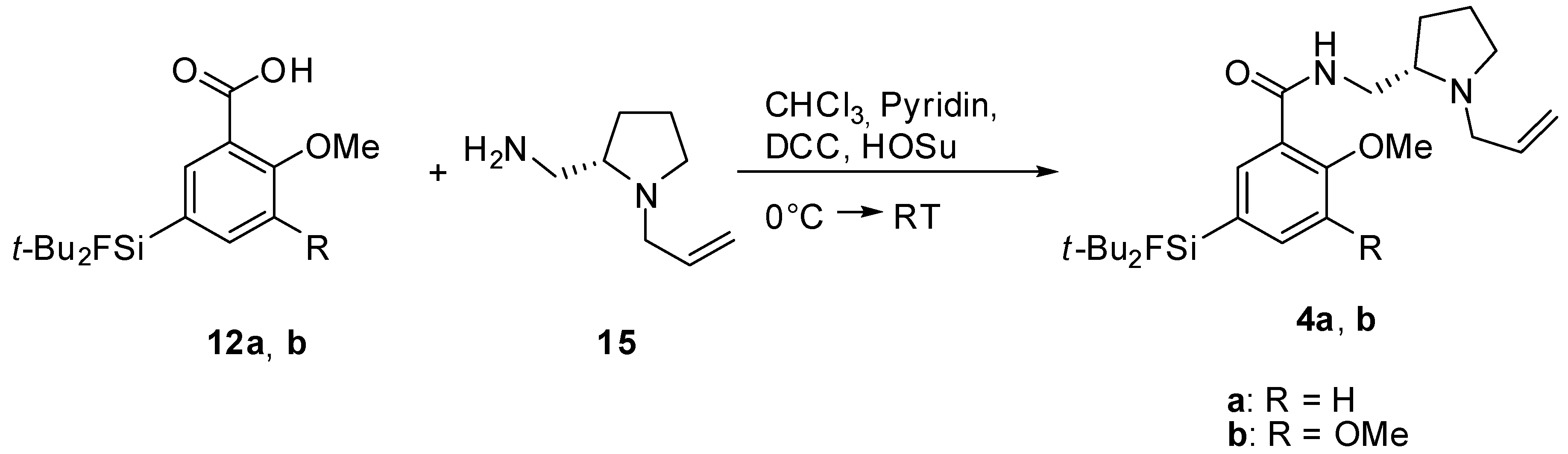
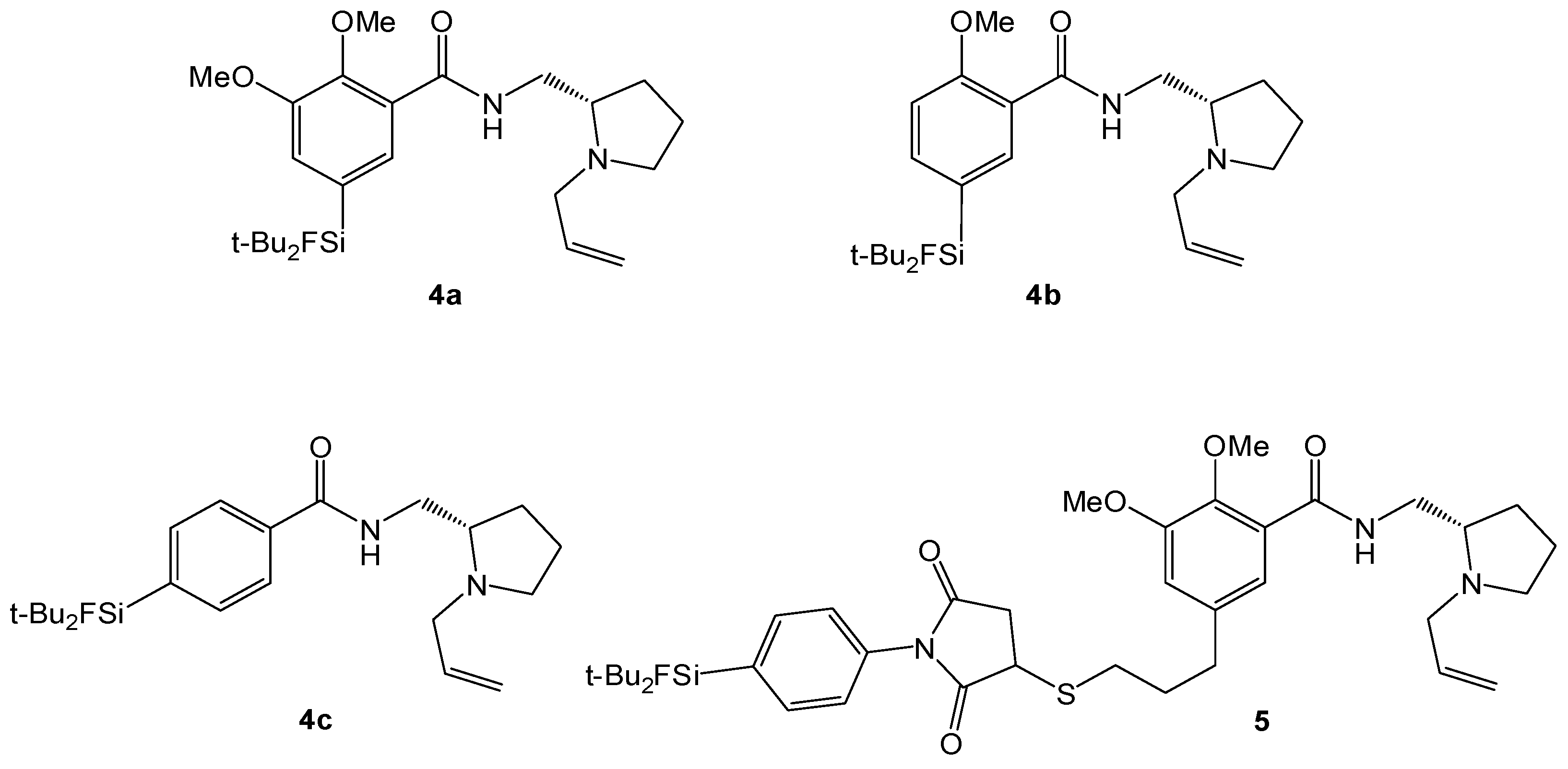
(S)-N-((1-Allylpyrrolidine-2-yl)methyl)-5-(di-tert-butylfluorosilyl)-2-methoxybenzamide (SiFA-DMFP 4a). To an ice-cooled solution in dry CHCl3 containing the substituted benzoic acid 12a (0.97 g, 3.10 mmol), (S)-(1-Allylpyrrolidine-2-yl)methanamine 15 (0.43 g, 3.10 mmol, 1.0 equiv.) and pyridine (0.25 mL, 3.10 mmol, 1.0 equiv.) dicyclohexylcarbodiimide (0.64 g, 3.10 mmol, 1.0 equiv.) and N-hydroxysuccinimide (0.36 g, 3.10 mmol, 1.0 equiv.) were added under stirring. The mixture was stirred at 0 °C for 5 h and 17 h at ambient temperature. After the white precipitate had been filtered, the filtrate was washed with saturated NaHCO3-solution (20 mL) and subsequently with H2O (20 mL). After extracting the aqueous phase with diethyl ether (20 mL) the combined organic layers were dried with MgSO4, filtered and the solvent was evaporated to give an oily residue. The latter was purified by column chromatography (CHCl3/Ethanol = 20/1 → CHCl3/Ethanol = 10/1) to afford benzamide 4a (0.55 g, 1.27 mmol, 41%) as a yellowish oil. 1H-NMR (400.13 MHz, CDCl3): δ (ppm) = 8.44 (d, 4J(1H-1H) = 1.4 Hz, 1H, C(6)H), 8.37 (d, 3J(1H-1H) = 3.8 Hz, 1H, NH), 7.68 (dd, 3J(1H-1H) = 8.2 Hz, 4J(1H-1H) = 1.3 Hz, 1H, C(4)H), 7.00 (d, 3J(1H-1H) = 8.2 Hz, 1H, C(3)H), 5.96–5.84 (m, 1H, CH=CH2), 5.24-5.06 (m, 2H, CH=CH2), 3.95 (s, 3H, OCH3), 3.75 (ddd, 2J(1H-1H) = 13.8 Hz, 3J(1H-1H) = 7.1 Hz, 3J(1H-1H) = 3.1 Hz, 1H, NHCH–H), 3.48 (dd, 2J(1H-1H) = 13.5 Hz, 3J(1H-1H) = 5.3 Hz, 1H, CH2=CHCH-H), 3.35 (ddd, 2J(1H-1H) = 13.9 Hz, 3J(1H-1H) = 3.7 Hz, 3J(1H-1H) = 3.7 Hz, 1H, NHCH–H), 3.19–3.13 (m, 1H, NCH–H), 2.91 (dd, 2J(1H-1H) = 13.5 Hz, 3J(1H-1H) = 7.5 Hz, 1H, CH2=CHCH–H), 2.75 (s, 1H, CH), 2.33–2.22 (m, 1H, NCH–H), 1.99–1.85 (m, 1H, CH2CH2CH2), 1.79–1.61 (m, 3H, CHCH2CH–H), 1.02 (s, 18H, CCH3). 13C[1H]-NMR (100.63 MHz, CDCl3): δ (ppm) = 165.5 (s, CONH), 158.7 (s, C(2)), 138.6 (d, 3J(13C-19F) = 3.8 Hz, C(4)), 137.7 (d, 3J(13C-19F) = 4.7 Hz, C(6)), 135.8 (s, CH=CH2), 125.3 (d, 2J(13C-19F) = 14.5 Hz, C(5)), 120.9 (s, C(1)), 116.2 (s, CH=CH2), 110.7 (s, C(3)), 61.9 (s, CH), 57.0 (s, CH2CH=CH2), 55.5 (s, OCH3), 54.2 (s, NCH2), 41.3 (s, CH2NH), 28.5 (s, CH2), 27.3 (s, CCH3), 22.9 (s, CH2), 20.2 (d, 2J(13C-19F) = 12.0 Hz, C(CH3)3). 19F-NMR (282.38 MHz, CDCl3): δ (ppm) = −188.8 (s, 1J(19F-29Si) = 298 Hz). 29Si-NMR (59.63 MHz, CDCl3): δ (ppm) = 14.4 (d, 1J(29Si-19F) = 298 Hz). Elemental analysis calculated (%) for C24H39FN2O2Si∙H2O (452.68 g/mol): C 63.7, H 9.1, N 6.2; found: C 64.1, H 9.1, N 6.3. HR-MS (LC-ESI): calculated for C24H40O2N2FSi [M+H]+ (m/z): 435.2838; found: 435.2832.
(S)-N-((1-Allylpyrrolidine-2-yl)methyl)-5-(di-tert-butylfluorosilyl)-2,3-dimethoxybenzamide (SiFA-FP, 4b). The procedure was analogous to the synthesis of 4a. The reaction of the benzoic acid derivative 12b (1.47 g, 4.28 mmol) with (S)-(1-Allylpyrrolidine-2-yl)methanamine 15 (0.60 g, 3.10 mmol, 1.0 equiv.) gave SiFA-FP 4b (0.81 g, 1.74 mmol, 41%) as a yellowish oil. 1H-NMR (400.13 MHz, CDCl3): δ (ppm) = 8.51 (s, br, 1H, NH), 7.93 (s, 1H, C(4)H), 7.24 (s, 1H, C(6)H), 5.99–5.87 (m, 1H, CH=CH2), 5.27-5.09 (m, 2H, CH=CH2), 3.93 (s, 3H, OCH3), 3.90 (s, 3H, OCH3), 3.80 (ddd, 2J(1H-1H) = 13.9 Hz, 3J(1H-1H) = 6.9 Hz, 3J(1H-1H) = 3.6 Hz, 1H, NHCH–H), 3.54 (dd, 2J(1H-1H) = 13.5 Hz, 3J(1H-1H) = 5.3 Hz, 1H, CH2=CHCH–H), 3.47–3.37 (m, 1H, NHCH–H), 3.19–3.13 (m, 1H, NCH–H), 2.98 (dd, 2J(1H-1H) = 13.2 Hz, 3J(1H-1H) = 7.6 Hz, 1H, CH2=CHCH–H), 2.86 (s, 1H, CH), 2.39–2.29 (m, 1H, NCH–H), 2.03–1.91 (m, 1H, CH2CH2CH2), 1.87–1.67 (m, 3H, CHCH2CH–H), 1.05 (s, 18H, CCH3). 13C[1H]-NMR (100.63 MHz, CDCl3): δ (ppm) = 165.5 (s, CONH), 151.9 (s, C(2)), 148.8 (s, C(3)), 134.6 (s, CH=CH2), 129.4 (d, 2J(13C-19F) = 14.0 Hz, C(5)), 128.4 (d, 3J(13C-19F) = 4.7 Hz, C(4)), 125.8 (s, C(1)), 120.3 (d, 3J(13C-19F) = 3.9 Hz, C(6)), 117.9 (s, CH=CH2), 62.2 (s, CH), 61.2 (s, OCH3), 57.0 (s, CH2CH=CH2), 56.0 (s, OCH3), 53.9 (s, NCH2), 41.0 (s, CH2NH), 28.4 (s, CH2), 27.3 (s, CCH3), 22.6 (s, CH2), 20.2 (d, 2J(13C-19F) = 12.2 Hz, C(CH3)3). 19F-NMR (282.38 MHz, CDCl3): δ (ppm) = −188.5 (s, 1J(19F-29Si) = 298 Hz). 29Si-NMR (59.63 MHz, CDCl3): δ (ppm) = 14.2 (d, 1J(29Si-19F) = 299 Hz). Elemental analysis calculated (%) for C25H41FN2O3Si∙H2O (482.70 g/mol): C 62.2, H 9.0, N 5.8; found (%): C 61.9, H 8.6, N 4.9. HR-MS (LC-ESI): calculated for C25H42O3N2FSi [M+H]+ (m/z): 465.2943; found: 465.2924.
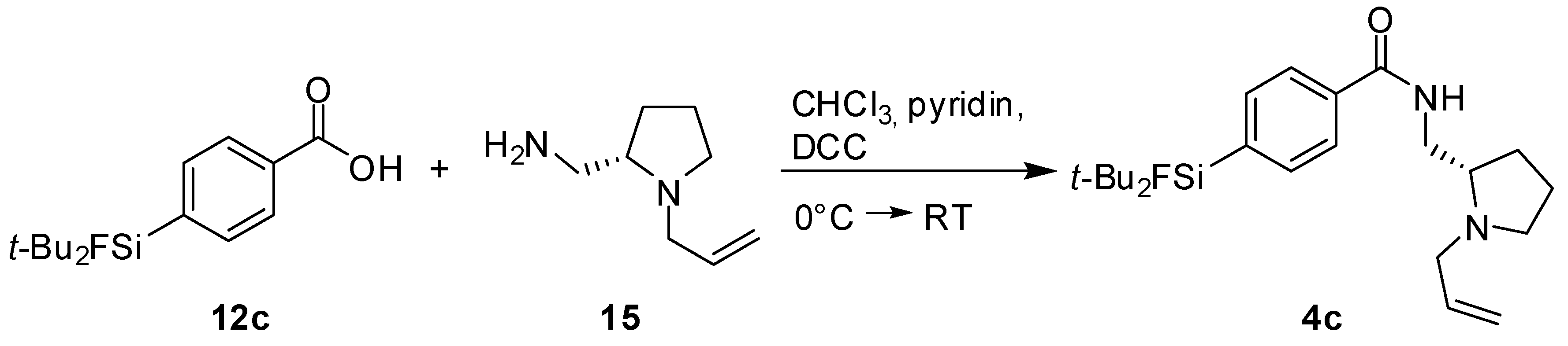
(S)-N-((1-Allylpyrrolidine-2-yl)methyl)-4-(di-tert-butylfluorosilyl) benzamide (SiFA-DDMFP, 4c). The procedure was analogous to the synthesis of 4a. The reaction of the benzoic acid derivative 12c [17] (0.50 g, 1.77 mmol) with (S)-(1-Allylpyrrolidine-2-yl)methanamine 15 (0.25 g, 1.77 mmol, 1.0 equiv.) gave SiFA-DDMFP 11c (0.08 g, 0.20 mmol, 11%) as a yellowish oil. 1H-NMR (400.13 MHz, CDCl3): δ (ppm) = 7.80 (d, 3J(1H-1H) = 7.7 Hz, 2H, Ho), 7.69 (d, 3J(1H-1H) = 8.0 Hz, 2H, Hm), 7.00 (s, 1H, NH) 5.99–5.86 (m, 1H, CH=CH2), 5.29–5.12 (m, 2H, CH=CH2), 3.73 (ddd, 2J(1H-1H) = 13.7 Hz, 3J(1H-1H) = 7.4 Hz, 3J(1H-1H) = 3.2 Hz, 1H, NHCH–H), 3.50–3.43 (m, 2H), 3.2 (s, 1H), 3.04–2.76 (m, 2H), 2.42–2.28 (m, 1H), 1.92–1.90 (m, 1H), 1.64–1.54 (m, 3H), 1.06 (s, 18H, CCH3). 13C[1H]-NMR (100.63 MHz, CDCl3): δ (ppm) = 167.8 (s, CONH), 137.9 (d, 2J(13C-19F) = 13.7 Hz, Cp), 135.2 (s, Ci), 134.1 (d, 3J(13C-19F) = 4.7 Hz, Cm), 134.1 (s, CH=CH2), 126.0 (s, Co), 117.4 (s, CH=CH2), 62.8 (s, CH), 57.4 (s, CH2CH=CH2), 54.1 (s, NCH2), 40.7 (s, CH2NH), 28.2 (s, CH2), 27.2 (s, CCH3), 23.1 (s, CH2), 20.2 (d, 2J(13C-19F) = 12.3 Hz, C(CH3)3). 19F-NMR (282.38 MHz, CDCl3): δ (ppm) = −189.2 (s, 1J(19F-29Si) = 299 Hz). 29Si-NMR (59.63 MHz, CDCl3): δ (ppm) = 14.0 (d, 1J(29Si-19F) = 299 Hz). Elemental analysis calculated (%) for C23H37FN2OSi∙H2O + C8H16N2 (562.88 g/mol): C 66.2, H 9.5, N 8.4; found (%): C 66.0, H 9.2, N 8.2. HR-MS (LC-ESI): calculated for C23H37ON2FSi [M+H]+ (m/z): 405.2732; found: 405.2726.
(S)-N-((1-Allylpyrrolidin-2-yl)methyl)-5-(3-(1-(4-(di-tert-butylfluorosilyl)phenyl)-2,5-dioxopyrrolidin-3-ylthio)propyl)-2,3-dimethoxybenzamide (SiFA-M-FP, 5). To a freshly prepared solution of 1-(4-(di-tert-butylfluorosilyl)phenyl)-1H-pyrrole-2,5-dione (SiFA-maleimide, [17]) (3 mg, 9 µmol) in phosphate buffer (PB, 0.1 M, pH 6.0) and acetonitrile (1:1) was added a solution of (S)-N-((1-allylpyrrolidin-2-yl)methyl)-5-(3-mercaptopropyl)-2,3-dimethoxybenzamide (FP-thiol) (3.4 mg, 9 µmol) in acetonitrile (100 µL) and the pH of the solution was adjusted to 7.2 using PB (0.1M, pH = 7.2, 200 µL). After 10 min, the product was isolated by semi-preparative HPLC applying a gradient of 40%–80% acetonitrile in 10 minutes. The product was obtained as white powder after lyophilization (3.1 mg, 4.4 µmol, 49% yield). ESI-MS calculated for [M+H]+ (m/z): 712.35, found: 712.36.

3.4. In Vitro Binding Assay

Evaluation of the dopamine D2-receptor binding affinity: The substances were dissolved in water or DMSO-water mixtures, respectively, and evaluated according to the [3H]spiperone binding protocol published by Malmberg and coworkers [21]. Source of the dopamine-D2-receptors was cell line HEK293 which expresses the D2 receptors stably. The Kd-value of [3H]spiperone was determined in three saturation experiments (41.0 ± 6.0 pM (± SEM), pKd: 9.398 ± 0.067). All substances were evaluated in 3 independent competition experiments in six concentrations (each in triplicate). The Ki-values were calculated from the IC50-values according to Cheng-Prusoff [22] as described in detail in [23].

3.5. Radiolabeling

General procedure for the labeling of SiFA-compounds. Aqueous [18F]fluoride (4000–7500 MBq) produced by the 18O(p,n)18F nuclear reaction on an enriched [18O]water (95%) target was loaded onto a Chromafix PS-HCO3 cartridge and eluted with a mixture of acetonitrile (800 µL), water (200 µL), potassium oxalate solution (1 M, 10 µL), and Kryptofix 2.2.2 (12.5 mg) (procedure a) or eluted with a mixture of 800 μL acetonitrile and 300 μL aqueous tetrabutylammonium hydrogen carbonate solution (0.075 mol/L, ABX, Radeberg, Germany) (procedure b). The solvents were removed by coevaporation to dryness under reduced pressure (650 mbar) using a stream of helium at 90 °C for 4 min. The drying step was repeated twice with acetonitrile (0.8 mL) (3 min) and full vacuum (~10 mbar) was applied in the final drying step (4 min). The dried [18F]F-complex was dissolved in dry acetonitrile, DMF or DMSO, respectively, (500–1000 µL) and used as stock solution for labeling. The labeling precursors (1–2 mg) were dissolved in dry acetonitrile, DMF or DMSO, respectively (1 µmol/mL) and aliquots containing 5–10 nmol of these stock solutions (5–10 µL) were used for labeling and incubated with 250 µL of the 18F stock solutions. After 5 min reaction time at ambient temperature (without stirring), samples were withdrawn from the reaction mixture and analyzed by analytical HPLC. For purification, the reaction mixture was diluted with 10 mL HEPES-buffer (1 M, pH = 4.0) and loaded on a SepPak C-18 light cartridge (Waters, Germany), preconditioned with 5mL ethanol and 5mL isotonic saline. The cartridge was washed with 10 mL isotonic saline and subsequently eluted with 1 mL ethanol. After dilution with 9 mL isotonic saline the radiotracers were measured, analyzed by radio-HPLC and used for plasma stability experiments.
In vitro stability in human plasma. To human plasma (500 µL) at 37 °C were added 10–100 MBq of the injectable solutions. The mixture was incubated at 37 °C. After 90 min, aliquots (75 µL, in triplicate) were removed and treated with acetonitrile (75 µL). Samples were then stored on ice for 5 min for complete precipitation of the plasma proteins. The precipitate was removed by centrifugation, and the supernatants were analyzed by radio-HPLC. Radioactivity in precipitate and supernatant was measured.

4. Conclusions

Motivated by our recent development of a kit-like radiolabeling strategy for the introduction of 18F-fluoride into proteins and target-specific peptides using Silicon-Fluoride-Acceptors (SiFAs) and their application in preclinical studies, we synthesized a series of SiFA-containing fallypride/ desmethoxyfallypride derivatives (Figure 2). Compared to other medium-affinity D2-receptor ligands such as DMFP 2, the compounds showed a reduced affinity to the targeted receptor. However, the affinity is still in the nanomolar range and should therefore be high enough for an in vivo application. The radiochemical synthesis of the potential radiotracers, most notably the synthesis time of only 10 min and the easy cartridge purification, could be a breakthrough in radiofluorinations of small-molecule radiotracers. However, the potential of these SiFA-radiotracers remains to be shown in ongoing in vitro studies regarding receptor subtype-selectivity as well as Pgp-activity and finally in preclinical in vivo studies.

Acknowledgments

This research was supported by the German Federal Ministry of Education and Research (Munich Biotech Cluster m4, project PM10) as part of the Leading Edge Cluster Initiative and by the Fonds der Chemischen Industrie.

Conflict of Interest

The authors declare no conflict of interest.

References and Notes

  1. Rosenthal, M.S.; Bosch, A.L.; Nickles, R.J.; Gatley, S.J. Synthesis and some characteristics of no-carrier added [F-18] fluorotrimethylsilane. Int. J. Appl. Radiat. Is. 1985, 36, 318–319. [Google Scholar] [CrossRef]
  2. Walsh, J.C.; Akhoon, K.M.; Satyamurthy, N.; Barrio, J.R.; Phelps, M.E.; Gambhir, S.S.; Toyokuni, T. Application of silicon-fluoride chemistry to fluorine-18 labeling agents for biomolecules: A preliminary note. J. Label. Compd. Rad. 1999, 42, S1–S3. [Google Scholar] [CrossRef]
  3. Schirrmacher, R.; Bradtmöller, G.; Schirrmacher, E.; Thews, O.; Tillmanns, J.; Siessmeier, T.; Buchholz, H.G.; Bartenstein, P.; Wängler, B.; Niemeyer, C.M.; et al. F-18-labeling of peptides by means of an organosilicon-based fluoride acceptor. Angew. Chem. Int. Ed. 2006, 45, 6047–6050. [Google Scholar]
  4. Schirrmacher, E.; Wängler, B.; Cypryk, M.; Bradtmoller, G.; Schäfer, M.; Eisenhut, M.; Jurkschat, K.; Schirrmacher, R. Synthesis of p-(di-tert-butyl[(18)f]fluorosilyl)benzaldehyde ([F-18]SiFA-A) with high specific activity by isotopic exchange: A convenient Labeling synthon for the F-18-labeling of n-amino-oxy derivatized peptides. Bioconjugate Chem. 2007, 18, 2085–2089. [Google Scholar] [CrossRef]
  5. Ametamey, S.M.; Mu, L.J.; Hohne, A.; Schubiger, R.A.; Graham, K.; Cyr, J.E.; Dinkelborg, L.; Stellfeld, T.; Srinivasan, A.; Voigtmann, U.; et al. Silicon-based building blocks for one-step F-18-radiolabeling of peptides for PET imaging. Angew. Chem. Int. Ed. 2008, 47, 4922–4925. [Google Scholar]
  6. Höhne, A.; Mu, L.; Honer, M.; Schubiger, P.A.; Ametamey, S.M.; Graham, K.; Stellfeld, T.; Borkowski, S.; Berndorff, D.; Klar, U.; et al. Synthesis, F-18-labeling, and in vitro and in vivo studies of bombesin peptides modified with silicon-based building blocks. Bioconjugate Chem. 2008, 19, 1871–1879. [Google Scholar] [CrossRef]
  7. Wängler, B.; Quandt, G.; Iovkova, L.; Schirrmacher, E.; Wängler, C.; Boening, G.; Hacker, M.; Schmoeckel, M.; Jurkschat, K.; Bartenstein, P.; et al. Kit-like 18F-labeling of proteins: Synthesis of 4-(di-tert-butyl[18F]fluorosilyl)benzenethiol (Si[18F]FA-SH) labeled rat serum albumin for blood pool imaging with PET. Bioconjugate Chem. 2009, 20, 317–321. [Google Scholar] [CrossRef]
  8. Rosa-Neto, P.; Wängler, B.; Iovkova, L.; Boening, G.; Reader, A.; Jurkschat, K.; Schirrmacher, E. [18F]SiFA-isothiocyanate: A new highly effective radioactive labeling agent for lysine-containing proteins. Chembiochem 2009, 10, 1321–1324. [Google Scholar] [CrossRef]
  9. Tietze, L.F.; Schmuck, K. SiFA azide: A new building block for PET imaging using click chemistry. Synlett 2011, 12, 1697–1700. [Google Scholar] [CrossRef]
  10. Szlosek-Pinaud, M.; Schulz, J.; Vimont, D.; Bordenave, T.; James, D.; Escudier, J.M.; Allard, M.; Fouquet, E. Silicon-based chemistry: An original and efficient one-step approach to [(18)F]-nucleosides and [(18)F]-oligonucleotides for PET imaging. Chem.-Eur. J. 2011, 17, 3096–3100. [Google Scholar] [CrossRef]
  11. Höhne, A.; Yu, L.; Mu, L.J.; Reiher, M.; Voigtmann, U.; Klar, U.; Graham, K.; Schubiger, P.A.; Ametamey, S.M. Organofluorosilanes as model compounds for F-18-labeled silicon-based PET tracers and their hydrolytic stability: Experimental data and theoretical calculations (PET = Positron Emission Tomography). Chem.-Eur. J. 2009, 15, 3736–3743. [Google Scholar]
  12. Mukherjee, J.; Yang, Z.Y.; Das, M.K.; Brown, T. Fluorinated benzamide neuroleptics. 3. Development of (S)-N-[(1-allyl-2-pyrrolidinyl)methyl]-5-(3-[F-18] fluoropropyl)-2,3-dimethoxy-benzamide as an improved dopamine D-2 receptor tracer. Nucl. Med. Biol. 1995, 22, 283–296. [Google Scholar] [CrossRef]
  13. Mukherjee, J.; Yang, Z.Y.; Brown, T.; Roemer, J.; Cooper, M. F-18-desmethoxyfallypride: A fluorine-18 labeled radiotracer with properties similar to carbon-11 raclopride for pet imaging studies of dopamine D-2 receptors. Life Sci. 1996, 59, 669–678. [Google Scholar] [CrossRef]
  14. de Paulis, T. The discovery of epidepride and its analogs as high-affinity radioligands for imaging extrastriatal dopamine D(2) receptors in human brain. Curr. Pharm. Des. 2003, 9, 673–96. [Google Scholar] [CrossRef]
  15. la Fougère, C.; Pöpperl, G.; Levin, J.; Wängler, B.; Böning, G.; Uebleis, C.; Cumming, P.; Bartenstein, P.; Botzel, K.; Tatsch, K. The value of the dopamine D(2/3) receptor ligand (18)F-desmethoxyfallypride for the differentiation of idiopathic and nonidiopathic parkinsonian syndromes. J. Nucl. Med. 2010, 51, 581–587. [Google Scholar] [CrossRef]
  16. Fehr, C.; Yakushev, I.; Hohmann, N.; Buchholz, H.G.; Landvogt, C.; Deckers, H.; Eberhardt, A.; Klaager, M.; Smolka, M.N.; Scheurich, A.; et al. Association of low striatal dopamine D-2 receptor availability with nicotine dependence similar to that seen with other drugs of abuse. Am. J. Psychiat. 2008, 165, 507–514. [Google Scholar] [CrossRef]
  17. Iovkova, L.; Wängler, B.; Schirrmacher, E.; Schirrmacher, R.; Quandt, G.; Boening, G.; Schurmann, M.; Jurkschat, K. para-Functionalized aryl-di-tert-butylfluorosilanes as potential labeling synthons for F-18 radiopharmaceuticals. Chem.-Eur. J. 2009, 15, 2140–2147. [Google Scholar]
  18. Wängler, C.; Waser, B.; Alke, A.; Iovkova, L.; Buchholz, H.G.; Niedermoser, S.; Jurkschat, K.; Fottner, C.; Bartenstein, P.; Schirrmacher, R.; et al. One-step (18)F-labeling of carbohydrate-conjugated octreotate-derivatives containing a silicon-fluoride-acceptor (SiFA): In vitro and in vivo evaluation as tumor imaging agents for positron emission tomography (PET). Bioconjugate Chem. 2010, 21, 2289–2296. [Google Scholar] [CrossRef]
  19. Sheldrick, G.M. Phase annealing in shelx-90-Direct methods for larger structures. Acta Crystallogr. A 1990, 46, 467–473. [Google Scholar] [CrossRef]
  20. Hain, T. International Tables for Crystallography; Kluwer Academic Publishers: Dordrecht, The Netherlands/Boston, MA, USA/London, UK, 1992. [Google Scholar]
  21. Malmberg, A.; Jerning, E.; Mohell, N. Critical reevaluation of spiperone and benzamide binding to dopamine D-2 receptors: Evidence for identical binding sites. Eur. J. Pharmacol. 1996, 303, 123–128. [Google Scholar] [CrossRef]
  22. Cheng, Y.; Prusoff, W.H. Relationship between inhibition constant (K1) and concentration of inhibitor which causes 50 per cent inhibition (I50) of an enzymatic-reaction. Biochem. Pharmacol. 1973, 22, 3099–3108. [Google Scholar] [CrossRef]
  23. Wängler, C.; Nada, D.; Hofner, G.; Maschauer, S.; Wängler, B.; Schneider, S.; Schirrmacher, E.; Wanner, K.T.; Schirrmacher, R.; Prante, O. In vitro and initial in vivo evaluation of (68)Ga-labeled transferrin receptor (TfR) binding peptides as potential carriers for enhanced drug transport into TfR expressing cells. Mol. Imaging Biol. 2011, 13, 332–341. [Google Scholar] [CrossRef]

