Article

Development of Plant–Fungal Endophyte Associations to Suppress Phoma Stem Canker in Brassica

1
Resilient Agriculture, AgResearch Limited, Grasslands Research Centre, Private Bag 11008, Palmerston North 4410, New Zealand
2
School of Agriculture & Environment, Massey University, Private Bag 11222, Palmerston North 4442, New Zealand
3
Khorasan Razavi Agricultural and Natural Resources Research and Education Center, Agricultural Research, Education and Extension Organization (AREEO), Mashhad 91769-83641, Iran
4
Digital Agriculture, AgResearch Limited, Invermay Agricultural Centre, Private Bag 50034, Mosgiel 9053, New Zealand
5
Bio-Protection Research Centre, P.O. Box 85084, Lincoln University, Lincoln 7647, New Zealand
*
Author to whom correspondence should be addressed.
Microorganisms 2021, 9(11), 2387; https://doi.org/10.3390/microorganisms9112387
Submission received: 1 November 2021 / Revised: 17 November 2021 / Accepted: 18 November 2021 / Published: 19 November 2021
(This article belongs to the Special Issue Microbial Endophytes: Functional Biology and Applications)

Abstract

:
Endophytic microorganisms are found within the tissues of many plants species, with some conferring several benefits to the host plant including resistance to plant diseases. In this study, two putative endophytic fungi that were previously isolated from wild seeds of Brassica, identified as Beauveria bassiana and Pseudogymnoascus pannorum, were inoculated into cultivars of three Brassica species—Brassica napus, Br. rapa and Br. oleracea. Both fungal endophytes were reisolated from above- and below-ground tissues of inoculated plants at four different plant-growth stages, including cotyledon, one-leaf, two-leaf, and four-leaf stages. None of the plants colonised by these fungi exhibited any obvious disease symptoms, indicating the formation of novel mutualistic associations. These novel plant–endophyte associations formed between Brassica plants and Be. bassiana significantly inhibited phoma stem canker, a devastating disease of Brassica crops worldwide, caused by the fungal pathogen Leptosphaeria maculans. The novel association formed with P. pannorum significantly suppressed the amount of disease caused by L. maculans in one out of two experiments. Although biological control is not a new strategy, endophytic fungi with both antiinsect and antifungal activity are a highly conceivable, sustainable option to manage pests and diseases of economically important crops.

1. Introduction

Crops from the genus Brassica were among the earliest plants to be widely cultivated by mankind [1]. Brassica displays enormous diversity and subsequently provides the widest assortment of products used by man from a single plant genus [2] with many parts of the plant being edible, including their buds, flowers, leaves, roots, seeds, stems and tubers [3,4]. Many species of Brassica are used as important animal and human food sources, as ornamentals, sources of medicines, soil conditioners, green manures, composting crops, and are valued in bioremediation and in the production of edible and industrial oils [2,3,5,6]. The cultivation of oilseed rape (Brassica napus) has now become the second most important oilseed crop after soybean, with a worldwide production of over 70 million metric tonnes in 2018 [7] with Canada, China and India amongst the top producers.
All the economically important species of Brassica, including oil or leafy types, are vulnerable to attack from a wide range of invertebrate pests and plant pathogens [8,9,10]. One of the most important fungal pathogens of oilseed rape is Leptosphaeria maculans, which causes phoma stem canker (also known as blackleg disease) [11,12]. This ascomycete causes large yield losses and is a major constraint in the production of oilseed rape in Australia, Europe, New Zealand and North America. In the UK alone, the disease can cause up to GBP 56 million worth of damage per season [13] and more than USD 900 million per season worldwide [14]. Integrated disease management practices, including crop rotation and stubble management, are recommended to control the pathogen, with many growers also routinely relying on chemical fungicides to minimise crop losses [11,12,15]. Although the use of resistant canola varieties was seen as promising, field populations of L. maculans can overcome major resistance genes within a few years, potentially accelerated by growers that have adopted shorter crop rotations to capitalise on the high profits from the crop [15].
Overuse of agrochemicals has extensively damaged our environment and contributed to steep losses in biodiversity [16,17]. In direct response, the European Union has now banned three types of neonicotinoids, systemic pesticides commonly used as a seed coat on many crops, due to their negative impact on pollinators vital for food production, e.g., bees [18]. Coupled with chemical resistance problems [19], and negative impacts on human health [17], this advocates the need for new and innovative pest and disease management strategies for crops including Brassica.
Symbiotic microorganisms that live inside plant tissues, termed endophytes, associate with the majority of plant species grown in natural and managed ecosystems [20,21]. Species of Brassicaceae are no exception, with many of their associated symbionts classified as mutualistic, conferring one or several advantageous traits to their hosts including improved plant growth, increased yield and resistance against pests and/or plant pathogens (reviewed by [22]). Biological control of plant pathogens is not a new concept [23], but the transfer of beneficial microbial endophytes from wild plant species to modern day cultivars to obtain additional traits is a novel strategy that may overcome many of the dilemmas faced by traditional biological control approaches [20]. This study focused on the colonisation of cultivated Brassica species by two fungal endophytes of wild Brassica and investigated whether this novel association could offer plant resistance against the disease of phoma stem canker.

2. Materials and Methods

2.1. Fungal Endophytes Isolated from Wild Brassica

Two accessions of Brassica rapa were imported on permit no. 2015058982 as seed into the Margot Forde Germplasm Centre, New Zealand’s national genebank of grassland plants. The accessions were catalogued as O2380 and O2377 and stored at 0 °C and 30% relative humidity until use. Both accessions were collected as wild material and originated from California, USA. Seeds from these accessions were subsequently screened for the presence of microbial endophytes according to a modified method of Roodi et al. [24]. Briefly, seed were surface disinfected and grown under sterile conditions within tissue culture pots containing Murashige & Skoog (MS) basal salts [25] with minimal organics (Sigma-Aldrich, Auckland, New Zealand), plus 3% sucrose and 1.5% agar [26]. After incubation, clean seedlings (i.e., those free of epiphytic microbial growth when inspected with a dissecting microscope (Carl Zeiss AG, Oberkochen, Germany)) were dissected into two components: shoot and root. These organs were further dissected into 2–3 mm2 pieces using sterile forceps and a scalpel. Ten pieces per organ type from each seedling were transferred to Petri plates containing PDA. Petri plates were incubated for 3 weeks at 22 °C in the dark and checked daily under a dissecting microscope for microbial growth. Fungal colonies arising from dissected tissue pieces were selected, subcultured and purified. Representative fungal isolates were then subcultured onto fresh PDA and stored on sterilized wheat grain submersed in 20% glycerol within an ultralow temperature freezer (ULT) at −80 °C (according to [27]).
Species identification of fungal endophytes was achieved initially by morphological examination of reproductive structures on water agar using a BX50 microscope and DP12 digital camera system (Olympus NZ Ltd., Auckland, New Zealand). Confirmation of species identity was conducted by PCR amplification of the Internal Transcribed Spacer (ITS) of rDNA gene sequences [28]. For PCR, both fungal isolates were incubated at 22 °C for two weeks and their DNA extracted using Quick-DNATM Fungal/Bacterial Kit (Zymo Research Corporation, Irvine, USA) and quantified using the Invitrogen QubitTM 4 Fluorometer (ThermoFisher Scientific Inc., Waltham, MA, USA). The PCR reaction contained 1 μL of DNA extract suspension (15–20 ng/μL) of each purified fungal colony, 5 μL 10X PCR buffer, 1.5 μL MgCl2 (50 mM), forward primer, ITS1 (5′ TCCGTAGGTGAACCTGCGG-3′) and reverse primer ITS4 (5′-TCCTCCGCTTATTGATATGC-3′), 0.4 μL dNTPS (25 mM), 0.25 μL Taq-polymerase and 39.85 μL sterile Milli-Q water, to make a 50 μL PCR reaction. PCR was performed in a thermocycler (Bio-Rad C1000 TouchTM, Bio-Rad Laboratories Inc., Hercules, USA) with the following conditions: an initial step of 95 °C for 5 min was followed by 36 cycles of 94 °C for 30 s, 56 °C for 30 s, 72 °C for 90 s and a final step of 72 °C for 10 min. The reaction mixture from each sample was electrophoresed on a 1.5% agarose gel containing ethidium bromide. The gel was viewed on a transilluminator (Gel Doc™ XR+, Bio-Rad Laboratories Inc., Hercules, USA) to identify samples with amplification products. These products were purified and concentrated using the DNA clean & concentrator kit (Zymo Research Corporation, Irvine, USA) prior to Sanger sequencing [29] (New Zealand Genomics Ltd., Dunedin, New Zealand). DNA sequences were analysed with the software package Geneious Prime® version 2019.1.1 (Biomatters Ltd., Auckland, New Zealand). Sequences greater than 600 bp were used in BLASTn searches against the NCBI nonredundant database, and those with greater than 98% identity were selected and named.
Further identification of Beauveria sp. was achieved via the 1-alpha (EF1-apha) elongation factor using primers EF-1 (forward) 5′ ATGGGTAAGGAGGACAAGAC and EF-2 (reverse) 5′ GGAAGTACCAGTGATCATGTT [30] with a 25 μL PCR reaction mixture that consisted of: 15.75 μL sterile water, 2.5 μL buffer (10×) plus MgCl2 (2 mM), 2 μL deoxynucleotide (dNTP’s) (2.5 mM), 0.25 μL Fast start polymerase Taq, 1 μL of each primer, 0.5 μL bovine serum albumin (Bio Labs® Inc., Lawrenceville, USA) and 2 μL of extracted DNA per sample. Thermocycling conditions were set as 95 °C for 5 min followed by 40 cycles of (95 °C for 45 s, 53 °C for 45 s, 72 °C for 1 min) and a final extension of 72 °C for 7 min. Elongation factor sequences were aligned with the programme ClustalW in the software package Geneious Prime®, with a gap cost set at 15. The sequence of strain O2380 was compared to previously published sequences, including those referred to by Rehner and Buckley [31], downloaded from GenBank. Sequences were aligned and trimmed to 687–707 bp for each isolate. After alignment, a tree was generated using Geneious Tree Builder, set to the Jukes-Cantor distance model and Neighbour-Joining with Be. hoplocheli Bt124 designated as an outgroup. Bootstrapping was set at 1000 replicates.

2.2. Development of Novel Brassica–Endophyte Associations

Seeds from three forage cultivars of Brassica, cv. Hunter, Titan and Regal, were sourced from PGG Wrightson Seeds Ltd. (Christchurch, New Zealand) for the development of novel, or artificial, plant–endophyte associations. Hunter, a leafy turnip, is an interspecific hybrid developed by crossing turnips with related Asiatic leaf vegetables of the same species. Titan, a forage rape, is an interspecies cross developed by crossing rape with kale, while Regal is a high-yielding, intermediate-height kale. In order to reduce any epiphytic microorganisms attached to the seed coats, seeds were surface disinfected by washing for five min in 5% aqueous Tween® 20 solution (Sigma-Aldrich Inc., Auckland, New Zealand), two min in 70% ethanol, 10 min in 2% sodium hypochlorite, one min in 70% ethanol and were rinsed three times in sterile tap water. Seeds were then dried on filter paper (110 mm, ThermoFisher Scientific Inc., LabServ®, Waltham, MA, USA) within a sterile environment and stored at 4 °C until use.
The two candidate fungi were removed from the ULT freezer and defrosted at room temperature before plating onto PDA (CM0139, Oxoid Ltd., Basingstoke, UK). Petri plates containing the fungi were then incubated for approximately two weeks at 22 °C in the dark to promote mycelial growth and sporulation. Subsequent spores of each fungal strain were dislodged by adding 50 mL of sterile water to the Petri plate and gently brushing the fungal colony with a sterile loop. The resulting crude suspension was passed through a single layer of sterile Miracloth (Sigma-Aldrich Inc., Auckland, New Zealand) to remove mycelial fragments, and one drop of Tween-20® was added to the solution to stop the spores adhering to each other. The concentration of each spore suspension was estimated using a haemocytometer and adjusted to 106 spores per mL. The viability of fungal spores was assessed by spraying diluted aliquots of the prepared spore suspensions onto fresh PDA and counting the subsequent colonies after 3 days of incubation at 22 °C in the dark.

2.3. Inoculation of Brassica Seed with Fungal Spore Suspensions

Disinfected seeds of each Brassica cultivar were soaked in a spore suspension of each fungal endophyte strain for 10 min at room temperature. Control seeds were soaked in aqueous Tween-20® solution. All treated and control seed then were transferred to sterile filter papers and allowed to dry at room temperature for 30 min. Seeds were sown in a vermiculite growth medium supplemented with essential nutrients to support plant growth, including necessary macro- and micro-nutrients in the form of a nutrient solution, following the manufacturer’s instructions (Thrive®, Yates New Zealand Ltd., Auckland, New Zealand). In each pot, measuring 10 × 15 cm, 10 seeds were sown, which were later thinned to three seedlings per pot after germination. Pots were subsequently placed in a glasshouse with natural light at 20–25 °C, and watered as required. Plant health, development, and any visible disease symptoms were assessed daily under a stereomicroscope.

2.4. Assessment of Endophyte Colonisation

The colonisation frequencies of the forage Brassica cultivars inoculated by either of the two fungal endophytes was determined in root and shoot (stem) tissues at four different plant growth stages, (1) cotyledon, (2) one-leaf, (3) three-leaf and (4) the four-leaf growth stage. For each of the two endophytes, for each Brassica cultivar at each growth stage, eight inoculated and eight uninoculated (control) plants were assessed as follows; each plant was removed from the growth medium, washed in tap water, and surface disinfected by washing in 5% aqueous Tween-20® solution for five min, 70% ethanol for 2 min, 5 min in 1% sodium hypochlorite followed by 70% ethanol for one min and finally three rinses in sterile tap water. At the cotyledon and one-leaf stage, a modified surface disinfection protocol was utilised as the above protocol was proven to be too harsh on the delicate plant tissues. In this protocol, 70% ethanol was used for one min and 30 s rather than two minutes and one minute. To assess the efficacy of the surface disinfection protocols, 3 × 20 μL drops of tap water from the last rinse were plated onto PDA and incubated at 22 °C. These PDA plates were subsequently observed every day for two weeks under a dissecting microscope for microbial growth. Following surface disinfection, seedlings were dried on filter papers (110 mm, LabServ®, Waltham, USA) within a sterile environment for 30 min at room temperature. Plant tissues were dissected from each plant with a sterile scalpel, and ten 1–2 mm2 pieces (of each root and shoot) transferred to Petri plates containing PDA. Petri plates were incubated at 22 °C and checked regularly (for up to three weeks) with the aid of a dissecting microscope for microbial growth. The presence of each fungal endophyte was assessed visually using a dissecting microscope. The number of fungal colonies that emerged from each tissue piece was recorded and the data presented as a percentage of tissue colonisation frequency (TCF) for each endophyte.
Confirmation of endophyte species was assessed using PCR as previously described. Statistical analyses for the endophyte frequency data were performed using the software R [32]. A generalised linear model (GLM) with a binomial distribution and logit link function was used to model the proportion of endophyte-infected tissue pieces (number of infected tissue pieces/total number of tissue pieces) for each fungal endophyte strain. The fixed effects used in the model were the growth stage, shoot/root location and Brassica cultivar. All two-way interaction terms between the variables were included in the model. Analysis of Deviance was used to assess the significance of the fixed effects and their interaction terms. The R package “emmeans” was used to generate back-transformed probabilities, standard errors and 95% confidence limits [33].

2.5. Bioactivity of Fungal Endophytes towards Leptosphaeria Maculans

The bioactivity of the novel Brassica–endophyte associations was assessed against L. maculans (strain LUPP2376), a highly pathogenic strain originally identified from a diseased swede collected in Gore, New Zealand [34], supplied by Dr Eirian Jones, Lincoln University. After subculturing on PDA, strain LUPP2376 was stored in 30% glycerol at −80 °C until use. Previous work noted that strain LUPP2376 was highly pathogenic towards oilseed rape (Br. napus), cv. Flash [34], and therefore initial bioactivity trials utilised this susceptible cultivar of Brassica. Strain LUPP2376 was defrosted at room temperature and plated onto PDA. Petri plates containing the pathogen were subsequently incubated for two weeks at 15–20 °C with a 16/8 h (light/dark) photoperiod. A spore suspension of the pathogen was then prepared as described for the candidate endophytes, Be. bassiana and G. pannorum, at a concentration of 107 spores per ml. The viability of L. maculans spores was assessed by spraying aliquots of the prepared spore suspension onto fresh PDA and observing the developing colonies after 5 days of incubation at 18 °C in the dark.
Seeds of oilseed rape were surface-disinfected and inoculated with the two fungal endophytes as described earlier. Control seeds were only treated with a sterile aqueous Tween-20® solution. Seeds from all treatment groups were then placed on sterile filter paper to dry, and later transferred to sterile plastic plant containers (product number 2105646, Alto Ltd., Auckland, New Zealand) containing autoclaved vermiculite. Subsequently, seedlings were planted in seedling trays containing autoclaved potting mix within Saxon mini greenhouses (Bunnings Group, Hawthorn East, Australia). The mini greenhouses were closed with a lid, sealed with plastic tape to keep humidity in the tray elevated and placed in a controlled environment (A1000, Conviron Asia Pacific Pty Ltd., Grovedale, Australia) at 18 °C with a 16/8 h (light/dark) photoperiod. At the cotyledon leaf stage, one cotyledon leaf per seedling was punctured with a sterile needle and 15 µL of the L. maculans spore suspension was placed on the wound site using a pipette. Plants were incubated, as described earlier, for two weeks to allow disease symptoms to appear and subsequently assessed using a 0–6 scale as described by Hammoudi et al. [35]: 0 = no symptoms; 1 = lesions on the infection site < 1.5 mm; 2 = lesions on the infection site 1.5–3.5 mm; 3 = lesions on the infection site > 3.0 mm; 4 = grey-to-green tissue collapse 3.1–5.0 mm; 5 = grey-to-green tissue collapse > 5.0 mm (≤10 pycnidia); 6 = grey-to-green tissue collapse > 5.0 mm (>10 pycnidia). The mean score from 10 infected seedlings from each tray was used in the analysis. Newly emerging leaves were carefully removed during the experimental period and the experiment was repeated once. There were three treatments (seed treated with two fungal endophytes, and an aqueous Tween-20® solution acting as a pathogen-only control), with 10 seedlings per treatment, arranged in a randomised complete block design.
Statistical analyses were performed using the software package R [32]. A cumulative link mixed model (CLMM) from the “ordinal” R package was used to model the lesion severity scores using an equidistant threshold [36]. The two experiments (1 + 2) were modelled separately. The treatment group was used as a fixed effect. Random intercepts were used for each block. Analysis of Deviance was used to assess the significance of the fixed effects. The R package “emmeans” was used to generate predicted means, standard errors and 95% confidence limits for lesion scores [33]. Multiple comparison p-value adjustment was performed using Tukey’s method.

3. Results

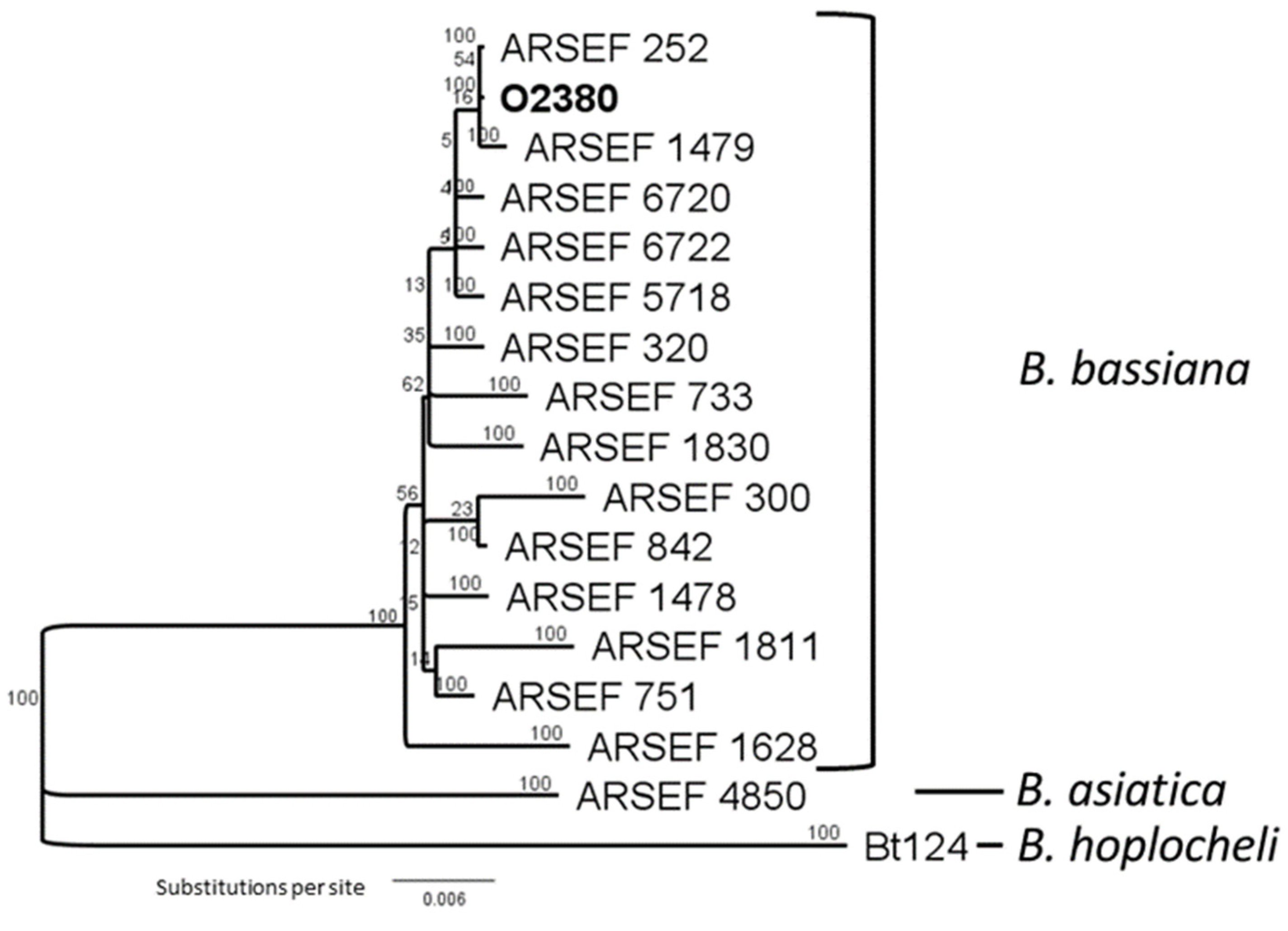
3.1. Fungal Endophytes Isolated from Wild Brassica

The fungal isolates from Br. rapa accessions O2380 and O2377 were identified as Be. bassiana and Pseudogymnoascus pannorum (syn. Geomyces pannorum), respectively. The isolate from accession O2380 displayed slow-growing white colonies on PDA at 22 °C. After 3–4 days incubation, the culture produced single-celled, near-spherical, hyaline conidia formed on a zig-zag conidiophore, or rachis, characteristic of Beauveria spp. A phylogenetic tree was produced comparing the Beauveria strain isolated from Br. Rapa accession O2380 to reference isolates, including strains sequenced by Rehner & Buckley [31]. Several strains of Be. Bassiana (sensu stricto) were similar to that from accession O2380 (Figure 1), based on partial sequences of the elongation factor gene, confirming its identity.
The fungal isolate from accession O2377 produced single-celled, hyaline, wedge-shaped conidia with a flat base on short conidiophores. These characteristics resembled those of P. pannorum and were distinguishable from P. destructans, a pathogen of bats that produces sickle-shaped conidia and is unable to grow above 22 °C [37,38,39]. The isolate from accession O2377 grew at temperatures above 22 °C but failed to grow above 37 °C. Subsequent PCR amplification of the ITS rDNA gene sequences confirmed its identity as P. pannorum.

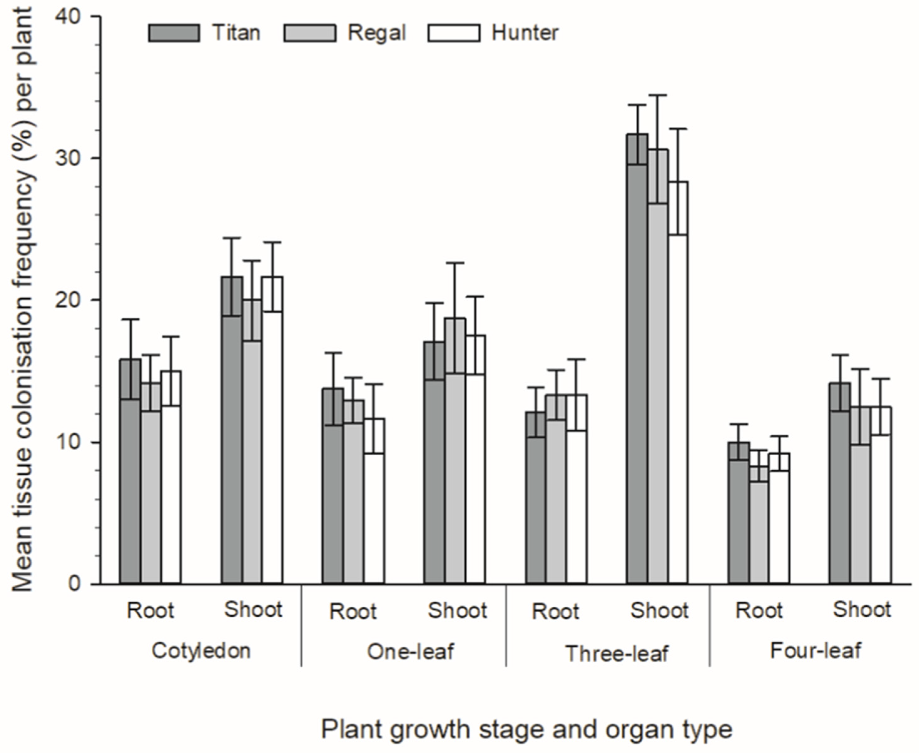
3.2. Development of Novel Plant–Endophyte Associations

Both Be. bassiana and P. pannorum strains were isolated from all plants, and all forage Brassica cultivars (rape, cv. Titan, kale, cv. Regal and a leafy turnip, cv. Hunter) that were intentionally inoculated with these endophytic fungi. Additionally, these fungi were isolated from all four plant growth stages (cotyledon, one-leaf, two-leaf and four-leaf) from all plants that were assessed. No plants exhibited any obvious disease symptoms related to colonisation by these two fungi. Overall, there was a greater abundance of P. pannorum isolated from the tissue pieces that were sampled from these artificially inoculated plants compared to Be. bassiana. When all cultivars were pooled from all plant growth stages, there was a significantly greater colonisation frequency recovered from shoot samples compared to those from the root of both P. pannorum and Be. bassiana (X21 = 34.62, p < 0.001 and X21 = 6.82, p = 0.009, respectively) (Figure 2 and Figure 3). For P. pannorum, this was especially evident at the three-leaf stage, where there was more than twice the number of tissue pieces from the shoot colonised in comparison to those from the root of Brassica (p = 0.015) and this was consistent across all three cultivars of Brassica (Figure 2). There were no effects of cultivar on the colonisation frequency of either P. pannorum or Be. bassiana (Figure 2 and Figure 3). All uninoculated (control) plants remained free of both P. pannorum and Be. bassiana (data not shown).

3.3. Bioactivity of Fungal Endophytes towards Leptosphaeria Maculans

In experiment 1, both P. pannorum and Be. bassiana significantly (p < 0.001, z = −2.63 and p < 0.001, z = −7.39, respectively) suppressed the amount of disease caused by L. maculans on leaves of oilseed rape (Br. napus), cv. Flash after wounding, compared to the pathogen-only control (Figure 4). In experiment 2, only Be. bassiana significantly (p < 0.001, z = −5.04) suppressed the amount of disease caused by L. maculans on leaves of oilseed rape (Br. napus) compared to the pathogen-only control (Figure 4).

4. Discussion

Our previous research identified that Methylobacterium was the dominant cultural bacterial genus inhabiting wild Brassica plants [24]. Within the same study, two fungal isolates were also recovered from accessions of wild Brassica, but this was not reported earlier. Utilising DNA-based and traditional mycology techniques, these fungal isolates were identified as Be. bassiana and P. pannorum. Both fungi were exceptionally rare amongst the wild Brassica accessions surveyed, only being recovered from single, separate accessions of Br. rapa, both originating from California, USA. Both fungi were isolated from multiple root and shoot samples dissected from several symptomless plants generated from surface disinfected seed.
Further study showed that both these fungi could colonise multiple cultivars of forage Brassica at high infection frequencies after artificial inoculation of Brassica seeds. Additionally, there were no effects of cultivar on the colonisation frequency by these fungal isolates, perhaps indicating a lack of host-specificity within the Brassica genus. Both fungi were also recovered from all four of the plant growth stages sampled (cotyledon, one-leaf, two-leaf and four-leaf), from all plants that were assessed, indicating that these fungi formed a self-sustaining and stable association with their plant hosts for many weeks post-inoculation. None of the plants colonised by these fungi exhibited any obvious disease symptoms, indicating the formation of novel mutualistic associations.
This is not the first time that these fungal species have been reported as being closely associated with vascular plants and their seeds. P. pannorum (syn. G. pannorum) associates with multiple plant species, exhibiting cellulolytic and keratinolytic abilities.
Pseudogymnoascus spp. have a global distribution, with most species being saprophytic exhibiting psychrophilic or psychrotolerant capabilities with P. pannorum present within temperate soils and permafrost within the Arctic and Antarctic [40,41]. The fungus has been identified from the rhizosphere of peat bog plants, the roots of Erica arborea and is described as an endophyte of Colobanthus quitensis, Rhododendron and Vaccinium [42,43,44,45]. Relatives of the fungus, such as species of Oidiodendron, are well-known ericoid mycorrhiza fungi that can improve nutrient uptake [44]. Vohník et al. [42] described P. pannorum as a putative ericoid mycorrhizal fungus due to its ability to produce intracellular coils in the rhizodermal cells of Vaccinium microcuttings. However, no obvious signs of improved fitness were observed for the host. The P. pannorum isolate from our study colonised shoot samples to a higher degree than the root. We did not investigate the formation of specialised structures within the original wild host accession or within artificial associations developed with the Brassica cultivars, but this will be incorporated in any future work.
Although Be. bassiana (family, Cordycipitaceae), is well-known for its association with insects and insect habitats, the species has been described as an endophyte of many dicot and monocot plant species [46]. Economically important crops endophytically colonised by the fungus include banana [47], broad bean [48], cauliflower [49], cocoa [50], cotton [51], grape [52], maize [53,54], pine [55], sorghum [56], sugarcane [57], tomato [58] and wheat [59]. Across this diverse host range, Be. bassiana has been observed colonising both vegetative (i.e., leaves, shoots and roots) and reproductive (i.e., seed) plant organs [51,60] providing further evidence for its mutualistic relationship with plants. Further microscopy study is required in order to determine whether this strain of Be. bassiana is endophytic within Brassica spp., as some fungal strains possess a saprophytic and epiphytic lifestyle as opposed to an endophytic one [61].
Beauveria spp. are entomopathogenic fungi. Be. bassiana is the most widely known member of the genus and is responsible for white muscardine disease, which affects various arthropod species [62]. For this reason, Be. bassiana has previously been developed as a biopesticide, with many products used in the biological control of a wide range of invertebrate pests including aphids, beetles, caterpillars, termites, thrips and whitefly [62,63,64]. The insect-killing ability of Be. bassiana has been known for around 200 years [65], however additional behaviours exhibited by the fungus have only been recently identified. These behaviours include the mutualistic symbiosis formed with many plant species. Traits conferred to plant hosts by Be. Bassiana are analogous to more commonly recognised plant symbionts, and include antifungal activity and plant promotional traits via mechanisms such as phosphate solubilization and siderophore production [66]. However, not all plant associations result in beneficial effects against invertebrates or plant pathogens [63]. The novel association formed between Brassica plants and Be. bassiana significantly inhibited phoma stem canker, a devastating disease of Brassica crops worldwide, caused by the fungal pathogen L. maculans. The novel association formed with P. pannorum significantly suppressed the amount of disease caused by L. maculans in one out of two experiments.
The teleomorphs of many Beauveria species are species of Cordyceps, with the teleomorph of Be. bassiana being Cordyceps bassiana, which has only been discovered in eastern Asia so far [67]. Be. bassiana and Be. brongniartii are the only species from this genus that have been shown to be endophytic [68]. However, Rehner and Buckley [31] report that Be. bassiana can be divided into two unrelated and morphologically indistinguishable clades formally described as Be. bassiana and Be. pseudobassiana. Be. bassiana sensu stricto is generally recognised as a globally distributed, genetically diverse species or species complex [69,70].
To our knowledge, this is the first study published on Be. bassiana providing protection against L. maculans, although strains of the entomopathogen have been reported to exhibit antifungal activity towards other phytopathogenic fungi. Pus [71] assessed three strains of Be. bassiana for their ability to control several pests and diseases of cabbage (Brassica oleracea var. capitata). Two strains of Be. bassiana significantly reduced the lifespan of green peach aphid (Myzus persicae) and disease caused by Sclerotinia sclerotiorum, but none inhibited infection caused by L. maculans [71]. Strains of Be. Bassiana have also provided potato with protection from Rhizoctonia solani [72] and tomato with protection from Fusarium oxysporum f. sp. lycopersici [73], Rhizoctonia solani and Pythium myriotylum [51]. Further antifungal activity has been observed in vitro towards a wider number of plant pathogens (see [51] for an extensive list). Mechanisms attributed to this bioactivity include antibiosis via multiple secondary metabolite compounds, competition, direct parasitism, and induced resistance [51,74,75,76]. We are yet to identify the mechanism/s attributed to the Be. bassiana strain investigated within our study.

5. Conclusions

Rapeseed is now the most widely cultivated crop in the family Brassicaceae and the third-most abundant oil crop worldwide [77]. Foliar fungicide applications to control L. maculans have been proven to be of limited value, as resistance is increasing in certain populations [78,79]. Furthermore, the timing of fungicide application is crucial, as minimal disease control is achieved once the pathogen has reached the plant stem. Be. bassiana as an endophyte offers an alternative, effective delivery mechanism for this biological control agent of invertebrate pests and plant pathogenic bacteria and fungi [47]. A biocontrol agent with dual protection, from herbivory and disease, increases the marketability of a product based on the fungus [51,80,81]. Additionally, Be. bassiana, like several other species within the order Hypocreales has a wide host range and is amenable to mass production [82]. With pesticides being withdrawn from many markets, driven by consumer demands for pesticide-free produce that is more sustainable while limiting damage to the environment, alternative pest and disease management options need to be addressed. Although biological control is not a new strategy, new techniques coupled with greater knowledge around the interactions between microbes and their hosts makes endophytic fungi a highly conceivable option to manage pests and diseases of important crops.

Author Contributions

Conceptualization and methodology, D.R. and S.D.C.; formal analysis, S.-Y.H., J.J.B. and T.R.G.; investigation, D.R. and S.D.C.; writing—original draft preparation, D.R and S.D.C.; writing—review and editing, D.R., J.P.M., C.R.M., R.D.J., J.J.B., T.R.G. and S.D.C.; supervision, J.P.M., C.R.M., R.D.J. and S.D.C.; funding acquisition, J.P.M., C.R.M. and S.D.C. All authors have read and agreed to the published version of the manuscript.

Funding

This project was funded by Grasslanz Technology Limited, the TR Ellet Agricultural Research Trust, a George Mason Sustainable Land Use scholarship to D.R. and The Agricultural Research, Education and Extension Organization (AREEO), Iran.

Institutional Review Board Statement

Not applicable.

Informed Consent Statement

Not applicable.

Data Availability Statement

All data are included in the present study.

Acknowledgments

We thank Eirian Jones (Lincoln University, New Zealand) for kindly supplying the culture of Leptosphaeria maculans.

Conflicts of Interest

The authors declare no conflict of interest. The funders had no role in the design of the study; in the collection, analyses, or interpretation of data; in the writing of the manuscript, or in the decision to publish the results.

References

  1. Snowdon, R.; Lühs, W.; Friedt, W. Oilseed Rape. In Genome Mapping and Molecular Breeding in Plants—Oilseeds; Kole, C., Ed.; Springer: Berlin/Heidelberg, Germany, 2006; pp. 54–114. [Google Scholar]
  2. Dixon, G.R. Vegetable Brassicas and Related Crucifers; CABI: Wallingford, UK, 2007. [Google Scholar]
  3. Gómez-Campo, C. Morphology and morpho-taxonomy of the tribe Brassiceae. In Brassica crops and Wild Allies. Biology and Breeding; Tsunoda, S., Hinata, K., Gomez-Campo, C., Eds.; Japan Scientific Societies Press: Tokyo, Japan, 1980; pp. 3–31. [Google Scholar]
  4. Warwick, S.I.; Francis, A.; Gugel, R.K. Guide to Wild Germplasm: Brassica and Allied Crops (Tribe Brassiceae, Brassicaceae); Agriculture and Agri-Food Canada: Ottawa, ON, Canada, 2009; pp. 1–6.
  5. Rao, S.; Horn, F. Cereals and Brassicas for forage. In Forages: An Introduction to Grassland Agriculture; Barnes, R., Miller, D., Nelson, C., Eds.; Iowa State University Press: Ames, IA, USA, 1995; Volume 5, pp. 451–462. [Google Scholar]
  6. Rakow, G. Species origin and economic importance of Brassica. In Brassica. Biotechnology in Agriculture and Forestry; Pua, E.C., Douglas, C.J., Eds.; Springer: Berlin/Heidelberg, Germany, 2004; Volume 54, pp. 3–11. [Google Scholar]
  7. USDA. World Agricultural Production; USDA: Washington, DC, USA, 2019; p. 33.
  8. Lamb, R.J. Entomology of oilseed Brassica crops. Annu. Rev. Entomol. 1989, 34, 211–229. [Google Scholar] [CrossRef]
  9. Dixelius, C.; Bohman, S.; Wretblad, S. Disease Resistance. In Biotechnology in Agriculture and Forestry; Douglas, C., Pua, E., Eds.; Brassica; Springer: Berlin/Heidelberg, Germany, 2004; Volume 54, pp. 253–271. [Google Scholar]
  10. Kimber, D.; McGregor, D. Brassica Oilseeds: Production and Utilization; CAB International: Wallingford, UK, 1995. [Google Scholar]
  11. West, J.S.; Kharbanda, P.D.; Barbetti, M.J.; Fitt, B.D.L. Epidemiology and management of Leptosphaeria maculans (phoma stem canker) on oilseed rape in Australia, Canada and Europe. Plant Pathol. 2001, 50, 10–27. [Google Scholar] [CrossRef] [Green Version]
  12. Salisbury, P.; Ballinger, D.; Wratten, N.; Plummer, K.; Howlett, B. Blackleg disease on oilseed Brassica in Australia: A review. Aust. J. Exp. Agric. 1995, 35, 665–672. [Google Scholar] [CrossRef]
  13. Fitt, B.D.; Brun, H.; Barbetti, M.; Rimmer, S. World-wide importance of phoma stem canker (Leptosphaeria maculans and L. biglobosa) on oilseed rape (Brassica napus). In Sustainable Strategies for Managing Brassica napus (Oilseed Rape) Resistance to Leptosphaeria maculans (Phoma Stem Canker); Springer: Berlin/Heidelberg, Germany, 2006; pp. 3–15. [Google Scholar]
  14. Fitt, B.D.; Hu, B.; Li, Z.; Liu, S.; Lange, R.; Kharbanda, P.; Butterworth, M.; White, R. Strategies to prevent spread of Leptosphaeria maculans (phoma stem canker) onto oilseed rape crops in China; costs and benefits. Plant Pathol. 2008, 57, 652–664. [Google Scholar] [CrossRef]
  15. Zhang, X.; Fernando, W.D. Insights into fighting against blackleg disease of Brassica napus in Canada. Crop Pasture Sci. 2018, 69, 40–47. [Google Scholar] [CrossRef]
  16. Lawrence, F. Agrichemicals and Ever More Intensive Farming Will not Feed the World. The Guardian 2016. Available online: https://www.theguardian.com/commentisfree/2016/oct/02/agrichemicals-intensive-farming-food-production-biodiversity (accessed on 5 April 2021).
  17. Keswani, C.; Singh, H.B.; Hermosa, R.; García-Estrada, C.; Caradus, J.; He, Y.-W.; Mezaache-Aichour, S.; Glare, T.R.; Borriss, R.; Vinale, F.; et al. Antimicrobial secondary metabolites from agriculturally important fungi as next biocontrol agents. Appl. Microbiol. Biotechnol. 2019, 103, 9287–9303. [Google Scholar] [CrossRef]
  18. Stokstad, E. European Union Expands Ban of Three Neonicotinoid Pesticides. ScienceInsider 2018. Available online: https://www.science.org/content/article/european-union-expands-ban-three-neonicotinoid-pesticides (accessed on 19 November 2020).
  19. Neik, T.X.; Barbetti, M.J.; Batley, J. Current status and challenges in identifying disease resistance genes in Brassica napus. Front. Plant Sci. 2017, 8, 1788. [Google Scholar] [CrossRef]
  20. Card, S.; Johnson, L.; Teasdale, S.; Caradus, J. Deciphering endophyte behaviour: The link between endophyte biology and efficacious biological control agents. FEMS Microbiol. Ecol. 2016, 92, fiw114. [Google Scholar] [CrossRef] [Green Version]
  21. Azevedo, J.L.; Maccheroni Jr, W.; Pereira, J.O.; de Araújo, W.L. Endophytic microorganisms: A review on insect control and recent advances on tropical plants. Electron. J. Biotechnol. 2000, 3, 15–16. [Google Scholar] [CrossRef]
  22. Card, S.D.; Hume, D.E.; Roodi, D.; McGill, C.R.; Millner, J.P.; Johnson, R.D. Beneficial endophytic microorganisms of Brassica–A review. Biol. Control 2015, 90, 102–112. [Google Scholar] [CrossRef]
  23. Finkel, O.M.; Castrillo, G.; Herrera Paredes, S.; Salas González, I.; Dangl, J.L. Understanding and exploiting plant beneficial microbes. Curr. Opin. Plant Biol. 2017, 38, 155–163. [Google Scholar] [CrossRef] [PubMed]
  24. Roodi, D.; Millner, J.P.; McGill, C.; Johnson, R.D.; Jauregui, R.; Card, S.D. Methylobacterium, a major component of the culturable bacterial endophyte community of wild Brassica seed. PeerJ 2020, 8, e9514. [Google Scholar] [CrossRef]
  25. Murashige, T.; Skoog, F. A revised medium for rapid growth and bioassays with tobacco tissue cultures. Physiol. Plant. 1962, 15, 473–497. [Google Scholar] [CrossRef]
  26. Ali, H.; Ali, Z.; Ali, H.; Mehmood, S.; Ali, W. In vitro regeneration of Brassica napus L., cultivars (Star, Cyclone and Westar) from hypocotyls and cotyledonary leaves. Pak. J. Bot. 2007, 39, 1251. [Google Scholar]
  27. Card, S.D. Biological Control of Botrytis cinerea in Lettuce & Strawberry Crops. Ph.D. Thesis, Lincoln University, Canterbury, New Zealand, 2005. [Google Scholar]
  28. White, T.J.; Bruns, T.; Lee, S.; Taylor, J. Amplification and direct sequencing of fungal ribosomal RNA genes for phylogenetics. PCR Protoc. Guide Methods Appl. 1990, 18, 315–322. [Google Scholar]
  29. Sanger, F.; Coulson, A.R. A rapid method for determining sequences in DNA by primed synthesis with DNA polymerase. J. Mol. Biol. 1975, 94, 441–448. [Google Scholar] [CrossRef]
  30. O’Donnell, K.; Kistler, H.C.; Cigelnik, E.; Ploetz, R.C. Multiple evolutionary origins of the fungus causing Panama disease of banana: Concordant evidence from nuclear and mitochondrial gene genealogies. Proc. Natl. Acad. Sci. USA 1998, 95, 2044–2049. [Google Scholar] [CrossRef] [Green Version]
  31. Rehner, S.A.; Buckley, E. A Beauveria phylogeny inferred from nuclear ITS and EF1-α sequences: Evidence for cryptic diversification and links to Cordyceps teleomorphs. Mycologia 2005, 97, 84–98. [Google Scholar] [CrossRef]
  32. R Core Team. A Language and Environment for Statistical Computing; R Core Team: Vienna, Austria, 2019. [Google Scholar]
  33. Lenth, R.V. Emmeans: Estimated Marginal Means, Aka Least-Squares Means. R Package Version 1.6.1. 2021. Available online: https://CRAN.R-project.org/package=emmeans (accessed on 17 December 2020).
  34. Lob, S. Leptosphaeria Diseases of Oilseed Rape and Swede: Identification and Epidemiology. Ph.D. Thesis, Lincoln University, Canterbury, New Zealand, 2014. [Google Scholar]
  35. Hammoudi, O.; Salman, M.; Abuamsha, R.; Ehlers, R.-U. Effectiveness of bacterial and fungal isolates to control Phoma lingam on oilseed rape Brassica napus. Am. J. Plant Sci. 2012, 3, 773. [Google Scholar] [CrossRef] [Green Version]
  36. Christensen, R.H.B. Ordinal—Regression Models for Ordinal Data. R Package Version 2019.12-10. 2019. Available online: https://CRAN.R-project.org/package=ordinal (accessed on 6 October 2021).
  37. Gianni, C.; Caretta, G.; Romano, C. Skin infection due to Geomyces pannorum var. pannorum. Mycoses 2003, 46, 430–432. [Google Scholar] [CrossRef] [PubMed]
  38. Verant, M.L.; Boyles, J.G.; Waldrep, W., Jr.; Wibbelt, G.; Blehert, D.S. Temperature-dependent growth of Geomyces destructans, the fungus that causes bat white-nose syndrome. PLoS ONE 2012, 7, e46280. [Google Scholar] [CrossRef] [PubMed]
  39. Chaturvedi, V.; Springer, D.J.; Behr, M.J.; Ramani, R.; Li, X.; Peck, M.K.; Ren, P.; Bopp, D.J.; Wood, B.; Samsonoff, W.A. Morphological and molecular characterizations of psychrophilic fungus Geomyces destructans from New York bats with white nose syndrome (WNS). PLoS ONE 2010, 5, e10783. [Google Scholar] [CrossRef]
  40. Marshall, W. Aerial transport of keratinaceous substrate and distribution of the fungus Geomyces pannorum in Antarctic soils. Microb. Ecol. 1998, 36, 212–219. [Google Scholar] [CrossRef]
  41. Sazanova, K.V.; Senik, S.V.; Kirtsideli, I.Y.; Shavarda, A.L. Metabolomic profiling and lipid composition of Arctic and Antarctic strains of micromycetes Geomyces pannorum and Thelebolus microsporus grown at different temperatures. Microbiology 2019, 88, 282–291. [Google Scholar] [CrossRef]
  42. Vohník, M.; Fendrych, M.; Albrechtová, J.; Vosátka, M. Intracellular colonization of Rhododendron and Vaccinium roots by Cenococcum geophilum, Geomyces pannorum and Meliniomyces variabilis. Folia Microbiol. 2007, 52, 407–414. [Google Scholar] [CrossRef]
  43. Domsch, K.H.; Gams, W.; Anderson, T.-H. Compendium of Soil Fungi; Academic Press Ltd.: London, UK, 1980; Volume 1. [Google Scholar]
  44. Lacourt, I.; Girlanda, M.; Perotto, S.; Del Pero, M.; Zuccon, D.; Luppi, A.M. Nuclear ribosomal sequence analysis of Oidiodendron: Towards a redefinition of ecologically relevant species. New Phytol. 2001, 149, 565–576. [Google Scholar] [CrossRef]
  45. Rosa, L.H.; Almeida Vieira, M.d.L.; Santiago, I.F.; Rosa, C.A. Endophytic fungi community associated with the dicotyledonous plant Colobanthus quitensis (Kunth) Bartl.(Caryophyllaceae) in Antarctica. FEMS Microbiol. Ecol. 2010, 73, 178–189. [Google Scholar] [CrossRef] [PubMed] [Green Version]
  46. Lohse, R.; Jakobs-Schönwandt, D.; Vidal, S.; Patel, A.V. Evaluation of new fermentation and formulation strategies for a high endophytic establishment of Beauveria bassiana in oilseed rape plants. Biol. Control 2015, 88, 26–36. [Google Scholar] [CrossRef]
  47. Akello, J.; Dubois, T.; Coyne, D.; Kyamanywa, S. Endophytic Beauveria bassiana in banana (Musa spp.) reduces banana weevil (Cosmopolites sordidus) fitness and damage. Crop Prot. 2008, 27, 1437–1441. [Google Scholar] [CrossRef]
  48. Jaber, L.R.; Enkerli, J. Effect of seed treatment duration on growth and colonization of Vicia faba by endophytic Beauveria bassiana and Metarhizium brunneum. Biol. Control 2016, 103, 187–195. [Google Scholar] [CrossRef]
  49. Gautam, S.; Mohankumar, S.; Kennedy, J. Induced host plant resistance in cauliflower by Beauveria bassiana. J. Entomol. Zool. Stud. 2016, 4, 476–482. [Google Scholar]
  50. Posada, F.; Vega, F.E. Establishment of the fungal entomopathogen Beauveria bassiana (Ascomycota: Hypocreales) as an endophyte in cocoa seedlings (Theobroma cacao). Mycologia 2005, 97, 1195–1200. [Google Scholar] [CrossRef]
  51. Ownley, B.H.; Griffin, M.R.; Klingeman, W.E.; Gwinn, K.D.; Moulton, J.K.; Pereira, R.M. Beauveria bassiana: Endophytic colonization and plant disease control. J. Invertebr. Pathol. 2008, 98, 267–270. [Google Scholar] [CrossRef] [PubMed]
  52. Rondot, Y.; Reineke, A. Endophytic Beauveria bassiana in grapevine Vitis vinifera (L.) reduces infestation with piercing-sucking insects. Biol. Control 2018, 116, 82–89. [Google Scholar] [CrossRef]
  53. Bing, L.A.; Lewis, L.C. Suppression of Ostrinia nubilalis (Hübner)(Lepidoptera: Pyralidae) by endophytic Beauveria bassiana (Balsamo) Vuillemin. Environ. Entomol. 1991, 20, 1207–1211. [Google Scholar] [CrossRef]
  54. Russo, M.L.; Scorsetti, A.C.; Vianna, M.F.; Cabello, M.; Ferreri, N.; Pelizza, S. Endophytic effects of Beauveria bassiana on corn (Zea mays) and its herbivore, Rachiplusia nu (Lepidoptera: Noctuidae). Insects 2019, 10, 110. [Google Scholar] [CrossRef] [Green Version]
  55. Lefort, M.-C.; McKinnon, A.; Nelson, T.L.; Glare, T. Natural occurrence of the entomopathogenic fungi Beauveria bassiana as a vertically transmitted endophyte of Pinus radiata and its effect on above-and below-ground insect pests. N. Z. Plant Prot. 2016, 69, 68–77. [Google Scholar] [CrossRef] [Green Version]
  56. Tefera, T.; Vidal, S. Effect of inoculation method and plant growth medium on endophytic colonization of sorghum by the entomopathogenic fungus Beauveria bassiana. BioControl 2009, 54, 663–669. [Google Scholar] [CrossRef] [Green Version]
  57. Kasambala Donga, T.; Vega, F.E.; Klingen, I. Establishment of the fungal entomopathogen Beauveria bassiana as an endophyte in sugarcane, Saccharum officinarum. Fungal Ecol. 2018, 35, 70–77. [Google Scholar] [CrossRef]
  58. Qayyum, M.A.; Wakil, W.; Arif, M.J.; Sahi, S.T.; Dunlap, C.A. Infection of Helicoverpa armigera by endophytic Beauveria bassiana colonizing tomato plants. Biol. Control 2015, 90, 200–207. [Google Scholar] [CrossRef]
  59. Sánchez-Rodríguez, A.R.; Raya-Díaz, S.; Zamarreño, Á.M.; García-Mina, J.M.; del Campillo, M.C.; Quesada-Moraga, E. An endophytic Beauveria bassiana strain increases spike production in bread and durum wheat plants and effectively controls cotton leafworm (Spodoptera littoralis) larvae. Biol. Control 2018, 116, 90–102. [Google Scholar] [CrossRef]
  60. Vega, F.E. Insect pathology and fungal endophytes. J. Invertebr. Pathol. 2008, 98, 277–279. [Google Scholar] [CrossRef]
  61. Koch, E.; Zink, P.; Ullrich, C.I.; Kleespies, R.G. Light microscopic studies on the development of Beauveria bassiana and other putative endophytes in leaf tissues. J. Kult. 2018, 70, 95–107. [Google Scholar]
  62. Mascarin, G.M.; Jaronski, S.T. The production and uses of Beauveria bassiana as a microbial insecticide. World J. Microbiol. Biotechnol. 2016, 32, 177. [Google Scholar] [CrossRef] [PubMed]
  63. McKinnon, A.C.; Saari, S.; Moran-Diez, M.E.; Meyling, N.V.; Raad, M.; Glare, T.R. Beauveria bassiana as an endophyte: A critical review on associated methodology and biocontrol potential. BioControl 2017, 62, 1–17. [Google Scholar] [CrossRef]
  64. Zimmermann, G. Review on safety of the entomopathogenic fungi Beauveria bassiana and Beauveria brongniartii. Biocontrol Sci. Technol. 2007, 17, 553–596. [Google Scholar] [CrossRef]
  65. Steinhaus, E.A. Disease in a Minor Chord: Being a Semihistorical and Semibiographical Account of a Period in Science When One Could Be Happily yet Seriously Concerned with the Diseases of Lowly Animals without Backbones, Especially the Insects; The Ohio State University Press: Columbus, OH, USA, 1975. [Google Scholar]
  66. Barra-Bucarei, L.; González, M.G.; Iglesias, A.F.; Aguayo, G.S.; Peñalosa, M.G.; Vera, P.V. Beauveria bassiana multifunction as an endophyte: Growth promotion and biologic control of Trialeurodes vaporariorum, (Westwood) (Hemiptera: Aleyrodidae) in tomato. Insects 2020, 11, 591. [Google Scholar] [CrossRef]
  67. Li, Z.; Li, C.; Huang, B.; Fan, M. Discovery and demonstration of the teleomorph of Beauveria bassiana (Bals.) Vuill., an important entomogenous fungus. Chin. Sci. Bull. 2001, 46, 751–753. [Google Scholar] [CrossRef]
  68. Jaber, L.R.; Enkerli, J. Fungal entomopathogens as endophytes: Can they promote plant growth? Biocontrol Sci. Technol. 2017, 27, 28–41. [Google Scholar] [CrossRef]
  69. Khonsanit, A.; Luangsa-ard, J.J.; Thanakitpipattana, D.; Noisripoom, W.; Chaitika, T.; Kobmoo, N. Cryptic diversity of the genus Beauveria with a new species from Thailand. Mycol. Prog. 2020, 19, 291–315. [Google Scholar] [CrossRef]
  70. Gasmi, L.; Baek, S.; Kim, J.C.; Kim, S.; Lee, M.R.; Park, S.E.; Shin, T.Y.; Lee, S.J.; Parker, B.L.; Kim, J.S. Gene diversity explains variation in biological features of insect killing fungus, Beauveria bassiana. Sci. Rep. 2021, 11, 91. [Google Scholar] [CrossRef]
  71. Pus, W. Plant-Mediated Effects of Trichoderma spp. and Beauveria bassiana Isolates on Insect and Pathogen Resistance. Master’s Thesis, Lincoln University, Lincoln, New Zealand, 2017. [Google Scholar]
  72. Tomilova, O.G.; Shaldyaeva, E.M.; Kryukova, N.A.; Pilipova, Y.V.; Schmidt, N.S.; Danilov, V.P.; Kryukov, V.Y.; Glupov, V.V. Entomopathogenic fungi decrease rhizoctonia disease in potato in field conditions. PeerJ 2020, 8, e9895. [Google Scholar] [CrossRef] [PubMed]
  73. Culebro-Ricaldi, J.; Ruíz-Valdiviezo, V.; Rodríguez-Mendiola, M.A.; Avila Miranda, M.E.; Gutiérrez-Miceli, F.; Cruz-Rodríguez, R.I.; Dendooven, L.; Montes-Molina, J. Antifungal properties of Beauveria bassiana strains against Fusarium oxysporum f. Sp. Lycopersici race 3 in tomato crop. J. Environ. Biol. 2017, 38, 821–827. [Google Scholar] [CrossRef]
  74. Bark, Y.G.; Lee, D.G.; Kang, S.; Kim, Y. Antibiotic properties of an entomopathogenic fungus, Beauveria bassiana, on Fusarium oxysporum and Botrytis cinerea. Korean J. Plant Pathol. 1996, 12, 245–250. [Google Scholar]
  75. Griffin, M.; Ownley, B.; Klingeman, W.; Pereira, R. Evidence of induced systemic resistance with Beauveria bassiana against Xanthomonas in cotton. Phytopathology 2006, 96, S42. [Google Scholar]
  76. Xu, Y.; Orozco, R.; Wijeratne, E.K.; Gunatilaka, A.L.; Stock, S.P.; Molnár, I. Biosynthesis of the cyclooligomer depsipeptide beauvericin, a virulence factor of the entomopathogenic fungus Beauveria bassiana. Chem. Biol. 2008, 15, 898–907. [Google Scholar] [CrossRef] [PubMed] [Green Version]
  77. Fetzer, A.; Müller, K.; Schmid, M.; Eisner, P. Rapeseed proteins for technical applications: Processing, isolation, modification and functional properties—A review. Ind. Crop. Prod. 2020, 158, 112986. [Google Scholar] [CrossRef]
  78. Van de Wouw, A.P.; Elliott, V.L.; Chang, S.; López-Ruiz, F.J.; Marcroft, S.J.; Idnurm, A. Identification of isolates of the plant pathogen Leptosphaeria maculans with resistance to the triazole fungicide fluquinconazole using a novel in planta assay. PLoS ONE 2017, 12, e0188106. [Google Scholar] [CrossRef] [Green Version]
  79. Wang, Y.; Akhavan, A.; Hwang, S.-F.; Strelkov, S.E. Decreased sensitivity of Leptosphaeria maculans to pyraclostrobin in Alberta, Canada. Plant Dis. 2020, 104, 2462–2468. [Google Scholar] [CrossRef]
  80. Parsa, S.; Ortiz, V.; Vega, F.E. Establishing fungal entomopathogens as endophytes: Towards endophytic biological control. J. Vis. Exp. 2013, 74, 50360. [Google Scholar] [CrossRef] [PubMed] [Green Version]
  81. Jaber, L.R.; Ownley, B.H. Can we use entomopathogenic fungi as endophytes for dual biological control of insect pests and plant pathogens? Biol. Control 2018, 116, 36–45. [Google Scholar] [CrossRef]
  82. Butt, T.M.; Coates, C.J.; Dubovskiy, I.M.; Ratcliffe, N.A. Chapter Nine—Entomopathogenic Fungi: New Insights into Host–Pathogen Interactions. In Advances in Genetics; Lovett, B., St. Leger, R.J., Eds.; Academic Press: Cambridge, MA, USA, 2016; Volume 94, pp. 307–364. [Google Scholar]
Figure 1. A phylogenetic tree comparing Be. bassiana strain O2380 isolated from Br. rapa to selected reference isolates using elongation factor sequences, including strains sequenced by Rehner & Buckley [31].
Microorganisms 09 02387 g001
Figure 2. Tissue colonisation frequency (TCF%) of Pseudogymnoascus pannorum O2377 within shoot and root tissues of three forage Brassica cultivars (rape, cv. Titan, kale, cv. Regal and a leafy turnip, cv. Hunter) at four plant growth stages (cotyledon, one-leaf, two-leaf and four-leaf).
Microorganisms 09 02387 g002
Figure 3. Tissue colonisation frequency (TCF%) of Beauveria bassiana O2380 within shoot and root tissues of three forage Brassica cultivars (rape, cv. Titan, kale, cv. Regal and a leafy turnip, cv. Hunter) at four plant growth stages (cotyledon, one-leaf, two-leaf and four-leaf).
Microorganisms 09 02387 g003
Figure 4. Mean disease score of oilseed rape (Br. napus), cv. Flash leaves after wounding and inoculation by Leptosphaeria maculans following treatment of seeds by Pseudogymnoascus pannorum O2377, Beauveria bassiana O2380 or an aqueous Tween-20® solution (pathogen-only control) (±SE). Results are presented from two replicate experiments, Experiment 1 (a) and Experiment 2 (b). Bars followed by the same letter are not significantly different (p < 0.05). Disease scores were assessed using a 0–6 scale whereby 0 = no symptoms on wound site; 1 = lesions on the wound site < 1.5 mm; 2 = lesions on the wound site 1.5–3.5 mm; 3 = lesions on the wound site > 3.0 mm; 4 = grey-to-green tissue collapse 3.1–5.0 mm; 5 = grey-to-green tissue collapse > 5.0 mm (≤10 pycnidia); 6 = grey-to-green tissue collapse > 5.0 mm (>10 pycnidia).
Microorganisms 09 02387 g004
Publisher’s Note: MDPI stays neutral with regard to jurisdictional claims in published maps and institutional affiliations.

