Article

Sustainable Pest Management Using Biodegradable Apitoxin-Loaded Calcium-Alginate Microspheres

by
Darija Lemic
1,*,
Matej Orešković
1,
Katarina M. Mikac
2,
Marijan Marijan
3,
Slaven Jurić
4,
Kristina Vlahoviček-Kahlina
4 and
Marko Vinceković
4
1
Department for Agricultural Zoology, Faculty of Agriculture, University of Zagreb, Svetošimunska 25, 10000 Zagreb, Croatia
2
Centre for Sustainable Ecosystem Solutions, School of Earth, Faculty of Science, Medicine and Health, Atmospheric and Life Sciences, University of Wollongong, Wollongong 2500, Australia
3
Department of Pharmacognosy, Faculty of Pharmacy and Biochemistry, University of Zagreb, Marulićev trg 20/II, 10000 Zagreb, Croatia
4
Department of Chemistry, Faculty of Agriculture, University of Zagreb, Svetošimunska 25, 10000 Zagreb, Croatia
*
Author to whom correspondence should be addressed.
Sustainability 2021, 13(11), 6167; https://doi.org/10.3390/su13116167
Submission received: 12 May 2021 / Revised: 21 May 2021 / Accepted: 28 May 2021 / Published: 30 May 2021
(This article belongs to the Special Issue Frontiers in Sustainable Agroecosystems Design and Management)

Abstract

:
Alternatives and replacements for synthetic chemical-based plant protectants are required. In this study biopolymeric microspheres containing arthropod-derived apitoxin are explored as a possible novel environmentally friendly formulation for plant protection. Here we document the optimization process for microencapsulation of apitoxin into a stable formulation, for ready use in agricultural applications. Efficacy trials were carried out on three different beetle species at various developmental stages (Leptinotarsa decemlineata (Say, 1824.), Tenebrio molitor (Linnaeus, 1758.), Sitophilus granarius (Linnaeus, 1758.)). The encapsulated apitoxin has a steady initial and long residual effect, due to the slow release of apitoxin which is one of its main advantages over other conventional control methods. Microspheres loaded with apitoxin have a detrimental effect on insects, of which it is significantly better gastric compared to contact action (due to pH). The results showed that the highest and fastest mortality was obtained when the highest concentrations (0.6%) were applied, chosen to be economically acceptable. These important findings contribute to knowledge on the application and development of encapsulated apitoxin formulations, and their effectiveness, as an alternative eco-friendly control method in agricultural production.

1. Introduction

The biggest problem associated with the long-term application of pesticides for plant protection is the eventual development of genetic/metabolic resistance in many pests of economic importance [1]. Today, the occurrence of insect resistance is known almost everywhere in the world. The first evidence of resistance—that of the common housefly to DDT—was reported in 1947. The latest research shows over 1000 different insect species that have developed resistance to one or more insecticides [2,3,4]. In the 1940s, U.S. farmers lost 7% of their crops due to resistant insects, while that percentage had increased to 13% by 1980 [5]. Today, due to the developed resistance, pests are a great threat to human health, which is confirmed by the fact that malaria-causing mosquitoes are resistant to almost all insecticides used to control them.
Besides the development of resistance, the use of agrochemicals in agriculture can have significant negative consequences on the environment, human and livestock food safety and health [6]. To reduce human and environmental exposure to agrochemicals, there is a growing trend and preference for environmentally friendly formulations such as biofertilizers and biopesticides worldwide [7]. The growing trend in the use of biopesticides is due to their low toxicity to the environment and the fact that they are biodegradable [8]. The next step in the process of discovering and testing environmentally sound biopesticides is to examine natural toxins produced by numerous arthropods such as apitoxin [9].
Bee venom or apitoxin is a product that honeybees (Apis mellifera, Linnaeus, 1758) excrete during a sting attack or for defense. Tens of thousands of bees are needed to produce 1 g of apitoxin [10]. Apitoxin itself is transparent and odorless, and one drop of it consists of 88% water and only 0.1 μg of dry venom [11,12]. Dried apitoxin has a powdery appearance and a light yellowish color. Apitoxin is produced by bees in the venom gland, which is present in the abdominal cavity [13,14,15]. Bees produce venom in the acid gland during the first 2–3 weeks of life, and it is stored in a venom sac for later use. There are more than 60 ingredients that can be recognized in apitoxin [16]. The main components of apitoxin are proteins and peptides [16] and the molecular formula of apitoxin is C129H224N38O31. The main toxic components in apitoxin are melittin and phospholipase A2 [17,18,19]. Melittin is the most important enzyme in bee venom because it allows other components to penetrate deeper into the tissue [20].
In recent years there has been increasing use of apitoxin in the cosmetic and pharmaceutical industries. Components of apitoxin are used as an analgesic, anticoagulant and anti-inflammatory agents for the treatment of chronic diseases such as arthritis, rheumatism, tendinitis, fibrosis, multiple sclerosis [21,22,23]. While apitoxin action on mammals has been studied, little research has been conducted on the effectiveness of apitoxin on insects [17]. Apitoxin has shown some insecticidal activities against cricket nymphs, corn earworm Heliothis zea Boddie [24], the tobacco hornworm Manduca sexta L. [17] and the lesser wax moth Achroia grisella Fabricius [25]. Apitoxin can negatively affect the larvae of Senotainia tricuspis Meigen, Mermis sp. and the parasitic mites Acarapis sp. and Varroa jacobsoni Oudemans [26,27,28].
More detailed research on the effectiveness of apitoxins on insects has not been conducted in the world or in Croatia. Our literature review also did not find research on the development of new formulations with apitoxin, which would be applicable in agriculture under the condition of economic viability and health safety. Of the new formulations that are in line with the European “Green Deal” action plan [29] special emphasis should be placed on microparticles/microspheres, which represent a safe option in terms of ecotoxicology for humans and domestic animals. The main role of microparticles/microspheres, which consist of the active substance and the coating, is the controlled release of the active substance and protection against premature release into the environment. For the applicable microparticle/microsphere with precise knowledge of the amount of applied and released active substance, it is necessary to perform encapsulation methods and then appropriate analysis to make the resulting formulation stable and more importantly safe for use and environmentally and economically acceptable.
Encapsulation technology allows sensitive materials (solids, liquids or gases) to be physically wrapped in protective material. The active ingredients are thus protected from adverse weather conditions, evaporation losses, unwanted interactions, etc. [30]. The microencapsulated material is called the core or active substance, while the material used for microencapsulation is called the shell/capsule/wall of the material or matrix [31,32]. Microparticles are micrometer in diameter (1–2000 μm) [33] and can be regular or irregular in shape. In general, given their morphological features, they can be divided into microspheres and mononuclear and polynuclear microcapsules [34]. Encapsulation methods are divided into physical (centrifugal extrusion, spray drying), physicochemical (solvent extraction by evaporation, ionic gelling, spray cooling, simple and complex coacervation) and chemical (in situ polymerization, boundary polymerization, etc.) [34,35]. The main advantage of encapsulation over the usual chemical methods is that the environment is less polluted if the active ingredients have been protected in the capsule [36].
Encapsulation of bioactive agents has been developed in recent years as a new potential tool for ecological and sustainable plant production [37]. Microsphere formulations are used in agriculture in plant nutrition applications, but most often in the application of plant protection products. The main advantage of microspheres in plant protection is that it improves pesticide utilization and contributes to the reduction of environmental pollution [38]. With better utilization of pesticides, the pest population is more easily kept in a low population size which means fewer treatments are needed. Consequently, less fieldwork (less soil compaction, no drift, etc.) [39]. The use of microspherical formulations would prevent many improper handling of preparations, which often lead to the application of extremely high concentrations of pesticides in a short time. In addition to these benefits, microencapsulation protects chemicals from degrading reactions (oxidation, dehydration), allows handling of liquids as solids, safe and practical handling of toxic substances, reduces phytotoxicity on the treated culture, reduces the flow of chemicals into groundwater, etc. [40,41,42].
Biosphere-based microparticles with a single bioactive substance are widely used in agriculture and have become standard formulations in many applications [43]. The release rate of the active substance can be controlled by the size of the microspheres, the thickness of the polymer membrane or the porosity of the polymer [40]. In addition to the listed advantages such as low toxicity, ease of manufacture and price, the disadvantages are low stability and high porosity, as well as reduced viscosity and strength when processing at higher temperatures [44]. Even though we can change the properties and characteristics of microspheres, the challenge in the application is the variability of environmental conditions, which can change at any time, leading to, for example, uneven soil coverage [45]. The main problem in the commercial use of a bioagent is to choose the appropriate formulation that ensures sustainability (correctness) during storage and application [7].
Considering the aforementioned, this study aimed to microencapsulate apitoxin into a formulation stable for application and to determine the effectiveness of apitoxin in common insect pests at economically viable doses. To achieve this aim it was necessary to: (i) optimize and analyze the formulation of microspheres loaded with apitoxin and determine physicochemical properties as a prerequisite for stability and applicability; (ii) then determine the effectiveness of digestive and contact action of apitoxin on Sitophilus granarius, Tenebrio molitor and Leptinotarsa decemlineata; and finally (iii) assess the economic viability of apitoxin microformulations for sustainable pest management.

2. Materials and Methods

Alginic acid sodium salt (Sigma-Aldrich, St. Louis, MO, USA, CAS Number: 58-08-2, MW 194.19 g/mol) and anhydrous calcium chloride (Sigma-Aldrich, CAS Number: 10043-52-4, MW 110.98 g/mol) were of analytical grade quality. A commercially available apitoxin (70% melittin) (LEONITUS d.o.o., Zagreb, Croatia) was acquired. All other chemicals were used without further purification.

2.1. Microsphere Formulation

2.1.1. Calibration Diagram for Apitoxin Determination

Apitoxin was dissolved in deionized water in various concentrations from 0.1–2.0 mg/mL (w/v). The optical absorbance was measured at λ = 300 nm (UV-1700, Shimadzu, Kyoto, Japan) against blank (deionized water). A six-point calibration diagram was constructed and apitoxin content was calculated according to the equation:
y = 0.3828 x   ( R 2 = 0.9999 )
and the data were expressed as mg of apitoxin per gram of microspheres.

2.1.2. Preparation of Microspheres

The microspheres (MS) were prepared in one step by ionic gelation at room temperature as described by Jurić et al. [46]. Apitoxin (0.2–0.6% (w/v)) was dissolved in 2% (w/v) CaCl2 solution. Alginic acid sodium salt solution (1.5% (w/v)) was dripped through the encapsulator nozzle size of 80 μm (Büchi-B390 Encapsulator, Bütchi Labortechnik AG, Flawil, Switzerland) into the solution containing Ca2+ and apitoxin under constant magnetic stirring. Encapsulation conditions (3000 Hz frequency, 0.6 bar pressure, 3 amplitude) were set up to obtain microspheres of optimal form and physicochemical properties. Formed microspheres were kept on a magnetic stirrer for an additional 30 min to promote gel strengthening. Microspheres were washed three times with deionized water to remove excess CaCl2. Control microspheres (only without apitoxin) were prepared using the same procedure. Microspheres were air-dried at room temperature for 48 h.

2.1.3. Encapsulation Efficiency (EE%) and Loading Capacity (Lc)

Encapsulation efficiency was calculated from the initial apitoxin concentration (mg/mL) versus encapsulated apitoxin in microspheres [7,47]. Encapsulation efficiency was expressed as the percentage of initial apitoxin concentration (ctot) and calculated by the equation:
E E % = ( c l o a d c t o t ) × 100
where c l o a d = c t o t c f and cf is an apitoxin concentration in the filtrate.
Loading capacity was defined as mg apitoxin per gram of wet or dry microcapsules. The dry and wet microbeads content was determined by dissolving 0.1 g of dry or 1.0 g of wet microspheres in 10 mL of a mixture of 0.2 mol/dm3 NaHCO3 (sodium carbonate) and 0.06 mol/dm3 Na3C6H5O7 × 2H2O (trisodium citrate dihydrate) at pH = 8.28. The resulting solution apitoxin concentration was determined by UV-VIS spectrophotometer. Loading capacity was calculated by the equation:
L c = c × V w
where c is a concentration of apitoxin in the sample, V is the volume of the sample and w is the weight of microspheres.

2.1.4. Microscopic Observations

Microphotographs and the average diameter of dry and wet microspheres were observed and recorded using optical microscopy analysed with the version E_LCmicro_09Okt2009 software (Olympus Soft Imaging Solutions GmbH, Münster, Germany). Fifty wet and dry microspheres were randomly selected from batches produced in triplicate, to determine the average particle size.

2.1.5. Dry Matter Content (%) and Swelling Degree (Sw%)

Wet microspheres (5 g) were dehydrated using a hygrometer (PMB 53 Moisture Analyzer, Adam Equipment Co Ltd, Kingston, Milton Keynes, U.K) to obtain dry matter content (%).
The swelling degree was determined by dispersing dry microspheres in deionized water. Microspheres (0.1 g) were dispersed in a glass tube containing 10 mL of deionized water and allowed to swell at room temperature for three hours. The wet weight of the swollen microcapsules was determined by weighing.
The swelling degree (Sw%) was calculated according to the following equation:
S w % = w s w o w o × 100
where ws is the weight of the swollen microspheres and wo is their initial weight. The measurements were replicated three times.

2.1.6. In Vitro Release Profiles of Apitoxin from Microspheres

Release kinetics parameters from wet and dry 0.5% apitoxin microspheres were monitored by dispersion of microspheres in deionized water and citrate buffer (pH 8.28) at room temperature to approximately simulate the digestive conditions of investigated insects. Samples were prepared by dispersing wet microspheres in deionized water (4 g/40 mL) and dry microspheres in deionized water or citrate buffer (0.4 g/40 mL). The release experiments from microspheres were carried out at room temperature (~20 °C). At appropriate intervals, aliquots of microspheres dispersion were taken and the concentration of apitoxin was determined. Results are presented as the fraction of released agents using the equation:
f = R t R t o t
where f represents the fraction of released apitoxin, Rt the amount of apitoxin (mol/dm3) released at a specific time t, and Rtot is the total amount of apitoxin loaded in microspheres.

2.2. Efficacy Estimation of Encapsulated Apitoxin on Selected Insect Pests

2.2.1. Insects

Three insect species with different development stages from three different families, that are important and harmful to agriculture have been procured for research. The list of insects and developmental stages tested in the study are presented in Table 1. The experiments were set up at a temperature of 25 °C and 40% RH at the zoological laboratory of the Zagreb Faculty of Agriculture.
A detailed description of the variants in the study and the amount of apitoxin applied per repetition is presented in Table 2.

2.2.2. Apitoxin Efficacy on Insects

Larval stages of L. decemlineata were collected in a potato field in the vicinity of Šašinovec (middle Croatia, 45°50′13.9″ N 16°11′38.9″ E). Collected insects were kept in entomological cages to recover overnight before testing, without additional feeding and previous contact with insecticides. From the same potato field, leaves have been collected for a digestive experiment. A trial was set up with adult stages of S. granarius. Laboratory insecticide-susceptible strains of S. granarius were used. These were cultured on a diet of insecticide-free whole wheat at 25 °C and 70% RH (photoperiod 14 h light:10 h dark). Weevils were removed from culture and held in clean glass tubes without food at the test conditions for 24 h before the experiment. A trial was set up with larval stages of Tenebrio molitor. Laboratory insecticide-susceptible strains of T. molitor were used. These were cultured on a diet of insecticide-free wheat flour at 25 °C and 70% RH.
Contact action experiment with all insects was set up equally. For all treatments, ten larvae of L. decemlineata, ten larvae of T. molitor and ten adults of S. granarius were placed in a Petri dish (r = 90 mm). Contact action was evaluated by applying encapsulated ingredients on the insects in the Petri dishes by spraying 0.2%, 0.4% and 0.6% sodium alginate solution containing microspheres with apitoxin using a laboratory sprayer in a volume of 2.6 mL per Petri dish. One Petri dish represented one replicate. Untreated food was added in all dishes to avoid starving.
Digestive action was evaluated by placing L. decemlineata into Petri dishes in which treated potato leaves were placed (IRAC method no. 007). The untreated control included a treatment in which L. decemlineata were placed into Petri dishes treated with water or in case of digestive action they were fed with potato leaves treated with water. Each application and the investigated action of tested ingredients occurred in four replicates (32 replicates in the experiment).
Digestive action on S. granarius was evaluated according to the modified IRAC method no. 007, where the grain was treated. Each application and the investigated action of tested ingredients occurred in four replicates (32 replicates in the experiment). In all Petri dishes the 10 adults were placed.
Dry apitoxin microcapsules have been used to determine the digestive action on T. molitor larvae. For each repetition, 6 g of flour were weighed and then mixed with microcapsules of certain concentrations of apitoxin. Treated flour is added to the Petri dishes with larvae. Pure untreated flour was added to the control variant.
The study was conducted over three consecutive days, i.e., 24, 48 and 72 h after the experiment was set up. When reading, all individuals in each petri dish were examined and classified as living or dead.

2.3. Statistical Analysis and Efficacy Assessment

Microsphere characterization experiments were carried out at room temperature in triplicate. One-way analysis of variance (ANOVA) was used for the determination of whether the means between samples differ significantly from each other. The significance (p < 0.05) was established using the posthoc t-tests with Bonferroni adjustment. Data were expressed as mean values with standard deviation.
The number of dead L. decemlineata, S. granarius and T. molitor in each Petri dish, was determined every 24 h for three days. Based on the number of dead insects found in the treatments and the untreated controls, the efficacy of the ingredients was determined according to Abbott’s formula [48]. Statistical data analysis (ANOVA, Tukey’s HSD test) was performed using ARM 2019® GDM software [49].

2.4. Economic Analysis of the Application of Apitoxin in Agriculture

The prices of commonly used chemical, biological and ecological control methods for these three insect pests were compared with the price of apitoxin microspheres. Data on the average price of the control measures were obtained from independent agricultural supply companies and data on recommended doses per hectare taken from associated chemical and other instruction and application manuals.

3. Results and Discussion

3.1. Physico-Chemical Characterization of Microspheres

3.1.1. Encapsulation Efficiency (EE%) and Loading Capacity (Lc)

Encapsulation efficiency and loading capacity analysis were performed to acquire information on the concentration and the yield of apitoxin in alginate-based microspheres. These parameters are crucial when considering the development of a new product of this type. The amount of active agents entrapped in alginate microparticles depends on the type and concentration of biopolymer, gelling cation, and active agent properties, as well as the method of preparation [50,51]. We have observed that apitoxin chemically reacts with sodium alginate solution so the standard methodology of active agent (apitoxin) dissolution in carrier solution was avoided. Thus, sodium alginate solution was dripped in the gelling cation (Ca2+) containing bath with apitoxin.
Significant differences were observed regarding the loading capacity (Lc) while encapsulation efficiency (EE%) was found to be similar for all the samples (Table 3). The EE% reflects the very similar microspheres composition and extent of electrostatic interactions and hydrogen bonds involved in interactions of apitoxin and with both, calcium chloride and sodium alginate during the encapsulation process [46]. Therefore, we can conclude that the change in the concentration of apitoxin in the given range does not affect the electrostatic (intermolecular) interactions in solution. In this case, differences in Lc values are connected to apitoxin concentration in wet microspheres and linear correlation (R2 = 0.993) between these two variables could be described with equation Lc = 0.0122 〖×c〗 _apitoxin + 0.0004, where c(apitoxin) represents the initial apitoxin concentration (0.2–0.6% (w/v)). This equation may be useful for the estimation of Lc values of apitoxin microspheres prepared under similar experimental conditions.

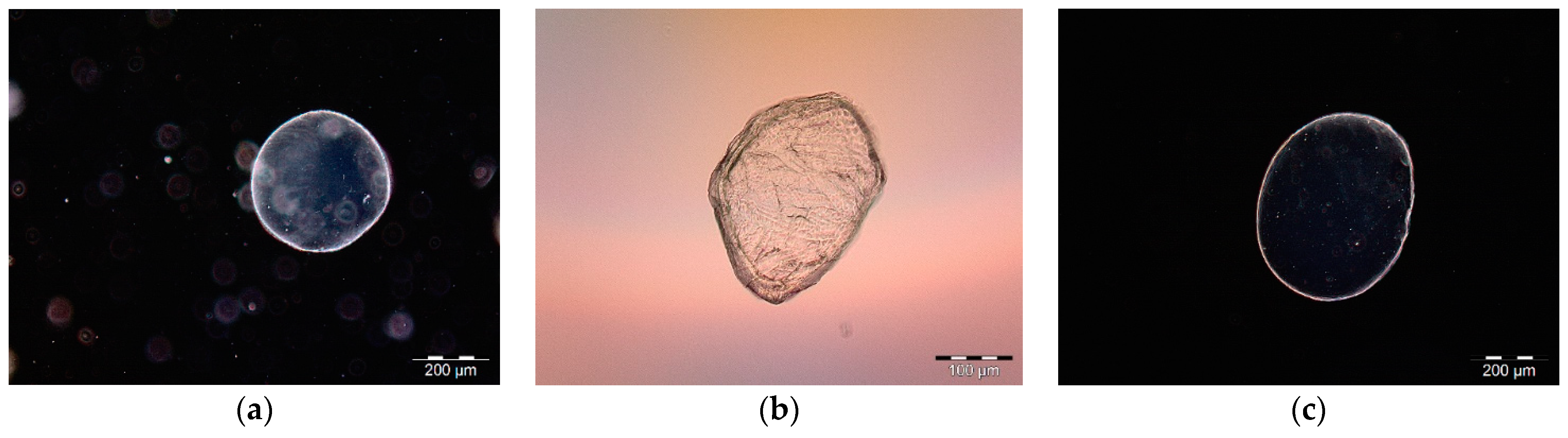
3.1.2. Morphological and Swelling Properties of Selected Microsphere Formulations

The morphology and size of the wet and dry microspheres were carried out by an optical microscope immediately after the preparation (Figure 1).
The diameter values of wet and dry microspheres with and without apitoxin after swelling in water, and the percentage of microsphere swelling after three hours in the water are presented in Table 4. All diameters were determined from 50 randomly selected microspheres.
The prepared microspheres containing apitoxin were spherical, but due to the small sizes and the presence of water molecules, they tended to coalesce together (Figure 1a). After drying to a constant mass the size of microspheres decreased. It was observed that the initial spherical shape was lost and became irregular resulting in a relatively wrinkled surface (Figure 1b). The wrinkled surface structure of the microsphere can be explained by a difference in polymer concentration on the surface and in the center of the gels, which results in uneven surfaces [46].
A non-significant difference was found between wet capsule size with and without apitoxin. However, after drying, the size of the microspheres formulation with apitoxin reduced to 226.95 μm, representing a significantly greater size compared to microspheres without apitoxin. The size reduction (about 25%) is a consequence of water and humidity loss associated with biopolymer strain-relaxation processes [7]. The difference between the radii of these capsules may also be explained by the interaction of apitoxin with the microsphere shell, which therefore does not collapse extensively during the drying process. The swelling behaviour indicated a rate at which this formulation absorbed water from the dissolution media and swelled. Because of swelling the microsphere formulations of (ALG/Api) become larger (~48%) and their Sw% considerably larger than microspheres without apitoxin (Table 4). These microsphere formulations are showing swelling behaviour in both media (deionized water pH = 6.8 and sodium-citrate buffer pH = 8.28) and from visual observation, the swelling started almost immediately and the hydrated layer was formed after their contact with the neutral or base medium (Figure 1c) [52].

3.1.3. In Vitro Release of Apitoxin from Microsphere Formulations

The possible use of alginate microspheres loaded with apitoxin in the plant protection process against insects requires research regarding their release capacity in certain physicochemical conditions. In this direction, the kinetics of the apitoxin release in deionized water and sodium citrate buffer from wet and dry microspheres prepared at a certain concentration of sodium alginate (1.5% (w/v)), calcium ions (2% (w/v)) and 0.5% (w/v) concentration of apitoxin was studied. Measuring the release of apitoxin from microspheres has proved problematic at short time intervals (1, 3, 5, 7, etc.) and therefore the monitoring interval was extended. Readings were conducted on a spectrophotometer at 60, 120, 180, 240, 1440, 2880, 4320, 8000, 10,080, and 11,520 min (192 h) periods until the maximum release from wet MS occurred (Figure 2a).
The release profiles of apitoxin from both wet microsphere formulations and dry microsphere formulations were different. In the case of wet microsphere formulations, the release of apitoxin in deionized water is characterized in three steps: (i) in the beginning it exhibits a rapid release; followed by (ii) a slower release [46]; and an (iii) third phase which comprises of a faster release of apitoxin due to the erosion of microsphere formulations. This initial burst release from wet microspheres could be attributed to the release of apitoxin contained on or near the surface of the microsphere formulations which is characteristic of amino acids and proteins [53].
In the dry microsphere formulations, the release of apitoxin in deionized water was much slower for overall measurements, obeying the power-law equation (f = 〖kt〗^n). The release profile (Figure 2a) shows an asymptotic pattern [54]. The slower release of apitoxin and asymptotic pattern could be explained as a consequence of the swelling of dry microspheres which influenced the release of the mechanism of apitoxin [52]. Some parts of the release function (i.e., after 12 h) obeyed the power-law equation [55]. The parameters of the power-law equation are presented in Table 5.
From the results of the power-law equation, in both cases (wet and dry microspheres formulations), the release of apitoxin was governed by the diffusion mechanism/anomalous transport. The release of apitoxin from both wet and dry microsphere formulations in deionized water was characterized by a very slow release whereafter as much as an hour the cumulative release was 11.22% and 6.6%, respectively. A nearly complete release of apitoxin was achieved only on the eighth day of measurement with a cumulative release of 98.12% and 26.1%. These results indicate the compatibility of water/microspheres with the apitoxin application during the plant treatment with a significant initial delay of its release. The slow release of apitoxin is important data for further assessing the effectiveness in pest control.
The release of apitoxin from wet microsphere formulations in the citrate buffer (pH = 8.28), was found to be complete after several minutes due to the complete degradation of spheres. The release of apitoxin from wet microspheres carried out in buffer is a representative simulation of the digestion of microspheres in insects, to assess the behaviour of apitoxin microspheres under certain conditions. Apitoxin was released more slowly from dry microspheres than from wet microspheres, respectively. After a minute and a half, the cumulative release was 48.31%, and after eighteen minutes there was a near-complete release from microspheres (98.74%). The release profile of apitoxin from dry calcium-alginate microparticles in the citric buffer is presented in Figure 2b.
The release fraction of apitoxin from dry microsphere formulation revealed the rapid release of apitoxin in comparison with releasing of the same formulation in deionized water. This could be mainly explained by the disintegration of the Ca-alginate structure as well as that at the pH = 8.28 there is a higher swelling ratio of microspheres which could be attributed to a chain expansion from the ionic carboxylate groups of alginate [56]. The parameters of the power-law equation are presented in Table 5. From the results of the power-law equation, the release of apitoxin from dry microparticles formulations in the sodium-citrate buffer (pH = 8.28) is governed by the diffusion mechanism which is related to the relaxation/dissolution process [56].
The pH of the digestive system of insects varies from species to species. Citrate buffer (pH = 8.28) was used in the investigation of insect digestion simulation because Vinokurov et al. [57] stated that within the specific family, all species have a morphologically similar digestive system, pH’s value varies from 5.9 to 9.0. Gayson [58] claims that the pH of L. decemlineata is 5.6–6.6, and the average pH gut value of all three investigated beetles in the paper is 6.3–8.3. Wet microspheres with apitoxin were mainly used, while apitoxin in dry microcapsules was only used in treatment with mealworm, where dry microcapsules are mixed with flour. Produced microspheres loaded with apitoxin are stable for application, characterized by a very slow release in the aqueous medium (important when preparing tank mixture), but very fast activation in the basic pH. This ensures the stability and applicability of microspheres formulations and their safe application without danger to humans and domestic animals.

3.2. The Efficiency of Apitoxin-Based Microspheres on Selected Inesct Pest Species

After encapsulation, we aimed to determine the effectiveness of apitoxin on harmful agricultural insect pests (notable pests in Croatian agriculture). This is the first study where the formulation of microspheres with apitoxin is used. All species used in the experiment, except the wheat weevil were treated with apitoxin for the first time, and in the study of Nassar [59], the wheat weevil was treated with apitoxin using a different method. The paper presents novel data on the effectiveness of apitoxin on harmful insect species using a new environmentally friendly formulation.
L. decemlineata is widely regarded as the most important insect defoliator of potatoes [60]. It is also the most important potato pest in Croatia. Without appropriate pest management, potato production would not be possible [60,61,62]. Due to the extensive damage it causes, manufacturers treat it intensively with chemicals, which creates a large burden on the environment and often leads to pest resistance [61,62,63,64,65,66]. Since the middle of the last century, L. decemlineata has developed resistance to 52 different compounds belonging to all major insecticide classes [60]. Insecticide resistance in this insect will likely remain a major challenge to pest control. The development of new insecticides is increasingly expensive, which affects their market price and available alternatives are less convenient and more environmentally damaging [60]. Therefore, it is essential to investigate a variety of novel techniques together with new active substances for the successful suppression of this pest in the years to come.
Conducted trials using apitoxin-based microspheres showed efficiency (E) against L. decemlineata larvae; higher efficiency was achieved after digestive treatment (75–97%). The first day after treatment, a difference between contact (E ~3%) and digestive (E ~27%) efficiency was observed. The same trend continued on the second and third days after treatment. On the third day after treatment, contact efficiency was 43 to 61% and no statistically significant difference was found between the concentrations applied. Digestive efficiency ranged from 75 to 97% without a significant difference between the concentrations applied.
The results of the study indicate the effectiveness of apitoxin on L. decemlineata larvae; approximately 61% mortality for the contact treatment and about 97% mortality for the digestive treatment. All applied concentrations of microspheres had satisfactory effects by the third day after treatment. The digestive activity was almost twice as good as contact treatment, which confirms the rapid release of apitoxin from microspheres introduced into the stomach of the larvae, as well as the potential of applying these formulations into potato protection against this pest (Table 6).
Sitophilus granarius is most commonly found infesting stored products in warehouses where it is capable of effecting considerable losses. It feeds on undamaged grains of cereals, but also on grains of corn, rice, chestnuts and pasta [67]. Contact insecticides have been widely used since the 1960s, alongside fumigants [68]. In warehouses, the use of contact insecticides or directly on grain is a very common way to kill this insect [68]. Over the past 40–50 years, the choice of insecticide compounds has moved from organochlorinated compounds such as dichlorvos, malathion, chlorpyrifos-methyl and pirimiphos-methyl, which act as contact or digestive insecticides [69,70]. Today the use of all compounds is becoming increasingly restricted [68].
Because of the great importance of this insect pest to agriculture and altered susceptibility (i.e., resistance), Nassar [59] tested apitoxin on S. granarius by contact application. Nassar [59] demonstrated mortality of 94% for adults after 72 h. However, the dosage applied was between 1.1 and 6.3 µg/insect; and the concentration was 6.3%. In comparison, in our study, several times lower concentrations (0.2–0.6%) of apitoxins in microspheres were used, which primarily ensure the economic acceptability of the application itself.
Apitoxin-based microspheres have efficiency against S. granarius adults (Table 6). On the first day after treatment, no significant efficiency was observed, neither contact nor digestive, the highest mortality was only 15%. Statistically significant differences between the concentrations applied were found on the second and third day of treatment. In both treatments, the highest efficiency was observed at the highest investigated apitoxin concentration (0.6%). On the third day after the treatment, maximum contact efficiency (E ~40%) and digestive efficiency (E ~48%) was observed at the highest tested concentration. These values are statistically higher than other concentrations applied, but they do not differ statistically from each other.
In our study, very low concentrations of apitoxin in microspheres were applied (0.2–0.6%) to ensure the economic acceptability of the application. In just three days, 40% (after contact application) and 47% efficiency (after digestive application) were found for the apitoxin formulated as wet microspheres. This result confirms the effectiveness of apitoxin in combating this type of insect.
T. molitor is the largest harmful beetle found in warehouses. It feeds on flour and flour products, grain, milk powder and meat, whereby the larvae damage the packaging of different products [67]. It is a polyphagous insect since it infests 46 commodities of animal and plant origin (e.g., grains, flour, amylaceous processed materials, pulses, nuts, dried meat, oilcake) [71]. Contrary to other stored-product pests, there is little knowledge on the management of T. molitor with contact insecticides [72]. Some synthetic insecticides are effective against this pest: α-cypermethrin with an efficiency of 97%; thiamethoxam with an efficiency of 94.4% [73]; α-cypermethrin in combination with entomopathogenic fungus Beauveria bassiana (Balsamo) had efficiency E = 100% the third day after treatment [74].
Apitoxin-based microspheres have low efficiency against T. molitor larvae (Table 6). The first three days after treatment, the apitoxin did not have a significant effect on the larvae, however, some efficiency was observed on contact variants (max. E ~22%). According to Table 6, on day 3, no significant difference was observed neither in contact or digested treatment. Whereas, the highest concentration in contact treatment was found to be significantly different as compared to the digestive treatment. The results of the study suggest that dry microcapsules of apitoxin mixed with flour did not affect T. molitor. As apitoxin was able to be released from dry microcapsules immediately after intake into the gastrointestinal tract of the insect (tested in different pH environments) additional investigations of digestive activity on this type of insect will be tested.
Investigations of this type are especially welcomed under the European “Green Deal” action plan [29]. Under this action plan, there is a call for an investigation into new formulations of plant protection products and microspheres fit this description because they are a safe option in terms of ecotoxicology for humans, livestock and sustainable agricultural production. The main advantage of microcapsules/microspheres is the controlled release of the active substance. Also, microcapsules/microspheres have enhanced usability which prevents premature release of the toxin into the environment and improved exploitation of active ingredients while minimizing environmental pollution [38].

3.3. Economic Analysis of the Apitoxin-Based Microsphere Uses in Agricultural Production

The third aim of this study was to assess the economic viability of apitoxin microformulations in integrated pest management (IPM). Due to insufficient market volumes and increasing demand, apitoxin achieves very high prices [10]. The market price of apitoxin is 23,000 euro/kg [75]. On the Croatian market, the price of apitoxin is 20 euros per gram.
Table 7 shows the costs of single-use of chemical and biological insecticides as well as the price of using microspheres with apitoxin in arable, vegetable and fruit production. All data on the prices of products were collected from an agricultural supply store in Zagreb, Croatia. Table 7 also shows that the use of chemical agents is the most financially favourable, followed by biological agents whose price is twice as high in arable production, but almost equated to the price of chemical preparations in vegetable and fruit production. Apitoxin-based microspheres are many times more expensive (5–15 times) than conventional chemical and biological preparations. The biggest price difference is in arable production, while the smallest price difference is between treatments in fruit production. During the analysis, the market of organic preparations used exclusively in organic production was also investigated. On the Republic of Croatia market, there are very few preparations for pest suppression, they mainly plant growth enhancers. Their prices are shown in grey and it is evident that they are 5 to 100 times more expensive than the price of apitoxin-based microspheres.
Since the price of apitoxin is the main limiting factor in the application of apitoxin microspheres, extremely low concentrations have been tested in the paper to make the application economically viable. The prices of apitoxin are many times higher than the prices of preparations allowed for the integrated production of crops (farming, vegetable farming, fruit farming), but they are also up to 100 times lower than insecticides licensed in organic production. Due to the high price of apitoxin, its future is in combining various environmentally friendly insecticides in low doses, which would achieve satisfactory efficiency, and the reduced dose reduces the cost of suppression and is less harmful to the environment [76]. Despite the price, the advantage of apitoxin microspheres is its characteristic slow release of active substance, which prolongs the period of action of apitoxin and provides longer-lasting protection, thus a smaller number of treatments is required.

4. Conclusions

Novel tests were conducted on the effectiveness of the application of encapsulated apitoxin to harmful agricultural insect pests, common in Croatia. Prepared microspheres loaded with apitoxin are stable and easily applicable. The encapsulated apitoxin has a steady initial and long residual effect, due to the slow release of apitoxin which is one of its main advantages over other conventional control methods. Apitoxin formulated as a microsphere has a detrimental effect on insects, of which it is significantly better gastric compared to contact action (due to pH). Here we demonstrated that the highest and fastest mortality was achieved when the highest concentrations were applied which have been shown to also be economically acceptable. Although the price of apitoxin microspheres is high, these new formulations have potential in organic and high-yield production. The collected data will contribute to the overall knowledge about the application and development of encapsulated formulations and the use of apitoxin microspheres as an ecofriendly-alternative in pest control.

Author Contributions

Conceptualization, D.L. and M.V.; methodology, D.L., M.V. and S.J.; software, S.J. and D.L.; validation, M.M. and M.V.; formal analysis, M.O., M.M. and K.V.-K.; investigation, M.O. and K.V.-K.; resources, M.V.; data curation, M.O., K.V.-K. and M.M.; writing—original draft preparation, M.O., D.L., S.J. and K.M.M.; writing—review and editing, K.M.M., D.L. and S.J.; visualization, S.J. and M.M.; supervision, M.V. and K.M.M.; project administration, M.V. and D.L.; funding acquisition, D.L. All authors have read and agreed to the published version of the manuscript.

Funding

This research received no external funding.

Institutional Review Board Statement

Not applicable.

Informed Consent Statement

Not applicable.

Data Availability Statement

The datasets and protocols used and/or analyzed during the current study are available from the corresponding author on reasonable request.

Acknowledgments

Authors thank Tvrtko Matijevic on apitoxin used in this study.

Conflicts of Interest

The authors declare no conflict of interest.

References

  1. Barzman, M.; Bàrberi, P.; Birch, A.N.E.; Boonekamp, P.; Dachbrodt-Saaydeh, S.; Graf, B.; Hommel, B.; Jensen, J.E.; Kiss, J.; Kudsk, P.; et al. Eight principles of integrated pest management. Agron. Sustain. Dev. 2015, 35, 1199–1215. [Google Scholar] [CrossRef]
  2. Callaghan, A. Mechanisms and detection of insecticide resistance: A review. Sci. Prog. 1991, 75, 423–438. [Google Scholar]
  3. Miller, G.T. Sustaining the Earth; Thompson Learning, Inc.: Pacific Grove, CA, USA, 2004. [Google Scholar]
  4. Štivičić, A.; Pajač Živković, I.; Lemic, D. Methods for determining resistance in entomological sciences. Fragm. Phytom. 2020, 34, 27–38. [Google Scholar]
  5. Javed, S.; Agurla, R. Molecular tools for detection of insecticide resistance. Int. J. Multidiscip. Adv. Res. Trends 2017, 4, 165–173. [Google Scholar]
  6. Zhang, L.; Yan, C.; Guo, Q.; Zhang, J.; Ruiz-Menjivar, J. The impact of agricultural chemical inputs on environment: Global evidence from informetrics analysis and visualization. Int. J. Low-Carbon Technol. 2018, 13, 338–352. [Google Scholar] [CrossRef] [Green Version]
  7. Jurić, S.; Đermić, E.; Topolovec-Pintarić, S.; Bedek, M.; Vinceković, M. Physicochemical properties and release characteristics of calcium alginate microspheres loaded with Trichoderma viride spores. J. Integr. Agric. 2019, 18, 2534–2548. [Google Scholar] [CrossRef]
  8. Pavela, R. Effectiveness of some botanical insecticides against Spodoptera littoralis Boisduvala (Lepidoptera: Noctudiae), Myzus persicae Sulzer (Hemiptera: Aphididae) and Tetranychus urticae Koch (Acari: Tetranychidae). Plant Prot. Sci. 2009, 45, 161–167. [Google Scholar] [CrossRef] [Green Version]
  9. Manzoli, M.; Gobbi, N.; Palma, M. Insects as biological models to assay spider and scorpion venom toxicity. J. Venom. Anim. Toxins 2003, 9, 174–185. [Google Scholar] [CrossRef]
  10. Levanić, M. Kemijska Karakterizacija Pčelinjeg Otrova Primjenom Infracrvene (FTIR-ATR) Spektroskopije. Master’s Thesis, University of Zagreb, Faculty of Agriculture, Zagreb, Croatia, 2019. [Google Scholar]
  11. Peiren, N.; de Graaf, D.C.; Vanrobaeys, F.; Danneels, E.L.; Devreese, B.; Van Beeumen, J.; Jacobs, F.J. Proteomic analysis of the honey bee worker venom gland focusing on the mechanisms of protection against tissue damage. Toxicon 2008, 52, 72–83. [Google Scholar] [CrossRef]
  12. Bellik, Y. Bee Venom: Its potential use in alternative medicine. Anti-Infect. Agents 2015, 13, 3–16. [Google Scholar] [CrossRef]
  13. Liu, X.; Chen, D.; Xie, L.; Zhang, R. Effect of honey bee venom on proliferation of K1735M2 mouse melanoma cells in-vitro and growth of murine B16 melanomas in-vivo. J. Pharm. Pharmacol. 2002, 54, 1083–1089. [Google Scholar] [CrossRef]
  14. Oršolić, N. Potentiation of Bleomycin lethality in HeLa and V79 cells by bee venom. Arch. Ind. Hyg. Toxicol. 2009, 60, 317–326. [Google Scholar] [CrossRef] [Green Version]
  15. Oršolić, N. Bee venom in cancer therapy. Cancer Metast. Rev. 2012, 3, 173–194. [Google Scholar] [CrossRef]
  16. Hossen, M.S.; Shapla, U.M. Impact of Bee Venom Enzymes on Diseases and Immune Responses. Molecules 2016, 22, 25. [Google Scholar] [CrossRef]
  17. Quistad, G.B.; Skinner, W.S.; Schooley, D.A. Venoms of social Hymenoptera-toxicity to the lepidopteran. Manduca sexta. Insect Biochem. 1988, 18, 511–514. [Google Scholar] [CrossRef]
  18. Lima, P.R.; Brochetto-Braga, M.R. Hymenoptera venom review focusing on Apis mellifera. J. Venom. Anim. Toxins 2003, 9, 149–162. [Google Scholar] [CrossRef]
  19. Bogdanov, S. Bee venom: Composition, health, medicine: A review. Bee Prod. Sci. 2017, 24, 1–20. [Google Scholar]
  20. Banks, B.; Shipolini, R. Chemistry and pharmacology of honey-bee venom. In Venoms of the Hymenoptera; Piek, T., Ed.; Academic Press: London, UK, 1986; pp. 330–416. [Google Scholar]
  21. Kim, H.; Lee, G.; Park, S.; Chung, H.S.; Lee, H.; Kim, J.Y.; Nam, S.; Kim, S.K.; Bae, H. Bee venom mitigates cisplatin-induced nephrotoxicity by regulating CD4+ CD5+ Foxp3+ Regulatory T cells in mice. Evid. Based Complementary Altern. Med. 2013, 10, 879845. [Google Scholar]
  22. Park, S.; Baek, H.; Jung, K.H.; Lee, G.; Lee, H.; Kang, G.H.; Lee, G.; Bae, H. Bee venom phospholipase a2 suppresses allergic airway inflammation in an ovalbumin-induced asthma model through the induction of regulatory t cells. Immun. Inflamm. Dis. 2014, 3, 386–397. [Google Scholar] [CrossRef] [PubMed]
  23. Sobral, F.; Sampaio, A.; Queiroz, M.J.; Calhelha, R.C.; Vilas-Boas, M.; Ferreira, I.C. Chemical characterization, antioxidant, anti-inflammatory and cytotoxic properties of bee venom collected in Northeast Portugal. Food Chem. Toxicol. 2016, 94, 172–177. [Google Scholar] [CrossRef] [Green Version]
  24. Ross, D.C.; Crim, J.W.; Brown, M.R.; Herzong, G.A.; Lea, A.O. Toxic and antifeeding actions of melittin in the corn earworm, Heliothis zea (Boddie): Comparisons to bee venom and insecticides chlorpyriphos and cyromazine. Toxicon 1987, 25, 307–313. [Google Scholar] [CrossRef]
  25. Mahgoub, M.O.; Lau, W.H.; Omar, D.B.; El Naim, A.M. Evaluation the Toxicity of Honey Bee Venom on Achroia grisella Developmental Stages. World J. Agric. Res. 2018, 6, 5–9. [Google Scholar]
  26. Hider, R.C. Honey bee venom: A rich source of pharmacologically active peptides. Endeavor 1988, 12, 60–65. [Google Scholar] [CrossRef]
  27. Hoffman, D.R. Hymenoptera venom proteins. Nat. Toxins 1996, 2, 169–186. [Google Scholar]
  28. Glinski, Z.; Jarosz, M. Infection and immunity in the honey bee Apis mellifera. Apiacata 2001, 36, 12–24. [Google Scholar]
  29. European Commision. Communication from the Commission to the European Parliament, the European Council, the Council, the European Economic and Social Committee and the Committee of the Regions; The European Green Deal COM/2019/640 final; European Commision: Brussels, Belgium, 2019. [Google Scholar]
  30. Ferrándiz, M.; Capablanca, L.; García, D.; Bonet, M.Á. Application of Antimicrobial Microcapsules on Agrotextiles. J. Agric. Chem. Environ. 2017, 6, 62–82. [Google Scholar] [CrossRef] [Green Version]
  31. Fang, Z.; Bhandari, B. Encapsulation of polyphenols—A review. Trends Food Sci. Technol. 2010, 21, 510–523. [Google Scholar] [CrossRef]
  32. Burgain, J.; Gaiani, C.; Linder, M.; Scher, J. Encapsulation of probiotic living cells: From laboratory scale to industrial applications. J. Food Eng. 2011, 104, 467–483. [Google Scholar] [CrossRef]
  33. Boras, A. Mikroinkapsulacija soja Lactobacillus Sakei MRS_296 S Potencijalom Primjene kao Starter Kkultura. Master’s Thesis, University of Zagreb, Faculty of Agriculture, Zagreb, Croatia, 2019. [Google Scholar]
  34. Gallo, M.; Corbo, M.R. Microencapsulation as a New Approach to Protect Active Compounds in Food. Application of Alternative Food-Preservation Technologies to Enhance Food Safety and Stability; Bentham Science Publisher: Sharjah, United Arab Emirates, 2010; pp. 188–195. [Google Scholar]
  35. Teixeira da Silva, P.; Martins Fries, L.L.; Ragagnin de Menezes, C.; Tasch Holkem, A.; Schwan, C.L.; Wigmann, É.F.; De Oliveira Bastos, J.; De Bona da Silva, C. Microencapsulation: Concepts, mechanisms, methods and some applications in food technology. Ciência Rural 2014, 44, 1304–1311. [Google Scholar] [CrossRef]
  36. Usmiati, S.; Richana, N.; Mangunwidjaja, D.; Noor, E.; Prangdimurti, E. The Using of Ionic Gelation Method Based on Polysaccharides for Encapsulating the Macromolecules. Int. Conf. Food Secur. Nutr. 2014, 67, 79–84. [Google Scholar]
  37. Vinceković, M.; Jalšenjak, N.; Topolovec-Pintarić, S.; Đermić, E.; Bujan, M.; Jurić, S. Encapsulation of Biological and Chemical Agents for Plant Nutrition and Protection: Chitosan/Alginate Microcapsules Loaded with Copper Cations and Trichoderma viride. J. Agric. Food Chem. 2016, 64, 8073–8083. [Google Scholar] [CrossRef] [PubMed]
  38. Liu, B.; Wang, Y.; Yang, F.; Cui, H.; Wu, D. Development of a chlorantraniliprole microcapsule formulation with a high loading content and controlled-release property. J. Agric. Food Chem. 2017, 66, 6561–6568. [Google Scholar] [CrossRef] [PubMed]
  39. Oxley, J. Bioencapsulation Innovations. In Microencapsulation: Guide to Industrial Applications; Bioencapsulation Research Group: Sucé sur Erdre, France, 2015; Volume 41, pp. 16–17. [Google Scholar]
  40. Knowles, A. Recent developments of safer formulations of agrochemicals. Environmentalist 2008, 28, 35–44. [Google Scholar] [CrossRef]
  41. Kumar Das, S.; Nakka David, S.R.; Rajabalaya, R.; Mukhopadhyay Kumar, H.; Halder, T.; Palanisamy, M.; Khanam, J.; Nanda, A. Microencapsulation techniques and its practice. J. Pharm. Sci. Technol. 2011, 2, 1–23. [Google Scholar]
  42. Bashir, O.; Claverie, J.P.; Lemoyne, P.; Vincent, C. Controlled-release of Bacillus thurigiensis formulations encapsulated in light-resistant colloidosomal microcapsules for the management of lepidopteran pests of Brassica crops. Peer J. 2016, 4, 2524. [Google Scholar] [CrossRef] [Green Version]
  43. Nuruzzaman, M.; Rahman, M.M.; Liu, Y.; Naidu, R. Nanoencapsulation, Nano-Guard for pesticides: A new window for safe application. J. Agric. Food Chem. 2016, 64, 1447–1483. [Google Scholar] [CrossRef] [PubMed]
  44. Haramija, F. Primjena Biopolimernih Kapsula u Konvencionalnom Uzgoju Salate. Master’s Thesis, University of Zagreb, Faculty of Agriculture, Zagreb, Croatia, 2019. [Google Scholar]
  45. Steinbrenner, U.; Bratz, M. Bioencapsulation Innovations. In Challenges for Microencapsulated Formulations in Agriculture; Bioencapsulation Research Group: Sucé sur Erdre, France, 2015; pp. 14–15. [Google Scholar]
  46. Jurić, S.; Tanuwidjaja, I.; Mrkonjić Fuka, M.; Vlahoviček-Kahlina, K.; Marijan, M.; Boras, A.; Kolić, N.U.; Vinceković, M. Encapsulation of two fermentation agents, Lactobacillus sakei and calcium ions in microspheres. Colloids Surf. B Biointerfaces 2021, 197, 111387. [Google Scholar] [CrossRef] [PubMed]
  47. Xue, W.-M.; Liu, X.; He, X.; Wang, W.; Ma, X.-J. Chemical Method of Breaking the Cell-Loaded Sodium Alginate/Chitosan Microcapsules. Gaodeng Xuexiao Huaxue Xuebao/Chem. J. Chin. Univ 2004, 55, 25. [Google Scholar]
  48. Abbott, W.S. A method of computing the effectiveness of an insecticide. J. Econ. Entomol. 1925, 18, 265–267. [Google Scholar] [CrossRef]
  49. Gylling Data Management Inc. ARM 2019® GDM Software; Revision 2019.4; August 5,2019 (B=25105); Gylling Data Management Inc.: Brookings, SD, USA, 2019. [Google Scholar]
  50. Mong Thu, T.T.; Krasaekoopt, W. Encapsulation of protease from Aspergillus oryzae and lipase from Thermomyces lanuginoseus using alginate and different copolymer types. Agric. Nat. Resour. 2016, 50, 155–161. [Google Scholar] [CrossRef] [Green Version]
  51. Aguilera, D.A.; Spinozzi Di Sante, L.; Pettignano, A.; Riccioli, R.; Roeske, J.; Albergati, L.; Corti, V.; Fochi, M.; Bernardi, L.; Quignard, F.; et al. Adsorption of a Chiral Amine on Alginate Gel Beads and Evaluation of its Efficiency as Heterogeneous Enantioselective Catalyst. Eur. J. Org. Chem. 2019, 2019, 3842–3849. [Google Scholar] [CrossRef]
  52. Sriamornsak, P.; Thirawong, N.; Korkerd, K. Swelling, erosion and release behavior of alginate-based matrix tablets. Eur. J. Pharm. Biopharm. 2007, 66, 435–450. [Google Scholar] [CrossRef] [PubMed]
  53. Alonso, M.J.; Gupta, R.K.; Min, C.; Siber, G.R.; Langer, R. Biodegradable microspheres as controlled-release tetanus toxoid delivery systems. Vaccine 1994, 12, 299–306. [Google Scholar] [CrossRef]
  54. Sánchez, A.; Villamayor, B.; Guo, Y.; McIver, J.; Alonso, M.J. Formulation strategies for the stabilization of tetanus toxoid in poly(lactide-co-glycolide) microspheres. Int. J. Pharm. 1999, 185, 255–266. [Google Scholar] [CrossRef]
  55. Higuchi, T. Drug Release. In Modeling in Biopharmaceutics, Pharmacokinetics, and Pharmacodynamics; Macheras, P., Iliadis, A., Eds.; Springer: New York, NY, USA, 2006; pp. 57–88. [Google Scholar]
  56. Lupo, B.; Maestro, A.; Porras, M.; Gutiérrez, J.M.; González, C. Preparation of alginate microspheres by emulsification/internal gelation to encapsulate cocoa polyphenols. Food Hydrocoll. 2014, 38, 56–65. [Google Scholar] [CrossRef]
  57. Vinokurov, K.; Taranushenko, Y.; Krishnan, N. Protease, amylase, and protease-inhibitor activities in the gut of six cockroach species. J. Insect Physiol. 2007, 53, 794–802. [Google Scholar] [CrossRef] [PubMed]
  58. Grayson, J. Digestive Tract pH of Six Species of Coleoptera; Virgina Agricultural Experminet Station: Blacksburg, VA, USA, 1958. [Google Scholar]
  59. Nassar, M.I. The potential of natural venom of Apis mellifera for the control of grain weevil adults (Sitophilus granarius Coleoptera Curculionidae). Int. J. Entomol. Res. 2013, 1, 25–31. [Google Scholar]
  60. Alyokhin, A.; Baker, M.; Mota-Sanchez, D.; Dively, G.; Grafius, E. Colorado Potato Beetle Resistance to Insecticides. Am. J. Pot. Res. 2008, 85, 395–413. [Google Scholar] [CrossRef]
  61. Bažok, R.; Čačija, M.; Lemić, D.; Virić Gašparić, H.; Drmić, Z. Rezistentnost štetnika na insekticide. Glas. Biljn. Zaštite 2017, 17, 429–438. [Google Scholar]
  62. Gluckselig, B. Rezistentnost Populacija Krumpirove Zlatice na Organofosforne Insekticide, Piretroide i Neonikotenoide. Master’s Thesis, University of Zagreb, Faculty of Agriculture, Zagreb, Croatia, 2019. [Google Scholar]
  63. Stewart, J.G.; Kennedy, G.G.; Sturz, A.V. Incidence of insecticide resistance in populations of Colorado potato beetle, Leptinotarsa decemlineata (Say) (Coleoptera: Chrysomelidae), on Prince Edward Island. Can. Entomol. 1997, 129, 21–26. [Google Scholar] [CrossRef]
  64. Stankovic, S.; Zabel, A.; Kostic, M.; Manojlovic, B.; Rajkovic, S. Colorado potato beetle [Leptinotarsa decemlineata (Say)] resistance to organophosphates and carbamates in Serbia. J. Pest Sci. 2004, 77, 11–15. [Google Scholar] [CrossRef]
  65. Mota-Sanchez, D.; Hollingworth, R.M.; Grafius, E.J.; Moyer, D.D. Resistance and cross-resistance to neonicotinoid insecticides and spinosad in the Colorado potato beetle, Leptinotarsa decemlineata (Say) (Coleoptera: Chrysomelidae). Pest Manag. Sci. 2006, 62, 30–37. [Google Scholar] [CrossRef]
  66. Benkovskaya, G.V.; Udalov, M.B.; Nikolenko, A.G.; Leontieva, T.L. Temporal and toxicological dynamics in the cover spot patterns of the Colorado potato beetle in South Ural. Resist. Pest Manag. 2006, 15, 13–15. [Google Scholar]
  67. Maceljski, M. Poljoprivredna Entomologija; Zrinski d.d.: Čakovec, Croatia, 2002. [Google Scholar]
  68. Kljajić, P.; Perić, I. Susceptibility to contact insecticides of granary weevil Sitophilus granarius (L.) (Coleoptera: Curculionidae) originating from different locations in the former Yugoslavia. J. Stored. Prod. Res. 2006, 42, 149–161. [Google Scholar] [CrossRef]
  69. White, N.D.G.; Leesch, J.G. Chemical control. In Integrated Management of Insects in Stored Products; Subramanyam, B., Hagstrum, D.W., Eds.; Marcel Dekker: New York, NY, USA; Basel, Switzerland; Hong Kong, China, 1996; pp. 287–330. [Google Scholar]
  70. Tomlin, C. The Pesticide Manual; British Crop Protection Council and The Royal Society of Chemistry: Cambridge, UK, 2000. [Google Scholar]
  71. Hagstrum, D.W.; Subramanyam, B. Stored-Product Insect Resource; AACC International: St. Paul, MN, USA, 2009. [Google Scholar]
  72. Kavallieratos, N.G.; Papanikolaou, N.E.; Kazani, A.N.; Boukouvala, M.C.; Malesios, C. Using multilevel models to explore the impact of abiotic and biotic conditions on the efficacy of pirimiphos-methyl against Tenebrio molitor L. Environ. Sci. Pollut. Res. 2021, 28, 17200–17207. [Google Scholar] [CrossRef] [PubMed]
  73. Athanassiou, C.G.; Kavallieratos, N.G.; Boukouvala, M.C.; Mavroforos, M.E.; Kontodimas, D.C. Efficacy of alpha-cypermethrin and thiamethoxam against Trogoderma granarium Everts (Coleoptera: Dermestidae) and Tenebrio molitor L. (Coleoptera: Tenebrionidae) on concrete. J. Stored. Prod. Res. 2015, 62, 101–107. [Google Scholar] [CrossRef]
  74. Meyling, N.V.; Arthur, S.; Pedersen, K.E.; Dhakal, S.; Cedergreen, N.; Fredensborg, B.L. Implications of sequence and timing of exposure for synergy between the pyrethroid insecticide alpha-cypermethrin and the entomopathogenic fungus Beauveria bassiana. Pest Manag. Sci. 2018, 74, 2488–2495. [Google Scholar] [CrossRef] [PubMed]
  75. Serrinha, V.; Correia, S.D.; Marques, G. Productivity and Economic Analysis of a New Intensive Collector in the Portuguese Market with Implication of Open Innovation Perspective. J. Open Innov. Technol. Mark. Complex. 2019, 5, 71. [Google Scholar] [CrossRef] [Green Version]
  76. Čačija, M.; Drmić, Z.; Kadoić Balaško, M.; Skendžić, S.; Lemić, D.; O’Keffee, J.; Jurada, I.; Bažok, R. Kombinacije Insekticida u Subletalnim Dozama-Antirezistentna Strategija u Suzbijanju Krumpirove Zlatice; Hrvatsko društvo biljne zaštite: Zagreb, Croatia, 2020; pp. 31–32. [Google Scholar]
Figure 1. Left to right, optical microscope microphotographs of wet(a), dry (b), swelled (c) microspheres.
Sustainability 13 06167 g001
Figure 2. The fraction (f) of released apitoxin from wet microsphere formulations (black figures) and dry microsphere formulations (red figures) in deionized water over time (min) observed at an initial apitoxin concentration of 0.5% (w/v) (a). The fraction of released apitoxin in Na-citrate buffer (pH = 8.28) with time (min) of dry microsphere formulations (red signs) at an initial apitoxin concentration of 0.5% (b).
Sustainability 13 06167 g002
Table 1. Description of the investigated insect pest species.
SpeciesDevelopmental Stage TestedInvestigated ActionNo. of Tested Individuals
Leptinotarsa decemlineataLarvaecontact, digestive320
Sitophilus granariusAdultscontact, digestive320
Tenebrio molitorLarvaecontact, digestive320
Table 2. Variants in the experiment and detailed calculation of apitoxin doses expressed per variant for all insects tested.
Variants (Amount of Apitoxin in Microspheres (%) Apitoxin Based Microspheres Per Repetition (mg) Dose of Apitoxin
Per Repetition (mg)
0.2%53.360.11
0.4%53.360.21
0.6%53.360.32
Control *--
* Deionized water was used as a control for L. decemlineata and S. granarius and flour was control variant for T. molitor.
Table 3. Encapsulation efficiency (EE%) and loading capacity (Lc) of dry and wet apitoxin microsphere formulations (prepared with the 1.5% (w/v) sodium alginate).
Apitoxin Content (%w/v)Lc Dry (mg/g)Lc Wet (mg/g)EE (%)
0.2%102.46 ± 3.07 a*2.96 ± 0.08 a73.87 ± 1.50 a
0.4%108.69 ± 8.82 a5.22 ± 0.23 b74.13 ± 0.92 a
0.5%219.11 ± 4.22 b6.33 ± 0.11 b76.53 ± 1.63 a
0.6%274.84 ± 6.14 c7.94 ± 0.16 c73.76 ± 2.00 a
* Values superscripted with the same letter within a column are not significantly different according to the post-hoc t-test with Bonferroni adjustment (p < 0.05).
Table 4. Diameters of wet and dry alginate microspheres (MS) with and without apitoxin.
MS CompositionWet MS Diameter (μm)Dry MS Diameter (μm)MS Diameter after Swelling (μm)Swelling Degree (%)
1.5% alginate/0.5% apitoxin301.58 ± 40.09 a*226.95 ± 29.06 a436.04 ± 37.79 a136.77 ± 4.17 a
1.5% alginate (control)294.13 ± 46.05 a178.44 ± 36.71 b320.32 ± 19.78 b33.90 ± 1.25 b
* Values superscripted with the same letter (a–c) within a column are not significantly different according to the posthoc t-test with Bonferroni adjustment (p < 0.05).
Table 5. The values of the release constant (k) and exponent (n) of encapsulated apitoxin for wet and dry microspheres in deionized water and sodium citrate buffer (pH = 8.28).
Microspheres Formulations (Ca-Alginate/Apitoxin)Release MediumK (min−1)nR2
wetdeionized water0.00910.48070.9996
dry0.00180.56540.9984
drysodium-citrate buffer0.57100.21270.9901
Table 6. The efficiency (%) of apitoxin-based microspheres on tested insects.
Treatment Dose of Apitoxin Per Repetition (mg)Days after the Treatment
123
Leptinotarsa decemlineata
contact0.110 ± 0 b22.81 ± 7.02 b43.86 ± 7.02 b
0.213.3 ± 3.3 ab40.35 ± 3.51 ab61.4 ± 3.51 ab
0.323.3 ± 3.3 ab26.32 ±6.08 b47.37 ± 12.15 b
digestive0.1116.7 ± 6.7 ab47.37 ± 6.08 ab96.49 ± 3.51 a
0.2126.7 ± 3.3 a57.89 ± 6.08 a75.44 ± 3.51 ab
0.3223.3 ± 8.8 ab47.37 ± 6.08 ab78.95 ± 12.16 ab
HSD p = 0.05 **25.3328.1135.35
Sitophilus granarius
contact0.112.5 ± 2.50.6 ± 4.6 b*12.2 ± 2 b
0.215.0 ± 59.1 ± 6.8 ab16.8 ± 3.6 b
0.3215.0 ± 2.922.4 ± 1.7 a40.0 ± 0 a
digestive0.110 ± 010.0 ± 0 b14.6 ± 2.3 b
0.210 ± 012.2 ± 2 b20.0 ± 0 b
0.325.0 ± 2.924.8 ± 1.9 a47.5 ± 1.4 a
HSD p = 0.05 ** 13.010.411.6
Tenebrio molitor
0.1110.0 ± 21.218.2 ± 2.218.2 ± 2.2 ab*
contact0.2110.0 ± 5.810.1 ± 0.919.3 ± 0.5 ab
0.325.0 ± 2.921.6 ± 0.534.8 ± 0.2 a
0.110 ± 00 ± 00 ± 0 b
digestive0.210 ± 00 ± 01.3 ± 0.6 b
0.320 ± 00 ± 00 ± 0 b
HSD p = 0.05 ** nsns31.4
* Mean values of the same column followed by the same letter (a, ab, b) are not significantly different (p > 0.05 HSD test). ** HSD was determined by comparing the effectiveness of apitoxin between the concentrations used for each reading.
Table 7. Economic analysis of pest control in different agricultural systems. Data are presented for a one-time treatment.
Agricultural ProductionType of PreparationRecommended
Dosage **
Price Per
Hectare (€/ha)
Arable cropsChemical 150 mL/ha22 *
Biological 150 mL/ha32
Microspheres with apitoxin16 g/ha318 ¥
Vegetables Chemical 300 mL/ha32
Biological 600 g/ha34
Microspheres with apitoxin16 g/ha318 ¥
OrchardsChemical 100 mL/100l69
Biological 1000 g/ha90
Microspheres with apitoxin16 g/ha318 ¥
Ecological Bioinsecticide50 g/m226,513
Booster10 L/ha1753
Microorganism1 kg/ha131
* Price of standard preparation in the use (information from the agricultural pharmacy). ** mean recommended dose according to user instructions. ¥ calculated for mean tested concentration (0.4%) and for water consumption of 200 L/ha.
Publisher’s Note: MDPI stays neutral with regard to jurisdictional claims in published maps and institutional affiliations.

