Article

Estradiol Upregulates the Expression of the TGF-β Receptors ALK5 and BMPR2 during the Gonadal Development of Schizothorax prenanti

College of Animal Science and Technology, Sichuan Agricultural University, Chengdu 611130, China
*
Author to whom correspondence should be addressed.
These authors contributed equally to the study.
Animals 2021, 11(5), 1365; https://doi.org/10.3390/ani11051365
Submission received: 16 March 2021 / Revised: 6 May 2021 / Accepted: 7 May 2021 / Published: 11 May 2021
(This article belongs to the Section Animal Genetics and Genomics)

Abstract

:

Simple Summary

Schizothorax prenanti, known as the ya-fish, is mainly distributed in regions adjacent to the Qinghai-Tibet Plateau (QTP) and is an endemic fish species with great economic importance in aquaculture in Western China. In the present study, we were aimed to explore the functions of ALK5 and BMPR2 during the gonadal development of S. prenanti. Our results suggest that ALK5 and BMPR2 may play a potentially vital role in both folliculogenesis and spermatogenesis in S. prenanti.

Abstract

TGF-β receptors play important roles in mediating TGF-β signals during gonadal development. To identify the functions of TGF-β receptors, including the type I receptor (activin receptor-like kinase 5, ALK5) and type II receptor (bone morphogenetic protein receptor 2, BMPR2), during the gonadal development of S. prenanti, the full-length cDNA sequences of ALK5 and BMPR2 were isolated and characterized. Their expression patterns in developing gonads and in the gonads of exogenous estradiol (E2) -fed fish were analyzed. The cDNAs of ALK5 and BMPR2 were 1925 bp and 3704 bp in length and encoded 501 and 1070 amino acid residues, respectively. ALK5 and BMPR2 were mostly expressed in gonads, particularly in cortical alveoli stage ovaries and mid-spermatogenic stage testes; however, the overall level of BMPR2 mRNA was higher than that of ALK5 during gonadal development. Furthermore, immunohistochemical signals of ALK5 and BMPR2 were mostly detected at chromatin nucleolar oocytes and perinuclear oocytes in ovaries and at spermatocytes and spermatogonia in testes. Exogenous E2 induces the gonadal expression of ALK5 and BMPR2, and BMPR2 is more responsive to E2 than ALK5. These results suggest that ALK5 and BMPR2 might play a potentially vital role in both folliculogenesis and spermatogenesis in S. prenanti.

1. Introduction

Transforming growth factor-beta (TGF-β) superfamily receptors, including the type I receptor (activin receptor-like kinase 5, ALK5) and type II receptor (bone morphogenetic protein receptor 2, BMPR2), are key factors in the transmission of signals that regulate gonadal development [1,2,3]. As a high-affinity signaling complex, BMPR2 and ALK5 transmit intracellular signals through the phosphorylation of downstream effectors during the reproductive process by stimulating and subsequently activating TGF-βs [2,4,5,6,7].
ALK5 is mainly expressed in mammalian gonads and expressed at high levels in cattle ovaries [8]. ALK5 has also been detected in human germ cells [9] and in oocytes and granulosa cells of mice and pigs at different developmental stages [10]. During the development of the corpus luteum (CL) from early to late CL, the expression of ALK5 continually increases [11]. In fish, ALK5, which is abundant in gonads and muscles, is distributed in the tissues of Oncorhynchus mykiss [12] and mainly expressed in the liver of Oreochromis niloticus [13] and Carassius gibelio [14]. These results indicated that the expression of ALK5 varies in different species.
Mammalian BMPR2 is widely expressed in various tissues, particularly in porcine oocytes [15] and granulosa cells of ewes [3]. The expression of BMPR2 in chicken exhibits a gradual increase during follicular development [16]. In addition, the expression of BMPR2 in pigs is higher at both the embryonic stage and pre- and late puberty than at other stages [17], but its expression is stable from early to late CL [11]. In fish, BMPR2 is widely distributed in Danio rerio and is mainly expressed in the testis and liver [18]. However, the abundant expression has been detected in the brain and liver of C. gibelio [14]. These results show that BMPR2 is expressed in different tissues and species, suggesting that BMPR2 plays different roles in various species.
ALK5 promotes the development of germ cells [9], oocytes and granulosa cells [10] in humans, inhibits pig granulosa cells apoptosis [19], and participates in establishing luteolysis in mare steroidogenic cells [11], and a lack of ALK5 induces severe defects in mice yolk sac development [20]. BMPR2 participates in embryonic development of porcine [17], follicular development of chicken [16] and cumulus cells expansion, proliferation and apoptosis of bovine [21] and maintains steroidogenesis in human [22], progesterone biosynthesis in rat granulosa cells [23], and ovarian cells functional homeostasis in hamster [24]. There is no doubt that ALK5 and BMPR2 are essential and vital factors and participate in these biological processes by enhancing or abating upstream signals. Therefore, these results are meaningful for obtaining further clarification of their roles in signal transduction.
Schizothorax prenanti, which belongs to the subfamily Schizothoracinae of the Cyprinidae family, is mainly distributed in regions adjacent to the Qinghai-Tibet Plateau (QTP) and an endemic fish species with great economic importance in aquaculture in Western China [25]. Our previous report showed that GDF9 plays an important role in folliculogenesis and spermatogenesis in S. prenanti [26], but the downstream signal transduction mechanism remains unclear. Because ALK5 and BMPR2 are important and necessary for signal transduction, their regulatory roles in the gonadal development process of S. prenanti should be unraveled. In the present study, we cloned the ALK5 and BMPR2 cDNA sequences, analyzed the expression patterns of ALK5 and BMPR2 at both the mRNA and protein levels, and detected their expression in juvenile fish gonads via the withdrawal of estradiol (E2) administration. All these results allow in-depth exploration of the functions of ALK5 and BMPR2 during the gonadal development of S. prenanti.

2. Materials and Methods

2.1. Animals

Adult S. prenanti (body length, 38.73 ± 3.52 cm; body weight, 1194.79 ± 197.86 g) were purchased from the Lu Shan Sword Fishing Company (Sichuan, China). All these fish were maintained in 60.0 × 50.0 × 40.0 cm3 indoor tanks at the Aquaculture Laboratory of Sichuan Agricultural University (Chengdu, China) under a natural photoperiod (16 h light: 8 h dark) at a temperature of 19.0 ± 0.1 °C. Fish were supplied with a flow of the freshwater system. All the experiments were performed following the Animal Research and Ethics Committees of Sichuan Agricultural University and followed the animal experiments guidelines of Sichuan Agricultural University (Approval No: 20170031).

2.2. Tissue Sampling

All adult fish were anesthetized with 0.02% tricaine buffer (80 µg/L) (Sigma, St. Louis, MO, USA) for 10 min after a 24 h fast. Tissues (n = 5), including the hypothalamus, telencephalon, epithalamus, heart, kidney, blood, liver, eyes, muscle, spleen, swim bladder, intestine, head kidney, medulla oblongata, skin, pituitary, gills, testes, and ovaries, were collected. Only the gonads were collected from the remaining individuals (n = 45). The tissues were snap-frozen in liquid nitrogen and stored at −80 °C until use. Fresh gonad samples were immobilized in Bouin’s solution and stored in 75% ethanol.
According to previous reports [25,27], the gonadal developmental stages of fish were identified through histological analysis of the gonad sections (the 5 μm sections were stained with hematoxylin–eosin). The gonads were classified as follows: ovaries at the chromatin nucleolar stage (CNS), perinucleolar stage (PS), cortical alveoli stage (CAS), mid-vitellogenic stage (MVS), and late vitellogenic stage (LVS) and testes at the spermatogonium stage (SGT), early spermatogenic stage (EST), mid-spermatogenic stage (MST) and late spermatogenic stage (LST).

2.3. Total RNA Isolation and Sequence Cloning

Total RNA was extracted using the TRIzol reagent (Invitrogen, Carlsbad, CA, USA) according to the manufacturer’s instructions. The integrity of total RNA was assessed by 1% agarose gel electrophoresis, and the optical density absorption ratios at 260 and 280 nm were quantified with a photometer (Bio-Rad, Hercules, CA, USA). Before reverse transcription, total RNA was enzymatically digested with gDNA Eraser (TaKaRa, Dalian, China).
The ovarian cDNAs were transcribed from 1.0 μg of total RNA from the ovary using a BD SMART™ PCR cDNA synthesis kit (Invitrogen, Carlsbad, CA, USA) according to the manufacturer’s instructions. Degenerate primers were designed according to the conserved regions of ALK5 and BMPR2. The 5′-RACE and 3′-RACE primers were designed based on their partial sequences (Table 1). Except for the first round of annealing (performed at 54 °C) and the second round of annealing (performed at 56 °C), the PCR methods and procedures were performed as described previously. The PCR mixtures comprised 5.0 μL of 2× Taq buffer (DSBIO, Guangzhou, China), forward and reverse primers (0.5 µM each), 3 μL of ddH2O, and 40 ng of cDNA, and the reactions were performed with the following program: pre-denaturation at 94 °C for 3 min, 30 cycles of denaturation at 94 °C for 30 s, annealing at 54 °C for 30 s, and extension at 72 °C for 90 s and elongation at 72 °C for 10 min. The PCR products were purified from a 1.5% agarose gel using a universal DNA purification kit (TIANGEN, Beijing, China), ligated into the pMD-19T vector (TaKaRa, Dalian, China), and then introduced into E. coli DH5α competent cells (Takara, Dalian, China). The positive clone was isolated and sequenced by Chengdu Tsingke Biotechnology Co., Ltd. (Chengdu, China).
The ALK5 and BMPR2 amino acid sequences of S. prenanti and other vertebrates were aligned by BLAST (http://bLast.ncbi.nLm.nih.gov/BLast.cgi, accessed on 24 September 2020). The phylogenetic tree was constructed using the neighbor-joining algorithm of MEGA 7.0. The reliability of the analysis was assessed by 1000 bootstrap replicates.

2.4. Real-Time Quantitative PCR Analysis

According to the manufacturer’s recommended protocol, the first-strand cDNA synthesis kit (Thermo, Waltham, MA, USA) was used for cDNA synthesis, and the cDNA quality was verified by the stable amplification of β-actin. As shown in our previous study, we used both β-actin and 18S rRNA as reference genes for controlling the error between samples in the analysis of ALK5 and BMPR2 expression. For all standard curves, the primer amplification efficiencies of the genes were 94.7–96.8% and 0.990 < R2 < 0.999. The target genes were normalized to the reference genes (geometric averaging of the β-actin and 18S rRNA Ct values), and the expression levels were compared using the relative Ct method.

2.5. Western Blotting Analysis

Primary antisera of BMPR2 (LS-C178875) and ALK5 (LS-C312882) were purchased from LifeSpan BioSciences (Seattle, WA, USA). Western blot analysis was performed according to the standard protocol. The ovarian and testicular homogenates were separated on a 12% SDS–PAGE gel, and the proteins were then transferred to PVDF (methanol-activated polyvinylidene difluoride) membranes (Merck Millipore, Darmstadt, Germany) using Bio-Rad trans-blot (Bio-Rad, Hercules, CA, USA). The membranes were incubated overnight in 5% nonfat milk powder in 10 mM PBST buffer at 4 °C, blocked and incubated again with the primary antiserum (1:800 dilution) diluted with 5% nonfat milk powder in 10 mM PBST, at 25 °C for 2 h. The membrane was subjected to three 5 min washes with PBST and incubated in horseradish peroxidase (HRP)-conjugated goat anti-rabbit IgG (Boster, Wuhan, China; 1:500 dilution) diluted with 5% nonfat milk powder in 10 mM PBST for 1 h at 25 °C. According to the protocol recommended by the manufacturer of the ECL kit (MXB, Fuzhou, China), the membranes were washed with PBST and detected using Bio-Rad ChemiDocTM MP (Bio-Rad, Hercules, CA, USA).

2.6. Immunohistochemistry Analysis

In 3% hydrogen peroxide, 5 μm-thick paraffin sections of the gonads were deparaffinized, dehydrated and incubated for 30 min. The sections were subjected to three 5 min washes with phosphate-buffered saline (PBS, 10 mM) and incubated with 10% normal goat serum (Boster, Wuhan, China) for 20 min to block nonspecific reactions. The sections were then incubated with rabbit BMPR2 and ALK5 (1:300 dilution) at 25 °C for 2 h, washed three times and incubated with a secondary antibody [horseradish peroxidase (HRP)-conjugated goat anti-rabbit IgG; 1:1000 dilution]. After rinsing with PBS, the sections were developed with an ECL reagent kit, mounted, examined under a microscope, and digitally photographed (Nikon, Tokyo, Japan).

2.7. Effect of ALK5 and BMPR2 Expression on E2-Fed Gonads

The fish were hatched in a laboratory at Sichuan Agricultural University (Chengdu, China) and supplied with continuously flowing fresh water at 19.0 ± 1.0 °C and natural light. Four hundred fifty fish (46.50 ± 1.55 mm) were randomly assigned to 15 indoor fiberglass tanks (60.0 × 50.0 × 40.0 cm3, 30 fish per tank) in the laboratory.
The feeding trial involved five diets: the blank and alcohol control diets and E2-supplemented diets (50, 100 and 150 mg/kg E2). Before being added to the diets, E2 (Sigma, St. Louis, MO, USA) was dissolved in alcohol. The fish in the alcohol control diet-fed group were treated like those in the E2 diet-fed groups. The fish belonging to the control group were fed a common diet throughout the experiment, which lasted 4 weeks. All fish were fed a common diet for 2 weeks before initiating the experiment to allow their acclimatization to the experimental diet and conditions. All fish were fed twice a day at 8:00 and 18:00, and the proportion was 3% of the body weight. The residual diets were removed with a siphon tubular. The survival ratio of every group was calculated daily.
Ten fish from each group were anesthetized via a tricaine buffer bath (80 µg/L), and gonad samples from each of these fish were collected and stored at −20 °C in RNAlater (Takara, Dalian, China). Total RNA extraction, cDNA synthesis, and target gene expression analysis were performed as described above.

2.8. Data Analysis

All the data are presented as the means ± SEMs and subjected to one-way analysis of variance (ANOVA) followed by Duncan’s multiple range test to determine the significance of the differences among the treatment groups or tissues at the level of p < 0.05 after the Kolmogorov–Smirnov test. If it does not fit the normality and homoscedasticity distribution, the data were used the mathematical transformation (logarithmic 10 + 10) to normalize the data before statistical analysis. The statistical analyses were performed using SPSS 21.0 software (SPSS Inc., Chicago, IL, USA).

3. Results

3.1. Nucleotide and Deduced Amino Acid Sequences

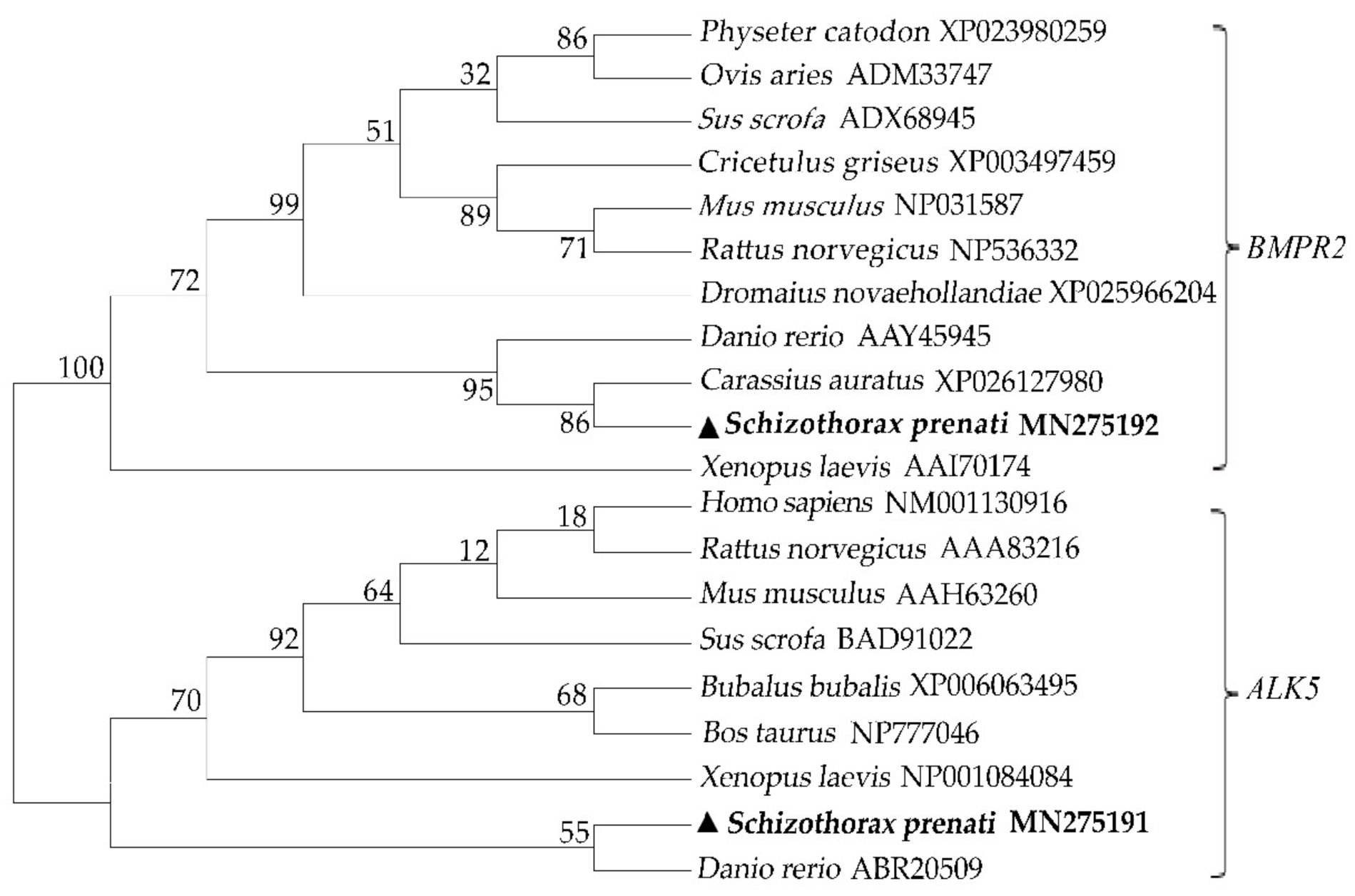
The full-length S. prenanti ALK5 cDNA sequence consisted of 1958 base pairs (bp), including a 62-bp 5′UTR and a 356-bp 3′UTR. The putative 501 amino acid residues contained two N-glycation sites, a transmembrane domain (Ala123-Ile145), a typical SGSGSG domain (Thr173-Lys230) and an S-TKc domain (Ile203-Leu490). The BMPR2 cDNA sequence of S. prenanti consisted of 3704 bp and encoded 1070 amino acids, including six N-glycosylation sites, one transmembrane domain (Ala157-Tyr179) and one STYKc (Lys210-Ser580) domain, and some low-complexity domains were deduced among the putative BMPR2 amino acid sequence. The phylogenetic analysis showed that ALK5 and BMPR2 of S. prenanti converged with those of fish and exhibited the highest identity with those of C. auratus and D. rerio, respectively (Figure 1).

3.2. Tissue Distribution of ALK5 and BMPR2

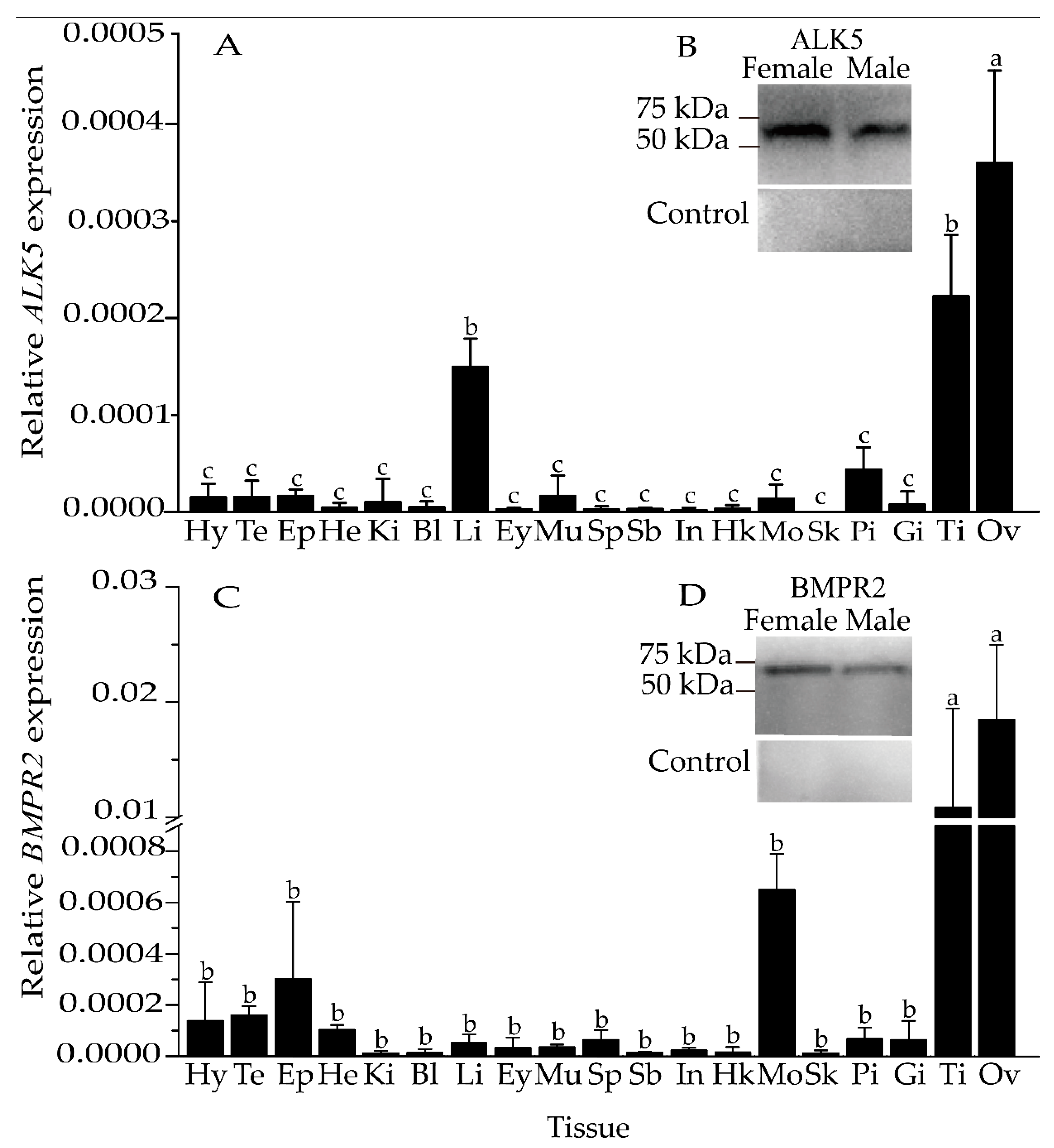
The tissue expression patterns of ALK5 and BMPR2 mRNA in adult females and males were analyzed by qRT–PCR. The ALK5 and BMPR2 mRNA expression levels in gonads were significantly higher than that in non-gonadal tissues (p < 0.05) (Figure 2A,C), and the ovarian expression levels were higher than those in the testes. Interestingly, the expression of BMPR2 in gonads was higher than that of ALK5.
Western blot analysis of tissue homogenates from the ovary and testis only detected specific bands of approximately 50 kDa and 75 kDa (Figure 2B,D). No bands were observed when the primary antiserum was replaced with PBS (Figure 2B,D).
ALK5 and BMPR2 protein bands with molecular weights of 50 kDa and 75 kDa were only detected in the ovary and testis. Among these bands, the size of BMPR2 was similar to that of the band sheared into the cytolemma.

3.3. Expression Levels of ALK5 and BMPR2 mRNAs during Gonad Development

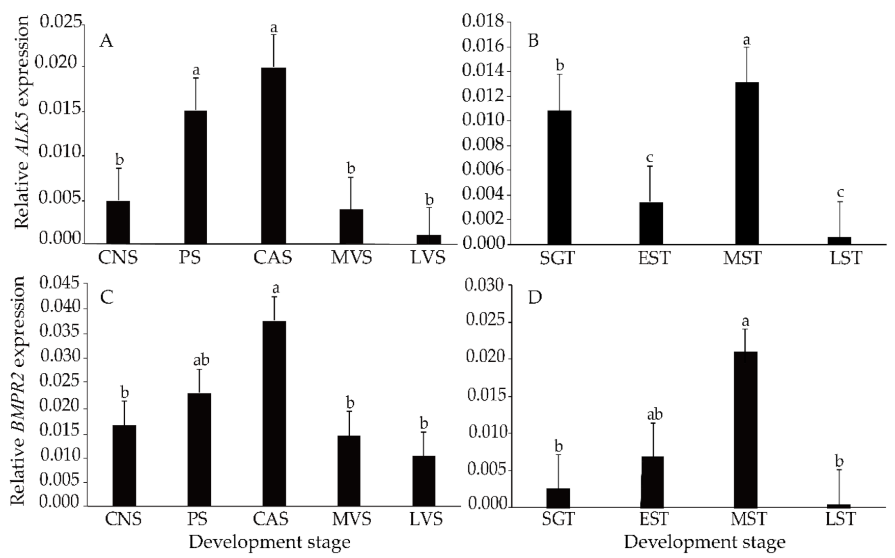
Similarly, the expression level of BMPR2 was higher than that of ALK5. During ovarian development, ALK5 and BMPR2 expression continued to increase until peaking at the CAS and then decreased (p < 0.05) (Figure 3A,C). However, some differences were detected during the testis development process. During testis development, the expression of BMPR2 increased gradually until reaching its maximum level at the MST and then decreased to its minimum at the LST (Figure 3D). ALK5 was higher at the SGT and MST and reached its minimum level at the EST and LST (Figure 3B).

3.4. Localization of ALK5 and BMPR2 during Gonadal Development

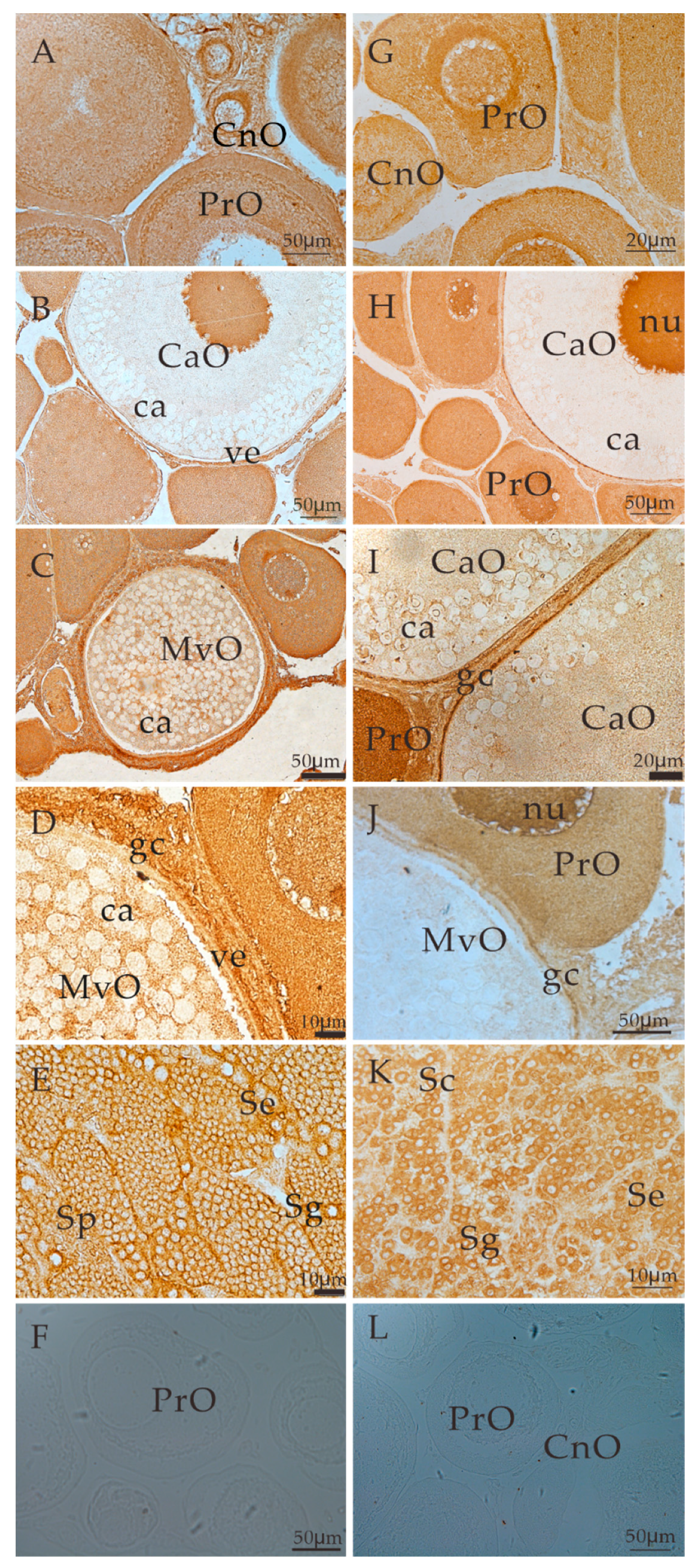
Immunohistochemical signals of ALK5 were detected in both the ovaries and testis of S. prenanti. Specifically, positive signals were detected in the cytoplasm of chromatin nucleolar oocytes and perinucleolar oocytes (Figure 4A,B) and the cytolemma and granulosa cells of cortical alveoli oocytes and mid-vitellogenic oocytes (Figure 4B–D). Strong signals were also detected in the cytoplasm of chromatin nucleolar oocytes and perinucleolar oocytes (Figure 4A) as well as in spermatogonia, spermatocytes, spermatids and Sertoli cells (Figure 4E). When PBS was used instead of the primary antiserum, no positive signals were observed in the gonads (Figure 4F).
In the ovary, BMPR2-positive signals were found in the cytoplasm of chromatin nucleolar oocytes and perinucleolar oocytes (Figure 4G). Signals were also detected in the cytodeme and granulosa cells of cortical alveoli oocytes and mid-vitellogenic oocytes (Figure 4H–J). Moreover, strong signals were detected in the cytolemma and cytoplasm of cortical alveoli oocytes (Figure 4H,I). In the testis, positive signals were detected in the cytoplasm of the spermatogonia, spermatocyte spermatids and Sertoli cells (Figure 4K). No positive signals were observed when the primary antiserum was replaced with PBS (Figure 4L).

3.5. Expression of ALK5 and BMPR2 Induced by E2

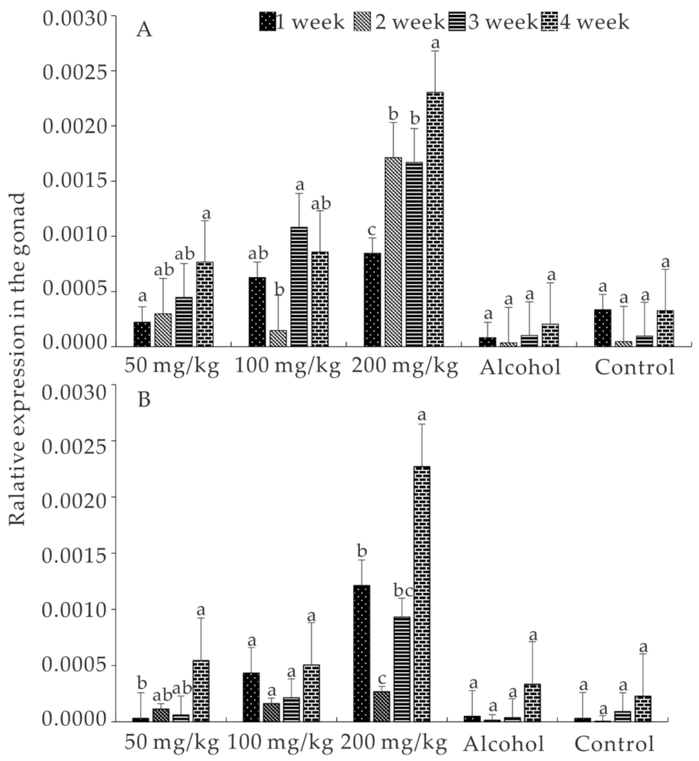
The survival rate, which was higher than 95% in all the groups, did not show significant differences during the experimental period (p > 0.05). As the E2 concentration and treatment time increased, the expression of ALK5 and BMPR2 gradually increased in all the treatment groups. In addition, ALK5 and BMPR2 expression in the group treated with 200 mg/kg E2 was higher than in the other groups (p < 0.05). Moreover, the expression of both ALK5 and BMPR2 was induced by E2, even though no significant difference was detected among the treatment groups on some weeks. All of these results showed that the expression of ALK5 and BMPR2 was induced by E2 and that the expression of BMPR2 is higher than that of ALK5 (Figure 5A,B).

4. Discussion

In the present study, the ALK5 and BMPR2 cDNA sequences of S. prenanti were characterized. Comparing the amino acid sequences showed that these ALK5 and BMPR2 cDNA sequences exhibited high homogeneity to those of other fish species. Both ALK5 and BMPR2 contained a conserved transmembrane domain, which is essential and plays a pivotal role in ligand activation [28,29]. Despite the divergence in their sequence, the serine/threonine kinase domains, which play a crucial role in ligand recognition, are conserved in the ALK5 and BMPR2 amino acid sequences, similar to the results observed in buffalo [8] and O. mykiss [12]. All of the results reveal the relatively high homogeneity of the different kinase domains and sequences with those of other vertebrates, suggesting that ALK5 and BMPR2 play conserved roles in signal transduction [13].
The expression of ALK5 and BMPR2 was detected in almost all tissues, particularly in gonads. Similarly, ALK5 is expressed in all tissues of O. mykiss [12] and is expressed at high levels in the buffalo ovary [8], the liver of O. niloticus [13] and C. gibelio follicle cells [14]. During oocytes development, the expression of ALK5 in ovaries at the early vitellogenic stage is higher than that at other stages [14]. ALK5 is more highly expressed in luteinized than in nonluteinized follicular cells of marmosets [30]. ALK5 expression in bovine granulosa cells is significantly increased after 12 h of treatment but decreased after 24 h of treatment [31]. In the mouse testes, ALK5 is involved in developing spermatogonia [32]. Although the expression of TGF-β and ALK5 is decreased in spermatogenesis [1], abundant ALK5 mRNA levels are detected during puberty, which confirms that ALK5 plays an irreplaceable role during this process [32,33]. Therefore, these results indicate that ALK5 is essential for oocytes growth and spermatogenesis.
Although it is not specific to non-gonadal tissues, BMPR2 plays a pivotal role during gonadal development. In C. gibelio, BMPR2a mRNA is mainly expressed in the brain, and BMPR2b is expressed in the liver [14]. However, in D. rerio, although expression of BMPR2a and BMPR2b mRNAs is universal, these mRNAs are mainly expressed in the testis and liver [18]. Importantly, BMPR2 gradually increases during oocytes and follicular development [16]. In piglets [34] and adult pigs [35,36], BMPR2 is significantly expressed in follicles and oocytes at various stages. Some differences have been detected in rats: BMPR2 has been detected in oocytes and granulosa cells at all stages, except for primitive follicular granulosa cells [37]. In contrast, BMPR2 is expressed at higher levels in primary oocytes than at other stages [14]. Moreover, BMPR2 expression decreases as germ cells enter the meiosis stage, but BMPR2 expression was detected at all stages of testicular development [38]. During mouse testes development, BMPR2 expression at the early developmental stage is significantly higher than that in mature testes [39]. The wide distribution of BMPR2 in gonadal and nongonadal tissues may involve a vast array of biological responses and functions.
The locations of ALK5 and BMPR2 further explain their functions in ovarian and testicular development. ALK5 and BMPR2 were detected in mouse oocytes and granulosa cells at all stages, similar to the results obtained with rats and pigs [10,37,40]. ALK5-positive signals have been detected in granulosa cells [41], and BMPR2 is detected in follicles at different developmental stages [17]. Moreover, ALK5 and BMPR2 are expressed in cattle and sheep oocytes [42] and in human primitive and primary follicles [10]. Some studies have shown that ALK5 and BMPR2 are more abundantly expressed at Mid-CL than at other stages [11] and that ALK5 and BMPR2 are expressed in both granulosa cells and theca cells of piglets [43]. ALK5 and BMPR2 are essential in inhibiting bovine granulosa cell apoptosis [19] and affecting cumulus cell expansion and proliferation [21]. All of these results demonstrate that ALK5 and BMPR2 might be closely related to regulating germ cells and somatic cell development.
The present study showed that the expression of ALK5 and BMPR2 was induced by E2 during S. prenanti gonad development, and the expression of BMPR2 was higher than that of ALK5. In addition, the ALK5 and BMPR2 mRNA levels generally increased with increases in the treatment time and dose. The expression of ALK5 and BMPR2 was particularly promoted in bovine granular cells by low and high concentrations of E2 [42]. Consistently, ALK5 and BMPR2 expression in ovine granular cells is significantly increased by daidzein, similar to the results obtained with E2 [44]. Analogously, treatment with FSH and E2 for different times and at different doses also promotes ALK5 and BMPR2 expression in granulosa cells of bovine [45]. Recently, some researchers have found that neonatal exposure to flutamide and 4-tertoctylphenol (OP, an estrogenic compound) can upregulate ALK5 and BMPR2 expression in small antral follicles of pigs [43]. Therefore, we hypothesized that it could result in differential activation of signaling pathways and an alteration of the gene expression profile in gonads. These results further confirmed that ALK5 and BMPR2 might be the key signaling factors during gonadal development and depend on precise regulation of the stages and hormone levels.
Interestingly, the fold change in BMPR2 expression obtained in this study was higher than that in ALK5 expression. Similarly, the expression of BMPR2 and ALK5 in ovarian follicles is stimulated by E2, and the stimulated expression of BMPR2 is clearly higher than that of ALK5 [42]. In addition, the expression of BMPR2 in bovine granular cells is higher than that of ALK5 [42,45]. In general, seven type I receptors, also known as activin receptor-like kinases (ALKs, ALK1–7), have been reported to date. TGF-β provides signals in most cells by binding to TGF-βRII and thus forming a complex with type I receptors. Moreover, some studies have found that TGF-β signaling is dependent on ALK5, and BMPR2 is not the only type I receptor [46,47,48]. In addition to activating ALK5 and BMPR2, as a prerequisite of many TGF-β and BMP-signaling pathways, participates in other signaling transduction pathways [4,5,6,47].

5. Conclusions

In summary, ALK5 and BMPR2 were characterized, and their mRNAs were prominently expressed in the ovary and testis, particularly at the CAS and MST. ALK5 and BMPR2 signals were observed in the cytoplasm of oocytes, spermatocytes, spermatids and somatic cells. Exogenous E2 promoted the expression of S. prenanti ALK5 and BMPR2, and the expression of BMPR2 was higher than that of ALK5. Combined with the findings from our previous study, the results obtained in the present study suggest that GDF9 signals transmitted via ALK5 and BMPR2 might participate in folliculogenesis and spermatogenesis in S. prenanti.

Author Contributions

Conceptualization, S.Z. and Z.H.; methodology, Y.C.; software, Z.M. and J.H.; formal analysis, T.Y.; investigation, Q.Z. and F.D.; resources, L.Y., H.C. and J.L.; writing—original draft preparation, S.Z., L.H., D.Y. and Z.H.; writing—review and editing, T.Y. and S.Z.; funding acquisition, S.Z. and Z.H. All authors have read and agreed to the published version of the manuscript.

Funding

This research was funded by the National Natural Science Foundation of China grant numbers 31402286 and 31972777.

Institutional Review Board Statement

The study was conducted according to the guidelines of the Animal Research and Ethics Committees of Sichuan Agricultural University and followed the animal experiments guidelines of Sichuan Agricultural University.

Informed Consent Statement

Not applicable.

Data Availability Statement

Data will be available upon request.

Acknowledgments

We thank Sen Xiong for their technical assistance, Song Li, Zhide He, Xiongyan Wang, Zhao Liu, Chengcheng Tian for the fish management and sample collection.

Conflicts of Interest

All authors declare no conflict of interest.

References

  1. Macias, M.J.; Martin, M.P.; Massagué, J. Structural determinants of Smad function in TGF-β signaling. Trends Biochem. Sci. 2015, 40, 296–308. [Google Scholar] [PubMed] [Green Version]
  2. Li, Q.; Agno, J.E.; Edson, M.A.; Nagaraja, A.K.; Nagashima, T.; Matzuk, M.M. Transforming growth factor β receptor type 1 is essential for female reproductive tract integrity and function. PLoS Genet. 2011, 7, e1002320. [Google Scholar] [CrossRef] [PubMed] [Green Version]
  3. Souza, C.J.H.; Campbell, B.K.; Mcneilly, A.S.; Baird, D.T. Effect of bone morphogenetic protein 2 (BMP2) on oestradiol and inhibin A production by sheep granulosa cells, and localization of BMP receptors in the ovary by immunohistochemistry. Reproduction 2002, 123, 363–369. [Google Scholar] [CrossRef] [PubMed]
  4. Derynck, R.; Zhang, Y.E. Smad-dependent and Smad-independent pathways in TGF-beta family signalling. Nature 2003, 425, 577–584. [Google Scholar] [PubMed]
  5. Moustakas, A.; Heldin, C.H. The regulation of TGFbeta signal transduction. Development 2009, 136, 3699–3714. [Google Scholar] [CrossRef] [PubMed] [Green Version]
  6. Xu, X.; Zheng, L.; Yuan, Q.; Zhen, G.; Crane, J.L.; Zhou, X.; Cao, X. Transforming growth factor-β in stem cells and tissue homeostasis. Bone Res. 2018, 6, 2. [Google Scholar] [PubMed] [Green Version]
  7. Yang, Q.; Ma, B.; Qiao, H.; Ma, H.; Dong, Y.; Cao, L.; Ma, J.; Li, Z. TGFB1 represses the expression of SF1 and LRH1 to inhibit E production in rat LCs. Reproduction 2017, 153, 621–629. [Google Scholar]
  8. Zhu, P.; Zhao, X.; Su, X.; Su, J.; Liu, J.; Liu, Q.; Shi, D. Cloning analysis of buffalo ALK5 gene and the study of its tissue expression pattern. Heilongjiang Anim. Sci. Vet. Med. 2015, 4, 9–13. [Google Scholar]
  9. Oron, G.; Fisch, B.; Ao, A.; Zhang, X.Y.; Farhi, J.; Ben-Haroush, A.; Haroush, A.B.; Kesseler-Icekson, G.; Abir, R. Expression of growth-differentiating factor 9 and its type 1 receptor in human ovaries. Reprod. Biomed. Online 2010, 21, 109–117. [Google Scholar]
  10. Sun, R.Z.; Lei, L.; Cheng, L.; Jin, Z.F.; Zu, S.J.; Shan, Z.Y.; Wang, Z.D.; Zhang, J.X.; Liu, Z.H. Expression of GDF-9, BMP-15 and their receptors in mammalian ovary follicles. J. Mol. Histol. 2010, 41, 325–332. [Google Scholar] [CrossRef]
  11. Galvão, A.; Wolodko, K.; Rebordão, M.R.; Skarzynski, D.; Ferreira-Dias, G. TGFB1 modulates in vitro secretory activity and viability of equine luteal cells. Cytokine 2018, 110, 316–327. [Google Scholar] [CrossRef]
  12. Maehr, T.; Wang, T.; González-Vecino, J.L.; Wadsworth, S.; Secombes, C.J. Cloning and expression analysis of the transforming growth factor-beta receptors type 1 and 2 in the rainbow trout Oncorhynchus mykiss. Dev. Comp. Immunol. 2012, 37, 115–126. [Google Scholar] [CrossRef]
  13. Zhan, X.L.; Ma, T.Y.; Wu, J.Y.; Yi, L.Y.; Wang, J.Y.; Gao, X.K.; Li, W.S. Cloning and primary immunological study of TGF-β1 and its receptors TβR I /TβR II in tilapia (Oreochromis niloticus). Dev. Comp. Immunol. 2015, 51, 134–140. [Google Scholar] [CrossRef]
  14. Chen, A.Q.; Liu, Z.; Wei, Q.L.; Yang, Z.; Zhong, Y.J. The expression pattern of GDF9 and MP15 I and II receptor genes during follicle development in gibel carp. J. Shanghai Ocean. Univ. 2015, 24, 656–661. [Google Scholar]
  15. Zhu, G.; Guo, B.; Pan, D.; Mu, Y.; Feng, S. Expression of bone morphogenetic proteins and receptors in porcine cumulus-oocyte complexes during in vitro maturation. Anim. Reprod. Sci. 2008, 104, 275–283. [Google Scholar] [CrossRef]
  16. Onagbesan, O.M.; Bruggeman, V.; Van-As, P.; Tona, K.; Williams, J.; Decuypere, E. BMPs and BMPRs in chicken ovary and effects of BMP-4 and -7 on granulosa cell proliferation and progesterone production in vitro. Am. J. Physiol. Endoc. Metab. 2003, 285, 973–983. [Google Scholar] [CrossRef] [Green Version]
  17. Quinn, R.L.; Shuttleworth, G.; Hunter, M.G. Immunohistochemical localization of the bone morphogenetic protein receptors in the porcine ovary. J. Anat. 2004, 205, 15–23. [Google Scholar] [CrossRef]
  18. Li, C.W.; Ge, W. Spatiotemporal expression of bone morphogenetic protein family ligands and receptors in the zebrafish ovary: A potential paracrine-signaling mechanism for oocyte-follicle cell communication. Biol. Reprod. 2011, 85, 977–986. [Google Scholar] [CrossRef] [Green Version]
  19. Zhou, J.; Liu, J.; Pan, Z.; Du, X.; Li, X.; Ma, B.; Yao, W.; Li, Q.; Liu, H. The let-7g microRNA promotes follicular granulosa cell apoptosis by targeting transforming growth factor-β type 1 receptor. Mol. Cell. Endocrinol. 2015, 409, 103–112. [Google Scholar] [CrossRef]
  20. Larsson, J.; Goumans, M.J.; Sjöstrand, L.J.; Van, R.M.A.; Ward, D.; Levéen, P.; Xu, X.; Ten, D.P.; Mummery, C.L.; Karlsson, S. Abnormal angiogenesis but intact hematopoietic potential in TGF-beta type I receptor-deficient mice. EMBO J. 2001, 20, 1663–1673. [Google Scholar] [CrossRef] [Green Version]
  21. Liu, C.; Yuan, B.; Chen, H.; Xu, M.; Sun, X.; Xu, J.; Gao, Y.; Chen, C.; Jiang, H.; Zhang, J. Effects of MiR-375-BMPR2 as a key factor downstream of BMP15/GDF9 on the Smad1/5/8 and Smad2/3 signaling pathways. Cell. Physiol. Biochem. 2018, 46, 213–225. [Google Scholar] [CrossRef] [Green Version]
  22. Iwata, N.; Hasegawa, T.; Fujita, S.; Nagao, S.; Nakano, Y.; Nada, T.; Nishiyama, Y.; Hosoya, T.; Otsuka, F. Effect of the interaction of metformin and bone morphogenetic proteins on ovarian steroidogenesis by human granulosa cells. Biochem. Biophys. Res. Commun. 2018, 503, 1422–1427. [Google Scholar] [CrossRef] [Green Version]
  23. Fujita, S.; Hasegawa, T.; Nishiyama, Y.; Fujisawa, S.; Nakano, Y.; Nada, T.; Iwata, N.; Kamada, Y.; Masuyama, H.; Otsuka, F. Interaction between orexin A and bone morphogenetic protein system on progesterone biosynthesis by rat granulosa cells. J. Steroid. Biochem. Mol. Biol. 2018, 181, 73–79. [Google Scholar] [CrossRef] [Green Version]
  24. Patiño, L.C.; Silgadom, D.; Laissue, P. A potential functional association between mutant BMPR2 and primary ovarian insufficiency. Syst. Biol. Reprod. Med. 2017, 63, 145–149. [Google Scholar] [CrossRef]
  25. Yan, T.; Cai, Y.; He, J.; Zhang, Q.; Wang, X.; Zhang, S.; He, L.; He, Z. Characterization and expression profiles of cyp19a1a in the schizothoracine fish Schizothorax prenanti. Tissue Cell 2019, 58, 70–75. [Google Scholar] [CrossRef]
  26. Yan, T.; Zhang, S.; Zhang, Q.; Deng, F.; Cai, Y.; He, J.; Ma, Z.; He, L.; Luo, J.; Yang, D.; et al. Expression patterns and oestradiol regulation of growth differentiation factor 9 in Schizothorax prenanti. Comp. Biochem. Physiol. Part B Biochem. Mol. Biol. 2020, 248–249, 110470. [Google Scholar] [CrossRef]
  27. Blazer, V.S. Histopathological assessment of gonadal tissue in wild fishes. Fish Physiol. Biochem. 2002, 26, 85–101. [Google Scholar] [CrossRef]
  28. Lin, H.Y.; Wang, X.F.; Ng-Eaton, E.; Weinberg, R.A.; Lodish, H.F. Expression cloning of the TGF-beta type II receptor, a functional transmembrane serine/threonine kinase. Cell 1992, 68, 775–785. [Google Scholar] [CrossRef]
  29. Zhu, H.J.; Sizeland, A.M. A pivotal role for the transmembrane domain in transforming growth factor-beta receptor activation. J. Biol. Chem. 1999, 274, 11773–11781. [Google Scholar] [CrossRef] [Green Version]
  30. Wehrenberg, U.; Giebel, J.; Rune, G.M. Possible involvement of transforming growth factor-β1 and transforming growth factor-β receptor type II during luteinization in the marmoset ovary. Tissue Cell 1998, 30, 360–367. [Google Scholar] [CrossRef]
  31. Haas, C.S.; Rovani, M.T.; Oliveira, F.C. Expression of growth and differentiation factor 9 and cognate receptors during final follicular growth in cattle. Anim. Reprod. 2016, 13, 756–761. [Google Scholar] [CrossRef]
  32. Wu, F.; Lin, T.; Sung, L.; Chang, W.; Wu, P.; Luo, C. BMP8A sustains spermatogenesis by activating both SMAD1/5/8 and SMAD2/3 in spermatogonia. Sci. Signal. 2017, 10, eaal1910. [Google Scholar] [CrossRef] [PubMed]
  33. Zhao, G.Q.; Hogan, B.L. Evidence that mouse Bmp8a (Op2) and Bmp8b are duplicated genes that play a role in spermatogenesis and placental development. Mech. Dev. 1996, 57, 159–168. [Google Scholar] [CrossRef]
  34. Shimizu, T.; Yokoo, M.; Miyake, Y.; Sasada, H.; Sato, E. Differential expression of bone morphogenetic protein 4–6 (BMP-4, -5, and -6) and growth differentiation factor-9 (GDF-9) during ovarian development in neonatal pigs. Domest. Anim. Endocrinol. 2004, 27, 397–405. [Google Scholar] [CrossRef]
  35. Brankin, V.; Quinn, R.L.; Webb, R.; Hunter, M.G. Evidence for a functional bone morphogenetic protein (BMP) system in the porcine ovary. Domest. Anim. Endocrinol. 2005, 28, 367–379. [Google Scholar] [CrossRef]
  36. Prochazka, R.; Nemcova, L.; Nagyova, E.; Kanka, J. Expression of growth differentiation factor 9 messenger RNA in porcine growing and preovulatory ovarian follicles. Biol. Reprod. 2004, 71, 1290–1295. [Google Scholar] [CrossRef] [Green Version]
  37. Erickson, G.F.; Shimasaki, S. The spatiotemporal expression pattern of the bone morphogenetic protein family in rat ovary cell types during the estrous cycle. Reprod. Biol. Endocrinol. 2003, 1, 9. [Google Scholar] [CrossRef] [Green Version]
  38. Young, J.C.; Wakitani, S.; Loveland, K.L. TGF-β superfamily signaling in testis formation and early male germline development. Semin. Cell Dev. Biol. 2015, 45, 94–103. [Google Scholar] [CrossRef]
  39. Ciller, I.M.; Palanisamy, S.K.; Ciller, U.A.; Mcfarlane, J.R. Postnatal expression of bone morphogenetic proteins and their receptors in the mouse testis. Physiol. Res. 2016, 65, 673–682. [Google Scholar] [CrossRef]
  40. Yi, S.E.; Lapolt, P.S.; Yoon, B.S.; Chen, J.Y.; Lu, J.K.; Lyons, K.M. The type I BMP receptor BmprIB is essential for female reproductive function. Proc. Natl. Acad. Sci. USA 2001, 98, 7994–7999. [Google Scholar] [CrossRef] [Green Version]
  41. Paradis, F.; Novak, S.; Murdoch, G.K.; Dyck, M.K.; Dixon, W.T.; Foxcroft, G.R. Temporal regulation of BMP2, BMP6, BMP15, GDF9, BMPR1A, BMPR1B, BMPR2 and TGFBR1 mRNA expression in the oocyte, granulosa and theca cells of developing preovulatory follicles in the pig. Reproduction 2009, 138, 115–129. [Google Scholar] [CrossRef]
  42. Jayawardana, B.C.; Shimizu, T.; Nishimoto, H.; Kaneko, E.; Tetsuka, M.; Miyamoto, A. Hormonal regulation of expression of growth differentiation factor-9 receptor type I and II genes in the bovine ovarian follicle. Reproduction 2006, 131, 545–553. [Google Scholar] [CrossRef]
  43. Knapczyk, S.K.; Grzesiak, M.; Witek, P.; Duda, M.; Koziorowski, M.; Slomczynska, M. Neonatal exposure to agonists and antagonists of sex steroid receptors induces changes in the expression of oocyte-derived growth factors and their receptors in ovarian follicles in gilts. Theriogenology 2019, 134, 42–52. [Google Scholar] [CrossRef]
  44. Chen, A.; Xu, Z.; Yu, S.; Yang, Z. Effects of daidzein on mRNA expression of bone morphogenetic protein receptor type I and II genes in the ovine granulosa cells. Asian-Australas. J. Anim. Sci. 2010, 23, 326–332. [Google Scholar] [CrossRef]
  45. Chen, A.Q.; Yu, S.D.; Wang, Z.G.; Xu, Z.R.; Yang, Z.G. Stage-specific expression of bone morphogenetic protein type I and type II receptor genes: Effects of follicle-stimulating hormone on ovine antral follicles. Anim. Reprod. Sci. 2009, 111, 391–399. [Google Scholar] [CrossRef]
  46. Reader, K.L.; Heath, D.A.; Lun, S.; Mcintosh, C.J.; Western, A.H.; Littlejohn, R.P.; Mcnatty, K.P.; Juengel, J.L. Signalling pathways involved in the cooperative effects of ovine and murine GDF9+BMP15-stimulated thymidine uptake by rat granulosa cells. Reproduction 2011, 142, 123–131. [Google Scholar] [CrossRef]
  47. Rol, N.; Kurakula, K.B.; Happé, C.; Bogaard, H.J.; Goumans, M.J. TGF-β and BMPR2 signaling in PAH: Two black sheep in one family. Int. J. Mol. Sci. 2018, 19, 2585. [Google Scholar] [CrossRef] [Green Version]
  48. Vitt, U.A.; Mazerbourg, S.; Klein, C.; Hsueh, A.J.W. Bone morphogenetic protein receptor type II is a receptor for growth differentiation factor-9. Biol. Reprod. 2002, 67, 473–480. [Google Scholar] [CrossRef] [Green Version]
Figure 1. Phylogenetic analysis of ALK5 and BMPR2 with those of other vertebrates. The ALK5 and BMPR2 sequences of the other vertebrates were downloaded from Entrez (NCBI). The phylogenetic tree was analyzed using the neighbor-joining algorithm of Mega 7.0. The numbers at the forks are the bootstrap proportions. S. prenanti is marked with black triangles.
Animals 11 01365 g001
Figure 2. Tissue distribution of ALK5 (A) and BMPR2 (B) in S. prenanti determined by qRT–PCR, and ALK5 and BMPR2 immunoreactivity in the ovary and testis (C,D). Hy, hypothalamus; Te, telencephalon; Ep, epithalamus; He, heart; Ki, kidney; Bl, blood; Li, liver; Ey, eye; Mu, muscle; Sp, spleen; Sb, swim bladder; In, intestine; Hk, head kidney; Mo, medulla oblongata; Sk, skin; Pi, pituitary; Gi, gill; Ti, testis; Ov, ovary. The data are represented as the means ± SEMs (n = 5). Means marked with different letters are significantly different (p < 0.05).
Animals 11 01365 g002
Figure 3. Expression of ALK5 (A,B) and BMPR2 (C,D) during gonadal development (n = 5). CNS, ovary at the chromatin nucleolar stage; PS, ovary at the perinuclear stage; CAS, ovary at the cortical alveoli stage; MVS, ovary at the mid-vitellogenic stage; LVS, ovary at the late vitellogenic stage; SGT, testis at the spermatogonium stage; EST, testis at the early spermatogenic stage; MST, testos at the mid-spermatogenic stage; LST, testis at the late spermatogenic stage. Means marked with different letters are significantly different (p < 0.05).
Animals 11 01365 g003
Figure 4. Localization of ALK5 (AF) and BMPR2 (GL) in the ovary and testis of S. prenanti. The sections of the ovary at the primary growth stage (A), the previtellogenic stage (C), and the mid- to the late-vitellogenic stage (D), and of the testis (E,K) were immunostained with anti-ALK5 (AE) and BMPR2 (GK) antiserum. Panel F and L: the ovary section immunostained with no antiserum. CnO, chromatin nucleolar oocytes; PrO, perinucleolar oocytes; CaO, cortical alveoli oocytes; MvO, mid-vitellogenic oocytes; nu, nucleolus; ve, vitelline envelope; ca, cortical alveoli; Sg, spermatogonia; Sp, spermatid; gc, granulosa cells; Se, Sertoli cells.
Animals 11 01365 g004
Figure 5. Concentration and time effects of ALK5 (A) and BMPR2 (B) expression in E2-fed S. prenanti. The data are represented as the means ± SEMs (n = 10). Means marked with different letters are significantly different (p < 0.05).
Animals 11 01365 g005
Table 1. Sequences of oligonucleotide primers used in this study.
PrimersSequence (5′-3′)Use
ALK5-F1GGACCATCGCCAGGACCATCSequence amplification
ALK5-F2CTTCTACATCTGCCACAGC
ALK5-R1GCCTCRCAGCTCTGCC
ALK5-R2GTTRGCGTACCAGCACTC
BMPR2-F1AACGAACGCTCCATATACSequence amplification
BMPR2-R1TGGGAGTGCTCCATCAGG
BMPR2-F2CHSHRGGRGGYGGAGC
BMPR2-F3GTCCGCCACAACTACAACG
RACE ALK5-F1TCCGTGCCAACATCCCASequence amplification
RACE ALK5-F2GAGGGAGTGCTGGTACGCTA
RACE ALK5-R1CCTTCCCTATGCTCTCCTG
RACE ALK5-R2GATGATGGTCCTGGCGA
β-Actin Q-F1GATTCGCTGGAGATGATGCTReal-time quantitative PCR analysis
β-Actin Q-R1CGTTCTAGAAGGTGTGATGCC
18S rRNA Q-F1ACCACCCACAGAATCGAGAAAReal-time quantitative PCR analysis
18S rRNA Q-R1GCCTGCGGCTTAATTTGACT
ALK5 Q-F1ACCCGAGGTGCTGGATGACTReal-time quantitative PCR analysis
ALK5 Q-R1TTGGGATGTTGGCACGGAG
BMPR2 Q-F1TGACGGGAAATCGCCTCReal-time quantitative PCR analysis
BMPR2 Q-R1CTCAAAGGAGGGGTGGTTC
Note: F, sense primer; R, antisense primer.
Publisher’s Note: MDPI stays neutral with regard to jurisdictional claims in published maps and institutional affiliations.