
Molecular Action of Polyphenols in Leukaemia and Their Therapeutic Potential


1
Biomolecular Sciences Research Centre, Department of Biosciences and Chemistry, Sheffield Hallam University, The Owen Building, City Campus, Howard Street, Sheffield S1 1WB, UK
2
Laboratory Medicine Department, Faculty of Applied Medical Sciences, Umm Al-Qura University, P.O. Box 715, Makkah 21955, Saudi Arabia
*
Author to whom correspondence should be addressed.
Int. J. Mol. Sci. 2021, 22(6), 3085; https://doi.org/10.3390/ijms22063085
Submission received: 27 January 2021
/
Revised: 8 March 2021
/
Accepted: 10 March 2021
/
Published: 17 March 2021
(This article belongs to the Special Issue Antitumor Activities of Natural Compounds From Plants)
Abstract
:Leukaemia is a malignant disease of the blood. Current treatments for leukaemia are associated with serious side-effects. Plant-derived polyphenols have been identified as potent anti-cancer agents and have been shown to work synergistically with standard chemotherapy agents in leukaemia cell lines. Polyphenols have multiple mechanisms of action and have been reported to decrease cell proliferation, arrest cell cycle and induce apoptosis via the activation of caspase (3, 8 and 9); the loss of mitochondrial membrane potential and the release of cytochrome c. Polyphenols have been shown to suppress activation of transcription factors, including NF-kB and STAT3. Furthermore, polyphenols have pro-oxidant properties, with increasing evidence that polyphenols inhibit the antioxidant activity of glutathione, causing oxidative DNA damage. Polyphenols also induce autophagy-driven cancer cell death and regulate multidrug resistance proteins, and thus may be able to reverse resistance to chemotherapy agents. This review examines the molecular mechanism of action of polyphenols and discusses their potential therapeutic targets. Here, we discuss the pharmacological properties of polyphenols, including their anti-inflammatory, antioxidant, anti-proliferative, and anti-tumour activities, and suggest that polyphenols are potent natural agents that can be useful therapeutically; and discuss why data on bioavailability, toxicity and metabolism are essential to evaluate their clinical use.
1. Introduction
1.1. Leukaemia
Leukaemia is described as a heterogeneous group of haematological cancers [1,2], defined as a malignant disease of the blood, characterised by uncontrolled proliferation and the development of leucocytes in the blood [3] and bone marrow [4] that accumulate in the liver and spleen [1].
Since its original identification by Virchow in 1847 [5], the general classification of leukaemia has become more complex [1]. At present, leukaemia is classified into two main types: myeloid or lymphoid, based on the predominant cell of origin and is sub-divided into acute or chronic, based on the rate of progression [6] with multiple sub-classifications within each group [7].
The current therapies for leukaemia include chemotherapy, radiotherapy, immuno-therapy, and bone marrow transplantation [6,8]. Chemotherapy remains the front-line treatment for most leukaemias [9]. Leukaemia chemotherapies include anti-metabolites (e.g., methotrexate) [10,11]; topoisomerase II inhibitors (e.g., doxorubicin) and alkaloids (e.g., omacetaxine) [12]. In addition to the general chemotherapy agents, targeted therapies are used for specific leukaemias, such as tyrosine kinase inhibitors (e.g., Imatinib), which are used for BCR/ABL positive leukaemia [13,14]. However, many chemotherapy agents are associated with serious side effects, such as liver damage, nerve damage, heart disorders and loss of immunity [6], which result in poor adherence to treatment regimens and poor prognosis [6]. Thus, new treatments for leukaemia are needed, which could be used individually or in combination with existing standard chemotherapy agents, in an attempt to potentiate anti-tumour effects, whilst reducing treatment doses and side effects. One potential source of therapeutics is polyphenols.
1.2. Polyphenols
Polyphenols are a family of phenolic phytochemicals [15] (Figure 1). They are an essential part of the diet, and are found in fruits, vegetables, cereals, nuts, and herbs [16], and drinks such as wine, beer, tea, and coffee [17]. Epidemiological and animal studies have shown potential benefits, with protective effects seen against a wide range of conditions, including cancers such as leukaemia [17,18]. Polyphenols’ anti-cancer activities include halting cell cycle, inducing apoptosis, modulation of angiogenesis pathways, and preventing metastasis [19]. Studies have shown that the consumption of polyphenol-rich foods can prevent 10 to 70% of cancer deaths [18,19]. A major advantage of the use of polyphenols as anti-cancer agents, is that they have low toxicity, and are safe to consume, and highly accessible [20]. This could offer opportunities for innovation in drug discovery [21] and could play a major role in cancer prevention [22]. Moreover, a number of polyphenols (e.g., quercetin, apigenin, rhein, emodin and resveratrol) have been shown to act synergistically with chemotherapy agents when used as combination treatments in vitro, enhancing cell cycle arrest and inducing apoptosis [23,24,25]. These synergistic effects were shown to be at least partly regulated by a decrease in glutathione levels and an increase in DNA damage, when polyphenols were combined with etoposide; doxorubicin [24,25,26]; cisplatin and 5-fluorouracil in leukaemia cell lines [24,26,27,28].
Here, we summarise the molecular actions of polyphenols and discuss their potential therapeutic use. We outline how they act at a cellular and molecular level in a wide variety of cell lines (Table 1) which represent the different forms of leukaemia, and ask the question: Is there sufficient evidence to progress towards clinical trials for polyphenols as an adjunctive treatment alongside standard chemotherapy agents?
2. Molecular Mechanisms of Polyphenols
Polyphenols target several molecular pathways leading to cell cycle arrest and induced apoptosis in leukaemia cell lines, while being protective to non-tumour control cells [67,68,154,155] (Figure 2). The key molecular targets of polyphenols include:
- 2.1.
- Induction of cellular stress and catabolism: through an increase in reactive oxygen species (ROS) and a decrease in cellular antioxidants such as glutathione (GSH).
- 2.2.
- Modulation of cell metabolic activity.
- 2.3.
- Cell cycle arrest.
- 2.4.
- Induction of cell death via:
- Apoptosis.
- Autophagy.
- 2.5.
- Interaction with chemotherapy agents and the reduction or reversal of multidrug resistance.
These targets make polyphenols an attractive potential therapy for use in leukaemia.
Abbreviations: Glutathione (GSH), Signal transducer and activator of transcription 3 (STAT3), Protein kinase B (Akt), Extracellular signal-regulated kinases (ERKs), Tumour necrosis factor-α (TNFα), inhibitor of NF-κB (IκBα), Mitogen-activated protein kinase (MAPKs), Nuclear factor κB (NF-κB), Phosphoinositide 3-kinase (PI3K), Mammalian target of rapamycin (mTOR), Interleukin-1 (IL-1), Interleukin-6 (IL-6), Interleukin-17 (IL-17), Interleukin-23 (IL-23), CC-chemokine ligand 2 (CCL2), Reactive oxygen species (ROS), Heme oxygenase-1 (HO-1), Glutathione S-transferases (GST), Adenosine triphosphate (ATP), Cyclin dependent kinases 1 (CDK1), Cyclin dependent kinases 2 (CDK2), Cyclin dependent kinases 4 (CDK4), Cyclin dependent kinases 6 (CDK6), Cyclin-dependent Kinase Inhibitor (p21CIP/WAF), Cyclin-dependent kinase inhibitor (p27Kip1), mitogen-activated protein (MAP), B-cell lymphoma 2 protein (Bcl-2), B-cell lymphoma extra-large (Bcl-XL), X-linked inhibitor of apoptosis protein (XIAP), Cellular Inhibitor of Apoptosis Protein 1(cIAP-1), Cellular Inhibitor of Apoptosis Protein 2 (cIAP-2), Myeloid cell leukaemia 1 (Mcl-1), Cysteine aspartic acid specific protease 3, 8 and 9 (Caspase 3, 8 and 9), APO-l/CD95/tumour necrosis factor superfamily 6 (TNFRSF6)/APT-1(Fas), Fas Ligand (FasL), Tumour necrosis factor receptor (TNF), Fas-associated Protein with Death Domain (FADD), Death receptor 4 and 5 (DR4 and DR 5), Bcl-2-like protein (BIM), Bcl-2-associated X protein (Bax), Bcl2-antagonist/killer (Bak), the Bcl2 associated agonist of cell death (Bad), BH3 interacting-domain death agonist (Bid), Poly (ADP-ribose) polymerase (PARP), Growth Arrest and DNA Damage (GADD45), Light chain I and II (LC3-I and LC3-II), Permeability glycoprotein (P-gp), ATP Binding Cassette Subfamily C Member 1 (ABCC1).
2.1. Polyphenols, Cellular Stress and Catabolism
Inflammation is associated with the development and initiation of most cancers, by activating cellular signals that lead to DNA damage and several epigenetic changes [156]. This is accompanied by an increase in inflammatory cell infiltration and the activation of immune cell signalling factors such as nuclear factor κB (NF-κB) and STATs [156,157]. This leads to increased activation of chemokines, cyclooxygenase (COX)-2 enzyme, prostaglandin E2 (PGE2), inducible nitric oxide synthase (iNOS), matrix metalloproteinase 9 (MMP-9), vascular endothelial growth factor (VEGF), and cytokine production (e.g., interleukin (IL)-1, IL-6 and IL-8) [157,158,159]. The role of inflammation and cancer progression has been reviewed extensively [156,157,158,159]. Targeting these signalling pathways represents an attractive strategy for the prevention and treatment of cancer [156,157,158,159].
Upregulation of signal transducer and activator of transcription 3 (STAT3) and NF-κB are two fundamental transcription factors that are associated with many types of cancer, including leukaemia [159,160,161,162]. NF-κB can affect all six hallmarks of cancer through the transcriptional activation of more than 200 genes associated with cell proliferation, angiogenesis, metastasis, tumour promotion, inflammation and suppression of apoptosis [163,164,165]. One of the most documented functions of NF-κB is its ability to promote cell survival through the induction of target genes, the products of which inhibit the apoptotic machinery in both normal and malignant cells [166,167]. NF-κB can also prevent programmed necrosis by inducing genes that encode antioxidant proteins [167]. In most cases, NF-κB is maintained in an active state within cancer cells through mutational the activation of upstream signalling molecules or in response to extracellular stimuli within the tumour microenvironment [168].
This increase in the activation of NF-κB in cancer cells increases cell survival and proliferation; and increases expression of inflammatory cytokines and growth factors in leukaemia cells [161,169]. The activation of NF-kB can be mediated by either the canonical or the alternative pathway (Figure 3). The canonical pathway is mainly activated by extracellular factors such as ligands [170]. The activation of NF-κB is most commonly mediated by the IκB kinase (IKK) complex, which is composed of two catalytic subunits, IKKα and IKKβ, and a regulatory subunit IKKγ/NEMO [171,172]. IKK-mediated IκB phosphorylation leading to successive ubiquitination by the SCF–βTrCP complex and its degradation by the proteasome [173], this means that it subsequently releases NF-κB dimers, which enter the nucleus [172]. The NF-kB heterodimers including p50, p65, and/or RelA [170], which translocate into the nucleus where it becomes phosphorylated [171,174], and regulated by other post-translational modifications, such as protein acetylations [175]. Since RelA/p50 complex binds to DNA, it becomes able to induce the expression of specific genes, such as anti-apoptotic genes cIAP2, Bcl-2 and Bcl-xL [174] to stimulate cell proliferation [176,177] (Figure 3).
The canonical or classical pathway is the most widely known route to NF-κB activation. It is essentially mediated by the action of the RelA/p50 subunits, while the non-canonical or alternative pathway mainly activates RelB-p52 complexes through the inducible processing of p100 [178]. In contrast to the canonical pathway, this pathway is activated by a more restricted number of ligands, such as the B-cell-activating factor (BAFF) belonging to the TNF family, CD40L, lymphotoxin β (LTβ), receptor activator nuclear factor ligand (RANKL), or CD30L [178]. The triggering of these cell surface molecules engages the assembly of a signalling complex that involves cellular inhibitor of apoptosis (cIAP1 and cIAP2), TRAF2, and TRAF3 [178]. Both canonical and non-canonical NF-κB activation as well as their dysregulations pathways have been implicated in human haematological malignancies [161,163,178,179].
NF-κB has been found to be constitutively activated in CLL [180,181,182], T-ALL [161,178] and AML [178]. In acute lymphoblastic leukaemia (ALL), the majority of patients present a constitutive activation of the canonical NF-κB pathway in the form of RelA/p50 complexes, which is an important switch to ensure the survival of ALL cells by blocking apoptosis or enhancing cell proliferation [161,178]. Furthermore, kinases acting on IκBα have been found to be activated in most ALL cases [161]. Bcr–Abl expression, as well as increasing the transactivation function of the RelA/p65 subunit of NF-κB, also leads to the activation of NF-κB-dependent transcription causing nuclear translocation of NF-κB in ALL and CML [183,184,185]. NF-κB has also been linked to the expression of multidrug resistance in leukaemia cell lines [186,187]. NF-κB is responsible for the enhancement of drug resistance through the control of the expression of the multidrug resistance gene 1 (mdr1) [187].
The activated form of NF-κB interacts and cooperates with other transcription factors such as STAT3 to modulate specific gene expression [188,189].
The STAT protein family includes seven members that can regulate cancer cell survival, proliferation, and angiogenesis [190]. STAT3 is an essential member of the signal transducer and activator of the transcription (STAT) family of signal responsive transcription factors [188]. STAT 3 remains in an inactive form in the cytoplasm like NF-κB [191,192]. STAT3 is a fundamental signalling intermediate in haematopoietic cells that is activated by recruitment to tyrosine-phosphorylated receptor complexes, including the granulocyte colony-stimulating factor (G-CSF) receptor [193]. STAT3 activation is mediated by the phosphorylation of a crucial tyrosine residue (Tyr 705) that induces STAT3 dimerization through phosphotyrosine-SH2 domain interaction [193,194]. Once dimerized, STAT3 transcription factors enter the nucleus and activate a broad array of target genes. However, unphosphorylated STAT3 is still capable for dimerization and induction of transcription [195,196]. The transcriptional activity of STAT 3 and its DNA binding are further enhanced through serine 727 residues (p-STAT3S727) in leukaemia cells [188,197,198] (Figure 3).
STAT3 has shown a direct link to the development of leukaemia [162], by promoting the proliferation of leukaemia cells, regulating the differentiation and blocking the apoptosis of leukaemia cells [162]. STAT3 is negatively regulated by two types of regulating factors, including the suppressor of cytokine signalling (SOCS) and the protein inhibitor of activated STAT (PIAS), which regulate the active status of STAT3 [199,200]. However, STAT3 can be activated through a number of mechanisms, including through the Ras/mitogen-activated protein kinase (MAPK), Janus kinase (JAK)/STAT3 and non-receptor tyrosine kinase signalling pathways [201]. In cancer cells, a number of cytokines including the IL-6 family, which signal through the gp130 common signalling subunit (IL-6, IL-11, oncostatin M, LIF, CNTF, IL-27 and IL-35), cytokines of the IL-10 family (IL-10, IL-22, IL-19 and IL-20) and growth factors including epidermal growth factor (EGF) family members, hepatocyte growth factor (HGF), VEGF, IL-23 and IL-21 are also capable of activating STAT3 [188,202,203]; moreover, it induces the cell surface growth factor and cytokine receptors, including (EGFR, c-Met, IL-23R) or cytoplasmic proto-oncogenes such as K-Ras, Src and c-Abl, which consequently led to STAT3 phosphorylation [204,205,206,207]. It is also activated by a variety of tyrosine kinase (TK) signalling pathways, such as the Src family kinases (SFKs) and JAK family kinases [201]. This confirms that STAT3 is one of the most commonly activated transcription factors in human cancer [188,191].
Activated nuclear STAT3 has been detected in multiple forms of leukaemia [193,197,208,209,210,211]. The STAT 3 activation has also shown to contribute to multidrug resistance (MDR). The elevation in the activity of STAT 3 enhances leukaemia cells such as AML and CML to be resistant to tyrosine kinase inhibitors (TKI) [212,213]. There are different suggested strategies for STAT3 inhibition, including: firstly, the inhibition of multiple tyrosine kinase pathways of many growth factor receptors such as JAKs, EGFR, and intracellular SFKs which are responsible for the activation/phosphorylation of STAT3 [208]. Secondly, inhibition of protein–protein interactions that involved in STAT3 signalling [208]. Furthermore, inhibition of STAT3-mediated transcription [208]. Finally, inhibition of nuclear import and export (translocation) of STAT3 [208].
NF-κB and STAT3 and their interactions are promising targets for leukaemia treatment [162]. The therapeutic inhibition of either pro-tumorigenic STAT3 or NF-κB signaling is currently being tested in clinical trials, for several cancer types including hepatocellular carcinoma (HCC), colorectal, prostate, breast cancer [156,214] and leukaemia [215,216].
Polyphenols including quercetin and curcumin have also been shown to act indirectly on inhibition of STAT3 in a number of leukaemia cell lines (HL-60, U-937 and K562) [113,125]. Flavopiridol, when used in combination with bortezomib, has also been shown to inhibit STAT3 and STAT5 activity, and induced apoptosis in K562 and LAMA84 leukaemia cell lines [128] (Figure 3).
Quercetin has also been shown to target protein kinases and inhibit protein kinase B (Akt) and extracellular signal-regulated kinases (ERKs) in NB4, HL-60 and THP-1 leukaemia cell lines [114]. Curcumin also inhibits the constitutive activation of pro-survival pathways, some of which are preferentially active in primary B-cell chronic lymphocytic leukaemia (B-CLL) cells, including STAT3, Akt, and NF-κB [63,64]. Resveratrol combined with bestatin downregulates PI3K, Akt and mTOR in K562 and K562/ADR cells [130]. Resveratrol, flavopiridol, and epsilon-viniferin (ε-viniferin) were also shown to induce apoptosis by the reduction of nitric oxide synthases (iNOS) levels in WSU-CLL, ESKOL and B-CLL leukaemia cells [65,66] (Figure 3).
A recent study confirmed that curcumin can affect the apoptosis and invasion of SHI-1 cells in vivo, by the activation of JNK and p38 and the inhibition of ERK and NF-κB signals [140]. Catechin and epicatechin reduce NF-κB activity in PMA-induced Jurkat cells [83]. Flavonoids can modulate NF-κB activation cascade at early phases by affecting IKK activation and the regulation of oxidant levels or at late phases by affecting binding of NF-κB to DNA in Jurkat cells [83]. Quercetin was also found to decrease STAT3 and p-STAT3 at the protein level, resulting in apoptosis in HL60 and U937 cell lines [113]. Apigenin has been shown to target JAK/STAT, inhibit the PI3K/PKB pathway and blocked proliferation through cell-cycle arrest in the G2/M phase of the cell cycle and induced caspase-dependent apoptosis in the HL-60 cell line [102] (Figure 3).
Icariside II, an active flavonoid, has been reported to suppress JAK2-dependent STAT3 activation through silencing SHP-1 and inducing apoptosis in U937 [147]. EGCG has been shown to induce apoptosis by targeting JAK2/STAT3/AKT and Bcr/Abl-mediated p38-MAPK/JNK signalling pathways in chronic myeloid leukaemia (CML) cells [217]. Curcumin has also been reported to inhibit anti-apoptotic proteins (e.g., Mcl-1 and XIAP) expression by inhibit STAT in CML [64] (Figure 3).
This suggests that a direct targeting of the pro-inflammatory pathways by polyphenols may have anti-cancer effects in leukaemia.
2.2. Polyphenols and Reactive Oxygen Species
The modulation of cellular reactive oxygen species (ROS) and hence cell oxidative stress (OS) is another potential therapeutic target for polyphenols. Oxidative stress is characterised by an imbalance between the production of ROS and a biological system’s ability to neutralize the reactive intermediates to repair oxidative damage [218]. Reactive oxygen species include: hydrogen peroxides (H2O2); superoxide (O¯2); hydroxyl (OH¯) [219]; hydroperoxyl (HOO¯); peroxyl (ROO¯); alkoxyl (RO¯) radicals; and reactive nitrogen species (RNS) such as nitric oxide (NO¯) and peroxynitrite anion (ONOO¯) [15]. Reactive oxygen species production is stimulated by both exogenous factors, such as radiation and therapeutic drugs [218,220]; and endogenous factors such as changes in cellular metabolism [218].
Reactive oxygen species play an important role in physiological and pathophysiological processes, including the regulation of cancer cell proliferation and survival [221]. A moderate increase in ROS production is one of the defining characteristics of cancer cells, including leukaemia cells [222]. This increase in ROS levels results in mitochondrial dysfunction, altered cell metabolism, genetic mutations [223], and an increase in peroxisome activity, cellular receptor signalling, and oncogene activation [224]. It also causes an increase of oxidases, cyclooxygenases, lipoxygenases, and thymidine phosphorylase [224]. This leads to an increase in cell proliferation, a resistance to cell death via apoptosis, and an increase in chemotherapy resistance [218,225].
Conversely, when ROS production is increased dramatically beyond the antioxidant function of cells, oxidative damage can occur, affecting DNA, proteins, and plasma and organelle membranes integrity [219]. The oxidative damage then reduces mitochondrial membrane potential (MMP), resulting in a compromised ATP production and increased cytochrome c release and subsequent apoptosis [226] or cellular senescence [223].
Under normal physiological conditions, the intracellular levels of ROS are carefully regulated, maintaining the internal cellular environment and preventing damage [224]. Levels of ROS are controlled via non-enzymatic molecules, such as glutathione (GSH), vitamins A, C, and E; dietary flavonoids [227]; as well as the induction of phase II detoxifying or antioxidative enzymes such as superoxide dismutase (SOD), catalase, and glutathione peroxidase (GSH-Px) and hemeoxygenase-1 (HO-1), all of which eliminate or inactivate the ROS [227].
Of these natural antioxidants, GSH is important in regulating many metabolic and cellular processes and increasing cell survival during periods of exposure to ROS [221,228]. Glutathione (GSH) is synthesised from glutamate (Glu), cysteine (Cys) and glycine (Gly) through two ATP-dependent steps (Figure 4). Firstly, L-glutamate and cysteine are converted to gamma-glutamylcysteine by glutamate-cysteine ligase (GCLC). Then, glycine is added to the C-terminal of gamma-glutamylcysteine in the presence of glutathione synthase (GS). Glutathione protects cells from ROS by reducing the disulphide bonds of the cytoplasmic proteins to cysteines [224]. During this process, GSH is oxidised to form glutathione disulphide (GSSG) in the presence of glutathione peroxidases (GPX) when it catalyses the breakdown of hydrogen peroxide and organic hydroperoxides [224]. The resulting forms of oxidized GSH (GSSG) are then reduced back to GSH by the NADPH-dependent catalysis of the flavoenzyme GSH reductase [229].
Glutathione reductase reduces GSSG back to GSH and refills the GSH pools. Under physiological conditions, GSH almost exclusively exists in its reduced form because of constitutive activity of glutathione reductase in cells [224]. Glutathione S-transferases (GST) are detoxification enzymes that catalyse the conjunction of GSH to a variety of exogenous and endogenous electrophilic compounds [230]. Glutathione S-transferases are overexpressed in a wide variety of tumour types, including leukaemia [231], resulting in increased proliferation via the action of mitogen-activated protein kinase (MAPK) pathways, and its overexpression is associated with increased resistance to chemotherapy [224].
Hence, enhancing the capacity of GSH and its associated enzymes has a fundamental role in protecting normal and cancer cells from redox-related changes or environmental toxins [221]. Therefore, the modulation of cellular GSH has great therapeutic potential in treating cancer. Depleting GSH and GSH-related detoxification pathways is an important strategy to sensitize cancer cells to chemotherapy [221]. Indeed, the depletion of GSH leads to increased ROS production, redox status alterations, and consequently cell death [232]. The type of cell death appears to be linked to the localisation of GSH depletion, with the depletion of cytosolic GSH triggering ferroptosis (Figure 4), whilst the depletion of mitochondrial GSH triggers apoptosis [233]. In contrast, GSH production is increased during oxidative stress and inflammation [232].
Ferroptosis is a form of regulated cell death (RCD) initiated by oxidative perturbations of the intracellular microenvironment that is under constitutive control by GPX4 and can be inhibited by iron chelators and lipophilic antioxidants [234].
Ferroptotic cell death differs from the classic programmed cell death such as apoptosis, autophagy, and necrosis [235]. This form of non-apoptotic cell death is characterised by unique cytological and morphological changes, including a reduction of cell volume and an increase in mitochondrial membrane density [235,236]. Ferroptotic cell death can be induced by iron-dependent lipid peroxide accumulation, caused by the depletion of essential glutathione (GSH) precursors. This GSH depletion can be caused by two classes of small-molecule substances [235]. Class I inhibit system XC– causes a reduction in the intracellular GSH content, causing an oxidation-reduction imbalance in cells through ferroptosis inducers, including erastin, sulfasalazine (SAS), diphenyleneiodonium chloride (DPI2) and buthionine sulfoximine. This causes an oxidation reduction imbalance in cells and cell death [236,237]. Class 2 blocks the GSH-dependent enzyme GPX4 [233,237], by ferroptosis inducers including Ras selective lethal 3 compound (RSL3), diphenyleneiodonium chloride (DPI7, DPI10, DPI12 and DPI13) [233,237] and ultimately leads to an accumulation of toxic lipid ROS (L-ROS) and induces cell death [233,235,236].
Glutathione plays a major role in the removal of ROS from cancer cells, especially leukaemia cells [222]. Many studies have reported that elevated levels of GSH are associated with chemotherapy resistance [221,238,239]. Glutathione binds to or reacts with chemotherapy agents and enhances the depletion of ROS created by the agents, and prevents protein and DNA damage, and can contribute to DNA repair [221].
The modulation of GSH levels has also been shown to influence drug resistant in cancer cells. Drug resistance is frequently associated with the over-expression of P-glycoprotein (P-gp) and/or multidrug resistance proteins (MRPs), which work as cellular pumps, extruding cytotoxic drugs out of tumour cells [232,240]. In contrast, the efflux of GSH, GSSG, and GSH S-conjugates (xenobiotics or metabolites) is achieved via different multidrug resistance proteins [241], with MRP-1, MRP-2 and MRP-4 being responsible for the transport of GSH [232,241,242].
When considering chemotherapy resistance, for example to cisplatin, there are three main mechanisms of action where GSH levels may be involved. Firstly, GSH may serve as a cofactor in facilitating MRP2-mediated efflux in mammalian cells for cisplatin. Secondly, GSH may serve as a redox regulating cryoprotection, based on the observations that many cisplatin-resistant cells overexpress GSH and gamma-glutamylcysteine synthetase (𝛾-GCS). Finally, GSH may function as a copper chelator, which as cisplatin is copper dependant, this will inhibit its actions [220]. For example, an elevated expression of GSTs, combined with high GSH levels, can increase the rate of conjugation and detoxification of chemotherapy agents, thus reducing their effectiveness, and increasing cancer cell survival [221,243].
Likewise, the depletion of GSH levels may also help to sensitize tumour cells to ionizing radiation. This was first demonstrated in CEM and HSB leukaemia cell lines, where it was found that the inhibition of glutathione synthesis, led to a reduction in intracellular GSH levels, and a decreased cell viability, and improved sensitivity to irradiation [244].
GSH levels have been shown to be modulated by polyphenols. Resveratrol has been shown to activate GSH efflux in U-937 cells, resulting in apoptosis [149]. Similarly, chrysin and apigenin have been reported to be the cause of a GSH efflux and to induce a depletion of intracellular GSH levels prostate (PC-3), lung (A549) and myeloid leukaemia (HL-60) cell lines [103] and to lead to an overexpression of Bcl-2-associated X protein (Bax) and apoptosis, without recruiting ROS mechanisms [149,232,245]. This suggests that the depletion of GSH, rather than an increase in ROS, is required for the induction of apoptosis in cancer cells.
The combinations of polyphenols and chemotherapeutic agents have been shown to act synergistically in leukaemia cell lines. Mahbub et al. 2015 showed a synergistic reduction of ATP levels (as a marker of cell viability) and induction of apoptosis when polyphenols (quercetin, apigenin, emodin, rhein and cis-stilbene) were combined with topoisomerase II inhibitors (doxorubicin and etoposide) [24] and alkylating agents (cisplatin, cyclophosphamide and chlorambucil) [24]. This was associated with a synergistic depletion in GSH levels and DNA damage in lymphoid (CCRF-CEM and Jurkat) cell lines. Although only quercetin and apigenin showed synergistic actions with these chemotherapy agents in myeloid (THP-1 and KG-1a) cell lines [24,71]. However importantly, when these polyphenols were used alone, there was an increase in DNA damage and an induction of apoptosis in all lymphoid and myeloid leukaemia cell lines tested [23,244]. This was facilitated by a polyphenol-induced decrease in GSH levels [24,67]. Similarly, quercetin has been reported to decrease GSH levels and increase the production of ROS in K562 [129] and MOLT-4 leukaemia cell lines [88]. Resveratrol and genistein also suppressed ROS levels in HL-60, NB4, THP-1, and Jurkat leukaemia cell lines [114,115,116].
However, in contrast, quercetin and curcumin have been shown to reduce, rather than increase ROS levels, in L1210 [97], and HL-60 leukaemia cell lines [108,109]. This dichotomy of action of polyphenols on ROS levels has been reported to be attributed to polyphenol dose within different cell lines [246,247,248,249]. An in vivo mouse model of lung cancer H1299 cell xenografts showed that the effects of epigallocatechin gallate (EGCG) on ROS levels was dose dependent [87]. Low dose treatments caused a protective effect and acted as a free radical scavenger, reducing ROS levels and increasing cell survival; whilst in high EGCG treatments, dose caused an increase in ROS, and a reduction in GSH levels, which led to cell apoptosis [87]. This evaluation included three biochemical parameters: the formation of the oxidative DNA-product, 8-hydroxyl-2′-deoxyguanosine (8-OHdG), which is commonly used as a marker of oxidative stress; the formation of phosphorylated histone 2A variant X (γ-H2AX) which is the cellular marker for the presence of double-strand DNA breaks, that can be caused by ROS; and finally, apoptotic activity, which was measured by the induction of caspase 3 [246]. Hence, there is significant evidence that polyphenols such as EGCG have therapeutic potential in the modulation of ROS and GSH levels, which can ultimately lead to the induction of apoptosis. However, it is important to consider treatment doses, to ensure that the dose is sufficiently high to induce cancer cell death (Figure 4).
2.3. Polyphenols and Metabolic Activity
Metabolic activity or more specifically ATP levels are commonly used as an indication of polyphenol treatment on total cell numbers, which is influenced by proliferation rates and viability [250]. A wide variety of polyphenols have been shown to decrease ATP levels in in vitro culture of leukaemia cells [23,24,67,68,69,97,111]. Punicalagin, quercetin, delphinidin, apigenin, emodin, rhein, and cis-stilbene have all been shown to decrease cell ATP levels in CCRF-CEM, MOLT-3, Jurkat and HL-60, THP-1, and KG-1a leukaemia cell lines [23,24,68]. Similarly, gallic acid, quercetin and tannic acid reduce ATP levels in L1210 cells [97] and flavonoids derived from thyme have also been shown to decrease ATP levels in CCRF-CEM, THP-1 [69], HL-60 [111] and U-937 [145] leukaemia cell lines.
Polyphenols (quercetin, apigenin, emodin and rhein) also have been shown to synergistically reduce ATP levels (as a marker of cell viability) when combined with alkylating agents (chlorambucil, cisplatin and cyclophosphamide) [71], antimetabolite agents (methotrexate, 6-mercaptopurine and 5-fluorouracil) [23] and topoisomerase II inhibitors (doxorubicin and etoposide) in Jurkat, CCRF-CEM and THP-1 cells [24]. However, in some cell lines, some antagonistic effects were observed when some of these polyphenols were used with some chemotherapy agents, thus care must be taken when selecting the appropriate combination therapies for each leukaemia type [23,24].
It is also important to point out that although polyphenols have been shown to decrease ATP levels in leukaemia cell lines, reduce cell viability and proliferation; they are reported to have limited or no detrimental effect on normal haematopoietic stem cells; suggesting that polyphenols are protective of non-cancerous healthy cells [67,68]. This suggests that metabolic changes seen in cancer cells [251] may be a possible target for polyphenol treatment.
2.4. Polyphenols and Cell Cycle Arrest
Polyphenols have also been shown to impact cell cycle progression. The cell cycle has four basic stages that cells pass through, in order to divide and produce new cells [154] (Figure 5). During this process, cells duplicate their DNA content in S phase, chromosome segregation and cell division occur in M phase, G1 is the gap between M phase and S phase, while G2 is the gap between S phase and M phase [252]. This process is tightly regulated by a family of serine/threonine protein dependent kinases known as cyclic dependant kinases (CDKs) [154,253]. These enzymes are controlled and activated by the cyclins. The levels of CDKs remain stable, however, their activity levels fluctuate according to the presence of cyclins and inhibitors [254] (Figure 5). CDK inhibitor proteins (CKI) regulate CDK activity via direct binding to CDKs or to CDK-cyclin complexes [253]. There are two families of CKI, the inhibitor of cyclin-dependent kinase 4 (INK4) family and CDK interacting protein/kinase inhibitory protein (Cip/Kip) family [255]. The INK4 family includes p15 (INK4b), p16 (INK4a), p18 (INK4c), and p19 (INK4d) [256], whilst the Cip/Kip family includes p21 (Cip1/Waf1), p27 (Cip2), and p57 (Kip2). These CKIs can inhibit the activity of CDK-cyclin complexes and stop the progression of the cell cycle and the production of new cells by mitosis [257,258] (Figure 5). The cell cycle is regulated by a number of key proteins and enzymes which regulate transfer between each phase. The proto-oncogene p53 regulates transcription factors via the induction of p21. The retinoblastoma protein (Rb) is an inhibitor of cell cycle progression from G1 to the S phase of the cell cycle by sequestering E2F transcription factors; this in turn is regulated by a cell-cycle dependent phosphorylation catalysed by cyclin-dependent kinases in the late G1 phase of the cell cycle [259] (Figure 5).
Emodin, quercetin, apigenin, rhein, aloe-emodin, trans- and cis-stilbene have all been shown to cause cell cycle arrest in G0/G1 phase of the cell cycle in CCRF-CEM, THP-1 and KG-1a leukaemia cell lines; and S phase arrest in Jurkat leukaemia cells [24,67]. Similarly, in 232B4 cells, quercetin caused G0/G1 cell cycle arrest [61]. Treatment of polyphenols from pomegranates: punicalagin, quercetin and delphinidin also showed G0/G1 and S phase cell cycle arrest in Jurkat, MOLT-3, HL-60, THP-1 and KG-1a leukaemia cell lines [68,260,261] (Figure 5). Likewise, treatment with quercetin has also been shown to cause cell cycle arrest in G2/M phase in both MOLT-4 [88,89] and L1210 leukaemia cells [98]. Similarly, resveratrol has also been shown to cause G0/G1 cell cycle arrest in HL-60, 232B4 and WIL2-NS leukaemia cells [61,117,153]; whilst S phase arrest was seen in KG-1a cells [135] (Figure 5).
At a molecular level, studies have reported the ability of polyphenols such as resveratrol to arrest cell cycle progression in HL-60 leukaemia cells by inducing the overexpression of cyclins A and E, resulting in the accumulation of the cells in the G1/S phases [118]. Flavopiridol has shown its ability to inhibit CDKs including CDK1, CDK2, CDK4, CDK6, and CDK7 in different solid tumour cell lines [118,262,263,264].
Quercetin in combination with ellagic acid has been shown to increase p21Cip1/Waf1 and MAP kinases in MOLT-4 cells [88], whilst resveratrol has been shown to enhance cell cycle arrest by inhibiting cyclins A and B in Jurkat, U-937, K562, WSU-CLL, HL-60, THP-1 and KCL22 cells [96].
Similarly, epigallocatechin gallate has been shown to increase the expression of p21 in NB4 and HL-60 cells [110], and p27Kip1 in both Jurkat [84] and Raji cells [144]. Meanwhile, carnosic acid induced G1 phase cell cycle arrest, which was associated with increased p21Cip/1Waf1 and p27Kip1 in HL-60 and U-937 cells [105]. CDK4 and CDK6 and their regulatory proteins (cyclin D1, cyclin D2 and cyclin E) expression levels have been reported to be downregulated by the polyphenol butein in MT-4 and TL-Oml cells [92]. This suggests that polyphenols are capable of both reducing CDKs, whilst increasing the production of CKIs, resulting in cell cycle arrest, highlighting the therapeutic potential of polyphenols in preventing cell cycle progression and cell division in leukaemia.
2.5. The Pro-Apoptotic Effect of Polyphenols in Leukaemia
There are two main types of apoptosis: type I or the extrinsic apoptotic pathway, which is death-receptor-mediated and dependent on caspase 8 activation; and the type II or the intrinsic apoptotic pathway which is triggered by intracellular signals (e.g., DNA damage) and is caspase 9-dependent and mitochondria-mediated [265,266] (Figure 6).
2.5.1. The Extrinsic Pathway
The Extrinsic Pathway is defined as a “specific variant of regulated cell death initiated by perturbations of the extracellular microenvironment detected by plasma membrane receptors, propagated by caspase 8 and accelerated via executioner caspases including caspase 3” [234].
Extracellular signals mediate apoptosis via the extrinsic pathway. Death receptors (DRs) are members of a subset of the tumour necrosis factor (TNF) receptor superfamily [265]. The extrinsic pathway is initiated via the binding of death ligands to their respective death receptors, such as FasL, tumour necrosis factor-alpha (TNF-α) [267], lymphotoxin-alpha (LT-α), TNF-like protein-1A (TL1A), and Apo2L/TNF-related apoptosis-inducing ligand (TRAIL) [268]. Adaptor proteins are then recruited to the Fas-associated death domain (FADD) and TNF receptor-associated death domain (TRADD) on the death receptor [269]. During the extrinsic pathway, procaspase 8 and 10 initiate and bind to the adaptor proteins to form the death-inducing signalling complex (DISC) which in turn causes their activation [269]. The formation and mechanism of DISC is regulated by the cellular FADD-like IL-1β-converting enzyme-inhibitory protein (c-FLIP) [265]. Next, caspase 3, 6 and 7 are activated together with a cascade of apoptosis proteins which degrade the cell, leading to cell death [265,270]. This causes direct activation of the executioner caspase 3, or alternatively, it may activate caspase 9 and activation of the intrinsic apoptosis cascade.
Polyphenols extracted from lyophilized Lonicera japonica (PELJ) and ellagic acid, together with all-trans retinoic acid, have been shown to trigger FasL signalling-dependent apoptosis in Jurkat, U-937 and HL-60 cells [122,150]. Resveratrol has been shown to enhance FasL expression, and Fas signalling and the activation of caspase 8 in HL-60, U-937 and Jurkat cells [75,115,271]. Resveratrol was also shown to cause S-phase cell cycle arrest, prior to Fas independent apoptosis in CCRF-CEM-C7H2 cells [74]. Similarly, Reis-Sobreiro et al. 2009 showed that the apoptosis induced by resveratrol in leukaemia cell lines (Jurkat, MM1S, MM144 and U-266) was associated with the activation of the Fas/CD95 death receptor [86]. However, in contrast Wang et al. 2005 found that apoptosis triggered by resveratrol in Jurkat cells, was associated with a FADD protein-dependent mechanism, without any involvement of CD95L, TNFα and TRAIL death receptors [85]. Resveratrol was also shown to increase the mRNA expression of TRAIL receptors in KG1-a cells [134] and induce Fas-mediated apoptosis in SEM, CEM, Nalm-6, REH, RS4;11 and MV4;11 [75], K562 and HSB-2 leukaemia cell lines [81]. Resveratrol also induced apoptosis in K562 and Adriamycin-resistant K562/ADR cells by triggering caspase 8 [130]. Moreover, (-)-Vitisin B, which is a resveratrol tetramer, was also shown to induce caspase 8 activity in HL-60 cells [120].
Other polyphenols have also been shown to induce apoptosis via the Fas-FasL system in leukaemia cells. Curcumin and carnosic acid have been shown to activate caspase 8 in KG-1a, HL-60, U-937, NB-4, murine C1498 and TIB-49 cells, and in vivo in a systemic AML model and peritoneal AML tumour model [106]. Likewise, curcumin, silibinin and carnosic acid have been reported to activate caspase 8 in HL-60 and KG-1a cells [107]. EGCG has also been shown to induce apoptosis by activating the Fas-associated receptor and caspase 8 in vitro in K562 [126,127], NB4, and HL-60 cells [110] and in vivo in a non-obese diabetic/severe combined immunodeficiency (NOD/SCID) mice model [272]. Gallic acid and ellagic acid alone [273] and in combination with all-trans retinoic acid [122] have been shown to trigger death receptor-induced apoptosis in HL-60 leukaemia cell lines. Likewise, butein-upregulated DR5 mRNA expression, induced TRAIL-mediated cell death and caspase 8 activation in K562, U-937, THP-1, HL-60 and Jurkat cells [82,104].
Piceatannol [148], green and black tea [274], woodfordin I extract (high in tannins) [123], and Allium cepa L. (PEAL) polyphenols [141] also increase Fas/FasL and induce apoptosis in K562, U-937, THP-1 and HL-60 cells. Meanwhile, quercetin [24,124], emodin, apigenin, rhein, aloe emodin and cis-stilbene [24] guggulsterone [146] and polyphenols that were extracted from lyophilized Lonicera japonica (PELJ) [150] induce both caspase 8- and 9-mediated apoptosis in U937, Jurkat, CCRF-CEM, THP-1, KG-1a cells.
These studies provide considerable evidence that polyphenols can induce extrinsic apoptosis, through the activation of death receptors, upregulation of pro-apoptotic proteins and induction of caspase 8 and 9.
2.5.2. The Intrinsic Pathway
The intrinsic pathway is defined as a “type of regulated cell death that is initiated by perturbations of whether the extracellular or intracellular microenvironment, determined by MOMP, and accelerated via executioner caspases including caspase 9 and 3” [234].
Intrinsic apoptosis is stimulated following internal cell signals such as DNA damage [269,275] by apoptosis inducing factor (AIF) and endonuclease G (ENDOG) relocation to the nucleus, where they mediate large-scale DNA fragmentation known as caspase-independent apoptosis [276]. Alternatively, caspase-dependent intrinsic apoptosis is initiated via the recruitment of caspases, Bcl-2 family of proteins and p53 [276,277]. B-cell lymphoma-2 (Bcl-2) protein family is one of the most important protein families responsible for the regulation of the intrinsic pathway [265]. Apoptotic stimuli result in the upregulation of the pro-apoptotic BH3-only proteins, which then inhibit the anti-apoptotic Bcl-2 proteins and reactivate both pro-apoptotic Bax and Bak proteins [269]. Bax is regulated by the tumour suppressor gene p53 [278]. Once Bax and Bak are activated, they result in mitochondrial outer membrane permeabilization (MOMP), which is the defining event of caspase-dependent intrinsic apoptosis and is considered as the point of no return [279]. The mitochondrial permeabilization leads to the release of three intermembrane proteins: cytochrome c, second mitochondria-derived activator of caspase (SMAC) and mitochondrial serine protease (Omi). As soon as cytochrome c is released, it binds to apoptotic protease-activating factor-1 (APAF-1) and dATP, to form an apoptosome. Now in the presence of the apoptosome, procaspase-9 is converted into caspase 9 [265,280]. Caspase 9 in turn activates caspases 3 and 7 [281]. These executioner caspases begin to break down proteins leading to cell death [279] (Figure 6).
Intrinsic apoptosis could include additional steps to confirm cell death. SMAC and Omi are activated during apoptosis to inhibit inhibitor of apoptosis proteins (IAP), so that apoptosis proceeds once the apoptosome is formed and X-linked inhibitor of apoptosis protein (XIAP), which is an endogenous inhibitor of caspase function [265]. Although the activation of caspases is important during the apoptosis process, most cells can undergo apoptosis by mitochondrial outer membrane permeabilization without the activation of caspases. Once the permeabilization of the mitochondrial membrane has occurred, it loses function, leading to cell death [279]. The permeabilization of the mitochondrial membrane can also be induced by the accumulation of calcium ions, deprivation of growth factors, DNA damage or oxidants, and microtubule targeting drugs [280]. However, some cells, particularly cancer cells and neurons, have the ability to survive mitochondrial outer membrane permeabilization [279] (Figure 6).
A number of polyphenols have been shown to induce intrinsic apoptosis in leukaemia cells. Quercetin has been shown to increase the release of cytochrome c in K562 and U-937 cells [124,129] and decrease Bcl-2, Bcl-xL, XIAP, cIAP-1 and cIAP-2 levels and activate caspase 8 and 3 in U-937 cells [124] and caspase 8, 9 and 3 in CCRF-CEM, Jurkat, KG-1a, THP-1, MOLT-3 and HL-60 cells [24,67,68]. Quercetin has also been reported to enhance apoptosis through the downregulation of Mcl-1 expression in B cells isolated from CLL patients [19]. Similarly, curcumin has been shown to promote pro-apoptotic activity through the inhibition of XIAP and Mcl-1, and increase Bim levels in BKS-2 and WEHI-231 cells [152]. Likewise, when curcumin was combined with silibinin and carnosic acid, levels of caspase 8, 9, 3 and Bid were increased in HL-60 and KG-1a cells [107]. Resveratrol has also been shown to activate caspase 8, 9 and 3, as well as increase Bax production and the release of cytochrome c in K562, HSB-2 [81], WSU-CLL and B-CLL cell lines [66]. These increases in caspase activity have also been shown to be accompanied by the cleavage of poly(ADP-ribose) polymerase (PARP), and cause growth arrest and DNA damage (measured as GADD45 expression) in HL-60, K562, OCI/AML3, MOLT-4, Jurkat, SEM, RS4:11, MV4:11, REH, NALM-6 and CEM cells [76]. Furthermore, resveratrol has been shown to increase the production of pro-apoptotic proteins Bax, Bad and Bim in MOLT-4, Jurkat, CEM-C-15 and CEM-C7-14 cells [77], and downregulate Bcl-2 expression in L1210 [99], KG-1a and HL-60 [119,135], Jurkat, Kasumi-1 and SUP-B15 [87], WSU-CLL, B-CLL and ESKOL cells [66].
(-)-Vitisin [120], luteolin [112] and epigallocatechin gallate (EGCG) [110] have also been shown to induce apoptosis via the activation of caspase 8, 9 and 3 and increase Bax in HL-60 cells. A combination of polyphenols, e-viniferin and vineatrol also downregulated Bcl-2 expression and activated caspase 3 in ESKOL and WSU-CLL cells [80]. Similarly, punicalagin and delphinidin activated caspase 8, 9 and 3 and induced apoptosis in CCRF-CEM, MOLT-3, HL-60 and THP-1 cells [68]. Meanwhile, emodin, apigenin, rhein, aloe-emodin, trans- and cis-stilbene also activated caspase 3, 8 and 9 in CCRF-CEM, Jurkat, THP-1 and KG-1a cells [24,67]. Finally, butein has also been shown to downregulate Bcl-2; increase Bad, Bid, and PARP cleavage; and activate caspase 8, 9 and 3 in K562, THP-1, HL-60 and U-937 cells [104].
These studies demonstrate that polyphenols can induce intrinsic apoptosis, via the upregulation of pro-apoptotic and the downregulation of anti-apoptotic proteins causing a decrease in mitochondrial membrane integrity and induction of caspases 9, 7 and 3.
2.5.3. Autophagy
Autophagy is a form of regulated cell death that depends on the autophagic machinery or components thereof in its mechanisms [234].
There is also evidence that polyphenols can induce autophagy. Autophagy is described as the molecular survival mechanism in which cells autodigest cellular components or organelles in order to survive periods of stress or starvation. These components can be recycled to aid cell survival [282,283]. However, if the stress levels continue, this will cause cells to progress from autophagy to apoptosis and cell death (Figure 7). Therefore, the modulation of autophagy is a potential target in cancer therapeutics.
Autophagy involves two main intracellular processes; firstly, there is the formation of an autophagosome. This is a double-membrane vesicle which surrounds the cellular structure or protein which is to be degraded [284]. Next is the production of lysosomes which fuse to the autophagosome and cause the degradation of the encapsulated structure [284]. Autophagy is a highly regulated process, involving many cellular functions, including the regulation of cell growth and survival [285]. Malfunctions in these processes can result in many pathophysiology changes, including cancer [284]. Therefore, autophagy is activated by tumour suppressor genes, and downregulated by oncogenes [286].
Autophagy is controlled by a group of autophagy-related genes (Atg genes) that are required for autophagosome formation [287,288,289]. There are also a number of proteins that play a central role in the regulation of initiation of autophagy; these include: mammalian target of rapamycin kinase (mTOR) which acts as a sensor for growth factors and nutrient availability and suppression of mTOR during starvation or cell stress will activate autophagy [290]. Similarly, AMP-activated protein kinase (AMPK) is a key energy sensor and regulates cellular metabolism to maintain energy homeostasis. An increase in AMPK can also induce autophagy [290].
Beclin 1 interacts with its target lipid kinase class III phosphatidylinositol 3-kinase (PIK3C3)/VPS34 and regulates its activity, thus it works as an essential regulator of autophagy downstream of mammalian target of rapamycin (mTOR) [287,291]. Beclin 1 is also important for the recruitment of autophagy proteins during the formation of autophagosome [287]. The Beclin 1-Vps34/PI3KC3 interaction is promoted by its activating molecule beclin1-regulated autophagy (Ambra1), which is another essential regulator during the process of autophagy [292,293].
The induction of autophagy occurs through mTOR inhibition or AMPK activation [290]. Autophagy is initiated by the Unc-51 like autophagy activating kinase 1 (ULK1) (human homolog of ATG1) complex. This is formed by the combination of ULK1, autophagy-related protein 13 (Atg13), and 17 (Atg17), and initiated by stress signals from the mammalian target of rapamycin (mTOR) complex 1 (mTORC1) [294] (Figure 7). Due to stress or nutrient deprivation, mTORC1 is inhibited, increasing ULK1 activity, and this leads to the initiation of autophagosome formation [295,296]. This is started with the creation of a vacuolar sorting protein 34 (Vps34), class III phosphoinositide 3-kinase (PI3K) and beclin 1 complex [297]. Now, during the initiation phase of phagosome formation, a complex of the Atg5-Atg12-Atg16 proteins is formed, which contributes to the conversion of cytosolic-associated protein light chain 3 (LC3-I) to its lipidated membrane-bound form LC3-II [298] (Figure 7). Then, LC3-II is attached to phosphatidylethanolamine (PE) and incorporated into the membrane by an Atg7- and Atg3-dependent activation and transfer cascade that leads to cleavage of LC3 by the cysteine protease Atg4 [299]. LC3 in turn combines with phosphatidylethanolamine and integrates into the membrane [299]. LC3-II remains attached to the surface of mature autophagosomes until the formation of autolysosomes is completed, which monitors autophagy [294] (Figure 7). After the outer autophagosomal membrane fuses with lysosomes, the inner autophagic body membrane and the cargo are degraded by a putative lipase and Atg15 and resident hydrolases respectively (Figure 7) [300]. The macromolecules derived from the cellular components are now recycled back into the cytoplasm by numerous permeases, including Atg22 (Figure 7) [300,301]. Alternatively, autophagy has been found to be promoted by AMPK through mTOR-dependent transcription factor EB (TFEB) activation and increasing the co-activator-associated arginine methyltransferase 1 (CARM1) levels, which is an important cofactor for TFEB transcription [295].
Other proteins that play a role in modulating autophagy include p53, which activates numerous autophagy inducers, including DNA damage-regulated autophagy modulator 1 (DRAM1), Sestrin 2 [286], and the unfolded protein response, which is a component of the endoplasmic reticulum (ER) stress pathway [294]. RAS membrane-anchored protein [286], Sequestosome 1 (SQSTM1)/p62 can assist mTORC1 activation on lysosomes, enables possesses LC3-interacting region and can also act as a cargo receptor for the autophagy of ubiquitinated proteins [302]. Following the binding to the ubiquitinated cargos, p62 undergoes oligomerization and delivers the cargo aggregates to the autophagosome via interacting with the autophagosomal membrane protein LC3 [303]. There is also evidence that the increases in p62 protein levels associates with induction of autophagy [90]. There are several preclinical studies which have shown that the inhibition of autophagy improved the chemosensitivity and enhanced tumour cell death [294].
2.5.4. Autophagic Cell Death
Autophagy is also believed to drive apoptosis [304,305,306,307,308,309]. This is described as autophagic cell death (ACD) [305]. During ACD, cells undergo apoptosis either by inhibition, depletion or deletion of autophagy genes and proteins [305,308]. For example, during autophagy, the antioxidant catalase can be reduced, resulting in an increase of ROS driven cell death, removing pro-apoptotic proteins such as Bax and Bak or the increase in caspase inhibitors [305,307,310,311,312]. Alternatively, apoptosis can be induced by the non-selective degradation of cellular components, such as mitochondria; to such a level that the cell can no longer survive [305,307,310,311,312].
One situation where autophagy may cause cell death is where cells no longer have the ability to activate intrinsic apoptosis, which is usually the preferred mechanism of death [306]. However, this scenario does not mean that autophagy is directly responsible for activating apoptosis; the process might be indirect [307]. For example, the autophagic removal of essential cellular components such as mitochondria will ultimately result in cell death [309]. Moreover, autophagy may contribute to an increase in ATP supplies, which are required for membrane blebbing during apoptosis [304]. Interestingly, both autophagy and apoptosis respond to similar stresses [313] and there is considerable evidence that autophagy, drives apoptosis under certain conditions [305,306]. Autophagy is likely to occur at early time points and during low levels of stress; whilst apoptosis occurs at late time points when stress levels are high [305,314].
Multiple direct and indirect interactions have been described, suggesting that there are mechanistic interactions between apoptosis and autophagy [307] and this can lead to apoptotic cell death [287,315] (Figure 8).
Firstly, autophagy machinery regulates apoptotic proteins. For example, a number of conjugates can be formed between autophagy and apoptotic proteins. These include: ATG3-ATG12-Bcl-xL, beclin 1-Bcl-2, beclin 1-Bim, beclin 1-Bak/Bax, beclin 1-mitochondria; beclin 1-cytochrome c; Bif1-Bak/Bax; ATG12-Bcl-2; ATG12-Mcl-1, ATG5-FADD [287,304,305,307,313,314,316,317]. This demonstrates how these two processes are interlinked and how ACD can be regulated (Figure 8).
Alternatively, ACD can be induced by the activation of caspase 8. This can occur by the formation of the death inducing signalling complex (DISC-like complex) on the autophagosomal membrane, through an interaction of the adaptor protein FADD and ATG5 [287]; or by the formation of an ATG5, LC3 and p62 platform; these subsequently lead to caspase 8 activation and extrinsic apoptosis [315]. Similarly, the inactivation of beclin-1 can cause an increase in caspase 9 and 3, and intrinsic apoptotic cell death [304,318,319] (Figure 8).
Apoptosis can also be induced by the autophagic removal of apoptotic proteins within the autophagosome. For example, the degradation inhibitor of apoptotic proteins (IAP) ultimately leads to apoptosis [287,315] (Figure 8). Plus, as soon as apoptosis is initiated, this will cause cytoprotective molecules to become cytotoxic, which prevents cytoprotective mechanisms such as autophagy [305].
Secondly, apoptosis machinery can be regulated by autophagy proteins. Autophagy proteins can mutate p53 and directly regulate Bcl-2 family proteins, PI3K, AKT, and FLIP [287,304,305,308,310,313,320]. Similarly, numerous autophagic proteins have been identified as targets for caspase-mediated cleavage. Those autophagy proteins targeted for caspase cleavage include: beclin 1, VPS34, ATG3, ATG4D and AMBRA1 [287,289,292,305,314,321,322].
Autophagy has also been shown to be a target for polyphenols in leukaemia cells. Resveratrol has been shown to increase in LC3-II and p62 protein expression and induced autophagy in MOLT-4 and HL-60 cells [90] (Figure 7). Resveratrol also induced autophagy in imatinib-sensitive (IM-S) and resistant (IM-R) chronic myelogenous leukaemia (K562) cells via JNK-mediated p62/SQSTM1 expression and AMPK activation [133] (Figure 7). Similarly, resveratrol increased the formation of autophagosomes and phagophore and LC3 production in K562, HL-60, OCI/AML3, OCIM2, MOLT-4, CEM, Jurkat, SEM, RS4:11, MV4:11, REH and NALM-6 cell lines [76] (Figure 7). Resveratrol and pinosylvin also showed enhancement of LC3-II and LC3 accumulation in U-937 and THP-1 cells [142]; and an increased ratio of LC3-II: LC3-I in MOLT-4, Jurkat, CEM-C1-15 and CEM-C7-14 cells [77]. Xanthohumol also led to LC3-II accumulation in HL-60 cells [121] (Figure 7). This demonstrates that polyphenols have the potential to induce autophagy, and drive autophagic cell death.
2.6. Interaction of Chemotherapy and Multidrug Resistance
One major problem with the use of chemotherapy treatments is the emergence of multidrug resistance (MDR) [323]. MDR affects drug uptake, drug efflux, activation of DNA repair mechanisms, activation of detoxifying systems and the evasion of drug-induced apoptosis [324]. In leukaemia, drug influx and efflux mechanisms decrease the intracellular concentration of chemotherapy agents [323,325]. This is mediated by ATP-binding cassette (ABC) transport proteins, which includes: ABCB1 (known also as P-Glycoprotein [P-gp] or multidrug resistance protein MDR1); ABCC1 (known also as MDR-associated protein [MRP] 1), and lung resistance protein (LRP) [325]. The ABCB1 protein is located in the plasma membrane [326], and decreases the intracellular concentration of drugs [327]. The expression of these proteins is associated with the obstruction of a wide range of hydrophobic anti-cancer drugs [328]. The ABCC1 protein is also a drug-efflux pump [329], which is ATP-dependant [330]. Meanwhile, LRPs are nuclear-cytoplasm transport proteins [327] and their expression is associated with resistance to chemotherapy agents, including doxorubicin, vincristine and platinum-based chemotherapy compounds such as cisplatin and carboplatin [331].
There is considerable evidence to suggest that polyphenols could prevent and reverse MDR. Resveratrol has been shown to inhibit the expression of MDR1 (or P-gp) in CCRF-CEM cells [70], arabinoside antimetabolite (Ara-C)-resistant AML-2/WT cells and doxorubicin resistant acute myeloid leukaemia cell lines (AML-2/DX30, AML-2/DX100 and AML-2/DX300) [100]. Furthermore, the combination of resveratrol and bestatin reduces MDR1 function and expression at mRNA and protein levels in K562 and K562/ADR cells [130]. EGCG, tannic acid and curcumin have also been shown to inhibit the activity of MDR1 proteins when combined with doxorubicin in CCRF-CEM cells [24]. Furthermore, EGCG alone has been reported to inhibit both MDR proteins: ABCB1 and ABCC1 in NB4 and HL-60 cells [110]. These findings suggest that polyphenols may help prevent and possibly reverse multidrug resistance.
3. Discussion
Polyphenols provide a broad spectrum of biological activity which could be utilised in the treatment of cancer. The pharmacokinetic, pharmacodynamic and safety properties of polyphenols are currently being investigated in clinical trials on different diseases and are still unknown [271,332,333,334]. However, based on limited biological data in humans, some polyphenols are considered pharmacologically safe [271], although little is known about the bioavailability or metabolic forms in which polyphenols are found in blood and tissues. In addition, several in vitro studies confirm their chemopreventive potential, by showing their antioxidant and anti-inflammatory activity. Furthermore, there is evidence that polyphenols are pro-apoptotic and pro-autophagic and sensitize cancer cells to anti-cancer drugs, enhancing their action [23,24].
Leukaemia might benefit from a treatment or pre-treatment with polyphenols during a combined therapy with standard chemotherapy agents. The pro-apoptotic and pro-oxidant effects represent a useful tool to sensitize cancer cells to a subsequent chemotherapy as well as the enhancement of different molecular pathways [76]. Polyphenols also show potential in protecting or possibly reversing multidrug resistance [335]. However, current evidence is predominately from in vitro studies on leukaemia cell lines and thus in vivo studies and clinical trials are needed to investigate the potential of these polyphenols.
4. Conclusions
The pharmacological properties of polyphenols, especially anti-tumour activity, antioxidant, and anti-inflammatory activities, support the use of these agents as complementary nutritional/pharmacological biomolecules. However, more attention should be paid to the bioavailability and toxicity in humans, as they are the key to developing treatments [335]. Given the fact that polyphenols regulate multiple molecular targets and signalling pathways, it is important to further elucidate these mechanisms to determine potential interactions with chemotherapy agents. In keeping with the evidence presented here, polyphenols can be considered as a therapeutic approach for the treatment of leukaemia alone and in combination with chemotherapy agents, although antagonistic actions must also be investigated [23,24,336].
Author Contributions
H.A.A., A.A.M., C.L.L.M. and N.J.-M.—all authors developed the program of research and took part in the writing of the manuscript. All authors have read and agreed to the published version of the manuscript.
Funding
This work was supported and funded by the Libyan Ministry of Higher Education-National Medical Research Centre (NMRC), Libya.
Conflicts of Interest
The authors declare no conflict of interest.
Abbreviations
Acute myeloid leukaemia (AML), Acute lymphoblastic leukaemia (ALL), Chronic myelogenous leukaemia (CML), Chronic lymphocytic leukaemia (CLL), Cyclooxygenases (COX-) 1 and (COX-) 2, Mitochondrial membrane potential (MMP), LRP (lung resistance protein), Glutathione (GSH), Signal transducer and activator of transcription 3 (STAT3), Granulocyte colony-stimulating factor (G-CSF), Suppressor of cytokine signaling (SOCS), inhibitor of activated STAT3 (PIAS3), lymphotoxin β (LTβ), receptor activator nuclear factor ligand (RANKL), IκB kinase (IKK), B-cell-activating factor (BAFF), Protein kinase B (Akt), Extracellular signal-regulated kinases (ERKs), Tumour necrosis factor-α (TNFα), inhibitor of NF-κB (IκBα), Mitogen-activated protein kinase (MAPKs), Nuclear factor κB (NF-κB), Phosphoinositide 3-kinase (PI3K), Mammalian target of rapamycin (mTOR), Interleukin-1 (IL-1), Interleukin-6 (IL-6), Interleukin-17 (IL-17), Interleukin-23 (IL-23), CC-chemokine ligand 2 (CCL2), Reactive oxygen species (ROS), Heme oxygenase-1 (HO-1), Glutathione S-transferases (GST), Adenosine triphosphate (ATP), Cyclin dependent kinases 1 (CDK1), Cyclin dependent kinases 2 (CDK2), Cyclin dependent kinases 4 (CDK4), Cyclin dependent kinases 6 (CDK6), Cyclin-dependent Kinase Inhibitor (p21Cip/Waf1), Cyclin-dependent kinase inhibitor (p27Kip1), mitogen-activated protein (MAP), B-cell lymphoma 2 protein (Bcl-2), B-cell lymphoma-extra-large (Bcl-xL), X-linked inhibitor of apoptosis protein (XIAP), Cellular Inhibitor of Apoptosis Protein 1(cIAP-1), Cellular Inhibitor of Apoptosis Protein 2 (cIAP-2), Myeloid cell leukaemia 1 (Mcl-1), Cysteine aspartic acid specific protease 3, 8 and 9 (Caspase 3, 8 and 9), APO-l/CD95/tumour necrosis factor superfamily 6 (TNFRSF6)/APT-1(Fas), Fas Ligand (FasL), Tumour necrosis factor receptor (TNF), Tumour necrosis factor-alpha (TNF-α), Fas-associated Protein with Death Domain (FADD), Death receptor 4 and 5 (DR4 and DR 5), Bcl-2-like protein (Bim), Bcl-2-associated X protein (Bax), Bcl-2-antagonist/killer (Bak), The Bcl-2 associated agonist of cell death (Bad), BH3 interacting-domain death agonist (Bid), Poly (ADP-ribose) polymerase (PARP), Light chain I and II (LC3-I and LC3-II), Permeability glycoprotein (P-gp), ATP Binding Cassette Subfamily C Member 1 (ABCC1), Unc-51 Like Autophagy Activating Kinase 1 (ULK1), and Atg17, WD-repeat domain phosphoinositide-interacting proteins (WIPIs) and double FYVE domain-containing protein 1 (DFCP1), AMP-activated kinase (AMPK), Growth Arrest and DNA Damage (GADD45), Second mitochondria-derived activator of caspase a mitochondrial protein (SMAC), Mitochondrial serine protease (Omi), Apoptotic protease-activating factor-1 (APAF-1), Lymphotoxin-alpha (LT-α), TNF-like protein-1A (TL1A), Apo2L/TNF-related apoptosis-inducing ligand (TRAIL), TNF receptor-associated death domain (TRADD), Death-inducing signalling complex (DISC), FADD-like IL-1β-converting enzyme-inhibitory protein (c-FLIP), SMAC (Second mitochondria-derived activator of caspase)/DIABLO (Direct Inhibitor of Apoptosis-Binding protein with LOw pI) (SMAC/DIABLO), Chemokine (C-C motif) ligand 2 (CCL2), The inhibitor of kappa B kinase epsilon (IKBKE), Sequestosome 1/ubiquitin-binding protein p62 (SQSTM1)/p62, (Beclin 1), vacuolar protein sorting 34 (VPS34), class III phosphatidylinositol 3-kinase (PIK3C3), autophagy-related genes (ATG genes), Activating Molecule in Beclin1-Regulated Autophagy (AMBRA1), DNA damage-regulated autophagy modulator 1 (DRAM1).
References
- Kampen, K.R. The discovery and early understanding of leukemia. Leuk. Res. 2012, 36, 6–13. [Google Scholar] [CrossRef]
- Jakobsen, N.A.; Vyas, P. From genomics to targeted treatment in haematological malignancies: A focus on acute myeloid leukaemia. Clin. Med. 2018, 18, 47. [Google Scholar] [CrossRef] [PubMed]
- Licht, J.D.; Sternberg, D.W. The Molecular Pathology of Acute Myeloid Leukemia. Hematology 2005, 2005, 137–142. [Google Scholar] [CrossRef] [PubMed] [Green Version]
- Neumann, E. Über die Bedeutung des Knochenmarks für die Blutbildung. Zent. Med. Wiss. 1868, 44, 122. [Google Scholar]
- Virchow, R. Leukamie, Gesammelte Abhandlungen zur Wissenschaftlichen Medicin; Meidinger: Frankfurt, Germany, 1862; pp. 190–212. [Google Scholar]
- Rafiq, S.; Raza, M.H.; Younas, M.; Naeem, F.; Adeeb, R.; Iqbal, J.; Anwar, P.; Sajid, U.; Manzoor, H.M. Molecular Targets of Curcumin and Future Therapeutic Role in Leukemia. J. Biosci. Med. 2018, 6, 33–50. [Google Scholar] [CrossRef] [Green Version]
- Arber, D.A.; Orazi, A.; Hasserjian, R.; Thiele, J.; Borowitz, M.J.; Le Beau, M.M.; Bloomfield, C.D.; Cazzola, M.; Vardiman, J.W. The 2016 revision to the World Health Organization classification of myeloid neoplasms and acute leukemia. Blood 2016, 127, 2391–2405. [Google Scholar] [CrossRef]
- Huang, A.; Cheng, H.; Lin, T.; Chen, W.; Lin, J.; Lin, J.; Lu, C.; Chiang, J.; Hsu, S.; Wu, P.; et al. Epigallocatechin gallate (EGCG), influences a murine WEHI-3 leukemia model in vivo through enhancing phagocytosis of macrophages and populations of T-and B-cells. Vivo 2013, 27, 627–634. [Google Scholar]
- Chen, P.; Wang, B.; Pan, B.; Guo, W. Resveratrol-4-O-D-(2′-galloyl)-glucopyranoside exerts an anticancer effect on leukemia cells via inducing apoptosis. Mol. Med. Rep. 2016, 13, 2281–2286. [Google Scholar] [CrossRef] [PubMed] [Green Version]
- Liu, L.; Gerson, S. Antimetabolite agent combinations in the treatment of cancer. US Patent Application No. 15,357,423, 16 March 2017. [Google Scholar]
- Moreno, C.; Greil, R.; Demirkan, F.; Tedeschi, A.; Anz, B.; Larratt, L.; Simkovic, M.; Samoilova, O.; Novak, J.; Ben-Yehuda, D.; et al. Ibrutinib plus obinutuzumab versus chlorambucil plus obinutuzumab in first-line treatment of chronic lymphocytic leukaemia (iLLUMINATE): A multicentre, randomised, open-label, phase 3 trial. Lancet Oncol. 2019, 20, 43–56. [Google Scholar] [CrossRef]
- Melo, J.V.; Chuah, C. Novel Agents in CML Therapy: Tyrosine Kinase Inhibitors and Beyond. Hematology 2008, 2008, 427–435. [Google Scholar] [CrossRef] [PubMed] [Green Version]
- Hochhaus, A.; Larson, R.A.; Guilhot, F.; Radich, J.P.; Branford, S.; Hughes, T.P.; Baccarani, M.; Deininger, M.W.; Cervantes, F.; Fujihara, S.; et al. Long-Term Outcomes of Imatinib Treatment for Chronic Myeloid Leukemia. N. Engl. J. Med. 2017, 376, 917–927. [Google Scholar] [CrossRef]
- Bitencourt, R.; Zalcberg, I.; Louro, I.D. Imatinib resistance: A review of alternative inhibitors in chronic myeloid leukemia. Rev. Bras. Hematol. Hemoter. 2011, 33, 470–475. [Google Scholar] [CrossRef] [PubMed]
- Lee, K.W.; Lee, H.J. The roles of polyphenols in cancer chemoprevention. BioFactors 2006, 26, 105–121. [Google Scholar] [CrossRef]
- Cas, M.D.; Ghidoni, R. Cancer Prevention and Therapy with Polyphenols: Sphingolipid-Mediated Mechanisms. Nutriments 2018, 10, 940. [Google Scholar] [CrossRef] [PubMed] [Green Version]
- Cipolletti, M.; Solar Fernandez, V.; Montalesi, E.; Marino, M.; Fiocchetti, M. Beyond the antioxidant activity of dietary poly-phenols in cancer: The modulation of estrogen receptors (ers) signaling. Int. J. Mol. Sci. 2018, 19, 2624. [Google Scholar] [CrossRef] [Green Version]
- Mojzer, E.B.; Hrnčič, M.K.; Škerget, M.; Knez, Ž.; Bren, U. Polyphenols: Extraction Methods, Antioxidative Action, Bioavailability and Anticarcinogenic Effects. Molecules 2016, 21, 901. [Google Scholar] [CrossRef] [PubMed]
- Spagnuolo, C.; Russo, M.; Bilotto, S.; Tedesco, I.; Laratta, B.; Russo, G.L. Dietary polyphenols in cancer prevention: The ex-ample of the flavonoid quercetin in leukemia. Ann. NY Acad. Sci. 2012, 1259, 95–103. [Google Scholar] [CrossRef]
- Rasouli, H.; Farzaei, M.H.; Khodarahmi, R. Polyphenols and their benefits: A review. Int. J. Food Prop. 2017, 20, 1–42. [Google Scholar] [CrossRef] [Green Version]
- Sak, K.; Everaus, H. Established Human Cell Lines as Models to Study Anti-leukemic Effects of Flavonoids. Curr. Genom. 2016, 18, 3–26. [Google Scholar] [CrossRef] [PubMed] [Green Version]
- Perveen, S.; Al-Taweel, A.M. Phenolic compounds from the natural sources and their cytotoxicity. In Phenolic Compounds Natural Sources, Importance and Applications; IntechOpen: London, UK, 2017; Volume 29. [Google Scholar]
- Mahbub, A.; Le Maitre, C.; Haywood-Small, S.; Cross, N.; Jordan-Mahy, N. Dietary polyphenols influence antimetabolite agents: Methotrexate, 6-mercaptopurine and 5-fluorouracil in leukemia cell lines. Oncotarget 2017, 8, 104877–104893. [Google Scholar] [CrossRef] [Green Version]
- Mahbub, A.A.; Le Maitre, C.L.; Haywood-Small, S.L.; Cross, N.; Jordanmahy, N. Polyphenols act synergistically with doxorubicin and etoposide in leukaemia cell lines. Cell Death Discov. 2015, 1, 15043. [Google Scholar] [CrossRef]
- Duraj, J.; Bodo, J.; Sulikova, M.; Rauko, P.; Sedlak, J. Diverse resveratrol sensitization to apoptosis induced by anticancer drugs in sensitive and resistant leukemia cells. Neoplasma 2006, 53, 384–392. [Google Scholar]
- Mahbub, A.A.; Le Maitre, C.L.; Haywood-Small, S.L.; Cross, N.A.; Jordan-Mahy, N. Glutathione is key to the synergistic en-hancement of doxorubicin and etoposide by polyphenols in leukaemia cell lines. Cell Death Discov. 2015, 6, e2028. [Google Scholar] [CrossRef] [Green Version]
- Li, S.-Z.; Qiao, S.-F.; Zhang, J.-H.; Li, K. Quercetin Increase the Chemosensitivity of Breast Cancer Cells to Doxorubicin Via PTEN/Akt Pathway. Anti-Cancer Agents Med. Chem. 2015, 15, 1185–1189. [Google Scholar] [CrossRef] [PubMed]
- Kuhar, M.; Imran, S.; Singh, N. Curcumin and quercetin combined with cisplatin to induce apoptosis in human laryngeal carcinoma Hep-2 cells through the mitochondrial pathway. J. Cancer Mol. 2007, 3, 121–128. [Google Scholar]
- Ismail, S.; Haris, K.; Abdul Ghani, A.R.I.; Abdullah, J.M.; Johan, M.F.; Mohamed Yusoff, A.A. Enhanced in-duction of cell cycle arrest and apoptosis via the mitochondrial membrane potential disruption in human U87 malignant glioma cells by aloe emodin. J. Asian Nat. Prod. Res. 2013, 15, 1003–1012. [Google Scholar] [CrossRef] [PubMed]
- Chen, X.; Wu, M.; Li, D.; You, J. Apigenin inhibits glioma cell growth through promoting microRNA-16 and suppression of BCL-2 and nuclear factor-κB/MMP 9. Mol. Med. Rep. 2016, 14, 2352–2358. [Google Scholar] [CrossRef] [PubMed]
- Zhang, Y.; Feng, J.; Cui, L.; Zhang, Y.; Li, W.; Li, C.; Shi, N.; Chen, Y.; Kong, W. Investigation Into Efficiency of a Novel Glycol Chitosan–Bestatin Conjugate to Protect Thymopoietin Oligopeptides From Enzymatic Degradation. J. Pharm. Sci. 2016, 105, 828–837. [Google Scholar] [CrossRef]
- Semwal, R.B.; Semwal, D.K.; Combrinck, S.; Viljoen, A. Butein: From ancient traditional remedy to modern nutraceutical. Phytochem. Lett. 2015, 11, 188–201. [Google Scholar] [CrossRef]
- Liu, P.; Dong, J. Protective effects of carnosic acid against mitochondria-mediated injury in H9c2 cardiomyocytes induced by hypoxia/reoxygenation. Exp. Med. 2017, 14, 5629–5634. [Google Scholar] [CrossRef]
- Isemura, M.; Miyoshi, N.; Pervin, M.; Suzuki, T.; Unno, K.; Nakamura, Y. Green tea catechins for well-being and therapy: Prospects and opportunities. Bot. Targets 2015, 5, 85–96. [Google Scholar] [CrossRef] [Green Version]
- Jiang, Y.; Gong, F.-L.; Zhao, G.-B.; Li, J. Chrysin Suppressed Inflammatory Responses and the Inducible Nitric Oxide Synthase Pathway after Spinal Cord Injury in Rats. Int. J. Mol. Sci. 2014, 15, 12270–12279. [Google Scholar] [CrossRef] [Green Version]
- Kawamori, T.; Lubet, R.; Steele, V.E.; Kelloff, G.J.; Kaskey, R.B.; Rao, C.V.; Reddy, B.S. Chemopreventive effect of curcumin, a naturally occurring anti-inflammatory agent, during the promotion/progression stages of colon cancer. Cancer Res. 1999, 59, 597–601. [Google Scholar]
- Masheta, D.Q.; Al-Azzawi, S.K. Antioxidant and Anti-Inflammatory Effects of Delphinidin on Glial Cells and Lack of Effect on Secretase Enzyme. IOP Conf. Ser. Mater. Sci. Eng. 2018, 454, 012061. [Google Scholar] [CrossRef]
- Eng, Q.Y.; Thanikachalam, P.V.; Ramamurthy, S. Molecular understanding of Epigallocatechin gallate (EGCG) in cardiovas-cular and metabolic diseases. J. Ethnopharmacol. 2018, 210, 296–310. [Google Scholar] [CrossRef] [PubMed]
- Cheshomi, H.; Bahrami, A.R.; Matin, M.M. Ellagic acid and human cancers: A systems pharmacology and docking study to identify principal hub genes and main mechanisms of action. Mol. Divers. 2021, 25, 333–349. [Google Scholar] [CrossRef] [PubMed]
- Meier, N.; Meier, B.; Peter, S.; Wolfram, E. In-Silico UHPLC Method Optimization for Aglycones in the Herbal Laxatives Aloe barbadensis Mill., Cassia angustifolia Vahl Pods, Rhamnus frangula L.; Bark, Rhamnus purshianus DC. Bark, and Rheum palmatum L. Roots. Molecules 2017, 22, 1838. [Google Scholar] [CrossRef] [PubMed] [Green Version]
- Hosseinimehr, S.J.; Rostamnezad, M.; Ghafari, R.V. Epicatechin Enhances Anti-Proliferative Effect of Bleomycin in Ovarian Cancer Cell. Res. Mol. Med. 2013, 1, 24–27. [Google Scholar] [CrossRef] [Green Version]
- Mattio, L.M.; Marengo, M.; Parravicini, C.; Eberini, I.; Dallavalle, S.; Bonomi, F.; Iametti, S.; Pinto, A. Inhibition of Pancreatic α-amylase by Resveratrol Derivatives: Biological Activity and Molecular Modelling Evidence for Cooperativity between Viniferin Enantiomers. Molecules 2019, 24, 3225. [Google Scholar] [CrossRef] [Green Version]
- Chohan, T.A.; Qayyum, A.; Rehman, K.; Tariq, M.; Akash, M.S.H. An insight into the emerging role of cyclin-dependent kinase inhibitors as potential therapeutic agents for the treatment of advanced cancers. Biomed. Pharm. 2018, 107, 1326–1341. [Google Scholar] [CrossRef] [PubMed]
- Fei, X.; Je, I.-G.; Shin, T.-Y.; Kim, S.-H.; Seo, S.-Y. Synthesis of Gallic Acid Analogs as Histamine and Pro-Inflammatory Cytokine Inhibitors for Treatment of Mast Cell-Mediated Allergic Inflammation. Molecules 2017, 22, 898. [Google Scholar] [CrossRef] [PubMed] [Green Version]
- Mei, L.; Zhang, H.; Zeng, X.; Huang, L.; Wang, Z.; Liu, G.; Wu, Y.; Yang, C. Fabrication of genistein-loaded biodegradable TPGS-b-PCL nanoparticles for improved therapeutic effects in cervical cancer cells. Int. J. Nanomed. 2015, 10, 2461–2473. [Google Scholar] [CrossRef] [PubMed] [Green Version]
- Kalariya, N.M.; Shoeb, M.; Reddy, A.B.M.; Zhang, M.; Van Kuijk, F.J.G.M.; Ramana, K.V. Prevention of Endotoxin-Induced Uveitis in Rats by Plant Sterol Guggulsterone. Investig. Opthalmology Vis. Sci. 2010, 51, 5105–5113. [Google Scholar] [CrossRef] [PubMed] [Green Version]
- Wu, J.; Guan, M.; Wong, P.F.; Yu, H.; Dong, J.; Xu, J. Icariside II potentiates paclitaxel-induced apoptosis in human melano-ma A375 cells by inhibiting TLR4 signaling pathway. Food Chem. Toxicol. 2012, 50, 3019–3024. [Google Scholar] [CrossRef] [PubMed]
- Imran, M.; Rauf, A.; Abu-Izneid, T.; Nadeem, M.; Shariati, M.A.; Khan, I.A.; Imran, A.; Orhan, I.E.; Rizwan, M.; Atif, M.; et al. Luteolin, a flavonoid, as an anticancer agent: A review. Biomed. Pharm. 2019, 112, 108612. [Google Scholar] [CrossRef] [PubMed]
- Ko, Y.-J.; Kim, H.-H.; Kim, E.-J.; Katakura, Y.; Lee, W.-S.; Kim, G.-S.; Ryu, C.-H. Piceatannol inhibits mast cell-mediated allergic inflammation. Int. J. Mol. Med. 2013, 31, 951–958. [Google Scholar] [CrossRef] [Green Version]
- Nascimento, M.D.; Santana, A.; Maranhão, C.A.; Oliveira, L.S.; Bieber, L. Phenolic extractives and natural resistance of wood. In Biodegradation: Life of Science; IntechOpen: Rijeka, Croatia, 2013; Volume 801. [Google Scholar]
- Li, J.; Wang, G.; Hou, C.; Li, J.; Luo, Y.; Li, B. Punicalagin and ellagic acid from pomegranate peel induce apoptosis and in-hibits proliferation in human HepG2 hepatoma cells through targeting mitochondria. Food Agric. Immunol. 2019, 30, 897–912. [Google Scholar] [CrossRef] [Green Version]
- Wu, C.W.; Nakamoto, Y.; Hisatome, T.; Yoshida, S.; Miyazaki, H. Resveratrol and its dimers ε-viniferin and δ-viniferin in red wine protect vascular endothelial cells by a similar mechanism with different potency and efficacy. Kaohsiung J. Med. Sci. 2020, 36, 535–542. [Google Scholar] [CrossRef] [Green Version]
- Crotti, S.; Posocco, B.; Marangon, E.; Nitti, N.; Toffoli, G.; Agostini, M. Mass spectrometry in the pharmacokinetic studies of anticancer natural products. Mass Spectrom. Rev. 2015, 36, 213–251. [Google Scholar] [CrossRef]
- Huang, J.; Zhang, Z.; Huang, P.; He, L.; Ling, Y. Design, synthesis and biological evaluation of rhein derivatives as anticancer agents. MedChemComm 2016, 7, 1812–1818. [Google Scholar] [CrossRef]
- Ezhilarasan, D. Lead compounds with the potentials for the treatment of chronic liver diseases. Phytochem. Lead Compd. New Drug Discov. 2020, 195–210. [Google Scholar] [CrossRef]
- Wonanke, A.D.D.; Ferguson, J.L.; Fitchett, C.M.; Crittenden, D.L.; Gulbrandsen, J.L.; Fitchett, C.M. Predicting the Outcome of Photocyclisation Reactions: A Joint Experimental and Computational Investigation. Chem. Asian J. 2018, 14, 1293–1303. [Google Scholar] [CrossRef]
- Takemoto, Y.; Ajiro, H.; Akashi, M. Hydrogen-Bonded Multilayer Films Based on Poly(N-vinylamide) Derivatives and Tannic Acid. Langmuir 2015, 31, 6863–6869. [Google Scholar] [CrossRef]
- He, F.; Liang, N.; Mu, L.; Pan, Q.; Wang, J.; Reeves, M.J.; Duan, C.Q. Anthocyanins and their variation in red wines, I.I. An-thocya-nin derived pigments and their color evolution. Molecules 2012, 17, 1483–1519. [Google Scholar] [CrossRef] [Green Version]
- Pacurari, M.; Brown, H.; Rieland, A. Hop-derived Xanthohumol Induces HL-60 Leukemia Cells Death. Int. J. Biochem. Res. Rev. 2020, 29, 61–72. [Google Scholar] [CrossRef] [Green Version]
- Wendel-Hansen, V.; Sällström, J.; De Campos-Lima, P.O.; Kjellström, G.; Sandlund, A.; Siegbahn, A.; Carlsson, M.; Nilsson, K.; Rosén, A. Epstein-Barr virus (EBV) can immortalize B-cll cells activated by cytokines. Leukemia 1994, 8, 476–484. [Google Scholar]
- Gokbulut, A.A.; Apohan, E.; Baran, Y. Resveratrol and quercetin-induced apoptosis of human 232B4 chronic lymphocytic leukemia cells by activation of caspase-3 and cell cycle arrest. Hematology 2013, 18, 144–150. [Google Scholar] [CrossRef] [Green Version]
- Hoogendoorn, M.; Wolbers, J.O.; Smit, W.M.; Schaafsma, M.R.; Barge, R.M.Y.; Willemze, R.; Falkenburg, J.H.F. Generation of B-cell chronic lymphocytic leukemia (B-CLL)-reactive T-cell lines and clones from HLA class I-matched donors using modified B-CLL cells as stimulators: Implications for adoptive immunotherapy. Leukemia 2004, 18, 1278–1287. [Google Scholar] [CrossRef] [PubMed] [Green Version]
- Angelo, L.S.; Kurzrock, R. Turmeric and green tea: A recipe for B-Chronic Lymphocytic Leukemia. Clin. cancer Res. Off. J. Am. Assoc. Cancer Res. 2009, 15, 1123. [Google Scholar] [CrossRef] [Green Version]
- Ghosh, A.K.; Kay, N.E.; Secreto, C.R.; Shanafelt, T.D. Curcumin inhibits prosurvival pathways in chronic lymphocytic leu-kemia B cells and may overcome their stromal protection in combination with EGCG. Clin. cancer Res. 2009, 15, 1250–1258. [Google Scholar] [CrossRef] [PubMed] [Green Version]
- Quiney, C.; Dauzonne, D.; Kern, C.; Fourneron, J.-D.; Izard, J.-C.; Mohammad, R.M.; Kolb, J.-P.; Billard, C. Flavones and polyphenols inhibit the NO pathway during apoptosis of leukemia B-cells. Leuk. Res. 2004, 28, 851–861. [Google Scholar] [CrossRef] [PubMed]
- Roman, V.; Billard, C.; Kern, C.; Ferry-Dumazet, H.; Izard, J.; Mohammad, R.; Mossalayi, D.M.; Kolb, J.P. Analysis of resvera-trol-induced apoptosis in human B-cell chronic leukaemia. Br. J. Haematol. 2002, 117, 842–851. [Google Scholar] [CrossRef]
- Mahbub, A.A.; Le Maitre, L.C.; Haywood-Small, L.S.; McDougall, J.G.; Cross, A.N.; Jordan-Mahy, N. Differential effects of polyphenols on proliferation and apoptosis in human myeloid and lymphoid leukemia cell lines. Anti-Cancer Agents Med. Chem. 2013, 13, 1601–1613. [Google Scholar] [CrossRef] [Green Version]
- Dahlawi, H.; Jordan-Mahy, N.; Clench, M.; McDougall, G.J.; Maitre, C.L. Polyphenols are responsible for the proapoptotic properties of pomegranate juice on leukemia cell lines. Food Sci. Nutr. 2013, 1, 196–208. [Google Scholar] [CrossRef] [PubMed]
- Galasso, S.; Pacifico, S.; Kretschmer, N.; Pan, S.-P.; Marciano, S.; Piccolella, S.; Monaco, P.; Bauer, R. Influence of seasonal variation on Thymus longicaulis C. Presl chemical composition and its antioxidant and anti-inflammatory properties. Phytochemistry 2014, 107, 80–90. [Google Scholar] [CrossRef] [PubMed]
- Heydarabad, M.Z.; Nikasa, M.; Vatanmakanian, M.; Azimi, A.; Hagh, M.F. Regulatory effect of resveratrol and prednisolone onMDR1gene expression in acute lymphoblastic leukemia cell line (CCRF-CEM): An epigenetic perspective. J. Cell. Biochem. 2018, 119, 4890–4896. [Google Scholar] [CrossRef]
- Mahbub, A.A.; Le Maitre, C.L.; Haywood-Small, S.; Cross, N.A.; Jordan-Mahy, N. Polyphenols enhance the activity of alkyl-ating agents in leukaemia cell lines. Oncotarget 2019, 10, 4570. [Google Scholar] [CrossRef] [PubMed]
- Li, H.; Krstin, S.; Wink, M. Modulation of multidrug resistant in cancer cells by EGCG, tannic acid and curcumin. Phytomedicine 2018, 50, 213–222. [Google Scholar] [CrossRef] [PubMed]
- Strasser-Wozak, E.M.; Hattmannstorfer, R.; Hála, M.; Hartmann, B.L.; Fiegl, M.; Geley, S.; Kofler, R. Splice site mutation in the glucocorticoid receptor gene causes resistance to glucocorticoid-induced apoptosis in a human acute leukemic cell line. Cancer Res. 1995, 55, 348–353. [Google Scholar] [PubMed]
- Bernhard, D.; Tinhofer, I.; Tonko, M.; Hübl, H.; Ausserlechner, M.J.; Greil, R.; Kofler, R.; Csordas, A. Resveratrol causes arrest in the S-phase prior to Fas-independent apoptosis in CEM-C7H2 acute leukemia cells. Cell Death Differ. 2000, 7, 834–842. [Google Scholar] [CrossRef] [Green Version]
- Dörrie, J.; Gerauer, H.; Wachter, Y.; Zunino, S.J. Resveratrol induces extensive apoptosis by depolarizing mitochondrial membranes and activating caspase-9 in acute lymphoblastic leukemia cells. Cancer Res. 2001, 61, 4731–4739. [Google Scholar] [PubMed]
- Frazzi, R.; Guardi, M. Cellular and Molecular Targets of Resveratrol on Lymphoma and Leukemia Cells. Molecules 2017, 22, 885. [Google Scholar] [CrossRef] [Green Version]
- Jiao, G.E.; Yan, L.; Qiang, L.I.; Xia, G.; Ling, G.U.; Gui, Z.; Zhu, Y.P. Resveratrol induces apoptosis and autophagy in T-cell acute lymphoblastic leukemia cells by inhibiting Akt/mTOR and activating p38-MAPK. Biomed. Environ. Sci. 2013, 26, 902–911. [Google Scholar]
- Gu, L.; Zhang, G.; Zhang, Y. A novel method to establish glucocorticoid resistant acute lymphoblastic leukemia cell lines. J. Exp. Clin. Cancer Res. 2019, 38, 1–12. [Google Scholar] [CrossRef] [PubMed]
- Harvey, W.; Srour, E.F.; Turner, R.; Carey, R.; Maze, R.; Starrett, B.; Kanagala, R.; Pereira, D.; Merchant, P.; Taylor, M.; et al. Characterization of a new cell line (ESKOL) resem-bling hairy-cell leukemia: A model for oncogene regulation and late B-cell differentiation. Leuk. Res. 1991, 15, 733–744. [Google Scholar] [CrossRef]
- Billard, C.; Quiney, C.; Tang, R.; Kern, C.; Ajchenbaum-Cymbalista, F.; Dauzonne, D.; Kolb, J. The Inducible NO Synthase Is Downregulated during Apoptosis of Malignant Cells from B-Cell Chronic Lymphocytic Leukemia Induced by Flavopiridol and Polyphenols. Ann. N. Y. Acad. Sci. 2003, 1010, 381–383. [Google Scholar] [CrossRef] [PubMed]
- Luzi, C.; Brisdelli, F.; Cinque, B.; Cifone, G.; Bozzi, A.; Cifone, M.G. Differential sensitivity to resveratrol-induced apoptosis of human chronic myeloid (K562) and acute lymphoblastic (HSB-2) leukemia cells. Biochem. Pharm. 2004, 68, 2019–2030. [Google Scholar] [CrossRef]
- Kim, N. Butein sensitizes human leukemia cells to apoptosis induced by tumor necrosis factor-related apoptosis inducing ligand (TRAIL). Arch. Pharmacal Res. 2008, 31, 1179–1186. [Google Scholar] [CrossRef]
- Mackenzie, G.G.; Carrasquedo, F.; Delfino, J.M.; Keen, C.L.; Fraga, C.G.; Oteiza, P.I. Epicatechin, catechin, and dimeric pro-cyanidins inhibit PMA-induced NF-κB activation at multiple steps in Jurkat T cells. FASEB J. 2004, 18, 167–169. [Google Scholar] [CrossRef] [Green Version]
- Nam, S.; Smith, D.M.; Dou, Q.P. Ester Bond-containing Tea Polyphenols Potently Inhibit Proteasome Activity in Vitro and in Vivo. J. Biol. Chem. 2001, 276, 13322–13330. [Google Scholar] [CrossRef] [Green Version]
- Wang, Y.; Wang, B.; Cheng, J.; Yang, L.; Liu, Z.; Balan, K.; Pantazis, P.; Wyche, J.H.; Han, Z. FADD-dependent apoptosis in-duction in Jurkat leukemia T-cells by the resveratrol analogue, 3, 4, 5-trihydroxy-trans-stilbene. Biochem. Pharmacol. 2005, 69, 249–254. [Google Scholar] [CrossRef]
- Reis-Sobreiro, M.; Gajate, C.; Mollinedo, F. Involvement of mitochondria and recruitment of Fas/CD95 signaling in lipid rafts in resveratrol-mediated antimyeloma and antileukemia actions. Oncogene 2009, 28, 3221–3234. [Google Scholar] [CrossRef] [Green Version]
- Li, T.; Wang, W.; Chen, H.; Li, T.; Ye, L. Evaluation of anti-leukemia effect of resveratrol by modulating SATA3 signaling. Int. Immunopharmacol. 2010, 10, 18–25. [Google Scholar] [CrossRef] [PubMed]
- Mertens-Talcott, S.U.; Percival, S.S. Ellagic acid and quercetin interact synergistically with resveratrol in the induction of apoptosis and cause transient cell cycle arrest in human leukemia cells. Cancer Lett. 2005, 218, 141–151. [Google Scholar] [CrossRef]
- Mertens-Talcott, S.U.; Talcott, S.T.; Percival, S.S. Low Concentrations of Quercetin and Ellagic Acid Synergistically Influence Proliferation, Cytotoxicity and Apoptosis in MOLT-4 Human Leukemia Cells. J. Nutr. 2003, 133, 2669–2674. [Google Scholar] [CrossRef] [PubMed]
- Siedlecka-Kroplewska, K.; Wozniak, M.; Kmiec, Z. The wine polyphenol resveratrol modulates autophagy and induces apoptosis in MOLT-4 and HL-60 human leukemia cells. J. Physiol. Pharmacol. 2020, 70, 825–838. [Google Scholar]
- Fernandez, M.V.; Delviks-Frankenberry, K.A.; Scheiblin, D.A.; Happel, C.; Pathak, V.K.; Freed, E.O. Authentication Analysis of MT-4 Cells Distributed by the National Institutes of Health AIDS Reagent Program. J. Virol. 2019, 93, 93. [Google Scholar] [CrossRef] [PubMed] [Green Version]
- Ishikawa, C.; Senba, M.; Mori, N. Butein inhibits NF-κB, AP-1 and Akt activation in adult T-cell leukemia/lymphoma. Int. J. Oncol. 2017, 51, 633–643. [Google Scholar] [CrossRef]
- Greil, J.; Gramatzki, M.; Burger, R.; Marschalek, R.; Peltner, M.; Trautmann, U.; Hansen-Hagge, T.E.; Bartram, C.R.; Fey, G.H.; Stehr, K.; et al. The acute lymphoblastic leukaemia cell line SEM with t(4;11) chromosomal rearrangement is biphenotypic and responsive to interleukin-7. Br. J. Haematol. 1994, 86, 275–283. [Google Scholar] [CrossRef] [PubMed]
- Sugamura, K.; Fujii, M.; Kannagi, M.; Sakitani, M.; Takeuchi, M.; Hinuma, Y. Cell surface phenotypes and expression of viral antigens of various human cell lines carrying human T-cell leukemia virus. Int. J. Cancer 1984, 34, 221–228. [Google Scholar] [CrossRef]
- Mohammad, R.M.; Mohamed, A.N.; Hamdan, M.Y.; Vo, T.; Chen, B.; Katato, K.; Abubakr, Y.A.; Dugan, M.C.; Al-Katib, A. Es-tablishment of a human B-CLL xen-ograft model: Utility as a preclinical therapeutic model. Leukemia 1996, 10, 130–137. [Google Scholar] [PubMed]
- Ferry-Dumazet, H.; Garnier, O.; Mamani-Matsuda, M.; Vercauteren, J.; Belloc, F.; Billiard, C.; Dupouy, M.; Thiolat, D.; Kolb, J.P.; Marit, G.; et al. Resveratrol inhibits the growth and induces the apoptosis of both normal and leukemic hematopoietic cells. Carcinogenesis 2002, 23, 1327–1333. [Google Scholar] [CrossRef] [PubMed]
- Sassi, A.; Bouhlel, I.; Mustapha, N.; Mokdad-Bzeouich, I.; Chaabane, F.; Ghedira, K.; Chekir-Ghedira, L. Assessment in vitro of the genotoxicity, antigenotoxicity and antioxidant of Ceratonia siliqua L. extracts in murine leukaemia cells L1210 by comet assay. Regul. Toxicol. Pharm. 2016, 77, 117–124. [Google Scholar] [CrossRef]
- Orzechowskiinsta, A.; Grzelkowska, K.; Zimowska, W.; Skierski, J.; Ploszaj, T.; Bachanek, K.; Motyl, T.; Karlik, W.; Filipecki, M. Induction of apoptosis and NF-kB by quercetin in growing murine L1210 lymphocytic leukaemic cells potentiated by TNF-α. Reprod. Nutr. Dev. 2000, 40, 441–465. [Google Scholar] [CrossRef] [PubMed] [Green Version]
- Li, T.; Fan, G.-X.; Wang, W.; Li, T.; Yuan, Y.-K. Resveratrol induces apoptosis, influences IL-6 and exerts immunomodulatory effect on mouse lymphocytic leukemia both in vitro and in vivo. Int. Immunopharmacol. 2007, 7, 1221–1231. [Google Scholar] [CrossRef]
- Kweon, S.H.; Song, J.H.; Kim, T.S. Resveratrol-mediated reversal of doxorubicin resistance in acute myeloid leukemia cells via downregulation of MRP1 expression. Biochem. Biophys. Res. Commun. 2010, 395, 104–110. [Google Scholar] [CrossRef] [PubMed]
- Song, J.H.; Kim, S.H.; Kweon, S.H.; Lee, T.H.; Kim, H.; Kim, H.; Kim, T.S. Defective expression of deoxycytidine kinase in cy-tarabine-resistant acute myeloid leukemia cells. International J. Oncol. 2009, 34, 1165–1171. [Google Scholar]
- Ruela-de-Sousa, R.R.; Fuhler, G.M.; Blom, N.; Ferreira, C.V.; Aoyama, H.; Peppelenbosch, M.P. Cytotoxicity of apig-enin on leukemia cell lines: Implications for prevention and therapy. Cell Death Dis. 2010, 1, 19. [Google Scholar] [CrossRef] [PubMed] [Green Version]
- Kachadourian, R.; Day, B.J. Flavonoid-induced glutathione depletion: Potential implications for cancer treatment. Free. Radic. Biol. Med. 2006, 41, 65–76. [Google Scholar] [CrossRef] [PubMed] [Green Version]
- Moon, D.; Kim, M.; Lee, J.; Choi, Y.H.; Kim, G. Butein suppresses c-Myc-dependent transcription and Akt-dependent phos-phorylation of hTERT in human leukemia cells. Cancer Lett. 2009, 286, 172–179. [Google Scholar] [CrossRef]
- Steiner, M.; Priel, I.; Giat, J.; Levy, J.; Sharoni, Y.; Danilenko, M. Carnosic acid inhibits proliferation and augments differentia-tion of human leukemic cells induced by 1, 25-dihydroxyvitamin dsub3 and retinoic acid. Nutr. Cancer 2001, 41, 135–144. [Google Scholar] [CrossRef]
- Pesakhov, S.; Nachliely, M.; Barvish, Z.; Aqaqe, N.; Schwartzman, B.; Voronov, E.; Sharoni, Y.; Studzinski, G.P.; Fishman, D.; Danilenko, M. Cancer-selective cytotoxic Ca2+ overload in acute myeloid leukemia cells and attenuation of disease progression in mice by synergistically acting polyphenols curcumin and carnosic acid. Oncotarget 2016, 7, 31847–31861. [Google Scholar] [CrossRef] [Green Version]
- Pesakhov, S.; Khanin, M.; Studzinski, G.P.; Danilenko, M. Distinct Combinatorial Effects of the Plant Polyphenols Curcumin, Carnosic Acid, and Silibinin on Proliferation and Apoptosis in Acute Myeloid Leukemia Cells. Nutr. Cancer 2010, 62, 811–824. [Google Scholar] [CrossRef]
- Derochette, S.; Franck, T.; Mouithys-Mickalad, A.; Deby-Dupont, G.; Neven, P.; Serteyn, D. Intra- and extracellular antioxidant capacities of the new water-soluble form of curcumin (NDS27) on stimulated neutrophils and HL-60 cells. Chem. Interact. 2013, 201, 49–57. [Google Scholar] [CrossRef] [PubMed]
- Liao, Y.; Hung, H.; Hour, T.; Hsu, P.; Kao, M.; Tsay, G.J.; Liu, G.Y. Curcumin induces apoptosis through an ornithine de-carboxylase-dependent pathway in human promyelocytic leukemia HL-60 cells. Life Sci. 2008, 82, 367–375. [Google Scholar] [CrossRef] [PubMed]
- Moradzadeh, M.; Roustazadeh, A.; Tabarraei, A.; Erfanian, S.; Sahebkar, A. Epigallocatechin-3-gallate enhances dif-ferentiation of acute promyelocytic leukemia cells via inhibition of PML-RARα and HDAC1. Phytotherapy Res. 2018, 32, 471–479. [Google Scholar] [CrossRef]
- Diab, A.E.K.; Shafik, R.E.; Yasuda, S. In Vitro Antioxidant and Antiproliferative Activities of Novel Orange Peel Extract and It’s Fractions on Leukemia HL-60 Cells. Asian Pac. J. Cancer Prev. 2015, 16, 7053–7060. [Google Scholar] [CrossRef] [PubMed] [Green Version]
- Cheng, A.-C.; Huang, T.-C.; Lai, C.-S.; Pan, M.-H. Induction of apoptosis by luteolin through cleavage of Bcl-2 family in human leukemia HL-60 cells. Eur. J. Pharm. 2005, 509, 1–10. [Google Scholar] [CrossRef]
- Alvarez, M.C.; Maso, V.; Torello, C.O.; Ferro, K.P.; Saad, S.T.O. The polyphenol quercetin induces cell death in leukemia by targeting epigenetic regulators of pro-apoptotic genes. Clin. Epigenetics 2018, 10, 1–11. [Google Scholar] [CrossRef]
- De Blas, E.; Estañ, M.C.; de Frutos, M.d.C.G.; Ramos, J.; del Carmen Boyano-Adánez, M.; Aller, P. Selected polyphenols potentiate the apoptotic efficacy of glycolytic inhibitors in human acute myeloid leukemia cell lines. Regulation by protein kinase activities. Cancer Cell Int. 2016, 16, 70. [Google Scholar] [CrossRef] [Green Version]
- Park, E.Y.; Kim, J.-I.; Leem, D.-G.; Shin, J.-S.; Kim, K.-T.; Choi, S.Y.; Lee, M.-H.; Choi, J.-H.; Lee, Y.S.; Lee, K.-T. Resveratrol analogue (E)-8-acetoxy-2-[2-(3,4-diacetoxyphenyl) ethenyl]-quinazoline induces apoptosis via Fas-mediated pathway in HL-60 human leukemia cells. Oncol. Rep. 2016, 36, 3577–3587. [Google Scholar] [CrossRef] [Green Version]
- Fan, Y.; Chen, M.; Meng, J.; Yu, L.; Tu, Y.; Wan, L.; Fang, K.; Zhu, W. Arsenic trioxide and resveratrol show synergistic an-ti-leukemia activity and neutralized cardiotoxicity. PLoS ONE 2014, 9, e105890. [Google Scholar]
- Komina, O.; Węsierska-Gądek, J. Action of resveratrol alone or in combination with roscovitine, a CDK inhibitor, on cell cycle progression in human HL-60 leukemia cells. Biochem. Pharm. 2008, 76, 1554–1562. [Google Scholar] [CrossRef]
- Abubakar, M.B.; Abdullah, W.Z.; Sulaiman, S.A.; Ang, B.S. Polyphenols as Key Players for the Antileukaemic Effects of Propolis. Evid. Based Complement. Altern. Med. 2014, 2014, 1–11. [Google Scholar] [CrossRef] [Green Version]
- Li, G.; He, S.; Chang, L.; Lu, H.; Zhang, H.; Zhang, H.; Chiu, J. GADD45α and annexin A1 are involved in the apoptosis of HL-60 induced by resveratrol. Phytomedicine 2011, 18, 704–709. [Google Scholar] [CrossRef]
- Wu, S.-S.; Chen, L.-G.; Lin, R.-J.; Lin, S.-Y.; Lo, Y.-E.; Liang, Y.-C. Cytotoxicity of (-)-vitisin B in human leukemia cells. Drug Chem. Toxicol. 2012, 36, 313–319. [Google Scholar] [CrossRef] [PubMed]
- Mi, X.; Wang, C.; Sun, C.; Chen, X.; Huo, X.; Zhang, Y.; Li, G.; Xu, B.; Zhang, J.; Xie, J.; et al. Xanthohumol induces paraptosis of leukemia cells through p38 mitogen activated protein kinase signaling pathway. Oncotarget 2017, 8, 31297–31304. [Google Scholar] [CrossRef] [PubMed] [Green Version]
- Hagiwara, Y.; Kasukabe, T.; Kaneko, Y.; Niitsu, N.; Okabe-Kado, J. Ellagic acid, a natural polyphenolic compound, induces apoptosis and potentiates retinoic acid-induced differentiation of human leukemia HL-60 cells. Int. J. Hematol. 2010, 92, 136–143. [Google Scholar] [CrossRef] [PubMed]
- Liu, M.-J.; Wang, Z.; Li, H.-X.; Wu, R.-C.; Liu, Y.-Z.; Wu, Q.-Y. Mitochondrial dysfunction as an early event in the process of apoptosis induced by woodfordin I in human leukemia K562 cells. Toxicol. Appl. Pharm. 2004, 194, 141–155. [Google Scholar] [CrossRef]
- Han, M.; Lee, W.S.; Nagappan, A.; Kim, H.J.; Park, C.; Kim, G.; Hong, S.H.; Kim, N.D.; Kim, G.; Ryu, C.H.; et al. Polyphenols from Korean prostrate spurge Eu-phorbia supina induce apoptosis through the Fas-associated extrinsic pathway and activation of ERK in human leukemic U937 cells. Oncology Rep. 2016, 36, 99–107. [Google Scholar] [CrossRef] [Green Version]
- Blasius, R.; Reuter, S.; Henry, E.; Dicato, M.; Diederich, M. Curcumin regulates signal transducer and activator of transcription (STAT) expression in K562 cells. Biochem. Pharm. 2006, 72, 1547–1554. [Google Scholar] [CrossRef] [PubMed]
- Chakraborty, S.; Kundu, T.; Dey, S.; Bhattacharya, R.K.; Siddiqi, M.; Roy, M. Tea-induced apoptosis in human leuke-mia K562 cells as assessed by comet formation. Asian Pac. J. Cancer Prev. 2006, 7, 201. [Google Scholar]
- Roy, M.; Chakrabarty, S.; Sinha, D.; Bhattacharya, R.K.; Siddiqi, M. Anticlastogenic, antigenotoxic and apoptotic activity of epigallocatechin gallate: A green tea polyphenol. Mutat. Res. Mol. Mech. Mutagen. 2003, 523-524, 33–41. [Google Scholar] [CrossRef]
- Dai, Y.; Rahmani, M.; Pei, X.-Y.; Dent, P.; Grant, S. Bortezomib and flavopiridol interact synergistically to induce apoptosis in chronic myeloid leukemia cells resistant to imatinib mesylate through both Bcr/Abl-dependent and -independent mechanisms. Blood 2004, 104, 509–518. [Google Scholar] [CrossRef] [PubMed]
- Mutlu Altundağ, E.; Yılmaz, A.M.; Koçtürk, S.; Taga, Y.; Yalçın, A.S. Synergistic induction of apoptosis by quercetin and curcumin in chronic myeloid leukemia (K562) cells. Nutr. Cancer 2018, 70, 97–108. [Google Scholar] [CrossRef] [PubMed]
- Wang, L.; Wang, C.; Jia, Y.; Liu, Z.; Shu, X.; Liu, K. Resveratrol Increases Anti-Proliferative Activity of Bestatin Through Downregulating P-Glycoprotein Expression Via Inhibiting PI3K/Akt/mTOR Pathway in K562/ADR Cells. J. Cell. Biochem. 2015, 117, 1233–1239. [Google Scholar] [CrossRef] [PubMed]
- Tsuruo, T.; Iida-Saito, H.; Kawabata, H.; Oh-Hara, T.; Hamada, H.; Utakoji, T. Characteristics of resistance to adriamycin in human myelogenous leukemia K562 resistant to adriamycin and in isolated clones. Jpn. J. Cancer Res. 1986, 77, 682–692. [Google Scholar]
- Grosso, S.; Puissant, A.; Dufies, M.; Colosetti, P.; Jacquel, A.; Lebrigand, K.; Barbry, P.; Deckert, M.; Cassuto, J.P.; Mari, B.; et al. Gene expression profiling of imatinib and PD166326-resistant CML cell lines identifies Fyn as a gene associated with resistance to BCR-ABL inhibitors. Mol. Cancer 2009, 8, 1924–1933. [Google Scholar] [CrossRef] [PubMed] [Green Version]
- Puissant, A.; Robert, G.; Fenouille, N.; Luciano, F.; Cassuto, J.-P.; Raynaud, S.; Auberger, P. Resveratrol Promotes Autophagic Cell Death in Chronic Myelogenous Leukemia Cells via JNK-Mediated p62/SQSTM1 Expression and AMPK Activation. Cancer Res. 2010, 70, 1042–1052. [Google Scholar] [CrossRef] [Green Version]
- Hu, L.; Cao, D.; Li, Y.; He, Y.; Guo, K. Resveratrol sensitized leukemia stem cell-like KG-1a cells to cytokine-induced killer cells-mediated cytolysis through NKG2D ligands and TRAIL receptors. Cancer Biol. 2012, 13, 516–526. [Google Scholar] [CrossRef] [Green Version]
- Li, Y.-J.; Xu, H.-J. Relationship between apoptotic effect of Resveratrol on KG-1 cells and expression of bcl-2/bax. Zhongguo shi yan xue ye xue za zhi 2008, 16, 1026–1029. [Google Scholar]
- Lanotte, M.; Martin-Thouvenin, V.; Najman, S.; Balerini, P.; Valensi, F.; Berger, R. NB4, a maturation inducible cell line with t (15; 17) marker isolated from a human acute promyelocytic leukemia (M3). Blood 1991, 77, 1080–1086. [Google Scholar] [CrossRef] [Green Version]
- Quentmeier, H.; Martelli, M.P.; Dirks, W.G.; Bolli, N.; Liso, A.; MacLeod, R.A.F.; Nicoletti, I.; Mannucci, R.; Pucciarini, A.; Bigerna, B.; et al. Cell line OCI/AML3 bears exon-12 NPM gene mutation-A and cytoplasmic expression of nucleophosmin. Leukemia 2005, 19, 1760–1767. [Google Scholar] [CrossRef] [PubMed] [Green Version]
- Papayannopoulou, T.; Nakamoto, B.; Kurachi, S.; Tweeddale, M.; Messner, H. Surface Antigenic Profile and Globin Phenotype of Two New Human Erythroleukemia Lines: Characterization and Interpretations. Blood 1988, 72, 1029–1038. [Google Scholar] [CrossRef] [Green Version]
- Chen, S.; Xue, Y.; Zhang, X.; Wu, Y.; Pan, J.; Wang, Y.; Ceng, J. A new human acute monocytic leukemia cell line SHI-1 with t(6;11)(q27;q23), p53 gene alterations and high tumorigenicity in nude mice. Haematology 2005, 90, 766–775. [Google Scholar]
- Zhu, G.; Shen, Q.; Jiang, H.; Ji, O.; Zhu, L.; Zhang, L. Curcumin inhibited the growth and invasion of human monocytic leu-kaemia SHI-1 cells in vivo by altering MAPK and MMP signalling. Pharm. Biol. 2020, 58, 25–34. [Google Scholar] [CrossRef] [PubMed] [Green Version]
- Han, M.H.; Lee, W.S.; Jung, J.H.; Jeong, J.; Park, C.; Kim, H.J.; Kim, G.; Jung, J.M.; Kwon, T.K.; Kim, G.Y.; et al. Polyphenols isolated from Allium cepa, L. induces apoptosis by suppressing IAP-1 through inhibiting PI3K/Akt signaling pathways in human leu-kemic cells. Food Chem. Toxicol. 2013, 62, 382–389. [Google Scholar] [CrossRef]
- Song, J.; Seo, Y.; Park, H. Pinosylvin enhances leukemia cell death via down-regulation of AMPKα expression. Phytother. Res. 2018, 32, 2097–2104. [Google Scholar] [CrossRef]
- Díaz-Rodríguez, E.; Álvarez-Fernández, S.; Chen, X.; Paiva, B.D.L.; López-Pérez, R.; García-Hernández, J.L.; Miguel, J.F.S.; Pandiella, A. Deficient Spindle Assembly Checkpoint in Multiple Myeloma. PloS ONE 2011, 6, e27583. [Google Scholar] [CrossRef] [PubMed] [Green Version]
- Osanai, K.; Landis-Piwowar, K.R.; Dou, Q.P.; Chan, T.H. A para-amino substituent on the D-ring of green tea poly-phenol epigallocatechin-3-gallate as a novel proteasome inhibitor and cancer cell apoptosis inducer. Bioorganic Med. Chem. 2007, 15, 5076–5082. [Google Scholar] [CrossRef] [Green Version]
- Zhamanbayeva, G.T.; Aralbayeva, A.N.; Murzakhmetova, M.K.; Tuleukhanov, S.T.; Danilenko, M. Cooperative an-ti-proliferative and differentiation-enhancing activity of medicinal plant extracts in acute myeloid leukemia cells. Biomed. Pharmacother. 2016, 82, 80–89. [Google Scholar] [CrossRef]
- Shishodia, S.; Sethi, G.; Ahn, K.S.; Aggarwal, B.B. Guggulsterone inhibits tumor cell proliferation, induces S-phase arrest, and promotes apoptosis through activation of c-Jun N-terminal kinase, suppression of Akt pathway, and downregulation of antiapoptotic gene products. Biochem. Pharm. 2007, 74, 118–130. [Google Scholar] [CrossRef] [Green Version]
- Kang, S.; Jeong, S.; Kim, S.; Kim, J.; Jung, J.H.; Koh, W.; Kim, J.H.; Kim, D.K.; Chen, C.Y.; Kim, S.H. Icariside II induces apoptosis in U937 acute myeloid leukemia cells: Role of inactivation of STAT3-related signaling. PLoS ONE 2012, 7, e28706. [Google Scholar] [CrossRef] [PubMed] [Green Version]
- Liu, W.-H.; Chang, L.-S. Piceatannol induces Fas and FasL up-regulation in human leukemia U937 cells via Ca2+/p38α MAPK-mediated activation of c-Jun and ATF-2 pathways. Int. J. Biochem. Cell Biol. 2010, 42, 1498–1506. [Google Scholar] [CrossRef]
- Guha, P.; Dey, A.; Sen, R.; Chatterjee, M.; Chattopadhyay, S.; Bandyopadhyay, S.K. Intracellular GSH depletion trig-gered mitochondrial Bax translocation to accomplish resveratrol-induced apoptosis in the U937 cell line. J. Pharmacol. Exp. Ther. 2011, 336, 206–214. [Google Scholar] [CrossRef] [PubMed] [Green Version]
- Park, C.; Lee, W.S.; Han, M.; Song, K.S.; Hong, S.; Nagappan, A.; Kim, G.Y.; Kim, G.S.; Jung, J.M.; Ryu, C.H.; et al. Lonicera japonica Thunb. Induces caspa-se-dependent apoptosis through death receptors and suppression of AKT in U937 human leukemic cells. Phytother. Res. 2018, 32, 504–513. [Google Scholar] [CrossRef]
- Udhayakumar, V.; Brodeur, P.H.; Rajagopalan, M.; Zimmer, S.; Pollok, K.E.; Subbarao, B. Isolation and immunological characterization of a group of new B lymphomas from CBA mice. Clin. Immunol. Immunopathol. 1989, 51, 240–251. [Google Scholar] [CrossRef]
- Bange, E.; Timlin, C.; Kabel, C.; Svoboda, J.; Roeker, L.; Mato, A.R. Evidence for and Against Green Tea and Turmeric in the Management of Chronic Lymphocytic Leukemia. Clin. Lymphoma Myeloma Leuk. 2018, 18, e421–e426. [Google Scholar] [CrossRef] [PubMed]
- Lee, S.K.; Zhang, W.; Sanderson, B.J.S. Selective Growth Inhibition of Human Leukemia and Human Lymphoblastoid Cells by Resveratrol via Cell Cycle Arrest and Apoptosis Induction. J. Agric. Food Chem. 2008, 56, 7572–7577. [Google Scholar] [CrossRef]
- Turrini, E.; Ferruzzi, L.; Fimognari, C. Potential Effects of Pomegranate Polyphenols in Cancer Prevention and Therapy. Oxidative Med. Cell. Longev. 2015, 2015, 1–19. [Google Scholar] [CrossRef] [Green Version]
- Mohan, A.; Narayanan, S.; Sethuraman, S.; Maheswari Krishnan, U. Combinations of plant polyphenols & anti-cancer mol-ecules: A novel treatment strategy for cancer chemotherapy. Anti-Cancer Agents Med. Chem. 2013, 13, 281–295. [Google Scholar]
- Todoric, J.; Antonucci, L.; Karin, M. Targeting Inflammation in Cancer Prevention and Therapy. Cancer Prev. Res. 2016, 9, 895–905. [Google Scholar] [CrossRef] [Green Version]
- Crusz, S.M.; Balkwill, F.R. Inflammation and cancer: Advances and new agents. Nat. Rev. Clin. Oncol. 2015, 12, 584–596. [Google Scholar] [CrossRef] [PubMed]
- Singh, N.; Baby, D.; Rajguru, J.P.; Patil, P.B.; Thakkannavar, S.S.; Pujari, V.B. Inflammation and cancer. Ann. Afr. Med. 2019, 18, 121–126. [Google Scholar] [CrossRef] [PubMed]
- Aggarwal, B.B.; Sethi, G.; Ahn, K.S.; Sandur, S.K.; Pandey, M.K.; Kunnumakkara, A.B.; Sung, B.; Ichikawa, H. Targeting sig-nal-transducer-and-activator-of-transcription-3 for prevention and therapy of cancer: Modern target but ancient solution. Ann. N. Y. Acad. Sci. 2006, 1091, 151–169. [Google Scholar] [CrossRef] [PubMed] [Green Version]
- Grivennikov, S.I.; Greten, F.R.; Karin, M. Immunity, Inflammation, and Cancer. Cell 2010, 140, 883–899. [Google Scholar] [CrossRef] [PubMed] [Green Version]
- Kordes, U.; Krappmann, D.; Heissmeyer, V.; Ludwig, W.D.; Scheidereit, C. Transcription factor NF-κB is constitutively acti-vated in acute lymphoblastic leukemia cells. Leukemia 2000, 14, 399–402. [Google Scholar] [CrossRef] [Green Version]
- Shi, Y.; Zhang, Z.; Qu, X.; Zhu, X.; Zhao, L.; Wei, R.; Guo, Q.; Sun, L.; Yin, X.; Zhang, Y.; et al. Roles of STAT3 in leukemia. Inter-Natl. J. Oncol. 2018, 53, 7–20. [Google Scholar]
- Baud, V.; Karin, M. Is NF-κB a good target for cancer therapy? Hopes and pitfalls. Nat. Rev. Drug Discov. 2009, 8, 33–40. [Google Scholar] [CrossRef] [Green Version]
- Basseres, D.S.; Baldwin, A.S. Nuclear factor-κ B and inhibitor of κ B kinase pathways in oncogenic initiation and pro-gression. Oncogene 2006, 25, 6817–6830. [Google Scholar] [CrossRef] [Green Version]
- Aggarwal, B.B. Nuclear factor-κB: The enemy within. Cancer Cell 2004, 6, 203–208. [Google Scholar] [CrossRef] [Green Version]
- Dutta, J.; Fan, Y.; Gupta, N.; Fan, G.; Gelinas, C. Current insights into the regulation of programmed cell death by NF-κB. Oncogene 2006, 25, 6800–6816. [Google Scholar] [CrossRef] [PubMed] [Green Version]
- Luo, J.; Kamata, H.; Karin, M. IKK/NF-κB signaling: Balancing life and death–a new approach to cancer therapy. J. Clin. Investig. 2005, 115, 2625–2632. [Google Scholar] [CrossRef] [PubMed] [Green Version]
- Karin, M.; Cao, Y.; Greten, F.R.; Li, Z. NF-κB in cancer: From innocent bystander to major culprit. Nat. Rev. Cancer 2002, 2, 301–310. [Google Scholar] [CrossRef]
- Karin, M. Nuclear factor-κB in cancer development and progression. Nature 2006, 441, 431. [Google Scholar] [CrossRef] [PubMed]
- Liu, Z.; Hazan-Halevy, I.; Harris, D.M.; Li, P.; Ferrajoli, A.; Faderl, S.; Keating, M.J.; Estrov, Z. STAT-3 activates NF-κB in chronic lym-phocytic leukemia cells. Mol. Cancer Res. 2011, 9, 507–515. [Google Scholar] [CrossRef] [PubMed] [Green Version]
- Hayden, M.S.; Ghosh, S. Shared principles in NF-κB signaling. Cell 2008, 132, 344–362. [Google Scholar] [CrossRef] [Green Version]
- Karin, M. The IκB kinase–a bridge between inflammation and cancer. Cell Res. 2008, 18, 334–342. [Google Scholar] [CrossRef]
- Vallabhapurapu, S.; Karin, M. Regulation and function of NF-κB transcription factors in the immune system. Annu. Rev. Immunol. 2009, 27, 693–733. [Google Scholar] [CrossRef]
- McFarland, B.C.; Gray, G.K.; Nozell, S.E.; Hong, S.W.; Benveniste, E.N. Activation of the NF-κB pathway by the STAT3 in-hibitor JSI-124 in human glioblastoma cells. Mol. Cancer Res. 2013, 11, 494–505. [Google Scholar] [CrossRef] [Green Version]
- Chen, L.; Fischle, W.; Verdin, E.; Greene, W.C. Duration of nuclear NF-κB action regulated by reversible acetylation. Science 2001, 293, 1653–1657. [Google Scholar] [CrossRef] [PubMed] [Green Version]
- Bollrath, J.; Greten, F.R. IKK/NF-κB and STAT3 pathways: Central signalling hubs in inflammation-mediated tumour promo-tion and metastasis. EMBO Rep. 2009, 10, 1314–1319. [Google Scholar] [CrossRef] [PubMed] [Green Version]
- Joyce, D.; Albanese, C.; Steer, J.; Fu, M.; Bouzahzah, B.; Pestell, R.G. NF-κB and cell-cycle regulation: The cyclin con-nection. Cytokine Growth Factor Rev. 2001, 12, 73–90. [Google Scholar] [CrossRef]
- Imbert, V.; Peyron, J. NF-κB in hematological malignancies. Biomedicines 2017, 5, 27. [Google Scholar] [CrossRef] [PubMed] [Green Version]
- Braun, T.; Carvalho, G.; Fabre, C.; Grosjean, J.; Fenaux, P.; Kroemer, G. Targeting NF-κB in hematologic malignancies. Cell Death Differ. 2006, 13, 748–758. [Google Scholar] [CrossRef] [PubMed] [Green Version]
- Hewamana, S.; Alghazal, S.; Lin, T.T.; Clement, M.; Jenkins, C.; Guzman, M.L.; Jordan, C.T.; Neelakantan, S.; Crooks, P.A.; Burnett, A.K.; et al. The NF-κB subunit Rel A is asso-ciated with in vitro survival and clinical disease progression in chronic lymphocytic leukemia and represents a promising therapeutic target. J. Am. Soc. Hematol. 2008, 111, 4681–4689. [Google Scholar]
- Cuni, S.; Perez-Aciego, P.; Perez-Chacon, G.; Vargas, J.A.; Sanchez, A.; Martin-Saavedra, F.M.; Ballester, S.; Garcia-Marco, J.; Jorda, J.; Durantez, A. A sustained acti-vation of PI3K/NF-κ B pathway is critical for the survival of chronic lymphocytic leukemia B cells. Leukemia 2004, 18, 1391–1400. [Google Scholar] [CrossRef]
- Schuh, K.; Avots, A.; Tony, H.-P.; Serfling, E.; Kneitz, C. Nuclear NF-ATp is a Hallmark of Unstimulated B Cells from B-CLL Patients. Leuk. Lymphoma 1996, 23, 583–592. [Google Scholar] [CrossRef] [PubMed]
- Delaval, B.; Lelièvre, H.; Birnbaum, D. Myeloproliferative disorders: The centrosome connection. Leukemia 2005, 19, 1739–1744. [Google Scholar] [CrossRef] [Green Version]
- Reuther, J.Y.; Reuther, G.W.; Cortez, D.; Pendergast, A.M.; Baldwin, A.S. A requirement for NF-κB activation in Bcr–Abl-mediated transformation. Genes Dev. 1998, 12, 968–981. [Google Scholar] [CrossRef] [Green Version]
- Hamdane, M.; David-Cordonnier, M.; D’Halluin, J.C. Activation of p65 NF-κB protein by p210 BCR–ABL in a myeloid cell line (P210 BCR–ABL activates p65 NF-κB). Oncogene 1997, 15, 2267–2275. [Google Scholar] [CrossRef] [Green Version]
- Davoudi, Z.; Akbarzadeh, A.; Rahmatiyamchi, M.; Movassaghpour, A.A.; Alipour, M.; Nejati-Koshki, K.; Sadeghi, Z.; Da-riushnejad, H.; Zarghami, N. Molecular target therapy of AKT and NF-kB signaling pathways and multidrug resistance by specific cell penetrating inhibitor peptides in HL-60 cells. Asian Pac. J. Cancer Prev. 2014, 15, 4353–4358. [Google Scholar] [CrossRef] [PubMed] [Green Version]
- Notarbartolo, M.; Cervello, M.; Dusonchet, L.; Cusimano, A.; D’Alessandro, N. Resistance to diverse apoptotic triggers in multidrug resistant HL60 cells and its possible relationship to the expression of P-glycoprotein, Fas and of the novel anti-apoptosis factors IAP (inhibitory of apoptosis proteins). Cancer Lett. 2002, 180, 91–101. [Google Scholar] [CrossRef]
- Grivennikov, S.I.; Karin, M. Dangerous liaisons: STAT3 and NF-κB collaboration and crosstalk in cancer. Cytokine Growth Factor Rev. 2010, 21, 11–19. [Google Scholar] [CrossRef] [PubMed] [Green Version]
- Chaturvedi, M.M.; Sung, B.; Yadav, V.R.; Kannappan, R.; Aggarwal, B.B. NF-κB addiction and its role in cancer:‘one size does not fit all’. Oncogene 2011, 30, 1615–1630. [Google Scholar] [CrossRef] [Green Version]
- Mankan, A.K.; Greten, F.R. Inhibiting signal transducer and activator of transcription 3: Rationality and rationale design of inhibitors. Expert Opin. Investig. Drugs 2011, 20, 1263–1275. [Google Scholar] [CrossRef]
- Yu, H.; Jove, R. The STATs of cancer—New molecular targets come of age. Nat. Rev. Cancer 2004, 4, 97–105. [Google Scholar]
- Darnell, J.E.; Kerr, I.M.; Stark, G.R. Jak-STAT pathways and transcriptional activation in response to IFNs and other extra-cellular signaling proteins. Science 1994, 264, 1415–1421. [Google Scholar] [CrossRef] [Green Version]
- Redell, M.S.; Ruiz, M.J.; Alonzo, T.A.; Gerbing, R.B.; Tweardy, D.J. Stat3 signaling in acute myeloid leukemia: Lig-and-dependent and-independent activation and induction of apoptosis by a novel small-molecule Stat3 inhibitor. Blood J. Am. Soc. Hematol. 2011, 117, 5701–5709. [Google Scholar] [CrossRef]
- Yoshimura, A.; Naka, T.; Kubo, M. SOCS proteins, cytokine signalling and immune regulation. Nat. Rev. Immunol. 2007, 7, 454–465. [Google Scholar] [CrossRef] [PubMed]
- Yang, J.; Liao, X.; Agarwal, M.K.; Barnes, L.; Auron, P.E.; Stark, G.R. Unphosphorylated STAT3 accumulates in re-sponse to IL-6 and activates transcription by binding to NFκB. Genes Dev. 2007, 21, 1396–1408. [Google Scholar] [CrossRef] [Green Version]
- Braunstein, J.; Brutsaert, S.; Olson, R.; Schindler, C. STATs Dimerize in the Absence of Phosphorylation. J. Biol. Chem. 2003, 278, 34133–34140. [Google Scholar] [CrossRef] [Green Version]
- Hazan-Halevy, I.; Harris, D.; Liu, Z.; Liu, J.; Li, P.; Chen, X.; Shanker, S.; Ferrajoli, A.; Keating, M.J.; Estrov, Z. STAT3 is constitu-tively phosphorylated on serine 727 residues, binds DNA, and activates transcription in CLL cells. Blood 2010, 115, 2852–2863. [Google Scholar] [CrossRef] [PubMed] [Green Version]
- Wen, Z.; Zhong, Z.; Darnell, J.E. Maximal activation of transcription by statl and stat3 requires both tyrosine and serine phosphorylation. Cell 1995, 82, 241–250. [Google Scholar] [CrossRef] [Green Version]
- Groner, B.; Lucks, P.; Borghouts, C. The function of Stat3 in tumor cells and their microenvironment. In Seminars in Cell and Developmental Biology; Elsevier: Amsterdam, The Netherlands, 2008; Volume 19, pp. 4341–4350. [Google Scholar]
- Kubo, M.; Hanada, T.; Yoshimura, A. Suppressors of cytokine signaling and immunity. Nat. Immunol. 2003, 4, 1169–1176. [Google Scholar] [CrossRef] [PubMed]
- Bowman, T.; Garcia, R.; Turkson, J.; Jove, R. STATs in oncogenesis. Oncogene 2000, 19, 2474–2488. [Google Scholar] [CrossRef] [Green Version]
- Kortylewski, M.; Xin, H.; Kujawski, M.; Lee, H.; Liu, Y.; Harris, T.; Drake, C.; Pardoll, D.; Yu, H. Regulation of the IL-23 and IL-12 Balance by Stat3 Signaling in the Tumor Microenvironment. Cancer Cell 2009, 15, 114–123. [Google Scholar] [CrossRef] [PubMed] [Green Version]
- Fu, X.Y. STAT3 in immune responses and inflammatory bowel diseases. Cell Res. 2006, 16, 214–219. [Google Scholar] [CrossRef] [PubMed] [Green Version]
- Taniguchi, K.; Karin, M. IL-6 and related cytokines as the critical lynchpins between inflammation and cancer. Semin. Immunol. 2014, 26, 54–74. [Google Scholar] [CrossRef] [PubMed]
- Haura, E.B.; Turkson, J.; Jove, R. Mechanisms of Disease: Insights into the emerging role of signal transducers and activators of transcription in cancer. Nat. Clin. Pr. Oncol. 2005, 2, 315–324. [Google Scholar] [CrossRef]
- Yang, J.; Chatterjee-Kishore, M.; Staugaitis, S.M.; Nguyen, H.; Schlessinger, K.; Levy, D.E.; Stark, G.R. Novel roles of un-phosphorylated STAT3 in oncogenesis and transcriptional regulation. Cancer Res. 2005, 65, 939–947. [Google Scholar] [PubMed]
- Darnell, J.E. Transcription factors as targets for cancer therapy. Nat. Rev. Cancer 2002, 2, 740–749. [Google Scholar]
- Mohassab, A.M.; Hassan, H.A.; Abdelhamid, D.; Abdel-Aziz, M. STAT3 transcription factor as target for anti-cancer therapy. Pharm. Rep. 2020, 72, 1101–1124. [Google Scholar] [CrossRef]
- Boudný, M.; Trbušek, M. The Important Role of STAT3 in Chronic Lymphocytic Leukaemia Biology. Klin. Onkol. 2020, 33, 32–38. [Google Scholar] [CrossRef] [Green Version]
- Liu, F.; Jia, L.; Wang, P.; Wang, H.; Farren, T.W.; Agrawal, S.G. STAT3 and NF-κB cooperatively control in vitro spon-taneous apoptosis and poor chemo-responsiveness in patients with chronic lymphocytic leukemia. Oncotarget 2016, 7, 32031. [Google Scholar] [CrossRef] [PubMed] [Green Version]
- Spiekermann, K.; Biethahn, S.; Wilde, S.; Hiddemann, W.; Alves, F. Constitutive activation of STAT transcription factors in acute myelogenous leukemia. Eur. J. Haematol. 2001, 67, 63–71. [Google Scholar] [CrossRef] [PubMed]
- Zhou, J.; Bi, C.; Janakakumara, J.V.; Liu, S.; Chng, W.; Tay, K.; Poon, L.F.; Xie, Z.; Palaniyandi, S.; Yu, H.; et al. Enhanced activation of STAT pathways and over-expression of survivin confer resistance to FLT3 inhibitors and could be therapeutic targets in AML. Blood 2009, 113, 4052–4062. [Google Scholar] [CrossRef]
- Bewry, N.N.; Nair, R.R.; Emmons, M.F.; Boulware, D.; Pinilla-Ibarz, J.; Hazlehurst, L.A. Stat3 contributes to resistance toward BCR-ABL inhibitors in a bone marrow microenvironment model of drug resistance. Mol. Cancer 2008, 7, 3169–3175. [Google Scholar] [CrossRef] [Green Version]
- Johnson, D.E.; O’Keefe, R.A.; Grandis, J.R. Targeting the IL-6/JAK/STAT3 signalling axis in cancer. Nat. Rev. Clin. Oncol. 2018, 15, 234–248. [Google Scholar] [CrossRef] [PubMed]
- Frelin, C.; Imbert, V.; Griessinger, E.; Peyron, A.; Rochet, N.; Philip, P.; Dageville, C.; Sirvent, A.; Hummelsberger, M.; Bérard, E.; et al. Targeting NF-κB activation via pharma-cologic inhibition of IKK2-induced apoptosis of human acute myeloid leukemia cells. Blood 2005, 105, 804–811. [Google Scholar] [CrossRef]
- Guzman, M.L.; Swiderski, C.F.; Howard, D.S.; Grimes, B.A.; Rossi, R.M.; Szilvassy, S.J.; Jordan, C.T. Preferential induction of apoptosis for primary human leukemic stem cells. Proc. Natl. Acad. Sci. USA 2002, 99, 16220–16225. [Google Scholar] [CrossRef] [Green Version]
- Xiao, X.; Jiang, K.; Xu, Y.; Peng, H.; Wang, Z.; Liu, S.; Zhang, G. (−)-Epigallocatechin-3-gallate induces cell apoptosis in chronic myeloid leukaemia by regulating Bcr/Abl-mediated p38-MAPK/JNK and JAK 2/STAT 3/AKT signalling pathways. Clin. Exp. Pharmacol. Physiol. 2019, 46, 126–136. [Google Scholar] [CrossRef]
- Udensi, U.K.; Tchounwou, P.B. Dual effect of oxidative stress on leukemia cancer induction and treatment. J. Exp. Clin. Cancer Res. 2014, 33, 1–15. [Google Scholar] [CrossRef] [PubMed] [Green Version]
- Perillo, B.; Di Donato, M.; Pezone, A.; Di Zazzo, E.; Giovannelli, P.; Galasso, G.; Castoria, G.; Migliaccio, A. ROS in cancer therapy: The bright side of the moon. Exp. Mol. Med. 2020, 52, 192–203. [Google Scholar] [CrossRef] [PubMed]
- Moskaug, J.Ø.; Carlsen, H.; Myhrstad, M.C.W.; Blomhoff, R. Polyphenols and glutathione synthesis regulation. Am. J. Clin. Nutr. 2005, 81, 277S–283S. [Google Scholar] [CrossRef]
- Traverso, N.; Ricciarelli, R.; Nitti, M.; Marengo, B.; Furfaro, A.L.; Pronzato, M.A.; Marinari, U.M.; Domenicotti, C. Role of Glutathione in Cancer Progression and Chemoresistance. Oxidative Med. Cell. Longev. 2013, 2013, 1–10. [Google Scholar] [CrossRef] [Green Version]
- Irwin, M.E.; Rivera-Del Valle, N.; Chandra, J. Redox control of leukemia: From molecular mechanisms to therapeutic oppor-tunities. Antioxid. Redox Signal. 2013, 18, 1349–1383. [Google Scholar] [CrossRef] [Green Version]
- Bansal, A.; Simon, M.C. Glutathione metabolism in cancer progression and treatment resistance. J. Cell Biol. 2018, 217, 2291–2298. [Google Scholar] [CrossRef] [Green Version]
- Liou, G.Y.; Storz, P. Reactive oxygen species in cancer. Free Radic. Res. 2010, 44, 479–496. [Google Scholar] [CrossRef] [Green Version]
- Maraldi, T.; Prata, C.; Sega, F.V.D.; Caliceti, C.; Zambonin, L.; Fiorentini, D.; Hakim, G. NAD(P)H oxidase isoform Nox2 plays a prosurvival role in human leukaemia cells. Free. Radic. Res. 2009, 43, 1111–1121. [Google Scholar] [CrossRef]
- Kwon, D.H.; Cha, H.; Lee, H.; Hong, S.; Park, C.; Park, S.; Kim, G.Y.; Kim, S.; Kim, H.S.; Hwang, H.J.; et al. Protective Effect of Glutathione against Oxidative Stress-induced Cytotoxicity in RAW 264.7 Macrophages through Activating the Nuclear Factor Erythroid 2-Related Fac-tor-2/Heme Oxygenase-1 Pathway. Antioxidants 2019, 8, 82. [Google Scholar] [CrossRef] [Green Version]
- Martindale, J.L.; Holbrook, N.J. Cellular response to oxidative stress: Signaling for suicide and survival. J. Cell. Physiol. 2002, 192, 1–15. [Google Scholar] [CrossRef] [PubMed]
- Rodríguez-González, J.; Wilkins-Rodríguez, A.A.; Gutiérrez-Kobeh, L. Role of glutathione, ROS, and Bcl-xL in the inhibition of apoptosis of monocyte-derived dendritic cells by Leishmania mexicana promastigotes. Parasitol. Res. 2018, 117, 1225–1235. [Google Scholar] [CrossRef] [PubMed]
- Aquilano, K.; Baldelli, S.; Ciriolo, M.R. Glutathione: New roles in redox signaling for an old antioxidant. Front. Pharm. 2014, 5, 196. [Google Scholar] [CrossRef] [Green Version]
- Townsend, D.M.; Tew, K.D. The role of glutathione-S-transferase in anti-cancer drug resistance. Oncogene 2003, 22, 7369–7375. [Google Scholar] [CrossRef] [Green Version]
- Kearns, P.; Pieters, R.; Rottier, M.; Veerman, A.; Schmiegalow, K.; Pearson, A. Hall AG. Glutathione in childhood acute leu-kaemias. In Drug Resistance in Leukemia and Lymphoma III. Advances in Experi-Mental Medicine and Biology; Kaspers, G.J.L., Pieters, R., Veerman, A.J.P., Eds.; Springer: Boston, MA, USA, 1999; Volume 457, pp. 211–216. [Google Scholar]
- Ortega, A.L.; Mena, S.; Estrela, J.M. Glutathione in Cancer Cell Death. Cancers 2011, 3, 1285–1310. [Google Scholar] [CrossRef] [PubMed] [Green Version]
- Cao, J.Y.; Dixon, S.J. Mechanisms of ferroptosis. Cell. Mol. Life Sci. 2016, 73, 2195–2209. [Google Scholar] [CrossRef] [PubMed] [Green Version]
- Galluzzi, L.; Vitale, I.; Aaronson, S.A.; Abrams, J.M.; Adam, D.; Agostinis, P.; Alnemri, E.S.; Altucci, L.; Amelio, I.; Andrews, D.W.; et al. Molecular mechanisms of cell death: Recommendations of the Nomenclature Committee on Cell Death 2018. Cell Death Differ. 2018, 25, 486–541. [Google Scholar] [CrossRef]
- Dixon, S.J.; Lemberg, K.M.; Lamprecht, M.R.; Skouta, R.; Zaitsev, E.M.; Gleason, C.E.; Patel, D.N.; Bauer, A.J.; Cantley, A.M.; Yang, W.S.; et al. Ferroptosis: An iron-dependent form of nonapoptotic cell death. Cell 2012, 149, 1060–1072. [Google Scholar] [CrossRef] [Green Version]
- Yu, H.; Guo, P.; Xie, X.; Wang, Y.; Chen, G. Ferroptosis, a new form of cell death, and its relationships with tumourous diseases. J. Cell. Mol. Med. 2016, 21, 648–657. [Google Scholar] [CrossRef]
- Yang, W.S.; SriRamaratnam, R.; Welsch, M.E.; Shimada, K.; Skouta, R.; Viswanathan, V.S.; Cheah, J.H.; Clemons, P.A.; Shamji, A.F.; Clish, C.B.; et al. Regulation of ferrop-totic cancer cell death by GPX4. Cell 2014, 156, 317–331. [Google Scholar] [CrossRef] [Green Version]
- Chen, H.H.W.; Kuo, M.T. Role of Glutathione in the Regulation of Cisplatin Resistance in Cancer Chemotherapy. Met. Drugs 2010, 2010, 1–7. [Google Scholar] [CrossRef]
- Zhang, K.; Mack, P.; Wong, K.P. Glutathione-related mechanisms in cellular resistance to anticancer drugs. Int. J. Oncol. 1998, 12, 871–953. [Google Scholar] [CrossRef]
- Baguley, B.C. Multidrug resistance in cancer. Mol. Biol. 2010, 596, 1–14. [Google Scholar]
- Cole, S.P.; Deeley, R.G. Transport of glutathione and glutathione conjugates by MRP1. Trends Pharm. Sci. 2006, 27, 438–446. [Google Scholar] [CrossRef]
- Keppler, D. Multidrug resistance proteins (MRPs, ABCCs): Importance for pathophysiology and drug therapy. In Drug Transporters. Handbook of Experimental Pharmacology; Fromm, M., Kim, R., Eds.; Springer: Berlin/Heidelberg, Germany, 2011; Volume 201, pp. 299–323. [Google Scholar]
- McLellan, L.I.; Wolf, C.R. Glutathione and glutathione-dependent enzymes in cancer drug resistance. Drug Resist. Updat. 1999, 2, 153–164. [Google Scholar] [CrossRef]
- Dethmers, J.K.; Meister, A. Glutathione export by human lymphoid cells: Depletion of glutathione by inhibition of its synthesis decreases export and increases sensitivity to irradiation. Proc. Natl. Acad. Sci. USA 1981, 78, 7492–7496. [Google Scholar] [CrossRef] [PubMed] [Green Version]
- Honda, T.; Coppola, S.; Ghibelli, L.; Cho, S.H.; Kagawa, S.; Spurgers, K.B.; Brisbay, S.M.; Roth, J.A.; Meyn, R.E.; Fang, B.; et al. GSH depletion enhances adenoviral bax-induced apoptosis in lung cancer cells. Cancer Gene 2004, 11, 249–255. [Google Scholar] [CrossRef] [PubMed] [Green Version]
- Babich, H.; Schuck, A.G.; Weisburg, J.H.; Zuckerbraun, H.L. Research Strategies in the Study of the Pro-Oxidant Nature of Polyphenol Nutraceuticals. J. Toxicol. 2011, 2011, 1–12. [Google Scholar] [CrossRef] [PubMed]
- Hsuuw, Y.-D.; Chan, W.-H. Epigallocatechin Gallate Dose-Dependently Induces Apoptosis or Necrosis in Human MCF-7 Cells. Ann. N. Y. Acad. Sci. 2007, 1095, 428–440. [Google Scholar] [CrossRef]
- Raza, H.; John, A. Green tea polyphenol epigallocatechin-3-gallate differentially modulates oxidative stress in PC12 cell compartments. Toxicol. Appl. Pharm. 2005, 207, 212–220. [Google Scholar] [CrossRef]
- Myhrstad, M.C.; Carlsen, H.; Nordström, O.; Blomhoff, R.; Moskaug, J.Ø. Flavonoids increase the intracellular gluta-thione level by transactivation of the γ- glutamylcysteine synthetase catalytical subunit promoter. Free Radic. Biol. Med. 2002, 32, 386–393. [Google Scholar] [CrossRef]
- Riss, T.L.; Moravec, R.A.; Niles, A.L.; Duellman, S.; Benink, H.A.; Worzella, T.J.; Minor, L. Cell Viability Assays. Assay Guidance Manual; Eli Lilly & Company and the National Center for Advancing Translational Sciences: Bethesda, MD, USA, 2016. [Google Scholar]
- Otto, W. On the origin of cancer cells. Science 1956, 123, 309–314. [Google Scholar]
- Alberts, B.; Johnson, A.; Lewis, J.; Raff, M.; Roberts, K.; Walter, P. Intracellular control of cell-cycle events. In Molecular Biology of the Cell, 4th ed.; Garland Science: New York, NY, USA, 2002. [Google Scholar]
- Vermeulen, K.; Van Bockstaele, D.R.; Berneman, Z.N. The cell cycle: A review of regulation, deregulation and thera-peutic targets in cancer. Cell Prolif. 2003, 36, 131–149. [Google Scholar] [CrossRef]
- Bertoli, C.; Skotheim, J.M.; De Bruin, R.A.M. Control of cell cycle transcription during G1 and S phases. Nat. Rev. Mol. Cell Biol. 2013, 14, 518–528. [Google Scholar] [CrossRef] [Green Version]
- Sherr, C.J.; Roberts, J.M. Inhibitors of mammalian G1 cyclin-dependent kinases. Genes Dev. 1995, 9, 1149–1163. [Google Scholar] [CrossRef] [Green Version]
- Carnero, A.; Hannon, G.J. The INK4 family of CDK inhibitors. In Cyclin Dependent Kinase (CDK) Inhibitors. Current Topics in Microbiology and Immunology; Vogt, P.K., Reed, S.I., Eds.; Springer: Berlin/Heidelberg, Germany, 1998; Volume 227, pp. 43–55. [Google Scholar]
- Harper, J.W.; Elledge, S.J.; Keyomarsi, K.; Dynlacht, B.; Tsai, L.; Zhang, P.; Dobrowolski, S.; Bai, C.; Connell-Crowley, L.; Swindell, E. Inhibition of cyclin-dependent kinases by p21. Mol. Biol. Cell 1995, 6, 387–400. [Google Scholar] [CrossRef] [Green Version]
- Lee, M.H.; Reynisdottir, I.; Massague, J. Cloning of p57KIP2, a cyclin-dependent kinase inhibitor with unique domain structure and tissue distribution. Genes Dev. 1995, 9, 639–649. [Google Scholar] [CrossRef] [Green Version]
- Goodger, N.; Gannon, J.; Hunt, T.; Morgan, P. Cell cycle regulatory proteins—An overview with relevance to oral cancer. Oral Oncol. 1997, 33, 61–73. [Google Scholar] [CrossRef]
- Hajleh, M.A.; Al-Dujaili, A. Anti-cancer activity of pomegranate and its biophenols; general review. EC Nutr. 2016, 6, 28–52. [Google Scholar]
- Dahlawi, H.; Jordan-Mahy, N.; Clench, M.R.; Le Maitre, C.L. Bioactive Actions of Pomegranate Fruit Extracts on Leukemia Cell Lines In Vitro Hold Promise for New Therapeutic Agents for Leukemia. Nutr. Cancer 2012, 64, 100–110. [Google Scholar] [CrossRef]
- Senderowicz, A.M. Flavopiridol: The First Cyclin-Dependent Kinase Inhibitor in Human Clinical Trials. Investig. New Drugs 1999, 17, 313–320. [Google Scholar] [CrossRef]
- Sedlacek, H. Mechanisms of action of flavopiridol. Crit. Rev. Oncol. 2001, 38, 139–170. [Google Scholar] [CrossRef]
- Shapiro, G.I. Preclinical and Clinical Development of the Cyclin-Dependent Kinase Inhibitor Flavopiridol. Clin. Cancer Res. 2004, 10, 4270–4275. [Google Scholar] [CrossRef]
- Zaman, S.; Wang, R.; Gandhi, V. Targeting the apoptosis pathway in hematologic malignancies. Leuk. Lymphoma 2014, 55, 1980–1992. [Google Scholar] [CrossRef]
- Aggarwal, B.B.; Bhardwaj, A.; Aggarwal, R.S.; Seeram, N.P.; Shishodia, S.; Takada, Y. Role of resveratrol in prevention and therapy of cancer: Preclinical and clinical studies. Anticancer. Res. 2004, 24, 2783–2840. [Google Scholar]
- Goldar, S.; Khaniani, M.S.; Derakhshan, S.M.; Baradaran, B. Molecular mechanisms of apoptosis and roles in cancer devel-opment and treatment. Asian Pac. J. Cancer Prev. 2015, 16, 2129–2144. [Google Scholar] [CrossRef] [Green Version]
- Yamada, A.; Arakaki, R.; Saito, M.; Kudo, Y.; Ishimaru, N. Dual role of Fas/FasL-mediated signal in peripheral im-mune tol-erance. Front. Immunol. 2017, 8, 403. [Google Scholar] [CrossRef] [Green Version]
- Pfeffer, C.M.; Singh, A.T.K. Apoptosis: A Target for Anticancer Therapy. Int. J. Mol. Sci. 2018, 19, 448. [Google Scholar] [CrossRef] [Green Version]
- Liu, H.; Su, D.; Zhang, J.; Ge, S.; Li, Y.; Wang, F.; Gravel, M.; Roulston, A.; Song, Q.; Xu, W.; et al. Improvement of pharmacokinetic profile of TRAIL via Tri-mer-Tag enhances its antitumor activity in vivo. Sci. Rep. 2017, 7, 8953. [Google Scholar] [CrossRef]
- Athar, M.; Back, J.H.; Kopelovich, L.; Bickers, D.R.; Kim, A.L. Multiple molecular targets of resveratrol: An-ti-carcinogenic mechanisms. Arch. Biochem. Biophys. 2009, 486, 95–102. [Google Scholar] [CrossRef] [Green Version]
- Torello, C.O.; Shiraishi, R.N.; Della Via, F.I.; De Castro, T.C.L.; Longhini, A.L.; Santos, I.; Bombeiro, A.L.; Silva, C.L.A.; Queiroz, M.L.D.S.; Rego, E.M.; et al. Reactive oxygen species production triggers green tea-induced anti-leukaemic effects on acute promyelocytic leukaemia model. Cancer Lett. 2018, 414, 116–126. [Google Scholar] [CrossRef]
- Maruszewska, A.; Tarasiuk, J. Antitumour effects of selected plant polyphenols, gallic acid and ellagic acid, on sensi-tive and multidrug-resistant leukaemia HL60 cells. Phytother. Res. 2019, 33, 1208–1221. [Google Scholar] [CrossRef] [PubMed]
- Kundu, T.; Dey, S.; Roy, M.; Siddiqi, M.; Bhattacharya, R. Induction of apoptosis in human leukemia cells by black tea and its polyphenol theaflavin. Cancer Lett. 2005, 230, 111–121. [Google Scholar] [CrossRef]
- Xu, W.; Jing, L.; Wang, Q.; Lin, C.; Chen, X.; Diao, J.; Liu, Y.; Sun, X. Bax-PGAM5L-Drp1 complex is required for intrinsic apop-tosis execution. Oncotarget 2015, 6, 30017. [Google Scholar] [CrossRef] [Green Version]
- Galluzzi, L.; Vitale, I.; Abrams, J.M.; Alnemri, E.S.; Baehrecke, E.H.; Blagosklonny, M.V.; Dawson, T.M.; Dawson, V.L.; El-Deiry, W.S.; Fulda, S.; et al. Molecular definitions of cell death subroutines: Recommendations of the Nomenclature Committee on Cell Death 2012. Cell Death Differ. 2011, 19, 107–120. [Google Scholar] [CrossRef] [PubMed]
- Papaliagkas, V.; Anogianaki, A.; Anogianakis, G.; Ilonidis, G. The proteins and the mechanisms of apoptosis: A mini-review of the fundamentals. Hippokratia 2007, 11, 108–113. [Google Scholar]
- Elmore, S. Apoptosis: A Review of Programmed Cell Death. Toxicol. Pathol. 2007, 35, 495–516. [Google Scholar] [CrossRef]
- Lopez, J.; Tait, S. Mitochondrial apoptosis: Killing cancer using the enemy within. Br. J. Cancer 2015, 112, 957–962. [Google Scholar] [CrossRef] [Green Version]
- Hassan, M.; Watari, H.; AbuAlmaaty, A.; Ohba, Y.; Sakuragi, N. Apoptosis and Molecular Targeting Therapy in Cancer. Biomed Res. Int. 2014, 2014, 1–23. [Google Scholar] [CrossRef] [PubMed] [Green Version]
- Green, D.R.; Llambi, F. Cell Death Signaling. Cold Spring Harb. Perspect. Biol. 2015, 7, a006080. [Google Scholar] [CrossRef]
- Shahrabi, S.; Paridar, M.; Zeinvand-Lorestani, M.; Jalili, A.; Zibara, K.; Abdollahi, M.; Khosravi, A. Autophagy regulation and its role in normal and malignant hematopoiesis. J. Cell. Physiol. 2019, 234, 21746–21757. [Google Scholar] [CrossRef] [PubMed]
- Menon, M.B.; Dhamija, S. Beclin 1 phosphorylation–at the center of autophagy regulation. Frontiers Cell Dev. Biol. 2018, 6, 137. [Google Scholar] [CrossRef] [PubMed] [Green Version]
- Chen, Y.; Klionsky, D.J. The regulation of autophagy—Unanswered questions. J. Cell Sci. 2010, 124, 161–170. [Google Scholar] [CrossRef] [Green Version]
- Das, G.; Shravage, B.V.; Baehrecke, E.H. Regulation and Function of Autophagy during Cell Survival and Cell Death. Cold Spring Harb. Perspect. Biol. 2012, 4, a008813. [Google Scholar] [CrossRef] [Green Version]
- Lorin, S.; Hamaï, A.; Mehrpour, M.; Codogno, P. Autophagy regulation and its role in cancer. Semin. Cancer Biol. 2013, 23, 361–379. [Google Scholar] [CrossRef]
- Rubinstein, A.D.; Kimchi, A. Life in the balance–a mechanistic view of the crosstalk between autophagy and apoptosis. J. Cell Sci. 2012, 125, 5259–5268. [Google Scholar] [CrossRef] [Green Version]
- Oral, O.; Oz-Arslan, D.; Itah, Z.; Naghavi, A.; Deveci, R.; Karacali, S.; Gozuacik, D. Cleavage of Atg3 protein by caspase-8 regulates autophagy during receptor-activated cell death. Apoptosis 2012, 17, 810–820. [Google Scholar] [CrossRef]
- Betin, V.M.S.; Lane, J.D. Caspase cleavage of Atg4D stimulates GABARAP-L1 processing and triggers mitochondrial targeting and apoptosis. J. Cell Sci. 2009, 122, 2554–2566. [Google Scholar] [CrossRef] [Green Version]
- Paquette, M.; El-Houjeiri, L.; Pause, A. mTOR Pathways in Cancer and Autophagy. Cancers 2018, 10, 18. [Google Scholar] [CrossRef] [Green Version]
- Zhu, H.; He, L. Beclin 1 Biology and its Role in Heart Disease. Curr. Cardiol. Rev. 2015, 11, 229–237. [Google Scholar] [CrossRef] [Green Version]
- Pagliarini, V.; Wirawan, E.; Romagnoli, A.; Ciccosanti, F.; Lisi, G.; Lippens, S.; Cecconi, F.; Fimia, G.M.; Vandenabeele, P.; Corazzari, M.; et al. Proteolysis of Ambra1 during apoptosis has a role in the inhibition of the autophagic pro-survival response. Cell Death Differ. 2012, 19, 1495–1504. [Google Scholar] [CrossRef] [PubMed] [Green Version]
- Fimia, G.M.; Stoykova, A.; Romagnoli, A.; Giunta, L.; Di Bartolomeo, S.; Nardacci, R.; Corazzari, M.; Fuoco, C.; Ucar, A.; Schwartz, P.; et al. Ambra1 regulates autophagy and development of the nervous system. Nature 2007, 447, 1121–1125. [Google Scholar] [CrossRef] [Green Version]
- Yang, Z.J.; Chee, C.E.; Huang, S.; Sinicrope, F.A. The Role of Autophagy in Cancer: Therapeutic Implications. Mol. Cancer 2011, 10, 1533–1541. [Google Scholar] [CrossRef] [Green Version]
- Schmeisser, K.; Parker, J.A. Pleiotropic Effects of mTOR and Autophagy During Development and Aging. Front. Cell Dev. Biol. 2019, 7, 192. [Google Scholar] [CrossRef] [Green Version]
- Rabinowitz, J.D.; White, E. Autophagy and Metabolism. Science 2010, 330, 1344–1348. [Google Scholar] [CrossRef] [Green Version]
- Pattingre, S.; Espert, L.; Biard-Piechaczyk, M.; Codogno, P. Regulation of macroautophagy by mTOR and Beclin 1 complexes. Biochimie 2008, 90, 313–323. [Google Scholar] [CrossRef]
- Matsushita, M.; Suzuki, N.N.; Obara, K.; Fujioka, Y.; Ohsumi, Y.; Inagaki, F. Structure of Atg5· Atg16, a complex es-sential for autophagy. J. Biol. Chem. 2007, 282, 6763–6772. [Google Scholar] [CrossRef] [Green Version]
- Satoo, K.; Noda, N.N.; Kumeta, H.; Fujioka, Y.; Mizushima, N.; Ohsumi, Y.; Inagaki, F. The structure of Atg4B–LC3 complex reveals the mechanism of LC3 processing and delipidation during autophagy. EMBO J. 2009, 28, 1341–1350. [Google Scholar] [CrossRef]
- Yin, Z.; Pascual, C.; Klionsky, D.J. Autophagy: Machinery and regulation. Microb. Cell 2016, 3, 588–596. [Google Scholar] [CrossRef]
- Zaffagnini, G.; Martens, S. Mechanisms of Selective Autophagy. J. Mol. Biol. 2016, 428, 1714–1724. [Google Scholar] [CrossRef] [PubMed] [Green Version]
- Alegre, F.; Moragrega, Á.B.; Polo, M.; Marti-Rodrigo, A.; Esplugues, J.V.; Blas-Garcia, A.; Apostolova, N. Role of p62/SQSTM1 beyond autophagy: A lesson learned from drug-induced toxicity in vitro. Br. J. Pharm. 2018, 175, 440–455. [Google Scholar] [CrossRef] [Green Version]
- Zhang, Y.; Mun, S.R.; Linares, J.F.; Ahn, J.; Towers, C.G.; Ji, C.H.; Fitzwalter, B.E.; Holden, M.R.; Mi, W.; Shi, X.; et al. ZZ-dependent regulation of p62/SQSTM1 in autophagy. Nat. Commun. 2018, 9, 1–11. [Google Scholar] [CrossRef] [Green Version]
- Fuchs, Y.; Steller, H. Live to die another way: Modes of programmed cell death and the signals emanating from dying cells. Nat. Rev. Mol. Cell Biol. 2015, 16, 329–344. [Google Scholar] [CrossRef] [PubMed]
- Marino, G.; Niso-Santano, M.; Baehrecke, E.H.; Kroemer, G. Self-consumption: The interplay of autophagy and apop-tosis. Nat. Rev. Mol. Cell Biol. 2014, 15, 81–94. [Google Scholar] [CrossRef] [Green Version]
- Tohru, Y.; Andrew, T. Autophagy and cell death. Essays Biochem. 2013, 55, 105–117. [Google Scholar]
- Gump, J.M.; Thorburn, A. Autophagy and apoptosis: What is the connection? Trends Cell Biol. 2011, 21, 387–392. [Google Scholar] [CrossRef] [Green Version]
- Fitzwalter, B.E.; Thorburn, A. Recent insights into cell death and autophagy. FEBS J. 2015, 282, 4279–4288. [Google Scholar] [CrossRef] [PubMed] [Green Version]
- Kulkarni, Y.M.; Kaushik, V.; Azad, N.; Wright, C.; Rojanasakul, Y.; Odoherty, G.A.; Iyer, A.K.V. Autophagy-Induced Apoptosis in Lung Cancer Cells by a Novel Digitoxin Analog. J. Cell. Physiol. 2016, 231, 817–828. [Google Scholar] [CrossRef] [Green Version]
- Shen, S.; Kepp, O.; Kroemer, G. The end of autophagic cell death? Autophagy 2012, 8, 1–3. [Google Scholar] [CrossRef] [Green Version]
- Shen, H.-M.; Codogno, P. Autophagic cell death: Loch Ness monster or endangered species? Autophagy 2011, 7, 457–465. [Google Scholar] [CrossRef] [Green Version]
- Kroemer, G.; Levine, B. Autophagic cell death: The story of a misnomer. Nat. Rev. Mol. Cell Biol. 2008, 9, 1004–1010. [Google Scholar] [CrossRef]
- Cooper, K.F. Till Death Do Us Part: The Marriage of Autophagy and Apoptosis. Oxidative Med. Cell. Longev. 2018, 2018, 1–13. [Google Scholar] [CrossRef] [Green Version]
- Delou, J.M.; Biasoli, D.; Borges, H.L. The Complex Link between Apoptosis and Autophagy: A Promising New Role for RB. An. Da Acad. Bras. De Ciências 2016, 88, 2257–2275. [Google Scholar] [CrossRef] [Green Version]
- Zhang, L.; Wang, K.; Lei, Y.; Li, Q.; Nice, E.C.; Huang, C. Redox signaling: Potential arbitrator of autophagy and apoptosis in therapeutic response. Free. Radic. Biol. Med. 2015, 89, 452–465. [Google Scholar] [CrossRef]
- Su, M.; Mei, Y.; Sinha, S. Role of the Crosstalk between Autophagy and Apoptosis in Cancer. J. Oncol. 2013, 2013, 1–14. [Google Scholar] [CrossRef] [PubMed] [Green Version]
- Radoshevich, L.; Murrow, L.; Chen, N.; Fernandez, E.; Roy, S.; Fung, C.; Debnath, J. ATG12 Conjugation to ATG3 Regulates Mitochondrial Homeostasis and Cell Death. Cell 2010, 142, 590–600. [Google Scholar] [CrossRef] [Green Version]
- Fu, L.-L.; Cheng, Y.; Liu, B. Beclin-1: Autophagic regulator and therapeutic target in cancer. Int. J. Biochem. Cell Biol. 2013, 45, 921–924. [Google Scholar] [CrossRef]
- Furuya, D.; Tsuji, N.; Yagihashi, A.; Watanabe, N. Beclin 1 augmented cis-diamminedichloroplatinum induced apop-tosis via enhancing caspase-9 activity. Exp. Cell Res. 2005, 307, 26–40. [Google Scholar] [CrossRef]
- Crighton, D.; Wilkinson, S.; O’Prey, J.; Syed, N.; Smith, P.; Harrison, P.R.; Gasco, M.; Garrone, O.; Crook, T.; Ryan, K.M. DRAM, a p53-Induced Modulator of Autophagy, Is Critical for Apoptosis. Cell 2006, 126, 121–134. [Google Scholar] [CrossRef] [Green Version]
- Wirawan, E.; Walle, L.V.; Kersse, K.; Cornelis, S.; Claerhout, S.; Vanoverberghe, I.; Roelandt, R.; De Rycke, R.; Verspurten, J.; Declercq, W.; et al. Caspase-mediated cleavage of Beclin-1 inactivates Beclin-1-induced autophagy and enhances apoptosis by promoting the release of proapoptotic factors from mitochondria. Cell Death Dis. 2010, 1, e18. [Google Scholar] [CrossRef]
- Luo, S.; Rubinsztein, D.C. Apoptosis blocks Beclin 1-dependent autophagosome synthesis: An effect rescued by Bcl-xL. Cell Death Differ. 2010, 17, 268–277. [Google Scholar] [CrossRef] [Green Version]
- Larsen, A.K.; Escargueil, A.E.; Skladanowski, A. Resistance mechanisms associated with altered intracellular distribution of anticancer agents. Pharmacol. Ther. 2000, 85, 217–229. [Google Scholar] [CrossRef]
- Gillet, J.-P.; Gottesman, M.M. Mechanisms of multidrug resistance in cancer. In Methods in Molecular Biology; Metzler, J.B., Ed.; Humana Press: New York, NY, USA, 2010; Volume 596, pp. 47–76. [Google Scholar]
- Leith, C. Multidrug resistance in leukemia. Curr. Opin. Hematol. 1998, 5, 287–291. [Google Scholar] [CrossRef]
- Gottesman, M.M.; Pastan, I. Biochemistry of multidrug resistance mediated by the multidrug transporter. Annu. Rev. Biochem. 1993, 62, 385–427. [Google Scholar] [CrossRef]
- Consoli, U.; Santonocito, A.; Stagno, F.; Fiumara, P.; Privitera, A.; Parisi, G.; Giustolisi, G.M.; Pavone, B.; Palumbo, G.A.; Di Raimondo, F.; et al. Multidrug resistance mechanisms in chronic lymphocytic leukaemia. Br. J. Haematol. 2002, 116, 774–780. [Google Scholar] [CrossRef]
- Szakács, G.; Paterson, J.K.; Ludwig, J.A.; Booth-Genthe, C.; Gottesman, M.M. Targeting multidrug resistance in cancer. Nat. Rev. Drug Discov. 2006, 5, 219–234. [Google Scholar] [CrossRef]
- Hyde, S.C.; Emsley, P.; Hartshorn, M.J.; Mimmack, M.M.; Gileadi, U.; Pearce, S.R.; Gallagher, M.P.; Gill, D.R.; Hubbard, R.E.; Higgins, C.F. Structural model of ATP-binding proteing associated with cystic fibrosis, multidrug resistance and bacterial transport. Nature 1990, 346, 362–365. [Google Scholar] [CrossRef]
- Van Der Deen, M.; De Vries, E.G.; Timens, W.; Scheper, R.J.; Timmer-Bosscha, H.; Postma, D.S. ATP-binding cassette (ABC) transporters in normal and pathological lung. Respir. Res. 2005, 6, 59. [Google Scholar] [CrossRef] [Green Version]
- Izquierdo, M.A.; Shoemaker, R.H.; Flens, M.J.; Scheffer, G.L.; Wu, L.; Prather, T.R.; Scheper, R.J. Overlapping phenotypes of multidrug resistance among panels of human cancer-cell lines. Int. J. Cancer 1996, 65, 230–237. [Google Scholar] [CrossRef]
- Cao, H.; Ou, J.; Chen, L.; Zhang, Y.; Szkudelski, T.; Delmas, D.; Daglia, M.; Xiao, J. Dietary polyphenols and type 2 diabetes: Human Study and Clinical Trial. Crit. Rev. Food Sci. Nutr. 2019, 59, 3371–3379. [Google Scholar] [CrossRef] [PubMed]
- Alam, N.; Almoyad, M.; Huq, F. Polyphenols in Colorectal Cancer: Current State of Knowledge including Clinical Trials and Molecular Mechanism of Action. Biomed Res. Int. 2018, 2018, 1–29. [Google Scholar] [CrossRef] [Green Version]
- Cory, H.; Passarelli, S.; Szeto, J.; Tamez, M.; Mattei, J. The Role of Polyphenols in Human Health and Food Systems: A Mini-Review. Front. Nutr. 2018, 5, 87. [Google Scholar] [CrossRef] [Green Version]
- Piotrowska, H.; Kucinska, M.; Murias, M. Biological activity of piceatannol: Leaving the shadow of resveratrol. Mutat. Res. Mutat. Res. 2012, 750, 60–82. [Google Scholar] [CrossRef]
- Kouhpeikar, H.; Butler, A.E.; Bamian, F.; Barreto, G.E.; Majeed, M.; Sahebkar, A. Curcumin as a therapeutic agent in leukemia. J. Cell. Physiol. 2019, 234, 12404–12414. [Google Scholar] [CrossRef]
Figure 1.
Chemical structures of all polyphenols discussed within this review. Aloe-emodin [29], Apigenin [30], Bestatin [31], Butein [32], Carnosic acid [33], Catechin [34], Chrysin [35], Curcumin [36], Delphinidin [37], Epigallocatechin gallate [38], Ellagic acid [39], Emodin [40], Epicatechin [41], Epsilon-viniferin [42], Flavopiridol [43], Gallic acid [44], Genistein [45], Guggulsterone [46], Icariside II [47], Luteolin [48], Piceatannol [49], Pinosylvin [50], Punicalagin [51], Quercetin [50], Resveratrol [52], Retinoic acid [53], Rhein [54], Silibinin [55], Cis-Stilbene [56], Trans-Stilbene [50], Tannic acid [57], Vitisin B [58], Xanthohumol [59].

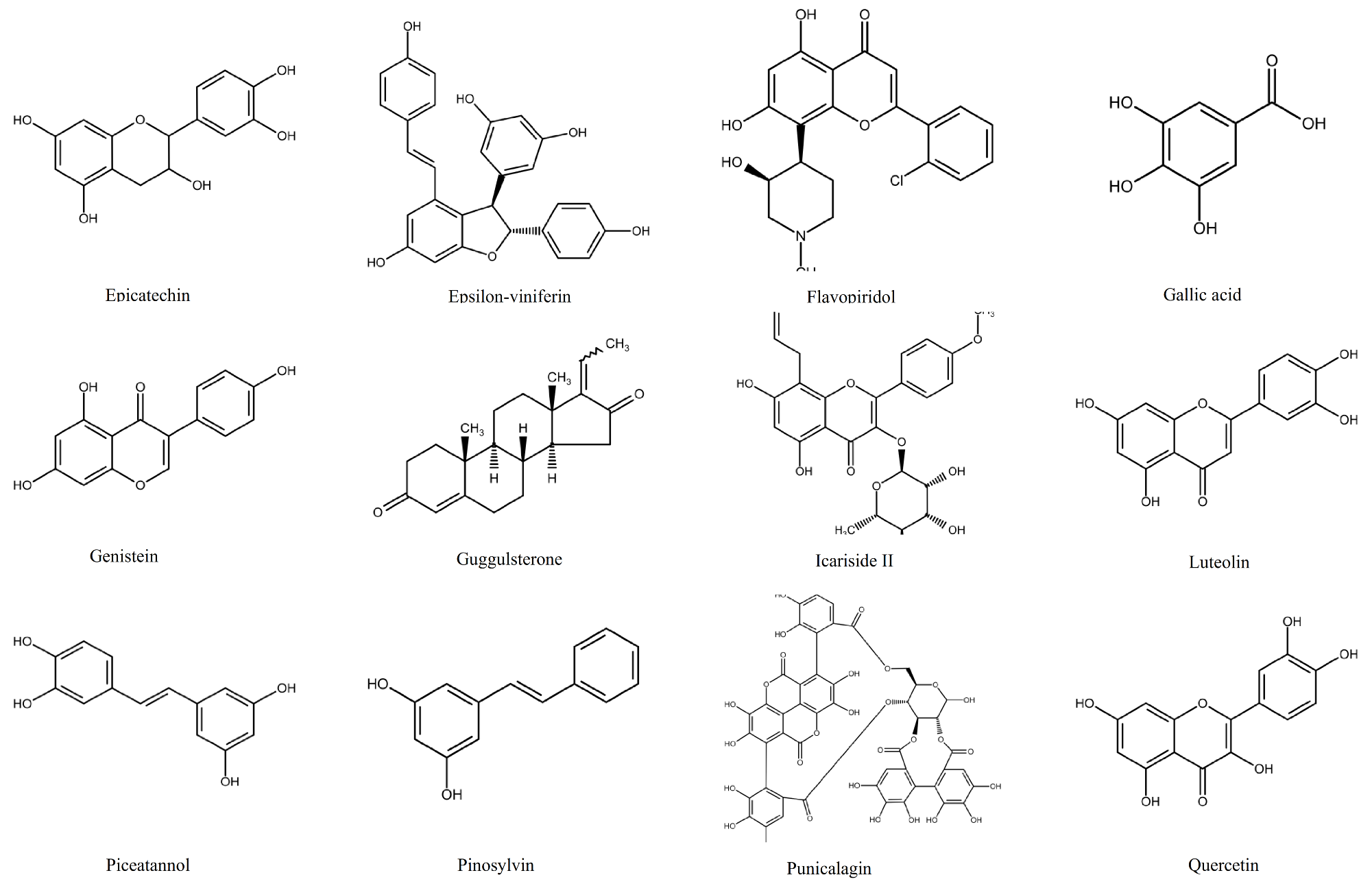
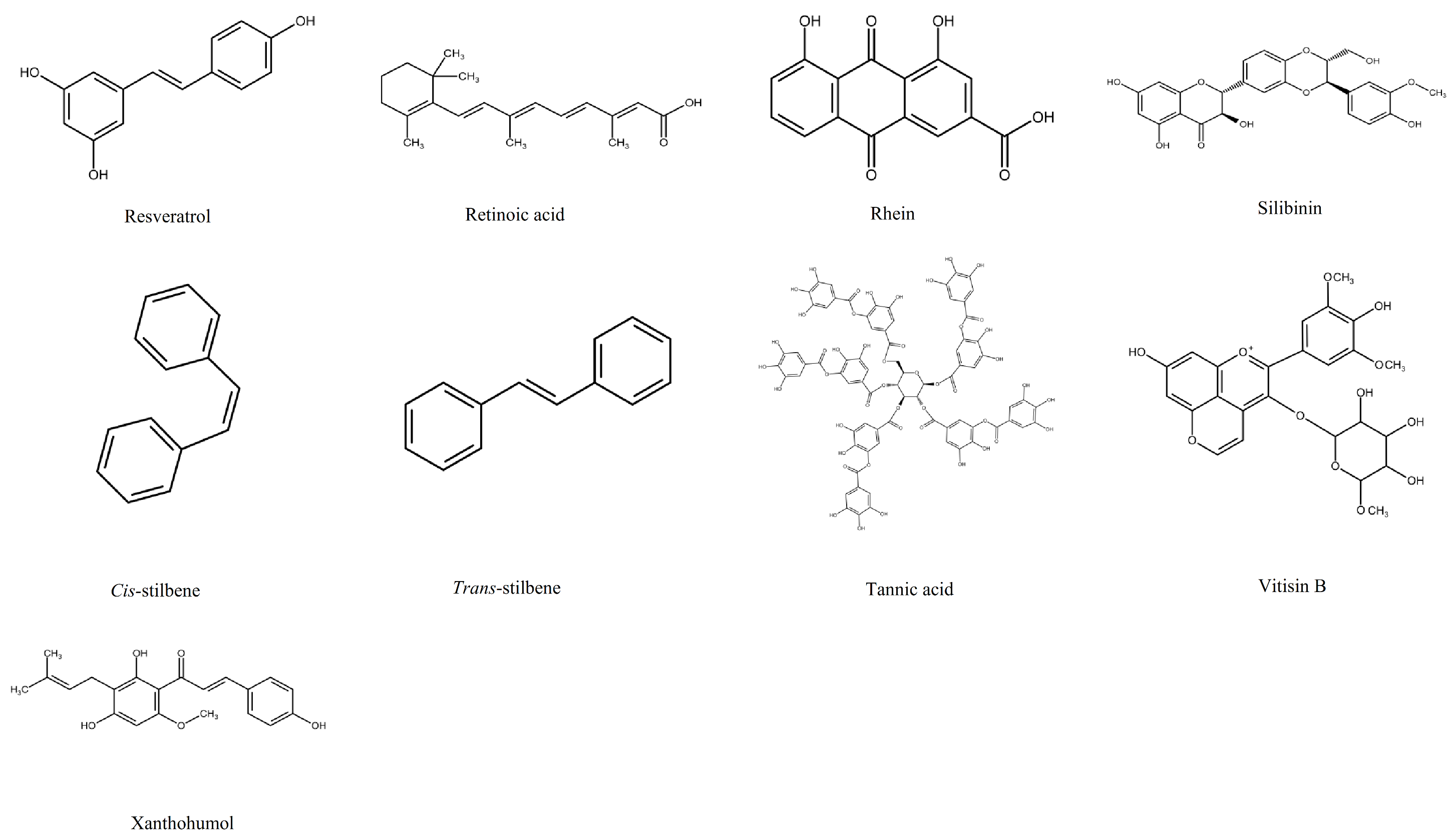
Figure 2.
Molecular targets of polyphenols in leukaemia. The arrows represent changes in the levels and activities of genes and proteins. The red arrow shows an inhibition or reduction in gene expression and/or protein production, whilst the blue arrow shows an induction or increase gene expression and protein production. The orange sign indicates cell cycle arrest. The figure does not indicate any hierarchy.

Figure 3.
The classical NF-κB (Right) and STAT3 (left) activation pathways. The NF-κB pathway is initiated by numerous stimuli including TNFα and IL1 and is mediated by the IκB kinase (IKK) complex. This results in the phosphorylation of IkBα and leads to degradation by the proteasome. This releases RelA/p50 complex to be translocated into the nucleus and binds to DNA to induce the expression of specific genes. STAT3 is activated by the Janus kinase (JAK)/signal transducer, non-receptor tyrosine kinase signalling pathways and Ras/mitogen-activated protein kinase (MAPK). Suppressor of cytokine signalling (SOCS) and protein inhibitor of activated STAT3 (PIAS3) can negatively regulate the activity status of STAT3. The activated form of STAT3 translocate into the nucleus and binds to the DNA of specific genes.

Figure 4.
The role of reactive oxygen species (ROS) and glutathione (GSH) in regulation of cell survival and cell death. Glutathione (GSH) (orange) is synthesised from glutamate (Glu), cysteine (Cys) and glycine (Gly) through two ATP-dependent steps. They are catalysed by glutamate-cysteine ligase (GCLC) and then glutathione synthase (GS) (green). Increased GSH levels (orange) lead to reduced ROS (purple) production and results in cell survival. In contrast, GSH depletion leads to increases ROS production, redox status alterations, and consequently cell death, whether apoptosis (blue) or ferroptosis (light pink). Stimulation of reactive oxygen species (ROS) can lead to induction of apoptotic signalling including permeabilisation of the mitochondrial membrane (brown), either directly or through Bcl2 (brown), which results in the release of cytochrome c (red) which in turn activates caspase 9 and 3 (yellow), which mediates cell death. Polyphenols (light green) alone or in combination with chemotherapy agents (light blue) drive an increase in ROS and decrease in GSH levels (blue lines) and cancer cell death via apoptosis or ferroptosis.

Figure 5.
The modulation effect of polyphenols on the cell cycle. Cyclin dependent kinases (CDK) regulate the cell cycle, with cyclins. The CDK inhibitors are regulated by regulatory proteins: p21, p16 and p27 (shown in orange). The proto-oncogene P53 (show in yellow) regulates transcription factors via induction of p21 which is a CDK inhibitor. The retinoblastoma protein (pRb) (shown in brown) is an inhibitor of cell cycle progression from G1 to the S phase of the cell cycle by interacting with E2F transcription factors (purple), this in turn is regulated by a cell-cycle dependent phosphorylation catalysed by cyclin-dependent kinases in the late G1 phase of the cell cycle. The modulation effect of a variety of polyphenols (lime green) is shown by the red lines.

Figure 6.
The modulation effect of polyphenols on the intrinsic and extrinsic pathways of apoptosis. The extrinsic death receptor pathway activates by the death receptor ligands such as FasL (dark red), TNF-α or TRAIL (light red). The binding of FasL to Fas (dark brown) recruits FADD (light blue) and pro-caspase-8 (orange and black), forming the DISC complex, which activates caspase-8 and then caspase 3. The binding of TNF-α (light red) to TNFR1 (light brown) recruits TRADD (dark green), RIP (red), TRAF2/5 (light green) and cIAP1/2 (blue) to form the complex, which in turn binds to pro-caspase 8 and then activates caspase 8. The intrinsic death receptor pathway is initiated by BH3-only protein (green), which can inactivate Bcl-2 (dark orange) and prevent Bcl-2 from effectively neutralizing Bax (dark purple) and Bak (light purple) and activates them. The Bax and Bak activation on the mitochondrial membrane leads to release of second mitochondria-derived activator of caspase a mitochondrial protein (SMAC) (dark grey) and cytochrome c (yellow) to the cytoplasm. The cytoplasmic cytochrome c then associates with Apaf-1 (red) and pro-caspase 9 to form the apoptosome, which activates caspase 9 and caspase 3, leading to apoptosis. Caspase 3 in turn initiates the cleavage of the nuclear enzyme PARP (yellow) which recognises damaged DNA, and initiates apoptosis if DNA damage is not repaired. Finally, SMAC is able to regulate apoptosis by inhibiting the inhibitor of apoptosis protein (XIAP). The effect of a variety of polyphenols (shown in lime green) is shown on both genes and proteins level regulating the intrinsic and extrinsic pathways of apoptosis. The blue arrows indicate the induction or activation of a protein or genes; whilst the red line shows the depletion or inhibition of a protein or gene expression.

Figure 7.
Stages and process of the autophagy. The process of the autophagy includes five stages (1) Initiation, (2) membrane nucleation and phagophore formation, (3) expansion of phagophore, (4) fusion with the lysosome, and (5) degradation. The autophagy initiation is mainly regulated by the mammalian target of rapamycin (mTOR) complex 1 mTOR (blue) as an inhibitor and AMP-activated kinase (AMPK) (light yellow) as an activator. The inhibition of mTOR and/or activation of AMPK leads to activation of the Unc-51-like kinase 1 (ULK1) complex (Purple), which includes ULK1, autophagy-related protein 13 (ATG13), and ATG101. This leads to triggering of the autophagy machinery and enables nucleation of the phagophore and regulated by three regulatory protein complexes. The class III PI3K (PI3KC3) complex (brown) involves vacuolar protein sorting 34 (VPS34), (VPS15), Beclin 1 and ATG14. The autophagosome-specific phosphatidyl-inositol-3-phosphate (PI3P)-binding complex (green) includes WD-repeat domain phosphoinositide-interacting proteins (WIPIs) and double FYVE domain-containing protein 1 (DFCP1). WIPIs then bind to ATG12-ATG5-ATG16L1 complex (light red), that in turn enhances the LC3 conjugation (orange). LC3 is converted into LC3-I by ATG4 (grey) and then conjugated with phosphatidylethanolamine (PE) (pink) and in the presence of ATG3 and ATG7 (grey) to form LC3-II, which is the characteristic signature of autophagosomal membranes. Different cellular membranes involve the elongation of the autophagosomal membrane such as the lipid bilayers, which are delivered by ATG9-containing vesicles (dark green). Now, the membrane of the autophagosomal seals around the structure are to be removed from the cell. The autophagosome fuses with the lysosome which destroys the contents. Finally, the autophagic cargo is degraded.
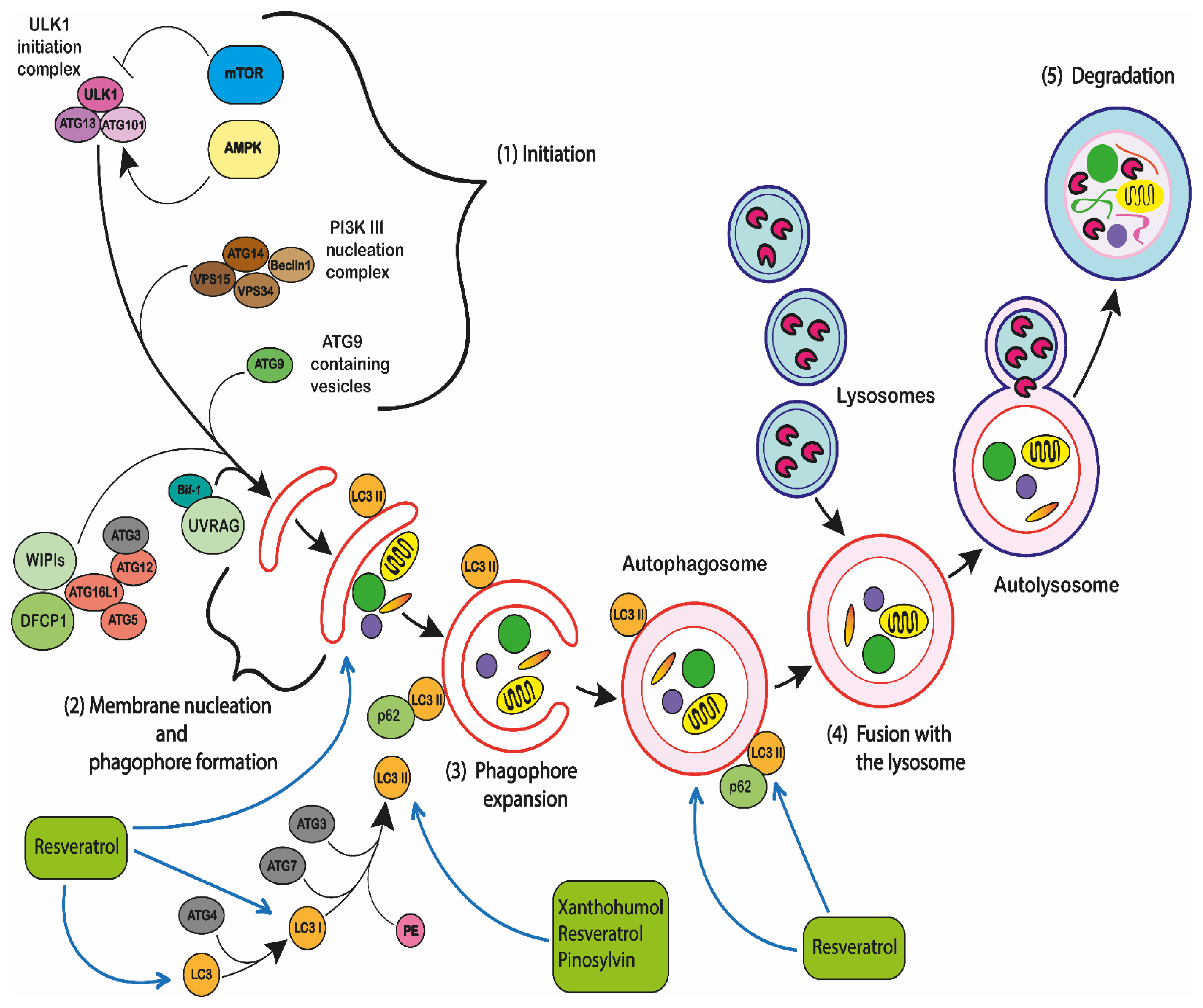
Figure 8.
Autophagy mediates programmed cell death. The extensive proteins and pathways crosstalk between autophagy and apoptosis, which include the Beclin 1-BCL-2 interaction; caspase-mediated Beclin 1 cleavage; ATG12- ATG3 conjugation; ATG12-Mcl-1 interaction; ATG5-FADD interaction. (red lines) represent inhibitory interactions, (while blue lines) with arrows represent stimulatory interactions.


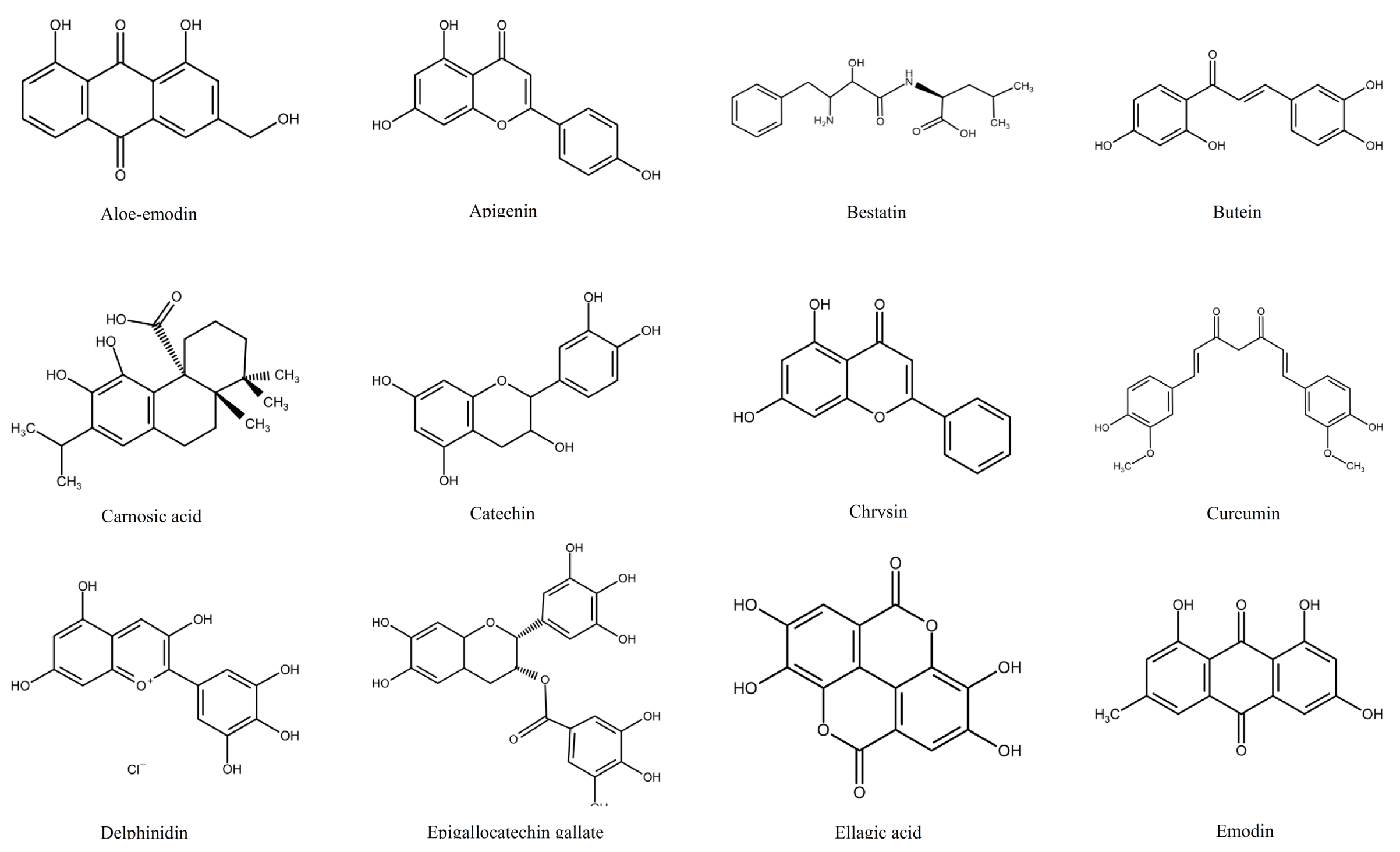
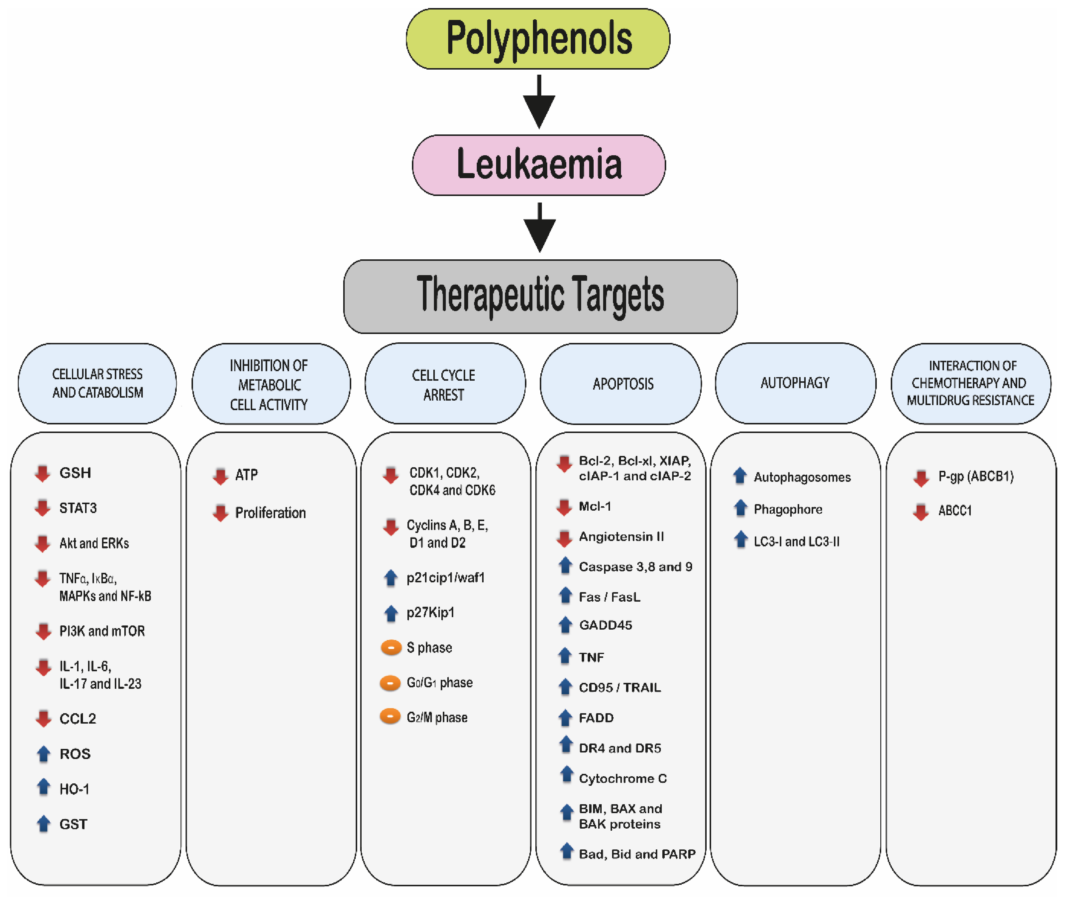
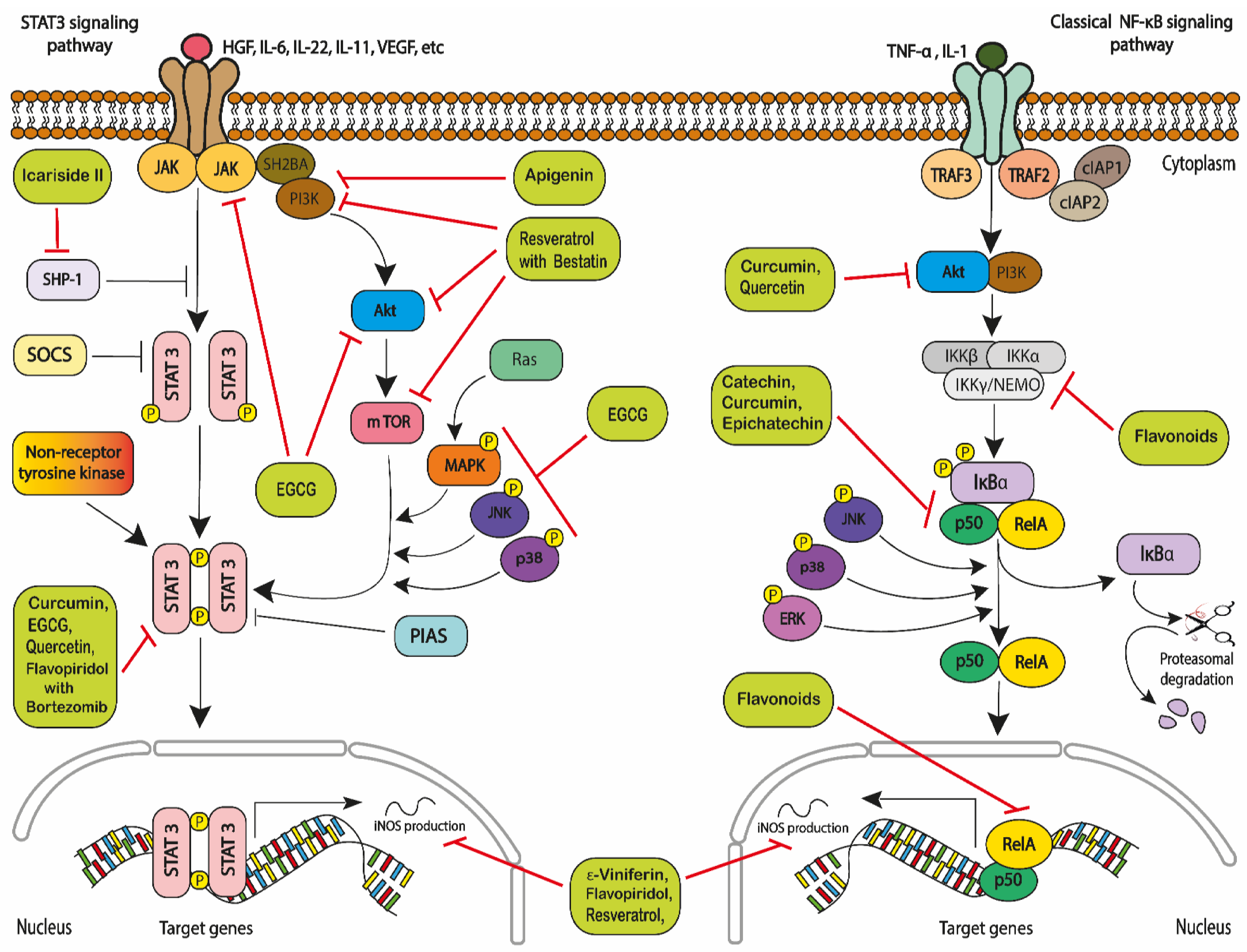
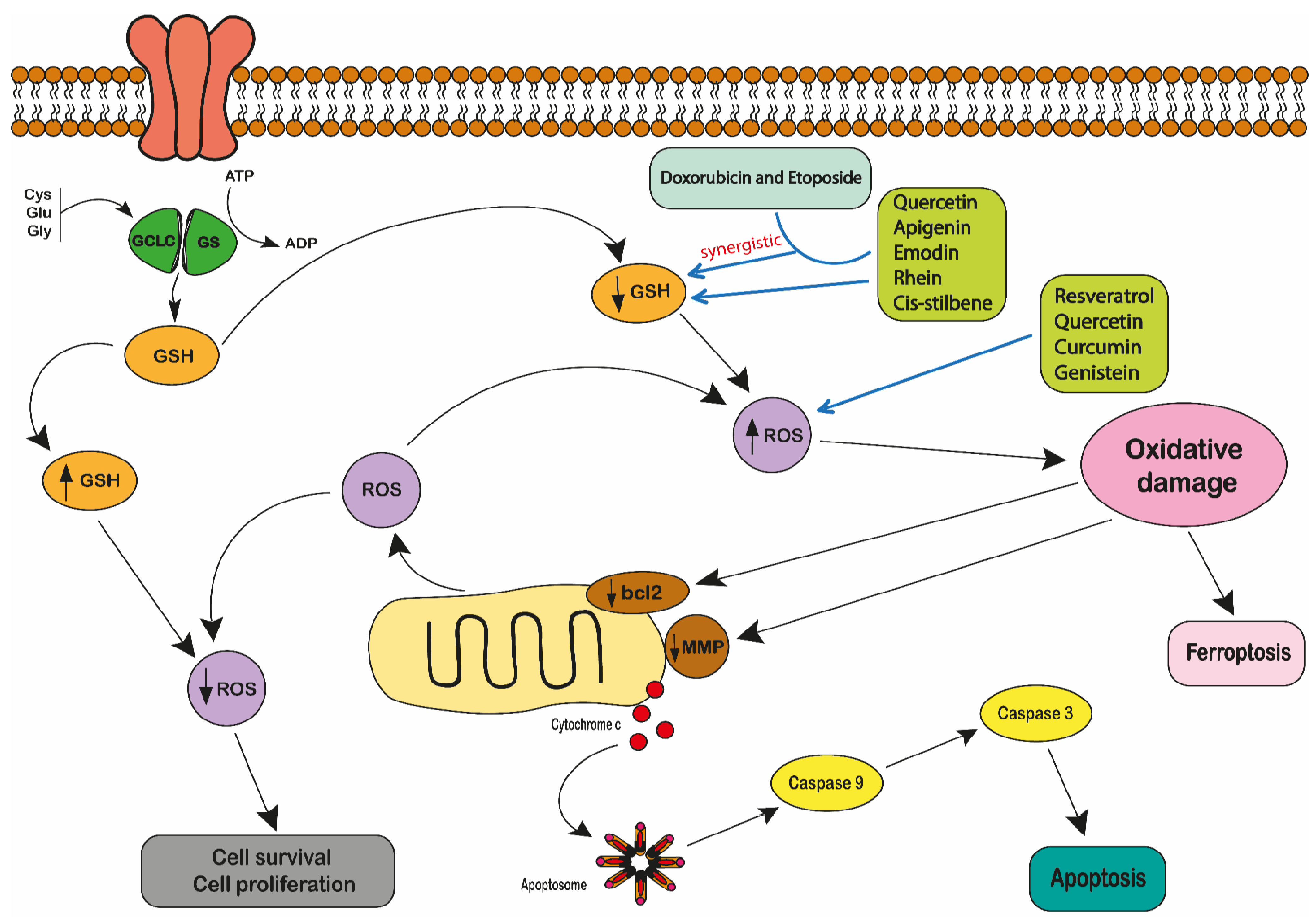
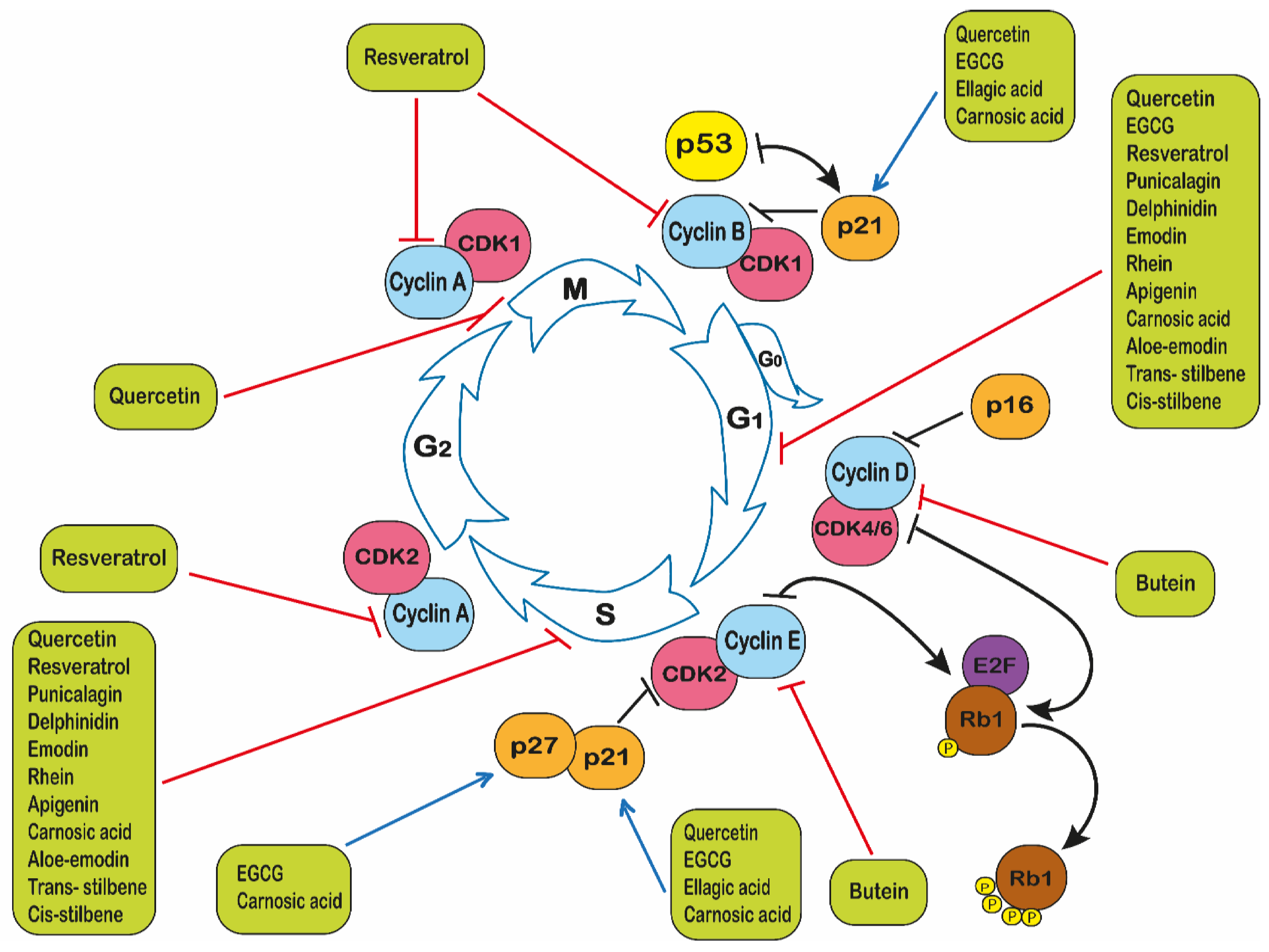
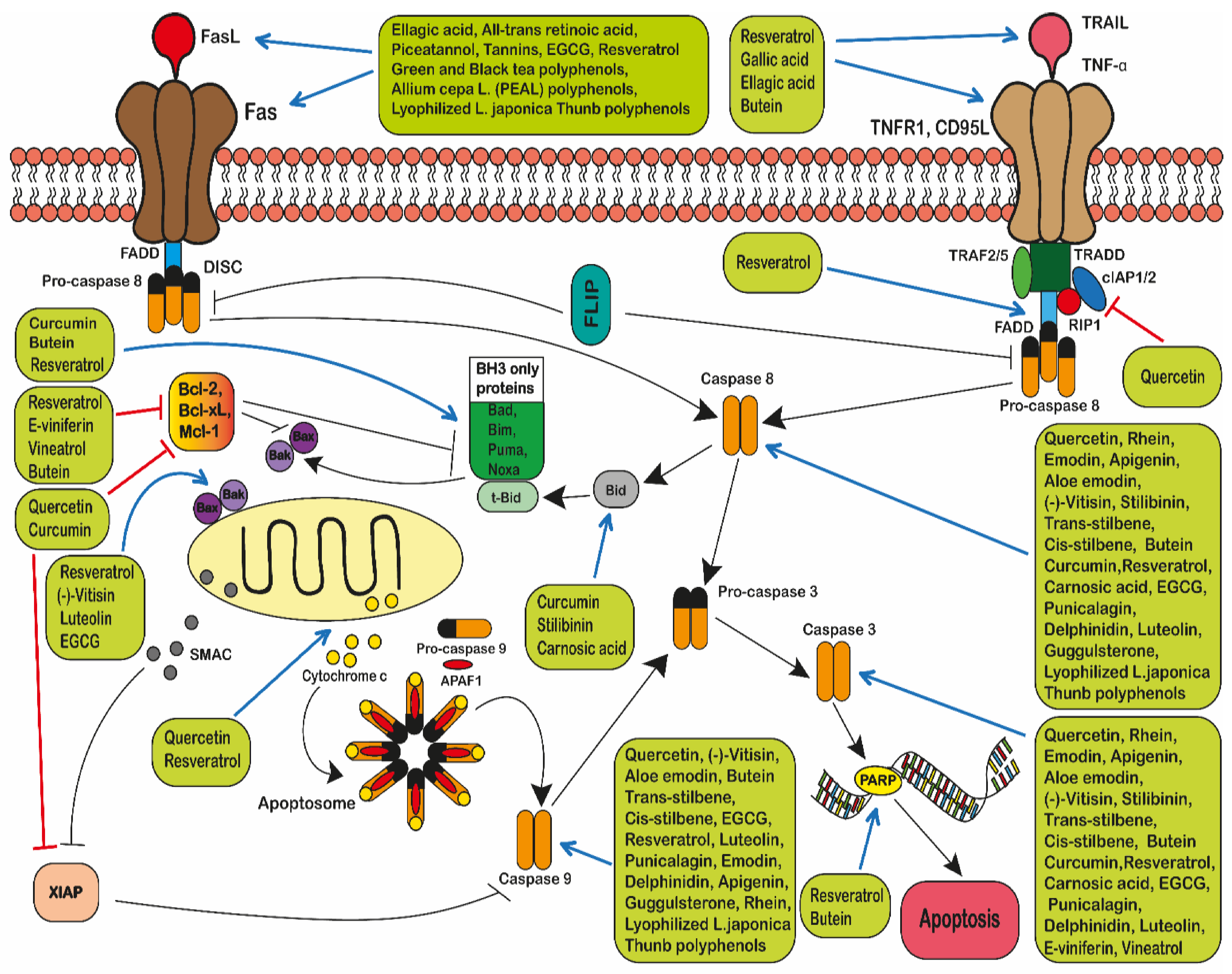
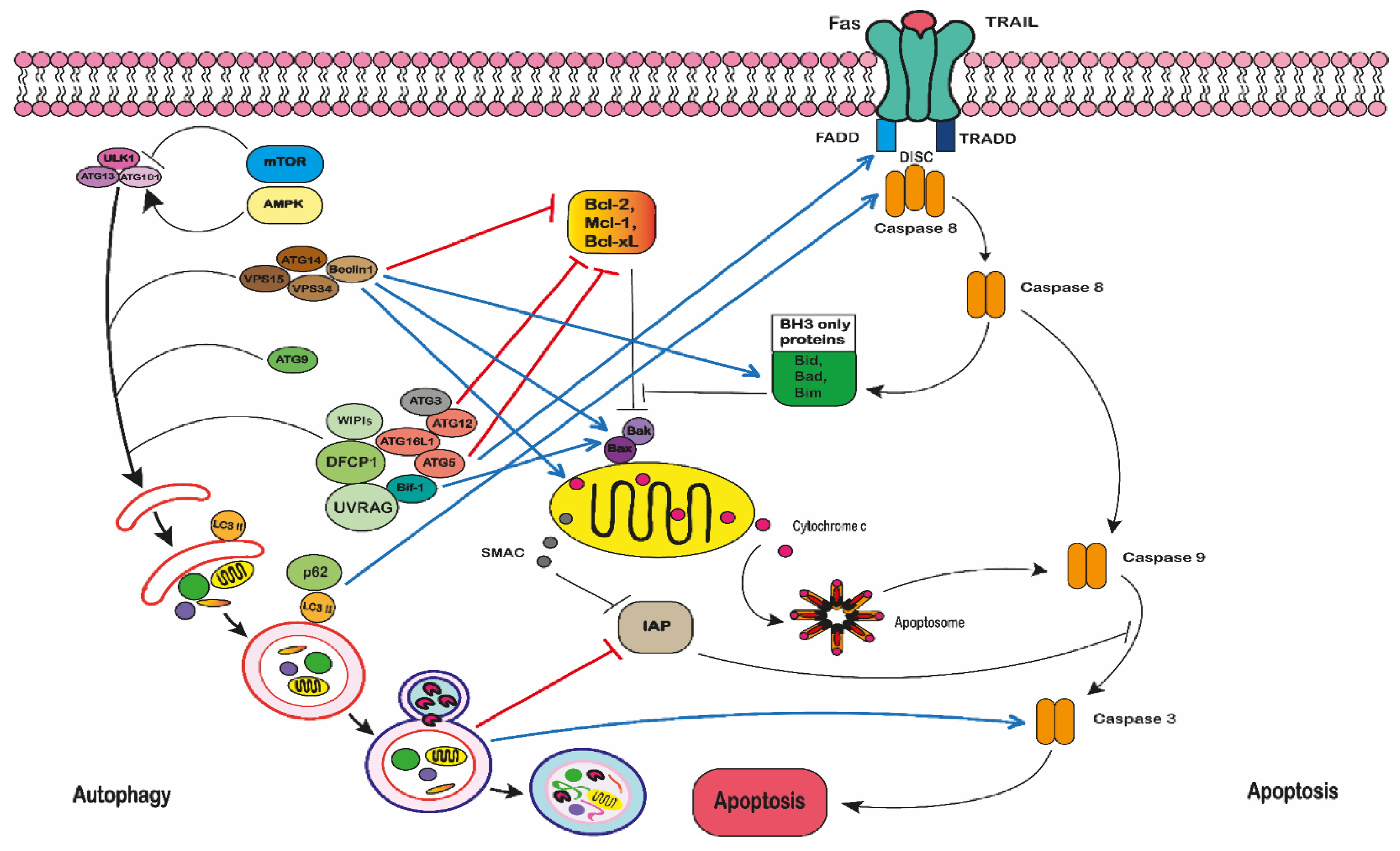
Table 1.
Summary of leukaemia cells lines discussed within this review, including the species of origin, designated abbreviation, commercial source or reference where the cell line was introduced and specific polyphenols that affect the cell lines.
Table 1.
Summary of leukaemia cells lines discussed within this review, including the species of origin, designated abbreviation, commercial source or reference where the cell line was introduced and specific polyphenols that affect the cell lines.
| CANCER TYPE | CELL LINE | DESCRIPTION | SPECIES | SOURCE OR REF | POLYPHENOLS |
|---|---|---|---|---|---|
| LYMPHOID LEUKAEMIA | 232B4 | Chronic lymphocytic leukaemia | Human | Wendel-Hansen et al. 1994 [60] | Quercetin [61] Resveratrol [61] |
| B-CLL | Chronic lymphocytic leukaemia | Human | Hoogendoorn et al. 2004 [62] | Curcumin [63,64] Epsilon-viniferin (ε-viniferin) [65] Flavopiridol [65] Quercetin [19] Resveratrol [66] | |
| CCRF-CEM | Acute lymphocytic leukaemia | Human | ATCC | Aloe-emodin [67] Apigenin [67] Delphinidin [68] Emodin [67] Flavonoids [69] Punicalagin [68] Quercetin [67,68] Rhein [67] Resveratrol [70] Trans-Stilbene [67] cis-Stilbene [67] Quercetin, apigenin, emodin, rhein or cis-stilbene plus doxorubicin or etoposide [24] Quercetin, apigenin, emodin, rhein, or cis-Stilbene plus chlorambucil, cisplatin or cyclophosphamide [71] Quercetin, apigenin, emodin, rhein, or cis-stilbene plus methotrexate, 6-mercaptopurine or 5-fluorouracil [23] EGCG, tannic acid or curcumin plus doxorubicin [72] | |
| CCRF-CEM-C7H2 | Acute lymphoblastic leukaemia | Human | Strasser-Wozak et al. 1995 [73] | Resveratrol [74] | |
| CEM | Acute lymphoblastic leukaemia | Human | ATCC | Resveratrol [75,76] | |
| CEM-C1-15 | Acute lymphoblastic leukaemia | Human | ATCC | Pinosylvin [77] Resveratrol [77] | |
| CEM-C7-14 | Acute lymphocytic leukaemia | Human | Gu et al. 2019 [78] | Pinosylvin [77] Resveratrol [77] | |
| ESKOL | B-lymphoblastoid hairy cell leukaemia cell lines | Human | Harvey et al. 1991 [79] | Resveratrol [66] e-Viniferin plus Vineatrol [80] | |
| HSB-2 | Acute lymphoblastic leukaemia | Human | ATCC | Resveratrol [81] | |
| Jurkat | Acute lymphocytic leukaemia | Human | ATCC | Aloe-emodin [67] Apigenin [67] Butein [82] Catechin [83] Emodin [67] Epichatechin [83] Epigallocatechin gallate [84] Flavonoids [83] Pinosylvin [77] Quercetin [67] Resveratrol [75,76,77,85,86,87] Rhein [67] trans- stilbene cis-Stilbene [24,67] Quercetin, apigenin, emodin, rhein, or cis-stilbene plus chlorambucil, cisplatin or cyclophosphamide [71] Quercetin, apigenin, emodin, rhein, or cis-Stilbene plus methotrexate, 6-mercaptopurine or 5-fluorouracil [23] Quercetin, apigenin, emodin, rhein, or cis-Stilbene plus doxorubicin or etoposide [24,67] | |
| MOLT-3 | Acute lymphocytic leukaemia | Human | ATCC | Delphinidin [68] Quercetin [68] | |
| MOLT-4 | Acute lymphocytic leukaemia | Human | ATCC | Pinosylvin [77] Quercetin [88,89] Resveratrol [76,77,90] Quercetin plus ellagic acid [88] | |
| MT-4 | Acute lymphoblastic leukaemia | Human | Fernandez et al. 2019 [91] | Butein [92] | |
| Nalm-6 | Acute lymphoblastic leukaemia | Human | ATCC | Resveratrol [66,75,76] | |
| REH | Acute lymphoblastic leukaemia | Human | ATCC | Resveratrol [75,76] | |
| RS4;11 | Acute lymphoblastic leukaemia | Human | ATCC | Resveratrol [75,76] | |
| SEM | Acute lymphoblastic leukaemia | Human | Greil et al. 1994 [93] | Resveratrol [75,76] | |
| SUP-B15 | Acute lymphocytic leukaemia | Human | ATCC | Resveratrol [87] | |
| TL-Oml | Acute lymphoblastic leukaemia | Human | Sugamura et al. 1984 [94] | Butein [92] | |
| WSU-CLL | Chronic lymphocytic leukaemia | Human | Mohammad et al. 1996 [95] | Resveratrol [66,96] e-Viniferin plus Vineatrol [80] | |
| L1210 | Lymphocytic leukaemia | Mouse | ATCC | Curcumin [97] Gallic acid [97] Quercetin [97,98] Tannic acid [97] Resveratrol [99] | |
| MYELOID LEUKAEMIA | AML-2/DX30, AML-2/DX100 AML-2/DX300 | Doxorubicin resistant acute myeloid leukaemia cell lines | Human | Kweon et al. 2010 [100] | Resveratrol [100] |
| AML-2/WT | Ara-C resistant acute myeloid leukaemia | Human | Song et al. 2009 [101] | Resveratrol [100] | |
| HL-60 | Acute promyelocytic leukaemia | Human | ATCC | Apigenin [102,103] Butein [104] Chrysin [103] Carnosic acid [105,106] Curcumin [106,107,108,109] Delphinidin [68] Ellagic acid [66] Epigallocatechin gallate [110] Flavonoids [111] Gallic acid [66] Luteolin [112] Punicalagin [68] Quercetin [68,108,109,113,114] Resveratrol [76,90,96,115,116,117,118,119] Silibinin [107] (-)-Vitisin [120] Xanthohumol [121] Curcumin plus silibinin or carnosic acid [107] Ellagic acid plus all-trans-retinoic acid [122] Gallic acid, ellagic acid plus all-trans retinoic acid [122] Green or black tea [123] | |
| K562 | Chronic myelogenous leukaemia | Human | ATCC | Allium cepa L. (PEAL) polyphenols [124] Butein [82,104] Curcumin [125] Epigallocatechin gallate [126,127] Flavopiridol [128] Quercetin [129] Resveratrol [76,81,96,130] Green or black tea [123] Resveratrol plus bestatin [130] Woodfordin I extract (high in tannins) [123] | |
| K562/ADR | Adriamycin-resistant chronic myeloid leukaemia cell line | Human | Tsuruo et al. 1986 [131] | Resveratrol plus bestatin [130] | |
| IM-S and IM-R K562 | Imatinib-sensitive and resistant chronic myelogenous leukaemia | Human | Grosso et al. 2009 [132] | Resveratrol [133] | |
| Kasumi-1 | Acute myeloblastic leukaemia | Human | ATCC | Resveratrol [87] | |
| KCL22 | Chronic myeloid leukaemia | Human | ATCC | Resveratrol [96] | |
| KG-1a | Acute myelogenous leukaemia | Human | ATCC | Aloe-emodin [67] Apigenin [23,67] Carnosic acid [106,107] Curcumin [106,107] Quercetin [23,67] Emodin [23,67] Rhein [23,67] Resveratrol [134,135] Silibinin [107] cis-Stilbene [23,67] trans-Stilbene [67] Curcumin plus silibinin or carnosic acid [107] Quercetin, apigenin plus doxorubicin or etoposide [24] Quercetin, apigenin plus cis-platin, cyclophosphamide or chlorambucil [71] | |
| LAMA84 | Chronic myeloid leukaemia | Human | ATCC | Flavopiridol [128] | |
| MV4:11 | Biphenotypic B myelomonocytic myeloid leukaemia | Human | ATCC | Resveratrol [75,76] | |
| NB4 | Acute promyelocytic leukaemia | Human | Lanotte et al. 1991 [136] | Carnosic acid [106] Curcumin [106] Epigallocatechin gallate [110] Genistein [114,115] Quercetin [114] Resveratrol [115] | |
| OCI/AML3 | Acute myeloid leukaemia | Human | Quentmeier et al. 2005 [137] | Resveratrol [76] | |
| OCIM2 | Acute myeloid leukaemia | Human | Papayannopoulou et al. 1988 [138] | Resveratrol [76] | |
| SHI-1 | Acute monocytic leukaemia | Human | Chen et al. 2005 [139] | Curcumin [140] | |
| THP-1 | Acute monocytic leukaemia | Human | ATCC | Allium cepa L. (PEAL) polyphenols [141] Aloe-emodin [67] Apigenin [67] Butein (104] Delphinidin [68] Emodin [67] Flavonoids [69] Genistein [114] Pinosylvin [142] Punicalagin [68] Quercetin [67,68,114] Resveratrol [96,142] Rhein [67] cis-stilbene [67] trans-Stilbene [67] Quercetin, apigenin, emodin, rhein, or cis-stilbene plus chlorambucil, cisplatin or cyclophosphamide [71] Quercetin, apigenin, emodin, rhein, or cis-Stilbene plus methotrexate, 6-mercaptopurine or 5-fluorouracil [23] Quercetin, apigenin, emodin, rhein, or cis-Stilbene plus doxorubicin or etoposide [24] | |
| C1498 (TIB-49) | Acute myeloid leukaemia | Mouse | ATCC | Carnosic acid [106] Curcumin [106] | |
| MYELOMA | MM144 | Plasma cell myeloma | Human | Díaz-Rodríguez et al. 2011 [143] | Resveratrol [86] |
| MM1S | Immunoglobulin A lambda myeloma | Human | ATCC | Resveratrol [86] | |
| U266 | Myeloma; plasmacytoma | Human | ATCC | Resveratrol [86] | |
| LYMPHOMA | Raji | Burkitt’s lymphoma cell line | Human | ATCC | Epigallocatechin-gallate [144] |
| U-937 | Histiocytic lymphoma | Human | ATCC | Butein [82,104] Carnosic acid [105,106] Curcumin [106] Flavonoids [145] Guggulsterone [146] Icariside II [147] Piceatannol [148] Pinosylvin [142] Quercetin [113,124] Resveratrol [96,115,142,149] Polyphenols extracted from lyophilized Lonicera japonica (PELJ) [150] | |
| BKS-2 | B lymphoma cell line | Mouse | Udhayakumar et al. 1989 [151] | Curcumin [152] | |
| WEHI-231 | B lymphoma cell line | Mouse | ATCC | Curcumin [152] | |
| HEREDITARY SPHEROCYTSIS | WIL2-NS | B lymphocyte | Human | ATCC | Resveratrol [153] |
Publisher’s Note: MDPI stays neutral with regard to jurisdictional claims in published maps and institutional affiliations. |
© 2021 by the authors. Licensee MDPI, Basel, Switzerland. This article is an open access article distributed under the terms and conditions of the Creative Commons Attribution (CC BY) license (http://creativecommons.org/licenses/by/4.0/).
Share and Cite
MDPI and ACS Style
Alaswad, H.A.; Mahbub, A.A.; Le Maitre, C.L.; Jordan-Mahy, N. Molecular Action of Polyphenols in Leukaemia and Their Therapeutic Potential. Int. J. Mol. Sci. 2021, 22, 3085. https://doi.org/10.3390/ijms22063085
AMA Style
Alaswad HA, Mahbub AA, Le Maitre CL, Jordan-Mahy N. Molecular Action of Polyphenols in Leukaemia and Their Therapeutic Potential. International Journal of Molecular Sciences. 2021; 22(6):3085. https://doi.org/10.3390/ijms22063085
Chicago/Turabian StyleAlaswad, Hamza A., Amani A. Mahbub, Christine L. Le Maitre, and Nicola Jordan-Mahy. 2021. "Molecular Action of Polyphenols in Leukaemia and Their Therapeutic Potential" International Journal of Molecular Sciences 22, no. 6: 3085. https://doi.org/10.3390/ijms22063085
Note that from the first issue of 2016, this journal uses article numbers instead of page numbers. See further details here.