
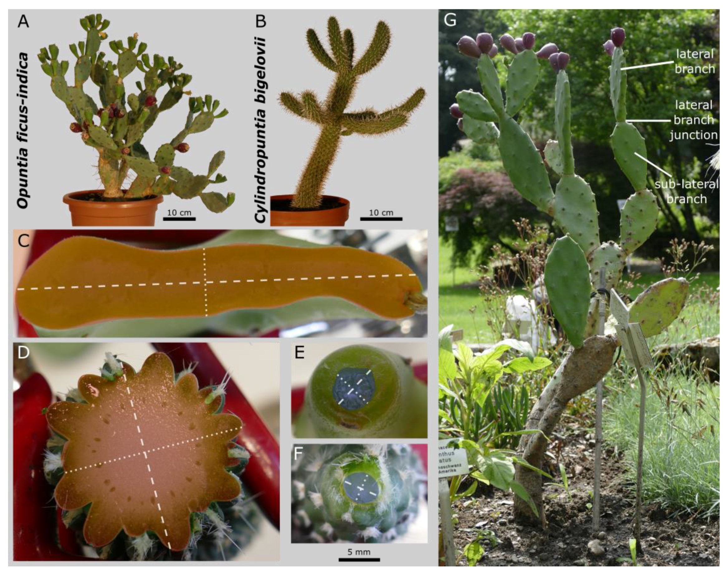
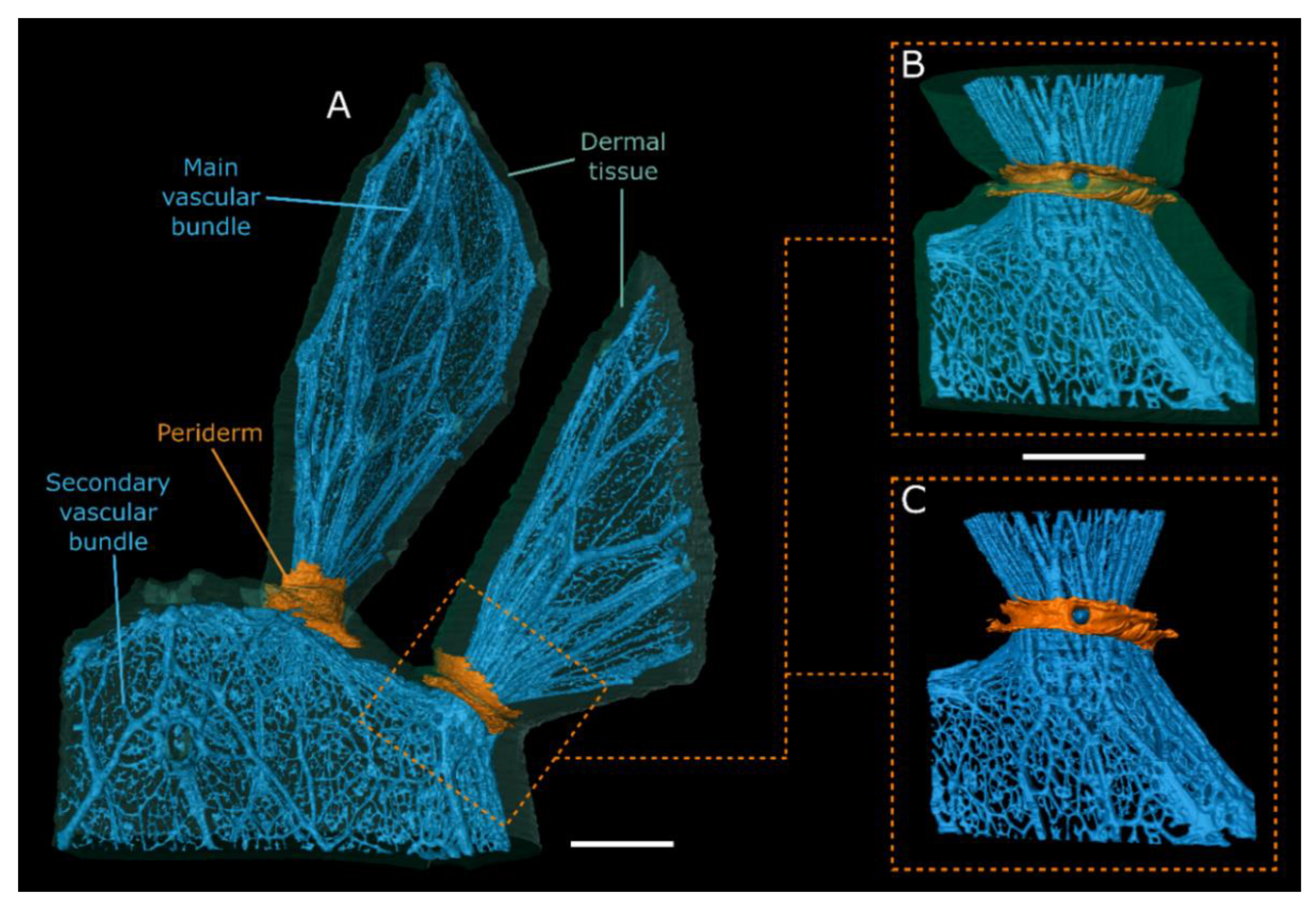
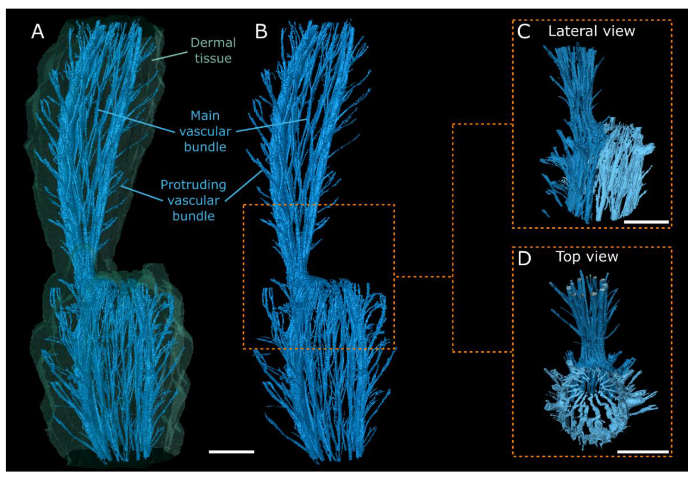
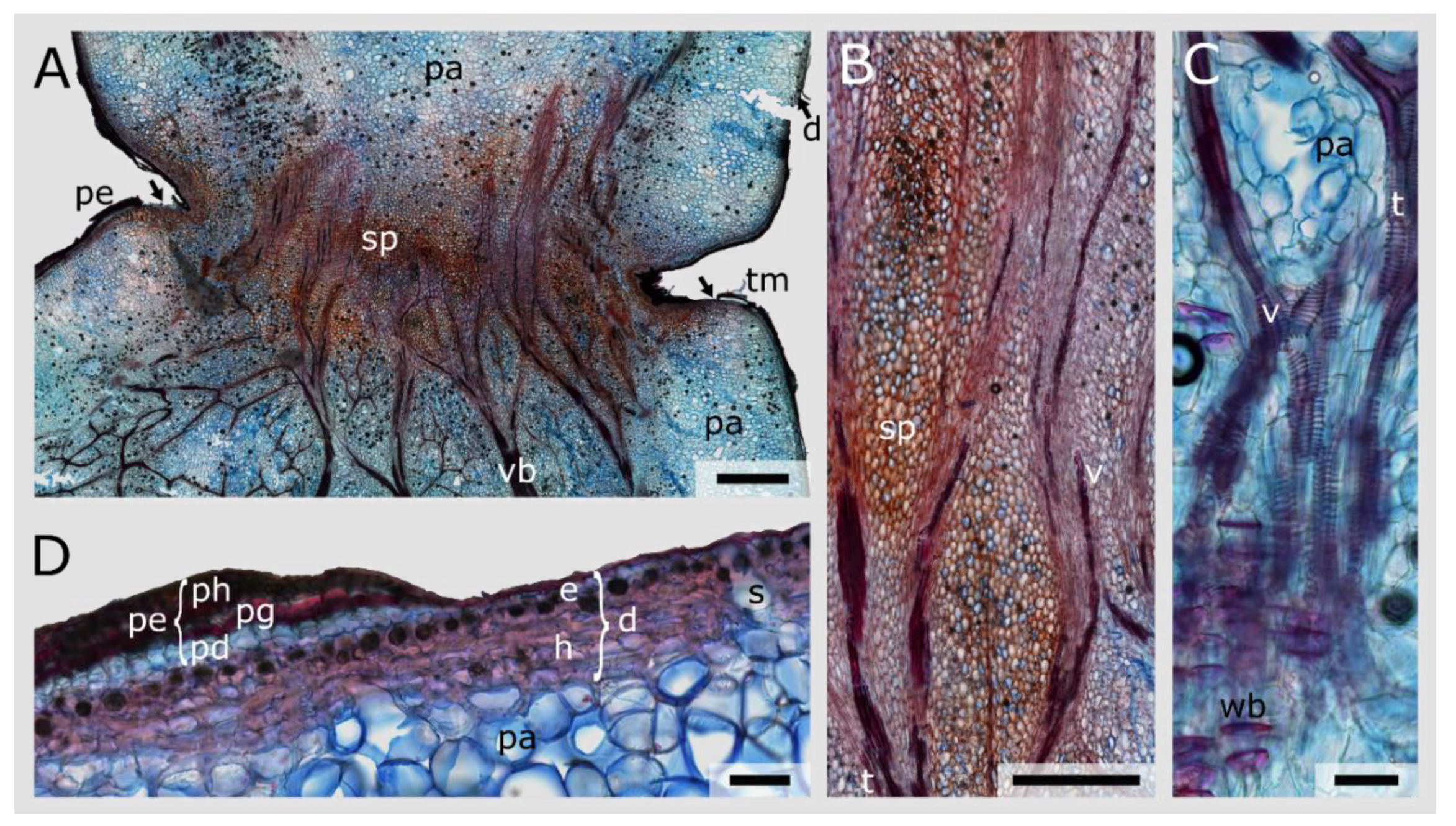
Morphology and Anatomy of Branch–Branch Junctions in Opuntia ficus-indica and Cylindropuntia bigelovii: A Comparative Study Supported by Mechanical Tissue Quantification

 ,
,  ,
,  and
and 
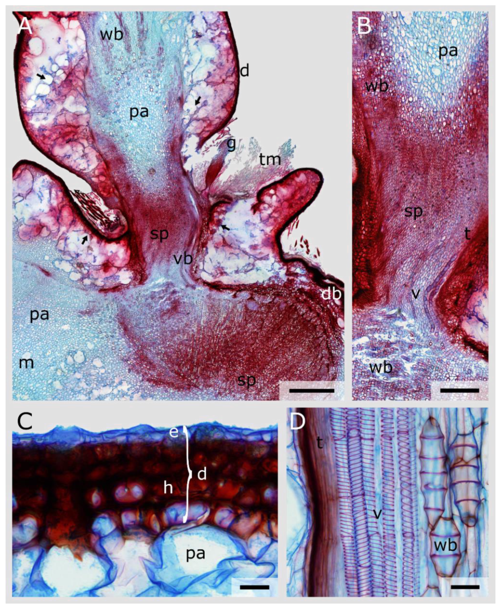
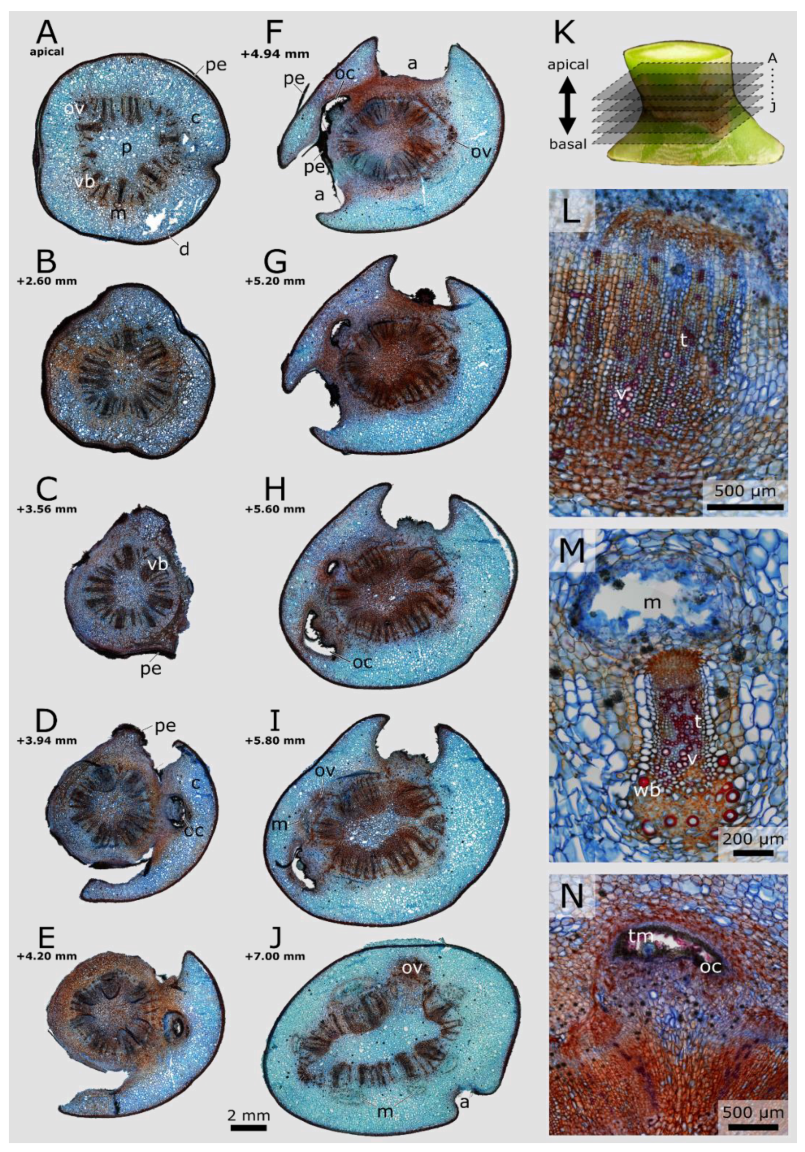
Abstract
Share and Cite
Mylo, M.D.; Hesse, L.; Masselter, T.; Leupold, J.; Drozella, K.; Speck, T.; Speck, O. Morphology and Anatomy of Branch–Branch Junctions in Opuntia ficus-indica and Cylindropuntia bigelovii: A Comparative Study Supported by Mechanical Tissue Quantification. Plants 2021, 10, 2313. https://doi.org/10.3390/plants10112313
Mylo MD, Hesse L, Masselter T, Leupold J, Drozella K, Speck T, Speck O. Morphology and Anatomy of Branch–Branch Junctions in Opuntia ficus-indica and Cylindropuntia bigelovii: A Comparative Study Supported by Mechanical Tissue Quantification. Plants. 2021; 10(11):2313. https://doi.org/10.3390/plants10112313
Chicago/Turabian StyleMylo, Max D., Linnea Hesse, Tom Masselter, Jochen Leupold, Kathrin Drozella, Thomas Speck, and Olga Speck. 2021. "Morphology and Anatomy of Branch–Branch Junctions in Opuntia ficus-indica and Cylindropuntia bigelovii: A Comparative Study Supported by Mechanical Tissue Quantification" Plants 10, no. 11: 2313. https://doi.org/10.3390/plants10112313
APA StyleMylo, M. D., Hesse, L., Masselter, T., Leupold, J., Drozella, K., Speck, T., & Speck, O. (2021). Morphology and Anatomy of Branch–Branch Junctions in Opuntia ficus-indica and Cylindropuntia bigelovii: A Comparative Study Supported by Mechanical Tissue Quantification. Plants, 10(11), 2313. https://doi.org/10.3390/plants10112313