Article

Evaluation of Inhibitory Activities of Sophora flavescens and Angelica gigas Nakai Root Extracts against Monoamine Oxidases, Cholinesterases, and β-Secretase

Department of Pharmacy, and Research Institute of Life Pharmaceutical Sciences, Sunchon National University, Suncheon 57922, Korea
*
Author to whom correspondence should be addressed.
Processes 2022, 10(5), 880; https://doi.org/10.3390/pr10050880
Submission received: 15 April 2022 / Revised: 27 April 2022 / Accepted: 28 April 2022 / Published: 29 April 2022
(This article belongs to the Section Pharmaceutical Processes)

Abstract

:
In this study, Sophora flavescens (SF) from Yeongcheon (YSF) and Mt. Jiri (JiSF), and Angelica gias (AG) from Yeongcheon (YAG), Mt. Jiri (JiAG), and Jecheon (JeAG) were extracted using three concentrations of ethanol, 95% (95Et), 70% (70Et), and 50% (50Et), and hot water (DW) to evaluate the inhibitions of monoamine oxidases (MAOs; MAO-A and B), cholinesterases (ChEs; AChE and BChE) and β-secretase (BACE1) for targeting depression and neurodegenerative diseases. There were no significant differences in constituent compounds depending on herbal origins, except that YSF-95Et and JiSF-95Et showed a distinct non-polar spot upper maackiain position, and JiAG and JeAG showed a higher amount of decursin than YAG. Ethanolic YAG and JeAG extracts showed the highest MAO-A inhibition, and YSF-95Et mostly inhibited MAO-B. JiSF-95Et showed the highest AChE inhibition and YSF-70Et, JiSF-95Et, and -70Et showed the highest BChE inhibition. Interestingly, ethanolic AG extracts showed extremely potent BACE1 inhibition, especially for JiAG-95Et and JeAG-50Et, whereas there have been no reports about BACE1 inhibition of decursin, the major compound, or AG extracts in other studies. All extracts were nontoxic to MDCK and SH-SY5Y with a low toxicity to HL-60. The results showed a different pattern of inhibitory activities of the extracts toward target enzymes depending on the origins, and multi-target abilities, especially for MAO-B and BChE by YSF-95Et, for AChE and BChE by JiSF-95Et, and for MAO-B and BACE1 by JiAG-95Et. It is suggested that those extracts are potential candidates for finding novel compounds with multi-target inhibitory activities, and herbal origin is an important factor to be considered in selection of the plants.

1. Introduction

Alzheimer’s disease (AD) is the most prevalent neurodegenerative disease, leading to dementia, which causes memory and cognitive dysfunction [1]. It brings about loss of neurons and degenerative changes of various neurotransmitter systems [2]. Most studies report that aggregation of amyloid-β (Aβ) and tau are considered the key pathologies in AD [3]. To date, most of the treatments to relieve symptoms of AD employ cholinesterase (ChE) inhibitors, which maintain the level of acetylcholine, a neurotransmitter involved in cognitive functions [4]. Recently, aducanumab, the monoclonal antibody for targeting the Aβ, received FDA approval for the first time as a treatment for AD [5]. In addition, there was a finding that elevated level of monoamine oxidase (MAO)-B, catalyzing oxidative deamination of monoamine neurotransmitters, was observed at neuronal cell of AD patients, and thus MAO-B was identified as a multi-target treatment (ChE and MAO-B inhibitors) for AD [6].
Parkinson’s disease (PD) is the second most common neurodegenerative disease, and causes movement disorder [7]. PD patients show a significant decrease in dopamine level caused by the death of the dopaminergic neuron in the substantia nigra [8]. Due to this fact, efforts to maintain the level of dopamine are employed for the treatment of PD such as dopamine agonists [9] and MAO-B inhibitors [10].
Depression is the most common psychiatric symptom, causing sadness, discouragement, and despair [11]. Depression is caused by a decrease in monoamine level, especially serotonin (5-HT) [12]. Based on the observations that MAO-A levels were elevated in patients of depression and that MAO-A is deeply involved in the breakdown of serotonin, MAO-A inhibitors have been employed for the treatment of depression [13].
MAO (EC 1.4.3.4) is a mitochondrial outer-membrane enzyme that catalyzes the oxidation of monoamine into aldehyde [14]. MAO has two isoforms, MAO-A and MAO-B; MAO-A has specificity for 5-HT and MAO-B has specificity for benzylamine and phenylethylamine [15]. It is known that MAO-A is associated with depression and cardiovascular diseases, and MAO-B is associated with neurodegenerative diseases such as AD and PD [16].
Cholinesterase (ChE) is classified into acetylcholinesterase (AChE, EC 3.1.1.7) and butyrylcholinesterase (BChE, EC 3.1.1.8). Because acetylcholine (ACh) exists in the synapse of the cerebral cortex and plays an important role as a neurotransmitter, especially in memory function, a selective inhibitor for AChE is a more efficient candidate for the treatment of AD [17]. In addition, a BChE inhibitor also showed increased choline levels and could be a candidate for the treatment of AD [18].
Beta-site amyloid precursor protein-cleaving enzyme 1 (BACE1, EC 3.4.23.46) cleaves the amyloid precursor protein (APP), which exists in the neuronal cell membrane, to cause Aβ aggregation. APP is cleaved by BACE1 followed by γ-secretase to form Aβ, which can be aggregated to form amyloid plaque [19,20]. On the basis of these facts, inhibitors of BACE1 and Aβ aggregation can be potential candidates for the treatment of AD [21,22].
On the other hand, Sophora flavescens (SF) is distributed in parts of east Asia such as Korea, China, and Japan, and is traditionally used as a medicine for allergy, inflammation, diarrhea, and gastrointestinal hemorrhage [23]. Maackiain, isolated from SF, has been studied with respect to numerous biological activities such as anti-inflammation [24] and anti-tumor [25]. Our previous study reported selective inhibition of maackiain for MAO-B, which can be used for AD and PD treatments [26].
Angelica gigas Nakai (AG) was traditionally used as sedative and blood tonic [27]. The biological activities of AG extract have been actively studied, and include anti-allergic [28] and antioxidant activity [29]. Decursin, well known as a main component of AG, has been reported to possess diverse biological activities, such as anti-tumor [30] and anti-inflammation [31]. In addition, our previous study revealed that decursin showed selective inhibition of MAO-A [32] and anti-depressant-like activity in mouse behavioral tests [33].
With this background, we extracted SF and AG using ethanol and water, analyzed their components, and evaluated their inhibitory activities against MAO-A, MAO-B, AChE, BChE, BACE1, and Aβ aggregation.

2. Materials and Methods

2.1. Materials

Prethanol A (95% ethanol, EtOH) was purchased from Duksan Science Corporation (Seoul, Korea). MAO-A, MAO-B, AChE, BChE, kynuramine, benzylamine, acetylthiocholine iodide (ATCI), S-butyrylthiocholine iodide (BTCI), 5,5′-dithiobis(2-nitrobenzoic acid) (DTNB), thioflavin T (ThT), and β-secretase (BACE1) activity detection kit (fluorescent) were purchased from Sigma-Aldrich (St. Louis, MO, USA). Dimethyl sulfoxide (DMSO), 2-mercaptoethanol (2-ME) were also obtained from Sigma Aldrich. 2,2-Diphenyl-1-picrylhydrazyl (DPPH) was purchased from Thermo Fisher Scientific (Middlesex, MA, USA). SF from Yeongcheon (YSF) in Gyeongbuk and AG from Jecheon in Chungbuk (JeAG) were purchased from Hanyakjae market (https://www.hanyakjae.net, accessed on 8 September 2021, Namyangju, Gyeonggi-do, Korea). SF (YSF) and AG from mountain Jiri (Mt. Jiri, JiAG) in Sancheong in Gyeongnam were purchased from Jirisan Herb Food (https://www.jirisanherbfood.com, accessed on 8 September 2021, Sancheong, Gyeongnam, Korea). AG from Yeongcheon in Gyeongbuk (YAG) was purchased from Hands Herb (https://www.handsherb.co.kr, accessed on 8 September 2021, Yeongcheon, Gyeongbuk, Korea). Beta-amyloid (1–42)/HFIP (hexafluoroisopropanol) was purchased from AnaSpec (Fremont, CA, USA). Madin-Darby canine kidney cells (MDCK; KCLB No. 10034), human promyelocytic leukemia cells (HL60; KCLB No.10240), and human neuroblastoma cells (SH-SY5Y; KCLB No.22266) were purchased from the Korean Cell Line Bank (KCLB, Seoul, Korea). Dulbecco’s Modified Eagle’s minimum essential medium (DMEM), minimum essential media (MEM), Roswell Park Memorial Institute (RPMI) 1640, fetal bovine serum (FBS), penicillin/streptomycin solution, and trypsin-EDTA solution were purchased from Hyclone Laboratories (Logan, UT, USA). Cell counting kit-8 (CCK-8) was acquired from Dojindo Laboratories (Kumamoto, Japan).

2.2. Extraction of SF and AG

Extraction was performed as described previously [26], i.e., the dried roots of SF or AG were ground to obtain each powder. The powder (20 g each) was added in 200 mL of three concentrations of prethanol A (95%, 70%, and 50% EtOH) or distilled water. In the case of 95%, 70%, and 50% EtOH extracts (95Et, 70Et, and 50Et, respectively), 20 g of powder was added in 200 mL of 95%, 70%, and 50% EtOH, respectively, and sonicated with medium level for 1 h at room temperature by using ultrasonic cleaner (POWERSONIC 520, 40 kHz, Hwashin Tech Corporation, Seoul, Korea). It was centrifuged at 6000× g for 15 min, the supernatant was collected and the respective 95%, 70%, and 50% EtOH was added onto the residual pellet to sonicate again. This process was performed 3 times. Until EtOH is gone, it was concentrated under rotary vacuum concentration at 45 °C and followed by 60 °C for the concentration of water. To obtain distilled water extract (DW), powder in distilled water was heated at 80 °C for 2 h, and then centrifuged at 6000× g for 15 min. The supernatant was collected, and the distilled water was added onto the residual pellet, and it was heated again. This procedure was repeated 3 times. The collected supernatant was filtered by Whatman No. 1 filter (GE healthcare, Uppsala, Sweden) and concentrated by vacuum at 65 °C.
All extracts were lyophilized to obtain dried extracts and their yields were calculated.

2.3. Thin-Layer Chromatography (TLC)

Thin-layer chromatography (TLC) was performed as described previously [34], with slight modifications to analyze active compounds in the extracts. Each extract was dissolved to be a concentration of 10 mg/mL in methanol, and 50 μg of the extract was loaded onto Prep TLC plates (PTLC Silica gel 60 F254, 0.5 mm, Merck, Darmstadt, Germany) and methanol (MeOH), ethyl acetate (EtOAc), and toluene (Tol) were used for developing solvents with different ratios. The extracts were developed in developing solvents (SF-95Et, MeOH:EtOAc:Tol = 1:2:7; SF-70Et, MeOH:EtOAc:Tol = 1:1:8; SF-50Et, MeOH:EtOAc:Tol = 1:1:8; AG-95Et, MeOH:EtOAc:Tol = 2:1:7; AG-70Et, MeOH:EtOAc:Tol = 1:1:8; AG-50Et, MeOH:EtOAc:Tol = 1:1:8) for 5 cm height and the spots were observed at 312 nm.

2.4. Enzyme Assays

MAO-A and MAO-B inhibitions were assayed as described previously [26,35] with slight modification. Briefly, MAO-A and MAO-B were reacted with 0.06 mM kynuramine and 0.3 mM benzylamine, respectively, in 50 mM sodium phosphate (pH 7.2). The extracts were dissolved with 10 mg/mL in DMSO and added to a final concentration of 5 to 50 μg/mL in the assay mixture. To avoid the organic solvent effect, the DMSO was combined with less than 1% in the assay mixture. The reaction was observed by a spectrophotometer (OPTIZEN, K-Lab, Daejeon, Korea); the absorbances of MAO-A and MAO-B assay mixtures were observed at 316 nm and 250 nm, respectively, in kinetic mode for 30 min.
The activity assays of AChE and BChE were performed according to the Ellman method [36] with slight modification [37]. In brief, AChE or BChE was reacted with 0.5 mM substrate (ATCI or BTCI, respectively) with 0.5 mM DTNB in 100 mM sodium phosphate (pH 7.5), and absorbance was observed at 412 nm for 15 min in the kinetic mode of the spectrophotometer.
For BACE1 assay, the BACE1 Activity Detection Kit (Fluorescent) (Sigma Aldrich, Saint Louis, MO, USA) was used as described previously [34,38]. BACE1 (0.3 units/μL) was added into the fluorescent assay buffer and reacted with 7-methoxycoumarin-4-acetyl-[Asn670, Leu671]-amyloid β/A4 precursor protein 770 fragment 667-676-(2,4 dinitrophenyl)Lys-Arg-Arg amide trifluoroacetate salt (BACE1 substrate). The fluorescent unit was observed at 320 nm for excitation and 405 nm for emission by fluorospectrophotometer (Fluoromate FS-2 Fluorospectrometer, Scinco, Seoul, Korea). After observing the time-zero value, the assay solution was incubated at 37 °C for 2 h and fluorescent units were observed to calculate the BACE1 activity.

2.5. DPPH Radical Scavenging Activity

DPPH radical scavenging activity was analyzed for observing antioxidant activity; a mixture of the extract (100 μg/mL) and DPPH (0.1 mM) was preincubated with ethanol, and after 15 min of preincubation, absorbance was measured at 517 nm [34,39].

2.6. Aβ42 Aggregation Assay

For measuring Aβ42 aggregation, a ThT assay was employed [40]; the extract (10 μg/mL) was incubated with 40 μM of Aβ42 and 0.5 mM of ThT at 37 °C, and the mixture was moved to fluorescent unit of fluorospectrophotometer (Synergy H1, BioTek, Winooski, VT, USA), and then fluorescence was measured at excitation 440 nm/emission 484 nm at every 5 min for 2 h.

2.7. Cell Culture and Viability

The cells, MDCK, HL60, and SH-SY5Y, were cultured in RPMI 1640, MEM, or DMEM, respectively, supplemented with 10% FBS, 100 units/mL penicillin/streptomycin solution, and 2-ME (50 μM), in a humidified atmosphere at 37 °C with 5% CO2. The cell viability was determined by CCK-8 assay [41]. Briefly, MDCK (1 × 10⁴), HL-60 (5 × 10⁴), and SH-SY5Y (5 × 10⁴) cells were seeded in 96 well plates, and then 1, 3, 10, and 30 μg/mL of the extract were added to the wells. The plate was then incubated for 24 h at 37 °C with 5% CO2. After that, 10 μL of CCK-8 solution was added to the wells and the cells were incubated for 3 h further. The absorbance was detected at 450 nm with a Micro Plate Reader (VersaMax, Molecular Devices, Sunnyvale, CA, USA). The cell viability was expressed as a percentage of the control culture.

3. Results

3.1. Yields of the Extracts

Two SF and three AG samples from different provinces were extracted with four solvents, 95Et, 70Et, 50Et, and DW. For SF, YSF-70Et showed the highest yield in YSF, 14.24%, and JiSF-DW showed the highest yield in JiSF, 15.30%. YSF and JiSF showed the lowest yield at 95Et (2.36% and 5.09%, respectively). For AG, all DW extracts showed a higher yield (21.60~23.35%) than 95Et (7.10~11.50%). In addition, YAG-70 or -50Et, JeAG-70 or -50Et, and JiAG-70Et showed the highest yield (28.00~33.82%), except JiAG-50Et (20.39%) (Table 1).

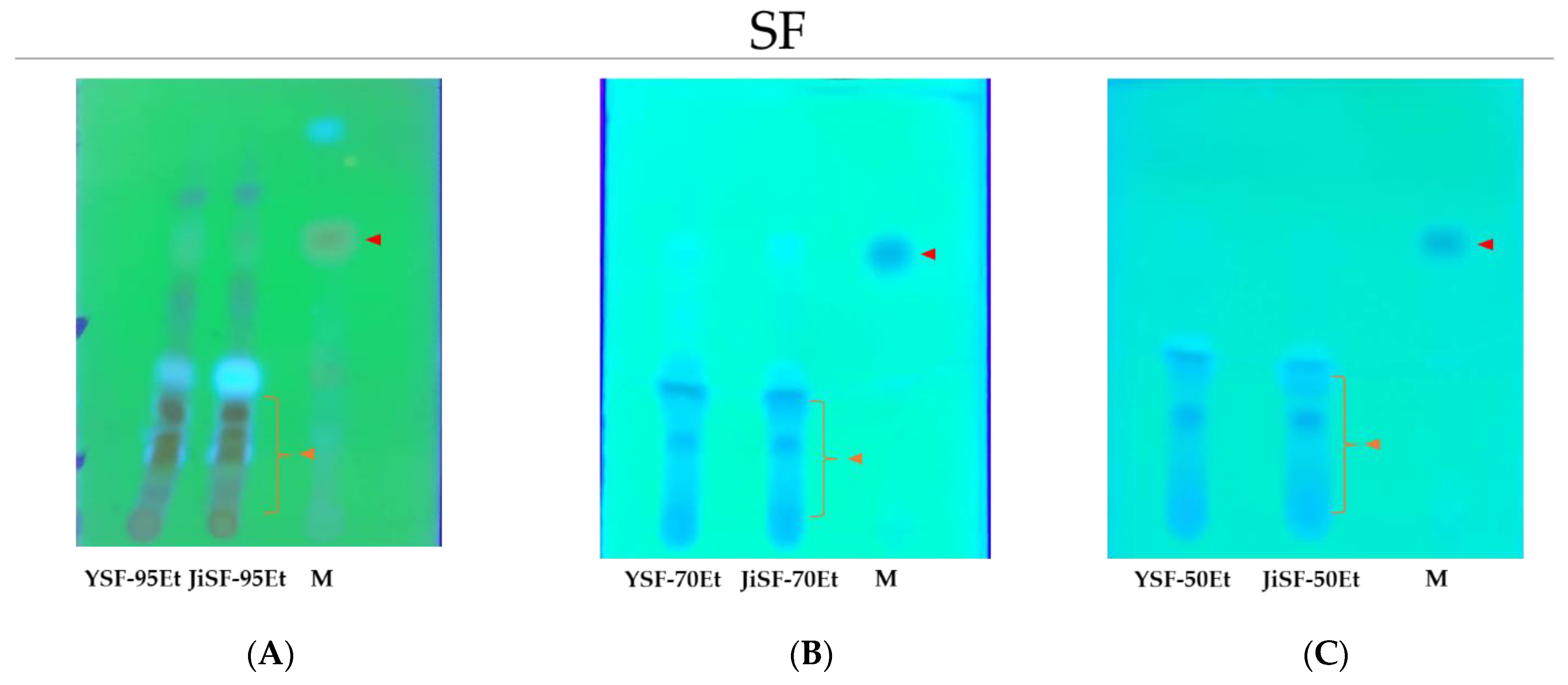
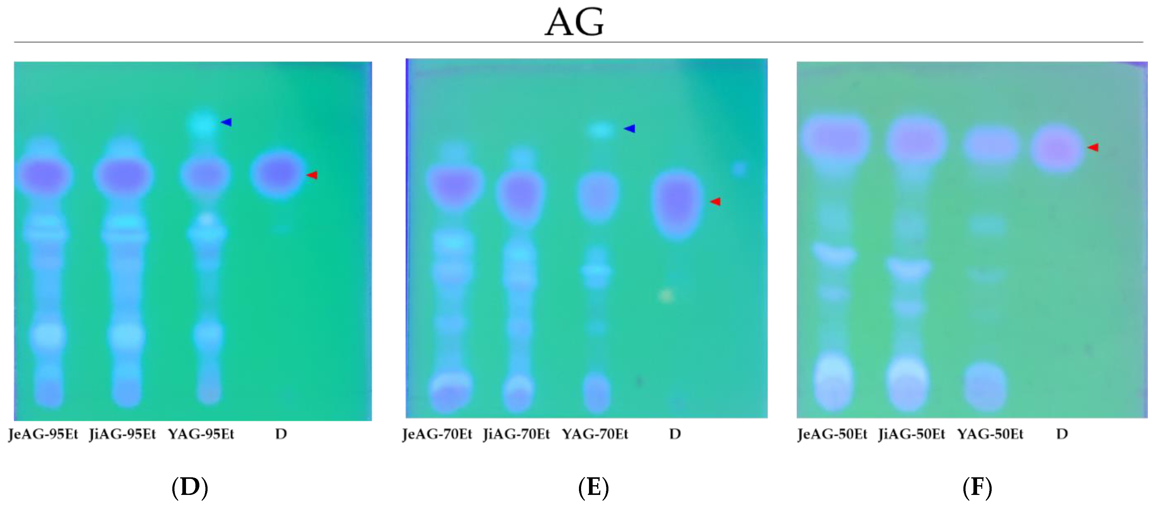
3.2. TLC Analysis of the Extracts

In TLC analysis of SF, there were no significant differences in YSF and JiSF, and it was revealed that SF extracts contained small amount of maackiain (Figure 1A–C). As the ethanol concentration decreased, polar components with low Rf values decreased due to the solvent polarity, as shown in Figure 1A–C with orange arrows. Interestingly, YSF-95Et and JiSF-95Et showed a distinct non-polar spot at the upper position of maackiain, which was not found in 70Et and 50Et.
In AG, all extracts showed that decursin was the main compound. However, JiAG and JeAG showed significantly higher amounts of decursin than YAG (Figure 1D–F). Interestingly, YAG contained a unique non-polar spot in YAG-95Et or -70Et, as shown in Figure 1D,E with a blue arrow. Furthermore, there were some differences in spots such as non-polar spots showing fluorescence. When the compounds were eluted from the spots using PTLC, no MAO-A inhibition was observed.
For all the DW extracts, the mobilities of the compounds were extremely low in the solvent systems used. For the best analysis, additional fractionation using organic solvents should be needed in further experiments. However, their inhibitory activities against the target enzymes were very low. Therefore, we decided to avoid the TLC analysis for the DW extracts.

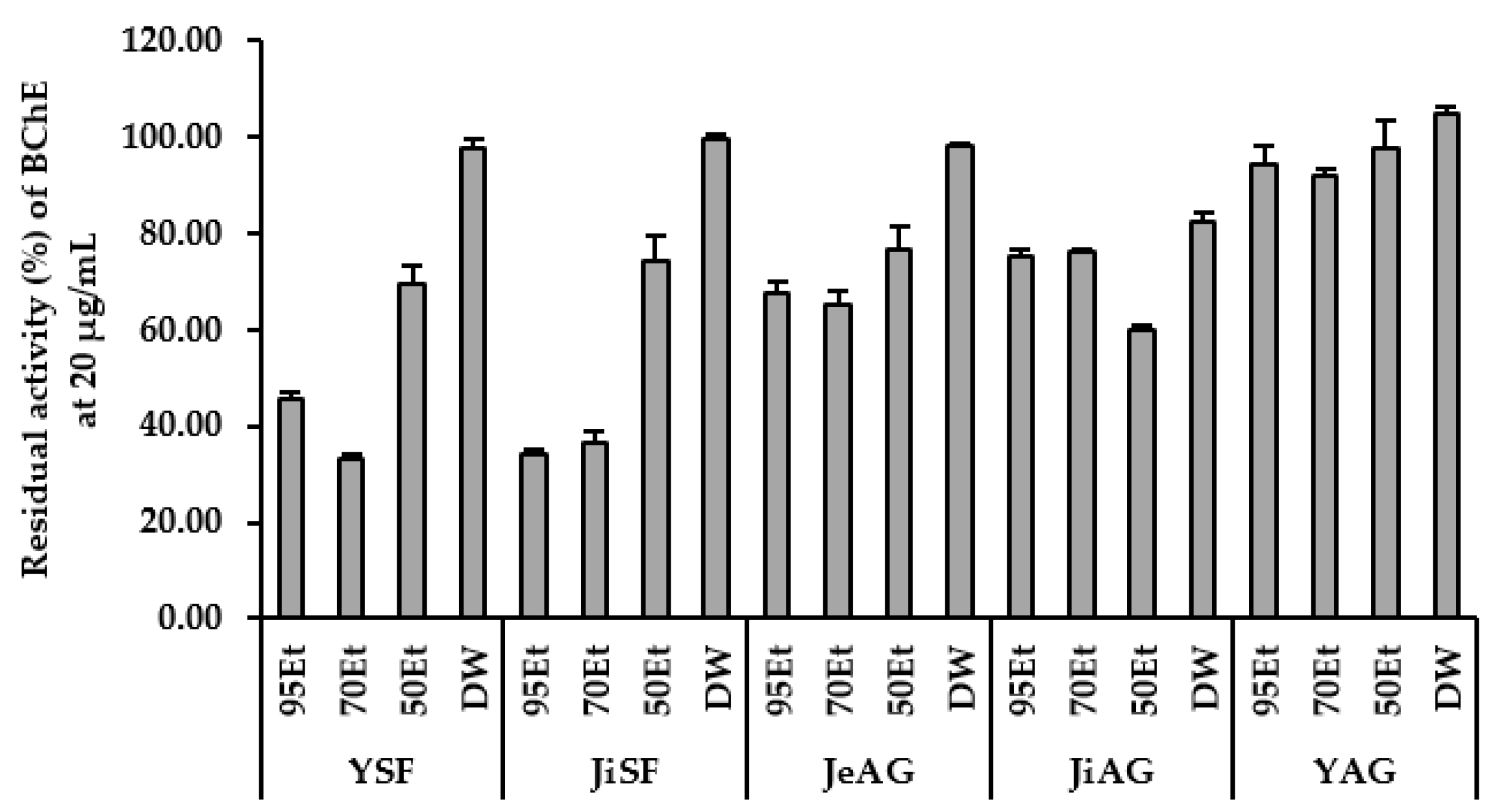
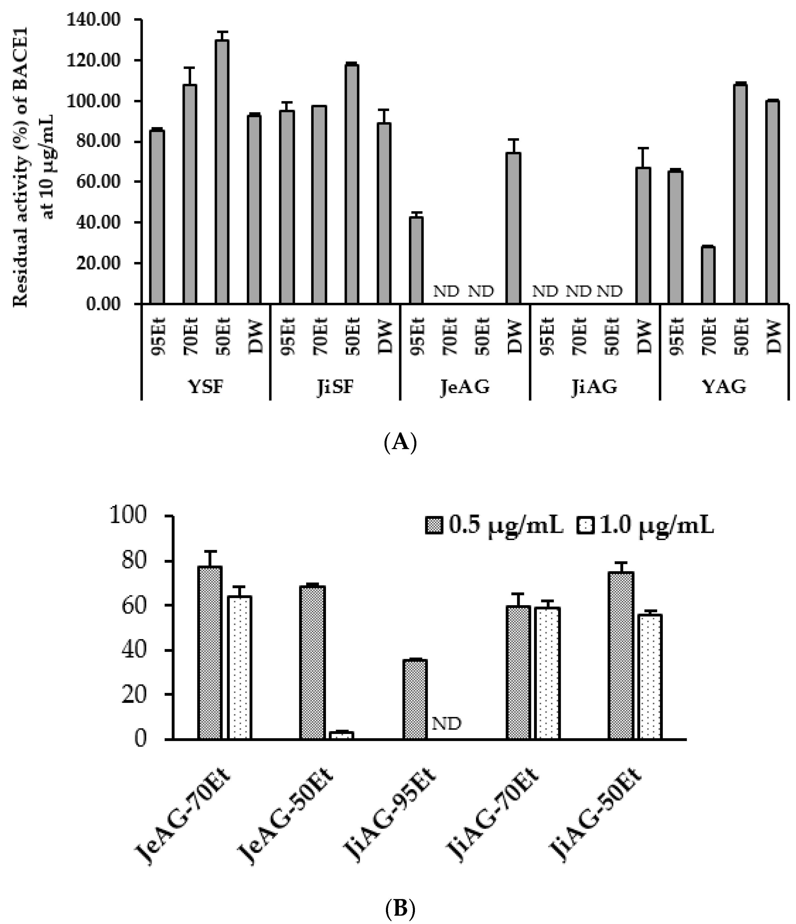
3.3. Inhibition of Enzymes by the Extracts

The inhibitory activities of extracts against MAOs and ChEs were assayed at 20 μg/mL, except for BACE1, which was assayed at 10 μg/mL.
In MAO-A inhibition analysis, JiSF-95Et and YSF-95Et showed effective inhibitory activity with the residual activities of 26.77% and 51.97%, respectively (Figure 2). As the concentration of DW increased, the inhibitory activity decreased. For AG, JeAG-70Et, YAG-70Et, and JiAG-50Et showed significant inhibition with the residual activities of 8.93%, 11.37%, and 16.22%, respectively (Figure 2). The ethanolic extracts of YAG and JeAG showed much higher inhibition (i.e., 95Et, 70Et, and 50Et of JeAG showed the residual activities of 11.59%, 8.93%, and 13.51%, respectively, and them of YAG showed 12.38%, 11.37%, and 14.11%, respectively) than that of JiAG (i.e., the residual activities of 57.38%, 53.02%, and 16.22%, respectively). In particular, JiAG-95Et and -70Et showed low MAO-A inhibition. All the DW extracts did not show effective inhibitory activities with the residual activities of 86.69~98.41%. Overall, MAO-A inhibitory activity of AG was higher than that of SF and the activity increased when concentration of EtOH was 50% or higher in AG.
In the MAO-B inhibition study, YSF-95Et, YSF-70Et, JiAG-95Et, and YSF-50Et showed effective inhibitory activity, with residual activities of 6.43%, 20.79%, 29.07%, and 42.08%, respectively (Figure 3). YSF showed significant MAO-B inhibition, and the inhibition increased with increasing EtOH concentration. For AG, JiAG-95Et showed the highest MAO-B inhibition, whereas it showed lower MAO-A inhibition compared to other AG EtOH extracts.
In AChE inhibition, JiSF-95Et showed the highest inhibitory activity, with a residual activity of 36.73%, followed by YSF-95Et with a residual activity of 57.01% (Figure 4). For AG, JeAG-95Et, JeAG-50Et, and YAG-50Et showed relatively effective inhibitory activity against AChE, with residual activities of 69.39%, 68.59%, and 71.33%, respectively, indicating that AG extracts showed significantly lower AChE inhibition than SF extracts. Other extracts did not exhibit effective AChE inhibitory activity.
In BChE inhibition, YSF-70Et, JiSF-95Et, JiSF-70Et, and YSF-95Et showed effective inhibitory activity, with residual activities of 33.26%, 34.16%, 36.28%, and 45.54%, respectively (Figure 5). SF showed higher BChE inhibition than AG, and 95Et and 70Et showed the highest BChE inhibition, compared to 50Et or DW (Figure 5).
In BACE1 inhibition, YAG-70Et showed effective inhibition, with a residual activity of 28.17% at 10 μg/mL (Figure 6A). Under these conditions, JiAG-95Et, JiAG-70Et, JiAG-50Et, JeAG-70Et, and JeAG-50Et gave negative values of the residual activities. Interestingly, when 1.0 μg/mL was used, JiAG-70Et, JiAG-50Et, JeAG-70Et, and JeAG-50Et showed extremely potent inhibition with the residual activities of 58.85%, 55.42%, 64.13%, and 3.15%, respectively (Figure 6B). Furthermore, JiAG-95Et showed the highest inhibition, with a residual activity of 35.5% at 0.5 μg/mL.

3.4. DPPH Radical Scavenging Activity and Aβ Aggregation Assay of the Extracts

In DPPH and Aβ aggregation assay, there were no significant inhibitions, with the highest inhibitions for DPPH being achieved by JiSF-70Et (18.83%) and Aβ aggregation by YSF-95Et (28.63%). In Aβ aggregation observations, JiSF-70Et, JiSF-50Et, JeAG-70Et, and JiAG-50Et showed negative values of % inhibition, probably due to interference of their components with the detection wavelength used in the assay (Table 2).

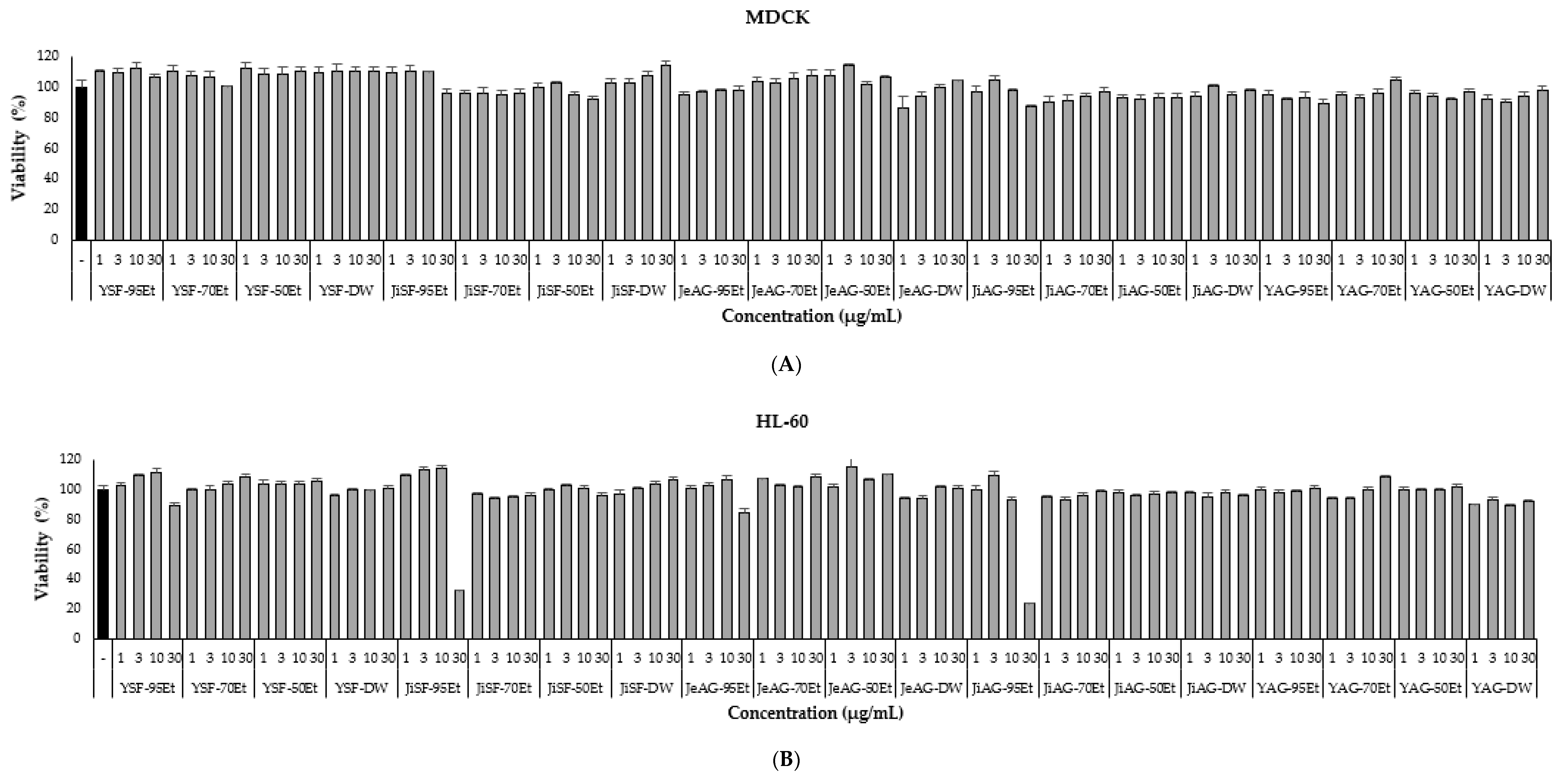
3.5. Cell Toxicity of the Extracts

MDCK was selected as a normal cell line, widely used for toxicity evaluation. HL-60 and SH-SY5Y were selected as human cell lines. To evaluate the toxicity of the extracts, cells were treated for 24 h and the CCK-8 assay was applied. None of the extracts showed significant toxicity to MDCK at any of the concentrations tested for each extract (Figure 7A). In addition, none of the extracts showed significant toxicity to SH-SY5Y with slight toxicity, from 85.79% viability by JiSF-95Et or higher at a high concentration of 30 μg/mL (Figure 7C). However, interestingly, JiSF-95Et and JiAG-95Et showed significant toxicity to HL-60, with viabilities of 32.68% and 23.94%, respectively, at a concentration of 30 μg/mL (Figure 7B). On the basis of these results, it could be confirmed that the extracts were non-toxic to the three cells in the range of concentration up to 20 μg/mL.

4. Discussion

Recently, SF extracts have been reported with respect to their antibacterial activities [42], metabolomic characterization [43], and toxicological evaluation [44]. Maackiain has been actively investigated with respect to its biological activities, with include anti-inflammation [24] and anti-tumor [25]. We reported that maackiain isolated from SF showed selective inhibition of MAO-B in the previous study [26]. Therefore, SF was selected as the subject of this study, and extracted with three concentrations of ethanol and distilled water. Interestingly, extracts of SF showed different patterns in enzyme inhibition, depending on the origin of the plants. YSF-95Et and YSF-70Et had strong MAO-B inhibition with residual activities of 6.43% and 20.79%, respectively, as we expected. However, JiSF-95Et exhibited lower MAO-B inhibition than YSF, with a residual activity of 51.22%. JiSF-95Et showed MAO-A, AChE, and BChE inhibitions with the residual activities of 26.77%, 36.73%, and 34.16%, respectively, whereas YSF-95Et and YSF-70Et showed BChE inhibition with residual activities of 45.54% and 33.26%, respectively. These differences might be a result of changes in their effective components due to different conditions of soils in their origins, similar to the case reported at Daphnes Cortex (Daphne giraldii Nitsche) in China [45]. We tracked differences of their major compounds by TLC; however, the content of maackiain was found to be present in low amount in both regions (Yeongcheon and Mt. Jiri), and there was no significant difference in other components. In addition, we observed that the inhibitory ability to MAO-B increased as the ethanol content of the extraction solvent increased, and the ratio of non-polar substances in the extracts also increased. These results suggest that the differences of inhibition pattern between the origins might be contributed by other minor compounds in the extracts, not by maackiain. A study reported that methanol extract of SF showed MAO-A and MAO-B inhibitions, contributed by formononetin and kushenol F, not by maackiain [46]. In addition, prenylated flavonoids from SF extract showed BACE1 inhibition [47], whereas our experiments did not exhibit BACE1 inhibition by SF. This difference might be a result of the extraction conditions, such as solvent, i.e., methanol vs. ethanol or water. These results also suggest that SF is a potential material for finding multi-target inhibitors of MAO-B, AChE, and BChE, which are attractive for the treatment of AD.
AG extracts have also been extensively investigated for medical applications such as anti-allergic [28] and antioxidant activity [29]. Decursin, which is a coumarin derivative and a main compound of AG, has been extensively investigated for biological activities such as anti-tumor [30] and anti-inflammation [31]. Recently, it was reported that selectivity of MAO inhibition of coumarin derivatives varied with their substituents [48]. In our previous study, decursin, isolated from AG, showed selective inhibition of MAO-A [32] and showed anti-depressant-like activities in mouse behavioral tests such as tail suspension test and forced swimming test [33]. In this study, AG was selected as a source, and its extracts were used in evaluation of inhibitory activities for target enzymes. Some of the ethanol extracts, such as YAG-95Et, YAG-70Et, YAG-50Et, JeAG-95Et, JeAG-70Et, and JeAG-50Et, showed significant MAO-A inhibition, with residual activities from 8.93% to 14.11%, as we predicted. However, JiAG-95Et and JiAG-70Et showed less MAO-A inhibition, with residual activities >50% compared to other ethanolic extracts. Interestingly, the results of TLC showed that the amount of decursin in YAG was lower than that of JeAG and JiAG, contrary to our prediction of decursin-dependent MAO-A inhibition. These results suggest that other components contribute to MAO-A inhibition, for example, a spot at upper position of decursin of YAG-95Et or -70Et in the TLC. The spot should be identified in further study. On the other hand, JiAG-95Et showed effective MAO-B inhibition with a residual activity of 29.07%, whereas other AG extracts did not. Furthermore, JiAG-95Et and JeAG-95Et showed significant BACE1 inhibition. There have been no other reports of BACE1 inhibition by AG extracts. On the other hand, coumarin derivatives from Angelica decursiva have shown BACE1 and AChE inhibition; however, there was no description about decursin [49]. Furthermore, BACE1 inhibition of natural coumarin derivatives has been reported; however, it was also mentioned that decursin and decursinol did not show BACE1 inhibition, with IC50 of >500 μM [50]. In addition, there have been no reports of BACE1 inhibition by AG extracts. Therefore, we can expect the potential novel compound having extremely effective BACE1 inhibition from JiAG and JeAG extracts. From these results, we concluded that AG extracts could be potential candidates for finding novel compounds with multi-target inhibition such as MAO-A, MAO-B, and BACE1.
The extracts were not effective for AChE and BChE inhibition, except JiSF-95Et for AChE, and JiSF-95Et, -70Et, and YSF-70Et for BChE inhibition. In the DPPH radical scavenging assay for antioxidant activity and Aβ aggregation inhibition assay, no significant activities were observed for the extracts.
On the other hand, the extracts were non-toxic to the normal cell line and neuroblast cell line, MDCK and SH-SY5Y, respectively, at all concentrations tested. However, 95% extracts such as JiSF-95Et and JiAG-95Et showed significant toxicity toward the cancer cell line HL-60 at a high concentration of 30 μg/mL.
There have been many studies on natural extracts aiming to find novel compounds in pharmacological applications for neurodegenerative diseases. For example, Woodfordia fruticose (L.) Kurz extract had AChE, BChE, and BACE1 inhibitory activities that could be potentially used for the AD treatment [51]. African mistletoe (Tapinanthus bangwensis Lor) from Moringa and Almond host plants showed MAO-A inhibition and antioxidant activity that could be used for multi-target inhibitor against depression [52]. These studies also stimulated interest in the people expecting novel medicines with multi-target inhibitor for neurodegenerative diseases. Those cases are similar to the extracts in this study, such as YSF-95Et, which inhibited MAO-B and BChE; JiSF-95Et, which inhibited AChE and BChE; and JiAG-95Et, which inhibited MAO-B and BACE1. We also previously reported on biologically active compounds using natural extracts, showing inhibitory activities against target enzymes such as MAOs, ChEs, and BACE1 from endogenous lichen fungi [34,53,54], marine bacteria [55,56], algae [37], and medicinal plants [57,58,59,60]. However, our previous studies focused on selective inhibitions of target enzymes by single compounds isolated from single origin and single extraction solvent, except that ellagic acid showed AChE and MAO-B inhibition [60], and glycyrol showed BChE and MAO-B inhibition [57]. Furthermore, we did not report BACE1 inhibitor from natural extracts. In this study, we observed inhibitory activities of the extracts of two plants, SF and AG, against the target enzymes, and compared them using the plant extracts derived from different origins, two and three sites, respectively. SF ethanolic extracts showed MAO-B, AChE, and BChE inhibitions, and AG extracts showed MAO-A, MAO-B, and BACE1 inhibitions and their inhibitory activities varied with their origins. We plan further studies to trace the reason these differences were occurred through identification of compounds using HPLC and other analytical methods.
From these results, AG and SF are suggested as potential herbal sources for the treatment of depression and neurodegenerative diseases, and it is suggested that we should consider not only the plant, but also its origin, when choosing a plant as an experimental source.

5. Conclusions

In this study, AG and SF were extracted using three concentrations of EtOH and distilled water. Ethanolic extracts of SF exhibited MAO-B, AChE, and BChE inhibitions and AG showed MAO-A, MAO-B, and BACE1 inhibition. In particular, YSF-95Et, which showed inhibitions of MAO-B and BChE, and JiSF-95Et, which exhibited AChE and BChE inhibitions, could be potential extracts for finding novel compounds with multi-target inhibition for the treatment of AD. In addition, JiAG-95Et showed effective MAO-B and extremely potent BACE1 inhibitory activities, which has not been reported in other studies, guiding to further experiments to find novel multi-target inhibitors. We used prethanol (95% ethanol) and distilled water, which is edible and can be used for making functional foods, contrary to other studies using methanol extraction. All these extracts were non-toxic to normal and neuroblast cells, thus making them safe. From these results, we suggest that AG and SF are promising candidates for the reservoirs of effective compounds to be isolated further or for making functional foods for the treatment of depression and neurodegenerative diseases, and that herbal origins should be seriously considered in these experiments and applications.

Author Contributions

Conceptualization, H.K.; Extraction, TLC analysis, and enzyme assays, J.E.P.; cell viability assay, S.-K.M.; data curation, J.E.P.; writing—original draft preparation, J.E.P.; writing—review and editing, H.K.; supervision, S.-T.Y. and H.K. All authors have read and agreed to the published version of the manuscript.

Funding

This study was supported by the National Research Foundation of Korea (NRF) grant funded by the Korea government (NRF-2022R1A2B5B01002536).

Institutional Review Board Statement

Not applicable.

Informed Consent Statement

Not applicable.

Data Availability Statement

The data presented in this study are available on request from the corresponding author.

Conflicts of Interest

The authors declare no conflict of interest.

References

  1. Burns, A.; Iliffe, S. Alzheimer’s disease. BMJ 2009, 338, b158. [Google Scholar] [CrossRef] [PubMed] [Green Version]
  2. Wenk, G.L. Neuropathologic changes in Alzheimer’s disease. J. Clin. Psychiatry 2003, 64 (Suppl. 9), 7–10. [Google Scholar] [PubMed]
  3. Bloom, G.S. Amyloid-β and Tau: The trigger and bullet in Alzheimer disease pathogenesis. JAMA Neurol. 2014, 71, 505–508. [Google Scholar] [CrossRef] [PubMed] [Green Version]
  4. Francis, P.T. The interplay of neurotransmitters in Alzheimer’s disease. CNS Spectr. 2005, 10, 6–9. [Google Scholar] [CrossRef] [PubMed]
  5. Dhillon, S. Aducanumab: First approval. Drugs 2021, 81, 1437–1443. [Google Scholar] [CrossRef]
  6. Schedin-Weiss, S.; Inoue, M.; Hromadkova, L.; Teranishi, Y.; Yamamoto, N.G.; Wiehager, B.; Bogdanovic, N.; Winblad, B.; Sandebring-Matton, A.; Frykman, S.; et al. Monoamine oxidase B is elevated in Alzheimer disease neurons, is associated with γ-secretase and regulates neuronal amyloid β-peptide levels. Alzheimer’s Res. Ther. 2017, 9, 57. [Google Scholar] [CrossRef]
  7. Jankovic, J. Parkinson’s disease: Clinical features and diagnosis. J. Neurol. Neurosurg. Psychiatry 2008, 79, 368–376. [Google Scholar] [CrossRef] [Green Version]
  8. Connolly, B.S.; Lang, A. Pharmacological treatment of parkinson disease. JAMA 2014, 311, 1670–1683. [Google Scholar] [CrossRef]
  9. Seppi, K.; Weintraub, D.; Coelho, M.; Perez-Lloret, S.; Fox, S.H.; Katzenschlager, R.; Hametner, E.-M.; Poewe, W.; Rascol, O.; Goetz, C.G.; et al. The movement disorder society evidence-based medicine review update: Treatments for the non-motor symptoms of Parkinson’s disease. Mov. Disord. 2011, 26, S42–S80. [Google Scholar] [CrossRef]
  10. Ives, N.J.; Stowe, R.; Marro, J.; Counsell, C.; Macleod, A.; Clarke, C.; Gray, R.; Wheatley, K. Monoamine oxidase type B inhibitors in early Parkinson’s disease: Meta-analysis of 17 randomised trials involving 3525 patients. BMJ 2004, 329, 593. [Google Scholar] [CrossRef] [Green Version]
  11. De Zwart, P.L.; Jeronimus, B.; de Jonge, P. Empirical evidence for definitions of episode, remission, recovery, relapse and recurrence in depression: A systematic review. Epidemiol. Psychiatr. Sci. 2018, 28, 544–562. [Google Scholar] [CrossRef] [PubMed]
  12. Daut, R.A.; Fonken, L.K. Circadian regulation of depression: A role for serotonin. Front. Neuroendocr. 2019, 54, 100746. [Google Scholar] [CrossRef] [PubMed]
  13. Meyer, J.H.; Ginovart, N.; Boovariwala, A.; Sagrati, S.; Hussey, D.; Garcia, A.; Young, T.; Praschak-Rieder, N.; Wilson, A.A.; Houle, S. Elevated monoamine oxidase a levels in the brain: An explanation for the monoamine imbalance of major depression. Arch. Gen. Psychiatry 2006, 63, 1209–1216. [Google Scholar] [CrossRef] [PubMed] [Green Version]
  14. Ramsay, R.R. Monoamine Oxidases: The biochemistry of the proteins as targets in medicinal chemistry and drug discovery. Curr. Top. Med. Chem. 2012, 12, 2189–2209. [Google Scholar] [CrossRef]
  15. Yeung, A.W.K.; Georgieva, M.G.; Atanasov, A.G.; Tzvetkov, N.T. Monoamine oxidases (MAOs) as privileged molecular targets in neuroscience: Research literature analysis. Front. Mol. Neurosci. 2019, 12, 143. [Google Scholar] [CrossRef] [Green Version]
  16. Youdim, M.B.H.; Edmondson, D.; Tipton, K.F. The therapeutic potential of monoamine oxidase inhibitors. Nat. Rev. Neurosci. 2006, 7, 295–309. [Google Scholar] [CrossRef]
  17. Birks, J.S. Cholinesterase inhibitors for Alzheimer’s disease. Cochrane Database Syst. Rev. 2006, 1, CD005593. [Google Scholar] [CrossRef]
  18. Lane, R.M.; Potkin, S.G.; Enz, A. Targeting acetylcholinesterase and butyrylcholinesterase in dementia. Int. J. Neuropsychopharmacol. 2005, 9, 101–124. [Google Scholar] [CrossRef]
  19. Vassar, R.; Bennett, B.D.; Babu-Khan, S.; Kahn, S.; Mendiaz, E.A.; Denis, P.; Teplow, D.B.; Ross, S.; Amarante, P.; Loeloff, R.; et al. β-Secretase cleavage of Alzheimer’s amyloid precursor protein by the transmembrane aspartic protease BACE. Science 1999, 286, 735–741. [Google Scholar] [CrossRef] [Green Version]
  20. Sinha, S.; Anderson, J.P.; Barbour, R.; Basi, G.S.; Caccavello, R.; Davis, D.; Doan, M.; Dovey, H.F.; Frigon, N.; Hong, J.; et al. Purification and cloning of amyloid precursor protein β-secretase from human brain. Nature 1999, 402, 537–540. [Google Scholar] [CrossRef]
  21. Ashrafian, H.; Zadeh, E.H.; Khan, R.H. Review on Alzheimer’s disease: Inhibition of amyloid beta and tau tangle formation. Int. J. Biol. Macromol. 2020, 167, 382–394. [Google Scholar] [CrossRef] [PubMed]
  22. Ghosh, A.K.; Osswald, H.L. BACE1 (β-secretase) inhibitors for the treatment of Alzheimer’s disease. Chem. Soc. Rev. 2014, 43, 6765–6813. [Google Scholar] [CrossRef] [PubMed] [Green Version]
  23. Krishna, P.M.; KNV, R.; S, S.; Banji, D. A review on phytochemical, ethnomedical and pharmacological studies on genus Sophora, Fabaceae. Rev. Bras. Farm. 2012, 22, 1145–1154. [Google Scholar] [CrossRef] [Green Version]
  24. Huh, J.; Lee, J.; Jeon, E.; Ryu, H.W.; Oh, S.; Ahn, K.; Jun, H.S.; Ha, U. Maackiain, a compound derived from Sophora flavescens, increases IL-1β production by amplifying nigericin-mediated inflammasome activation. FEBS Open Biol. 2020, 10, 1482–1491. [Google Scholar] [CrossRef] [PubMed]
  25. Peng, F.; Wang, L.; Xiong, L.; Tang, H.; Du, J.; Peng, C. Maackiain modulates miR-374a/GADD45A axis to inhibit triple-negative breast cancer initiation and progression. Front. Pharmacol. 2022, 13, 806869. [Google Scholar] [CrossRef] [PubMed]
  26. Lee, H.W.; Ryu, H.W.; Kang, M.-G.; Park, D.; Oh, S.-R.; Kim, H. Potent selective monoamine oxidase B inhibition by maackiain, a pterocarpan from the roots of Sophora flavescens. Bioorg. Med. Chem. Lett. 2016, 26, 4714–4719. [Google Scholar] [CrossRef]
  27. Yan, J.-J.; Kim, D.-H.; Moon, Y.-S.; Jung, J.-S.; Ahn, E.-M.; Baek, N.-I.; Song, D.-K. Protection against β-amyloid peptide-induced memory impairment with long-term administration of extract of Angelica gigas or decursinol in mice. Prog. Neuro-Psychopharmacol. Biol. Psychiatry 2004, 28, 25–30. [Google Scholar] [CrossRef]
  28. Ji, K.-Y.; Jung, D.H.; Pyun, B.-J.; Kim, Y.J.; Lee, J.Y.; Choi, S.; Jung, M.-A.; Song, K.H.; Kim, T. Angelica gigas extract ameliorates allergic rhinitis in an ovalbumin-induced mouse model by inhibiting Th2 cell activation. Phytomedicine 2021, 93, 153789. [Google Scholar] [CrossRef]
  29. Kwon, D.-A.; Kim, Y.S.; Kim, S.-K.; Baek, S.H.; Kim, H.K.; Lee, H.S. Antioxidant and antifatigue effect of a standardized fraction (HemoHIM) from Angelica gigas, Cnidium officinale, and Paeonia lactiflora. Pharm. Biol. 2021, 59, 389–398. [Google Scholar] [CrossRef]
  30. Joo, M.; Heo, J.B.; Kim, S.; Kim, N.; Jeon, H.J.; An, Y.; Song, G.-Y.; Kim, J.-M.; Lee, H.J. Decursin inhibits tumor progression in head and neck squamous cell carcinoma by downregulating CXCR7 expression in vitro. Oncol. Rep. 2021, 47, 39. [Google Scholar] [CrossRef]
  31. Lee, W.; Sim, H.; Choi, Y.-J.; Seo, J.Y.; Yun, M.-Y.; Song, G.Y.; Bae, J.-S. The decursin analog, CYJ-27, suppresses inflammation via the downregulation of NF-κB and STAT-J. Med. Food 2021, 24, 852–859. [Google Scholar] [CrossRef] [PubMed]
  32. Lee, H.W.; Ryu, H.W.; Kang, M.-G.; Park, D.; Lee, H.; Shin, H.M.; Oh, S.-R.; Kim, H. Potent inhibition of monoamine oxidase A by decursin from Angelica gigas Nakai and by wogonin from Scutellaria baicalensis Georgi. Int. J. Biol. Macromol. 2017, 97, 598–605. [Google Scholar] [CrossRef] [PubMed]
  33. Oh, J.M.; Lee, H.-S.; Baek, S.C.; Lee, J.P.; Jeong, G.S.; Paik, M.-J.; Kim, H. Antidepressant-like activities of hispidol and decursin in mice and analysis of neurotransmitter Monoamines. Neurochem. Res. 2020, 45, 1930–1940. [Google Scholar] [CrossRef] [PubMed]
  34. Jeong, G.-S.; Kang, M.-G.; Han, S.-A.; Noh, J.-I.; Park, J.-E.; Nam, S.-J.; Park, D.; Yee, S.-T.; Kim, H. Selective inhibition of human monoamine oxidase B by 5-hydroxy-2-methyl-chroman-4-one isolated from an endogenous lichen fungus Daldinia fissa. J. Fungi 2021, 7, 84. [Google Scholar] [CrossRef] [PubMed]
  35. Baek, S.C.; Lee, H.W.; Ryu, H.W.; Kang, M.-G.; Park, D.; Kim, S.H.; Cho, M.-L.; Oh, S.-R.; Kim, H. Selective inhibition of monoamine oxidase A by hispidol. Bioorganic Med. Chem. Lett. 2018, 28, 584–588. [Google Scholar] [CrossRef]
  36. Ellman, G.L.; Courtney, K.D.; Andres, V., Jr.; Featherstone, R.M. A new and rapid colorimetric determination of acetylcholinesterase activity. Biochem. Pharmacol. 1961, 7, 88–95. [Google Scholar] [CrossRef]
  37. Lee, J.P.; Kang, M.-G.; Lee, J.Y.; Oh, J.M.; Baek, S.C.; Leem, H.H.; Park, D.; Cho, M.-L.; Kim, H. Potent inhibition of acetylcholinesterase by sargachromanol I from Sargassum siliquastrum and by selected natural compounds. Bioorganic Chem. 2019, 89, 103043. [Google Scholar] [CrossRef]
  38. Ali, S.; Bin Asad, M.H.H.; Maity, S.; Zada, W.; Rizvanov, A.; Iqbal, J.; Babak, B.; Hussain, I. Fluoro-benzimidazole derivatives to cure Alzheimer’s disease: In-silico studies, synthesis, structure-activity relationship and in vivo evaluation for β secretase enzyme inhibition. Bioorganic Chem. 2019, 88, 102936. [Google Scholar] [CrossRef]
  39. Brand-Williams, W.; Cuvelier, M.E.; Berset, C. Use of a free radical method to evaluate antioxidant activity. LWT Food Sci. Technol. 1995, 28, 25–30. [Google Scholar] [CrossRef]
  40. Bolder, S.G.; Sagis, L.M.C.; Venema, P.; Van Der Linden, E. Thioflavin T and birefringence assays to determine the conversion of proteins into fibrils. Langmuir 2007, 23, 4144–4147. [Google Scholar] [CrossRef]
  41. Noh, J.-I.; Mun, S.-K.; Lim, E.; Kim, H.; Chang, D.-J.; Hur, J.-S.; Yee, S.-T. Induction of apoptosis in MDA-MB-231 cells treated with the methanol extract of lichen Physconia hokkaidensis. J. Fungi 2021, 7, 188. [Google Scholar] [CrossRef] [PubMed]
  42. Li, P.; Chai, W.C.; Wang, Z.-Y.; Tang, K.-J.; Chen, J.-Y.; Venter, H.; Semple, S.J.; Xiang, L. Bioactivity-guided isolation of compounds from Sophora flavescens with antibacterial activity against Acinetobacter baumannii. Nat. Prod. Res. 2021, 1–9. [Google Scholar] [CrossRef] [PubMed]
  43. Jiang, P.; Sun, Y.; Cheng, N. Liver metabolomic characterization of Sophora flavescens alcohol extract-induced hepatotoxicity in rats through UPLC/LTQ-Orbitrap mass spectrometry. Xenobiotica 2019, 50, 670–676. [Google Scholar] [CrossRef] [PubMed]
  44. Wu, C.; Huang, Y.; Huang, H.; Ma, Y.; Lin, Q.; Yang, X.; Pang, K. Acute and 13 weeks subchronic toxicological evaluation of the flavonoid-rich extract of Sophora flavescens. Drug Chem. Toxicol. 2021, 1–8. [Google Scholar] [CrossRef] [PubMed]
  45. Mu, Q.-R.; Jiang, D.; He, Y.; Geng, L.; Ren, G.-X.; Bai, Z.-F.; Zhang, X.; Zhang, Z.-Y.; Liu, C.-S. Correlation between chemical composition, ecological factors and soil factors of Chinese herbal medicine Daphnes Cortex. Zhongguo Zhong Yao Za Zhi 2020, 45, 1059–1063. [Google Scholar]
  46. Hwang, J.-S.; Lee, S.A.; Hong, S.S.; Lee, K.S.; Lee, M.K.; Hwang, B.Y.; Ro, J.S. Monoamine oxidase inhibitory components from the roots of Sophora flavescens. Arch. Pharmacal Res. 2005, 28, 190–194. [Google Scholar] [CrossRef]
  47. Jung, H.A.; Yokozawa, T.; Kim, B.-W.; Jung, J.H.; Choi, J.S. Selective Inhibition of Prenylated Flavonoids from Sophora flavescens against BACE1 and Cholinesterases. Am. J. Chin. Med. 2010, 38, 415–429. [Google Scholar] [CrossRef]
  48. Mzezewa, S.C.; Omoruyi, S.I.; Zondagh, L.S.; Malan, S.F.; Ekpo, O.E.; Joubert, J. Design, synthesis, and evaluation of 3,7-substituted coumarin derivatives as multifunctional Alzheimer’s disease agents. J. Enzym. Inhib. Med. Chem. 2021, 36, 1606–1620. [Google Scholar] [CrossRef]
  49. Ali, Y.; Jannat, S.; Jung, H.A.; Choi, R.J.; Roy, A.; Choi, J.S. Anti-Alzheimer’s disease potential of coumarins from Angelica decursiva and Artemisia capillaris and structure-activity analysis. Asian Pac. J. Trop. Med. 2016, 9, 103–111. [Google Scholar] [CrossRef] [Green Version]
  50. Marumoto, S.; Miyazawa, M. Structure–activity relationships for naturally occurring coumarins as β-secretase inhibitor. Bioorganic Med. Chem. 2012, 20, 784–788. [Google Scholar] [CrossRef]
  51. Raghuvanshi, R.; Nuthakki, V.K.; Singh, L.; Singh, B.; Bharate, S.S.; Bhatti, R.; Bharate, S.B. Identification of plant-based multitargeted leads for Alzheimer’s disease: In-vitro and in-vivo validation of Woodfordia fruticosa (L.) Kurz. Phytomedicine 2021, 91, 153659. [Google Scholar] [CrossRef] [PubMed]
  52. Oyeniran, O.H.; Ademiluyi, A.O.; Oboh, G. Host–parasite relationship modulates the effect of African mistletoe leaves on the cholinergic, monoaminergic and carbohydrate hydrolyzing enzymes in fruit fly. J. Basic Clin. Physiol. Pharmacol. 2021. [Google Scholar] [CrossRef] [PubMed]
  53. Jeong, G.S.; Hillman, P.F.; Kang, M.-G.; Hwang, S.; Park, J.-E.; Nam, S.-J.; Park, D.; Kim, H. Potent and Selective Inhibitors of Human Monoamine Oxidase A from an Endogenous Lichen Fungus Diaporthe mahothocarpus. J. Fungi 2021, 7, 876. [Google Scholar] [CrossRef] [PubMed]
  54. Jeong, G.-S.; Lee, E.-Y.; Kang, M.-G.; Nam, S.-J.; Park, D.; Kim, H. (S)-5-Methylmellein isolated from an endogenous lichen fungus Rosellinia corticium as a potent inhibitor of human monoamine oxidase A. Processes 2022, 10, 166. [Google Scholar] [CrossRef]
  55. Oh, J.M.; Lee, C.; Nam, S.-J.; Kim, H. Chromenone derivatives as monoamine oxidase inhibitors from marine-derived MAR4 clade Streptomyces sp. CNQ-031. J. Microbiol. Biotechnol. 2021, 31, 1022–1027. [Google Scholar] [CrossRef]
  56. Lee, H.W.; Choi, H.; Nam, S.-J.; Fenical, W.; Kim, H. Potent inhibition of monoamine oxidase B by a piloquinone from marine-derived Streptomyces sp. CNQ-027. J. Microbiol. Biotechnol. 2017, 27, 785–790. [Google Scholar] [CrossRef] [Green Version]
  57. Jeong, G.; Kang, M.-G.; Lee, J.; Lee, S.; Park, D.; Cho, M.; Kim, H. Inhibition of butyrylcholinesterase and human monoamine oxidase-B by the coumarin glycyrol and liquiritigenin isolated from Glycyrrhiza uralensis. Molecules 2020, 25, 3896. [Google Scholar] [CrossRef]
  58. Oh, J.M.; Jang, H.-J.; Kim, W.J.; Kang, M.-G.; Baek, S.C.; Lee, J.P.; Park, D.; Oh, S.-R.; Kim, H. Calycosin and 8-O-methylretusin isolated from Maackia amurensis as potent and selective reversible inhibitors of human monoamine oxidase-B. Int. J. Biol. Macromol. 2020, 151, 441–448. [Google Scholar] [CrossRef]
  59. Heo, J.H.; Eom, B.H.; Ryu, H.W.; Kang, M.-G.; Park, J.E.; Kim, D.-Y.; Kim, J.-H.; Park, D.; Oh, S.-R.; Kim, H. Acetylcholinesterase and butyrylcholinesterase inhibitory activities of khellactone coumarin derivatives isolated from Peucedanum japonicum Thurnberg. Sci. Rep. 2020, 10, 21695. [Google Scholar] [CrossRef]
  60. Oh, J.M.; Jang, H.-J.; Kang, M.-G.; Song, S.; Kim, D.-Y.; Kim, J.; Noh, J.-I.; Park, J.E.; Park, D.; Yee, S.-T.; et al. Acetylcholinesterase and monoamine oxidase-B inhibitory activities by ellagic acid derivatives isolated from Castanopsis cuspidata var. sieboldii. Sci. Rep. 2021, 11, 13953. [Google Scholar] [CrossRef]
Figure 1. TLC analysis of SF (AC) and AG (DF) extracts. The standard compounds used for SF and AG extracts were maackiain and decursin, respectively. Amounts of extracts and standards loaded were 50 µg and 10 µg, respectively. Solvent compositions were established using methanol (MeOH), ethyl acetate (EtOAc), and toluene (Tol): (A) MeOH:EtOAc:Tol = 1:2:7; (B) MeOH:EtOAc:Tol = 1:1:8; (C) MeOH:EtOAc:Tol = 1:1:8; (D) MeOH:EtOAc:Tol = 2:1:7; (E) MeOH:EtOAc:Tol = 1:1:8; (F) MeOH:EtOAc:Tol = 1:1:8. SF from Yeongcheon (YSF) and Mt. Jiri (JiSF), and AG from Yeongcheon (YAG), Mt. Jiri (JiAG), and Jecheon (JeAG) were extracted using hot water (DW) and three concentrations of ethanol, 95% (95Et), 70% (70Et), and 50% (50Et). The red arrows represent the standard compounds maackiain or decursin. The orange lines and arrows represent polar components with low Rf values in the solvents (A). The blue arrows represent the unique spots of the YAG extracts (D,E). M, maackiain; D, decursin.
Processes 10 00880 g001aProcesses 10 00880 g001b
Figure 2. Inhibition of MAO-A by the extracts. The different concentrations of DMSO were used as solvent for dissolving extracts due to their solubility, i.e., all the 95Et extracts in 100%; YSF-70Et, YSF-50Et, JiAG-70Et, JiAG-50Et, JeAG-70Et, and JeAG-50Et in 80%; JiSF-70Et, YAG-70Et, and YAG-50Et in 50%; and JiSF-50Et in 30% DMSO. All the DW extracts were dissolved in water. Activity of MAO-A was observed as the procedure described in the ‘Materials and Methods’ section, by adding the extract (20 μg/mL) to the reaction mixture containing 0.06 mM of kynuramine.
Processes 10 00880 g002
Figure 3. Inhibition of MAO-B by the extracts. Activity of MAO-B was observed as the procedure described in the ‘Materials and Methods’ section, by adding the extract (20 μg/mL) to the reaction mixture containing 0.30 mM of benzylamine.
Processes 10 00880 g003
Figure 4. Inhibition of AChE by the extracts. Activity of AChE was observed by Ellman method as described in the ‘Materials and Methods’ section, by adding the extract (20 μg/mL) to the reaction mixture containing ATCI and DTNB (0.5 mM each).
Processes 10 00880 g004
Figure 5. Inhibition of BChE by the extracts. Activity of BChE was observed by Ellman method as described in the ‘Materials and Methods’ section, by adding the extract (20 μg/mL) to the reaction mixture containing BTCI and DTNB (0.5 mM each).
Processes 10 00880 g005
Figure 6. Inhibition of BACE1 by the extracts at 10 μg/mL (A) and 1.0 or 0.5 μg/mL (B). Activity of BACE1 was observed by FRET-assay using the BACE1 activity detection kit. ND, not-detectable due to its negative value.
Processes 10 00880 g006
Figure 7. Effect of the extracts on the viability of three cell lines of MDCK (A), HL-60 (B), and SH-SY5Y (C). The cells were treated with the extracts (1, 3, 10, and 30 μg/mL) for 24 h. Culture supernatants were removed, and cell counting kit-8 (CCK-8) was applied. Viability was quantified using the micro plate reader. Data are presented as mean ± SD from three independent experiments.
Processes 10 00880 g007aProcesses 10 00880 g007b
Table 1. Comparison of the yield of extract according to the plant, origin, and solvent.
PlantOriginSolventYield (%)Abbreviation
Sophora
flavescens
Yeongcheon95% EtOH2.36YSF-95Et
70% EtOH14.24YSF-70Et
50% EtOH10.74YSF-50Et
DW12.82YSF-DW
Jiri95% EtOH5.09JiSF-95Et
70% EtOH12.37JiSF-70Et
50% EtOH8.09JiSF-50Et
DW15.30JiSF-DW
Angelica
gigas Nakai
Jecheon95% EtOH7.10JeAG-95Et
70% EtOH28.00JeAG-70Et
50% EtOH31.10JeAG-50Et
DW23.35JeAG-DW
Jiri95% EtOH8.00JiAG-95Et
70% EtOH33.82JiAG-70Et
50% EtOH20.39JiAG-50Et
DW21.60JiAG-DW
Yeongcheon95% EtOH11.50YAG-95Et
70% EtOH33.78YAG-70Et
50% EtOH31.24YAG-50Et
DW21.70YAG-DW
Table 2. Inhibitions of the extracts for DPPH radical scavenging activity and Aβ aggregation.
ExtractsInhibitions at 20 μg/mL (%)
DPPHAβ Aggregation
YSF-95Et8.02 ± 0.9728.63 ± 3.35
YSF-70Et8.4 ± 2.3912.45 ± 0.05
YSF-50Et4.61 ± 1.730.08 ± 1.25
YSF-DW12.25 ± 0.2222.68 ± 1.26
JiSF-95Et6.45 ± 2.1328.19 ± 1.58
JiSF-70Et18.83 ± 0.73−1.85 ± 0.00
JiSF-50Et3.57 ± 2.66−8.69 ± 0.56
JiSF-DW0.91 ± 1.734.64 ± 5.16
JeAG-95Et2.44 ± 2.2212.26 ± 1.21
JeAG-70Et17.23 ± 2.22−2.36 ± 0.00
JeAG-50Et4.89 ± 4.166.07 ± 6.12
JeAG-DW−4.51 ± 1.0625.74 ± 1.11
JiAG-95Et2.22 ± 4.2112.96 ± 1.25
JiAG-70Et3.13 ± 0.808.09 ± 5.46
JiAG-50Et1.16 ± 3.50−7.85 ± 0.73
JiAG-DW17.64 ± 2.359.21 ± 4.25
YAG-95Et0.323 ± 2.706.62 ± 2.45
YAG-70Et0.72 ± 0.312.35 ± 3.33
YAG-50Et3.26 ± 0.9717.10 ± 3.31
YAG-DW3.60 ± 3.419.37 ± 1.69
Publisher’s Note: MDPI stays neutral with regard to jurisdictional claims in published maps and institutional affiliations.

