
Characterization and Topical Study of Aloe Vera Hydrogel on Wound-Healing Process

 , , and
Abstract

1. Introduction
2. Materials and Methods
2.1. Restauder® Hydrogel
2.2. Characterization of the Hydrogel
2.3. In Vivo Assay
2.3.1. Surgical Procedure
2.3.2. Macroscopic Analysis
2.3.3. Microscopy Study
3. Results
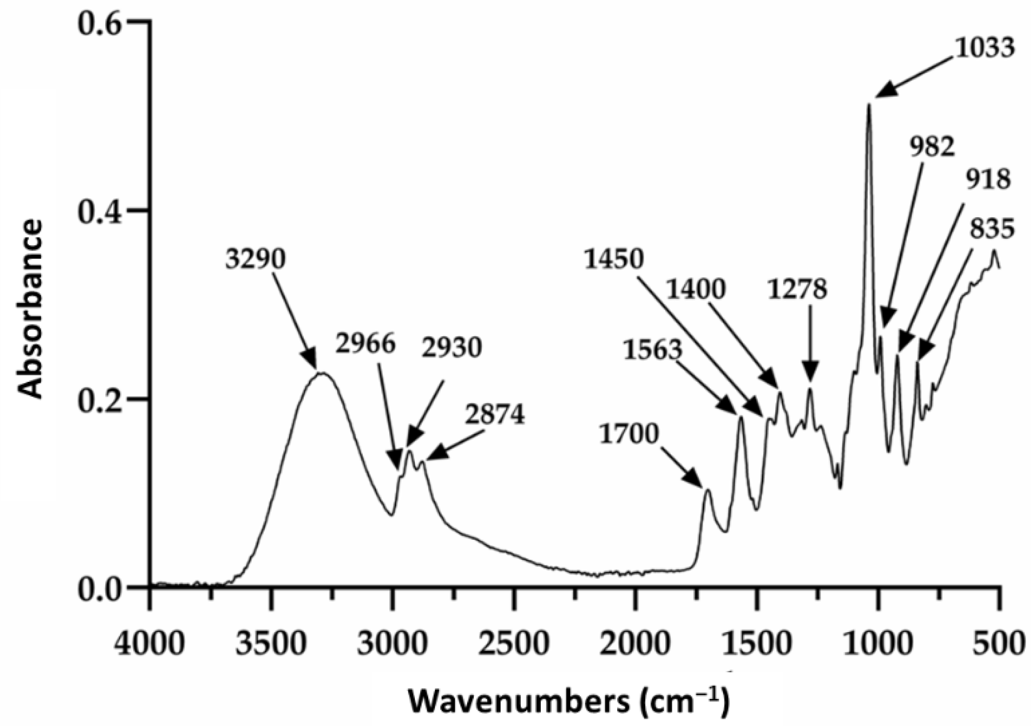
3.1. FTIR Analysis
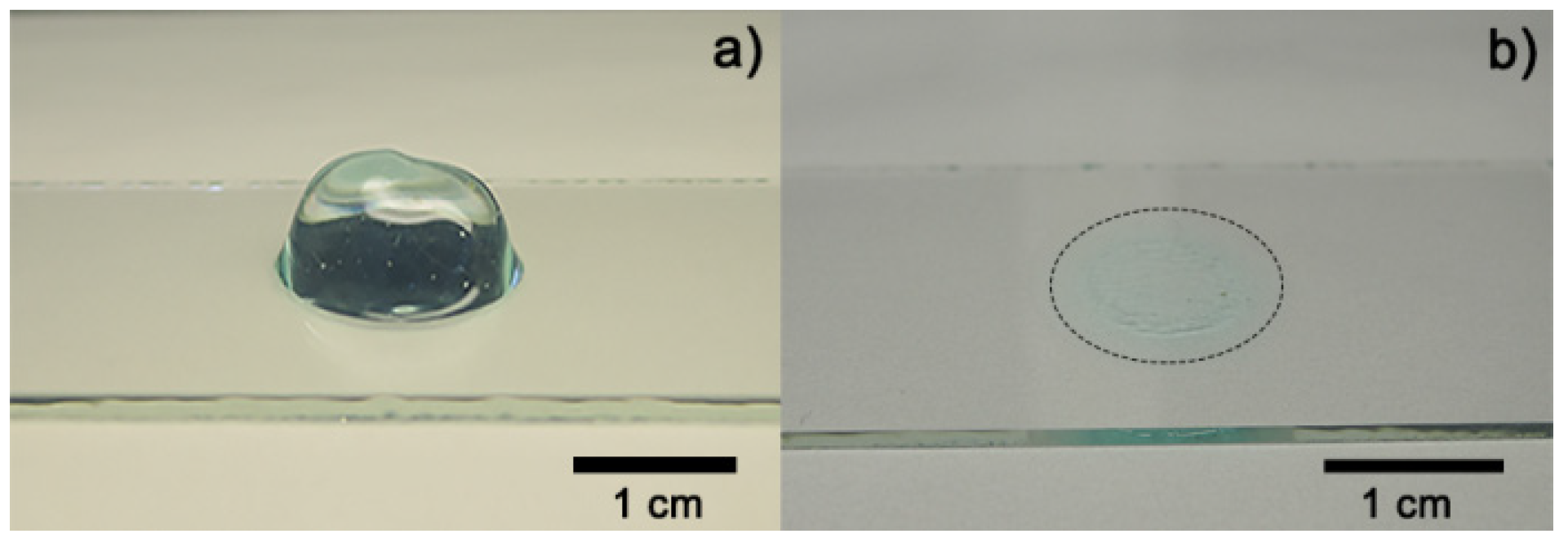
3.2. Contact Angle
3.3. Surface Analysis
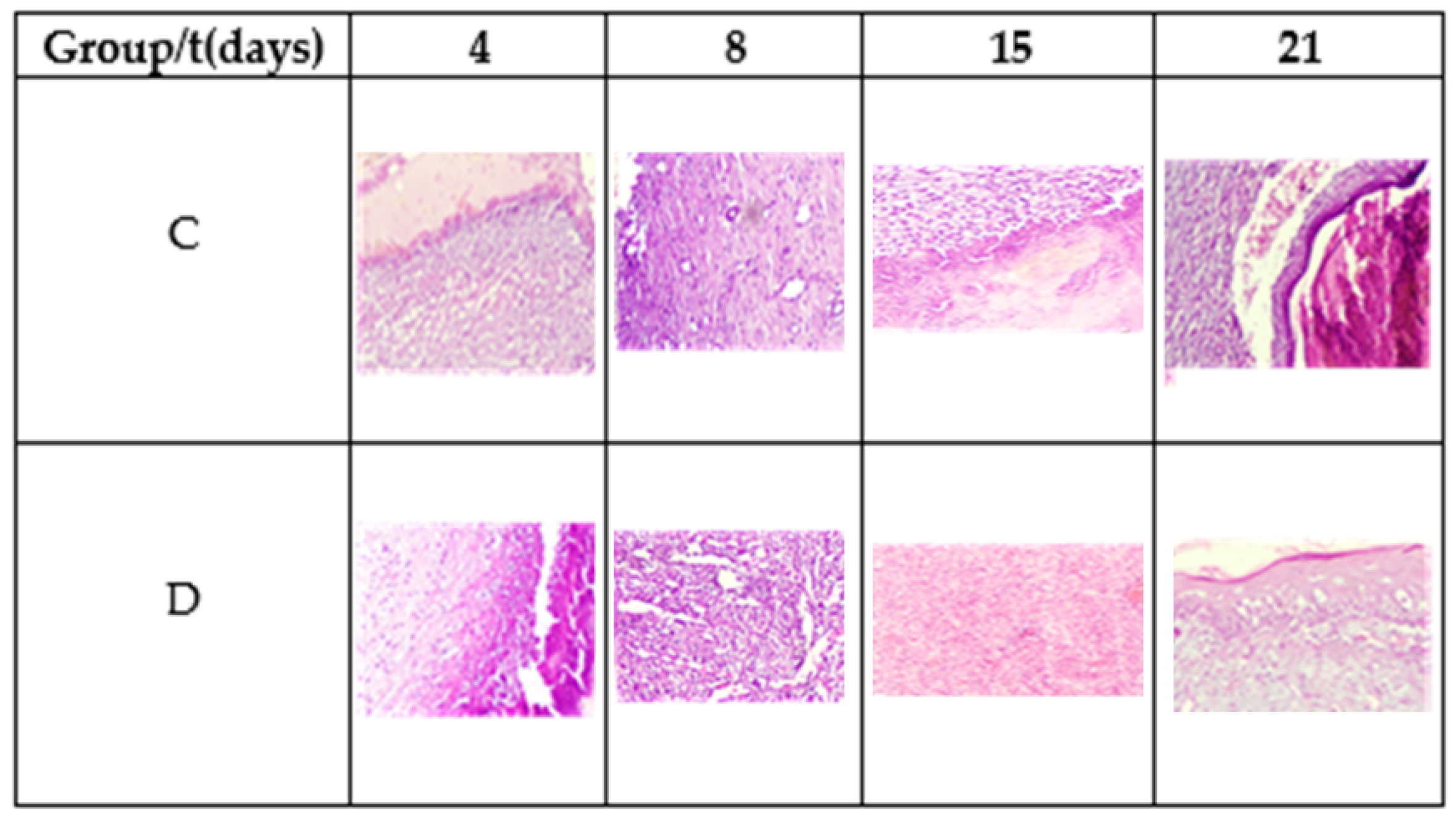
3.4. Healing Process
4. Discussion
5. Conclusions
Author Contributions
Funding
Institutional Review Board Statement
Informed Consent Statement
Data Availability Statement
Acknowledgments
Conflicts of Interest
References
- Mano, J.F.; Silva, G.A.; Azevedo, H.S.; Malafaya, P.B.; Sousa, R.A.; Silva, S.S.; Boesel, L.F.; Oliveira, J.M.; Santos, T.C.; Marques, A.P.; et al. Natural origin biodegradable systems in tissue engineering and regenerative medicine: Present status and some moving trends. J. R. Soc. Interface 2006, 4, 999–1030. [Google Scholar] [CrossRef] [PubMed]
- Kesharwani, P.; Bisht, A.; Alexander, A.; Dave, V.; Sharma, S. Biomedical applications of hydrogels in drug delivery system: An update. J. Drug Deliv. Sci. Technol. 2021, 66, 102914. [Google Scholar] [CrossRef]
- Carrillo, K.L.T.; Tamayo, G.; Donohue, A.; Kobayashi, T.; Acuna, R.A.S. Obtaining of Hydrogels using PVA and HEC for Adipose Tissue Regeneration. J. Tissue Sci. Eng. 2015, 6, 2. [Google Scholar] [CrossRef]
- Kong, F.; Mehwish, N.; Niu, X.; Lin, M.; Rong, X.; Hu, F.; Lee, B. Personalized hydrogels for individual health care: Preparation, features, and applications in tissue engineering. Mater. Today Chem. 2021, 22, 100612. [Google Scholar] [CrossRef]
- Maan, A.A.; Ahmed, Z.F.R.; Khan, M.K.I.; Riaz, A.; Nazir, A. Aloe vera gel, an excellent base material for edible films and coatings. Trends Food Sci. Technol. 2021, 116, 329–341. [Google Scholar] [CrossRef]
- Jadhav, A.S.; Patil, O.A.; Kadam, S.V.; Bhutkar, M.A. Review on Aloe Vera is used in Medicinal Plant. Asian J. Res. Pharm. Sci. 2020, 10, 26. [Google Scholar] [CrossRef]
- Zeng, W.M.; Parus, A.; Barnes, C.W.; Hiro, M.E.; Robson, M.C.; Payne, W.G. Aloe vera—Mechanisms of Action, Uses, and Potential Uses in Plastic Surgery and Wound Healing. Surg. Sci. 2020, 11, 312–328. [Google Scholar] [CrossRef]
- Anand, U.; Tudu, C.K.; Nandy, S.; Sunita, K.; Tripathi, V.; Loake, G.J.; Dey, A.; Proćków, J. Ethnodermatological use of medicinal plants in India: From ayurvedic formulations to clinical perspectives—A review. J. Ethnopharmacol. 2021, 284, 114744. [Google Scholar] [CrossRef]
- Anywar, G.; Tugume, P.; Kakudidi, E.K. A review of Aloe species used in traditional medicine in East Africa. South Afr. J. Bot. 2021. [Google Scholar] [CrossRef]
- Farid, A.; Tawfik, A.; Elsioufy, B.; Safwat, G. In vitro and in vivo anti-Cryptosporidium and anti-inflammatory effects of Aloe vera gel in dexamethasone immunosuppressed mice. Int. J. Parasitol. Drugs Drug Resist. 2021, 17, 156–167. [Google Scholar] [CrossRef]
- Sánchez, M.; González-Burgos, E.; Iglesias, I.; Gómez-Serranillos, M.P. Pharmacological Update Properties of Aloe Vera and its Major Active Constituents. Molecules 2020, 25, 1324. [Google Scholar] [CrossRef] [PubMed]
- Nizam, N.H.M.; Rawi, N.F.M.; Ramle, S.F.M.; Aziz, A.A.; Abdullah, C.; Rashedi, A.; Kassim, M.H.M. Physical, thermal, mechanical, antimicrobial and physicochemical properties of starch based film containing aloe vera: A review. J. Mater. Res. Technol. 2021, 15, 1572–1589. [Google Scholar] [CrossRef]
- Chokboribal, J.; Tachaboonyakiat, W.; Sangvanich, P.; Ruangpornvisuti, V.; Jettanacheawchankit, S.; Thunyakitpisal, P. Deacetylation affects the physical properties and bioactivity of acemannan, an extracted polysaccharide from Aloe vera. Carbohydr. Polym. 2015, 133, 556–566. [Google Scholar] [CrossRef] [PubMed]
- Yagi, A.; Takeo, S. Anti-inflammatory Constituents, Aloesin and Aloemannan in Aloe Species and Effects of Tanshinon VI in Salvia miltiorrhiza on Heart. Yakugaku Zasshi 2003, 123, 517–532. [Google Scholar] [CrossRef] [PubMed][Green Version]
- Sierra-García, G.D.; Castro-Ríos, R.; González-Horta, A.; Lara-Arias, J.; Chávez-Montes, A. Acemannan, an Extracted Polysaccharide from Aloe vera: A Literature Review. Nat. Prod. Commun. 2014, 9, 1217–1221. [Google Scholar] [CrossRef]
- Adlakha, K.; Koul, B.; Kumar, A. Value-added products of Aloe species: Panacea to several maladies. S. Afr. J. Bot. 2021. [Google Scholar] [CrossRef]
- Wahedi, H.M.; Jeong, M.; Chae, J.K.; Gil Do, S.; Yoon, H.; Kim, S.Y. Aloesin from Aloe vera accelerates skin wound healing by modulating MAPK/Rho and Smad signaling pathways in vitro and in vivo. Phytomedicine 2017, 28, 19–26. [Google Scholar] [CrossRef]
- Maan, A.A.; Nazir, A.; Khan, M.K.I.; Ahmad, T.; Zia, R.; Murid, M.; Abrar, M. The therapeutic properties and applications of Aloe vera: A review. J. Herb. Med. 2018, 12, 1–10. [Google Scholar] [CrossRef]
- Valle, K.Z.M.; Acuña, R.A.S.; Arana, J.V.R.; Lobo, N.; Rodriguez, C.; Cuevas-Gonzalez, J.C.; Tovar-Carrillo, K.L. Natural Film Based on Pectin and Allantoin for Wound Healing: Obtaining, Characterization, and Rat Model. BioMed Res. Int. 2020, 2020, 6897497. [Google Scholar] [CrossRef]
- Araya-Quintanilla, F.; Gutiérrez-Espinoza, H.; Cuyul-Vásquez, I.; Pavez, L. Effectiveness of aloe vera in patients with type 2 Diabetes Mellitus and pre-diabetes: An overview of systematic reviews. Diabetes Metab. Syndr. Clin. Res. Rev. 2021, 15, 102292. [Google Scholar] [CrossRef]
- Abdollahiyan, P.; Oroojalian, F.; Mokhtarzadeh, A. The triad of nanotechnology, cell signalling, and scaffold implantation for the successful repair of damaged organs: An overview on soft-tissue engineering. J. Control. Release 2021, 332, 460–492. [Google Scholar] [CrossRef]
- Majumder, R.; Das, C.K.; Mandal, M. Lead bioactive compounds of Aloe vera as potential anticancer agent. Pharmacol. Res. 2019, 148, 104416. [Google Scholar] [CrossRef]
- Liu, X.; You, L.; Tarafder, S.; Zou, L.; Fang, Z.; Chen, J.; Lee, C.H.; Zhang, Q. Curcumin-releasing chitosan/aloe membrane for skin regeneration. Chem. Eng. J. 2018, 359, 1111–1119. [Google Scholar] [CrossRef]
- Pathalamuthu, P.; Siddharthan, A.; Giridev, V.; Victoria, V.; Thangam, R.; Sivasubramanian, S.; Savariar, V.; Hemamalini, T. Enhanced performance of Aloe vera incorporated chitosan-polyethylene oxide electrospun wound scaffold produced using novel Spirograph based collector assembly. Int. J. Biol. Macromol. 2019, 140, 808–824. [Google Scholar] [CrossRef] [PubMed]
- Rodrigues, L.L.O.; de Oliveira, A.C.L.; Tabrez, S.; Shakil, S.; Khan, M.I.; Asghar, M.N.; Matias, B.D.; Batista, J.M.A.D.S.; Rosal, M.M.; de Lima, M.M.D.F.; et al. Mutagenic, antioxidant and wound healing properties of Aloe vera. J. Ethnopharmacol. 2018, 227, 191–197. [Google Scholar] [CrossRef] [PubMed]
- Drudi, D.; Tinto, D.; Ferranti, D.; Fiorelli, F.; Pozzo, M.D.; Capitani, O. Aloe barbadensis miller versus silver sulfadiazine creams for wound healing by secondary intention in dogs and cats: A randomized controlled study. Res. Veter. Sci. 2018, 117, 1–9. [Google Scholar] [CrossRef]
- Mirzaalizadeh, B.; Sharif, M.; Daryani, A.; Ebrahimzadeh, M.A.; Zargari, M.; Sarvi, S.; Mehrzadi, S.; Rahimi, M.T.; Mirabediny, Z.; Golpour, M.; et al. Effects of Aloe vera and Eucalyptus methanolic extracts on experimental toxoplasmosis in vitro and in vivo. Exp. Parasitol. 2018, 192, 6–11. [Google Scholar] [CrossRef] [PubMed]
- Araújo, L.U.; Grabe-Guimarães, A.; Mosqueira, V.; Carneiro, C.M.; Silva-Barcellos, N.M. Profile of wound healing process induced by allantoin. Acta Cir. Bras. 2010, 25, 460–461. [Google Scholar] [CrossRef]
- Bialik-Wąs, K.; Pluta, K.; Malina, D.; Barczewski, M.; Malarz, K.; Mrozek-Wilczkiewicz, A. Advanced SA/PVA-based hydrogel matrices with prolonged release of Aloe vera as promising wound dressings. Mater. Sci. Eng. C 2020, 120, 111667. [Google Scholar] [CrossRef]
- Costa, F.D.C.; Vasconcelos, E.M.; Azevedo, V.A.N.; Paulino, L.R.F.M.; Soares, M.D.; Silva, J.R.V.; Silva, A.W.B.; Souza, A.L.P. Aloe vera increases mRNA expression of antioxidant enzymes in cryopreserved bovine ovarian tissue and promotes follicular growth and survival after in vitro culture. Cryobiology 2021, 102, 104–113. [Google Scholar] [CrossRef]
- Ghorbani, M.; Nezhad-Mokhtari, P.; Ramazani, S. Aloe vera-loaded nanofibrous scaffold based on Zein/Polycaprolactone/Collagen for wound healing. Int. J. Biol. Macromol. 2020, 153, 921–930. [Google Scholar] [CrossRef] [PubMed]
- Yin, J.; Xu, L. Batch preparation of electrospun polycaprolactone/chitosan/aloe vera blended nanofiber membranes for novel wound dressing. Int. J. Biol. Macromol. 2020, 160, 352–363. [Google Scholar] [CrossRef] [PubMed]
- Wang, Y.; Zhang, Y.; Lin, Z.; Huang, T.; Li, W.; Gong, W.; Guo, Y.; Su, J.; Wang, J.; Tu, Q. A green method of preparing a natural and degradable wound dressing containing aloe vera as an active ingredient. Compos. Part B Eng. 2021, 222, 109047. [Google Scholar] [CrossRef]
- Abdel-Mohsen, A.; Frankova, J.; Abdel-Rahman, R.M.; Salem, A.; Sahffie, N.; Kubena, I.; Jancarabh, J. Chitosan-glucan complex hollow fibers reinforced collagen wound dressing embedded with aloe vera. II. Multifunctional properties to promote cutaneous wound healing. Int. J. Pharm. 2020, 582, 119349. [Google Scholar] [CrossRef] [PubMed]
- Chakraborty, T.; Gupta, S.; Nair, A.; Chauhan, S.; Saini, V. Wound healing potential of insulin-loaded nanoemulsion with Aloe vera gel in diabetic rats. J. Drug Deliv. Sci. Technol. 2021, 64, 102601. [Google Scholar] [CrossRef]
- Kudłacik-Kramarczyk, S.; Drabczyk, A.; Głąb, M.; Alves-Lima, D.; Lin, H.; Douglas, T.; Kuciel, S.; Zagórska, A.; Tyliszczak, B. Investigations on the impact of the introduction of the Aloe vera into the hydrogel matrix on cytotoxic and hydrophilic properties of these systems considered as potential wound dressings. Mater. Sci. Eng. C 2021, 123, 111977. [Google Scholar] [CrossRef]
- Praseetha, P.K.; Vibala, B.V.; Sreedevy, K.; Vijayakumar, S. Aloe-vera conjugated natural Carbon Quantum dots as Bio-enhancers to accelerate the repair of chronic wounds. Ind. Crop. Prod. 2021, 174, 114152. [Google Scholar] [CrossRef]
- Akhdar, M.; Abedini, R.; Tavakolpour, S.; Gholibeigian, Z.; Azizpour, A. A randomized, double-blind, placebo-controlled trial of a commercial Aloe vera gel for mitigation of phototherapy side-effects in vitiligo patients. J. Herb. Med. 2021, 28, 100442. [Google Scholar] [CrossRef]
- Sharifi, E.; Chehelgerdi, M.; Fatahian-Kelishadrokhi, A.; Yazdani-Nafchi, F.; Ashrafi-Dehkordi, K. Comparison of therapeutic effects of encapsulated Mesenchymal stem cells in Aloe vera gel and Chitosan-based gel in healing of grade-II burn injuries. Regen. Ther. 2021, 18, 30–37. [Google Scholar] [CrossRef] [PubMed]
- Poordast, T.; Ghaedian, L.; Ghaedian, L.; Najib, F.S.; Alipour, S.; Hosseinzadeh, M.; Vardanjani, H.M.; Salehi, A.; Hosseinimehr, S.J. Aloe Vera; A new treatment for atrophic vaginitis, A randomized double-blinded controlled trial. J. Ethnopharmacol. 2020, 270, 113760. [Google Scholar] [CrossRef]
- Cavasana, A.L.; dos Santos, C.H.M.; Dourado, D.M.; Guimarães, F.D.S.; Barros, F.H.R.; de Campos, G.C.O.; Leme, G.A.L.; da Silva, L.D.M.; Wahl, L.M.; Gutterres, N.B.D.A.; et al. Effectiveness of the Aloe Vera extract in the treatment of fistula-in-ano. J. Coloproctol. 2019, 40, 067–072. [Google Scholar] [CrossRef]
- Paul, K.; Darzi, S.; Del Borgo, M.P.; Cousins, F.L.; Werkmeister, J.A.; Gargett, C.E.; Mukherjee, S. Vaginal delivery of tissue engineered endometrial mesenchymal stem/stromal cells in an aloe vera-alginate hydrogel alleviates maternal simulated birth injury. Appl. Mater. Today 2020, 22, 100890. [Google Scholar] [CrossRef]
- Alamolhoda, S.H.; Mirabi, P.; Mojab, F. Effects of both Aloe Vera gel and breast milk on the improvement of nipple soreness in lactating women—A randomized controlled trial. J. Herb. Med. 2020, 21, 100327. [Google Scholar] [CrossRef]
- Gharaboghaz, M.N.Z.; Farahpour, M.R.; Saghaie, S. Topical co-administration of Teucrium polium hydroethanolic extract and Aloe vera gel triggered wound healing by accelerating cell proliferation in diabetic mouse model. Biomed. Pharmacother. 2020, 127, 110189. [Google Scholar] [CrossRef] [PubMed]
- Guleken, Z.; Depciuch, J.; Ege, H.; Ilbay, G.; Kalkandelen, C.; Ozbeyli, D.; Bulut, H.; Sener, G.; Tarhan, N.; Kuruca, S.E. Spectrochemical and biochemical assay comparison study of the healing effect of the Aloe vera and Hypericum perforatum loaded nanofiber dressings on diabetic wound. Spectrochim. Acta Part A Mol. Biomol. Spectrosc. 2021, 254, 119639. [Google Scholar] [CrossRef] [PubMed]
- Liebert, M.A. Final report on the safety assessment of methylparaben, ethylparaben, propylparaben, and butylparaben. J. Am. Coll. Toxicol. 1984, 3, 147–209. [Google Scholar]
- Nowak, K.; Jabłońska, E.; Ratajczak-Wrona, W. Controversy around parabens: Alternative strategies for preservative use in cosmetics and personal care products. Environ. Res. 2020, 198, 110488. [Google Scholar] [CrossRef] [PubMed]
- Howard, K. Reviewed by Rachel Nazarian, MD, FAAD Board-Certified Dermatologist on 24 July 2021. Carbomers Are in Thousands of Skincare Products—Here’s What They Do by. Available online: https://www.byrdie.com/carbomer-4777780 (accessed on 22 October 2021).
- Purnamawati, S.; Indrastuti, N.; Danarti, R.; Saefudin, T. The Role of Moisturizers in Addressing Various Kinds of Dermatitis: A Review. Clin. Med. Res. 2017, 15, 75–87. [Google Scholar] [CrossRef] [PubMed]
- National Center for Biotechnology Information. PubChem Compound Summary for CID 10442, 1,3-Propanediol. Updated 23 January 2021. Available online: https://pubchem.ncbi.nlm.nih.gov/compound/1_3-Propanediol (accessed on 22 October 2021).
- Shunatona, B. Reviewed by D. Engelman Board-Certified Dermatologist on 29 June 2020. What You Should Know about Propanediol in Your Skincare Products. A Complete Guide. Available online: https://www.byrdie.com/propanediol-for-skin-4773491#citation-2 (accessed on 22 October 2021).
- Shunatona, B. Reviewed by R. Nazarian, MD, FAAD Board-Certified Dermatologist on 27 June 2020. If You’ve Never Heard of Triethanolamine, Here’s What to Know. A Dermatologist and Cosmetic Chemist Explain. Available online: https://www.byrdie.com/triethanolamine-for-skin-4777052#citation-1 (accessed on 22 October 2021).
- Fiume, M.M.; Heldreth, B.; Bergfeld, W.F.; Belsito, D.V.; Hill, R.A.; Klaassen, C.D.; Liebler, D.C.; Marks, J.G.; Shank, R.C.; Slaga, T.J.; et al. Safety Assessment of Diethanolamine and Its Salts as Used in Cosmetics. Int. J. Toxicol. 2017, 36, 89S–110S. [Google Scholar] [CrossRef] [PubMed]
- Derseg Co. Casos de Éxito. Available online: http://www.derseg.com/exito.php (accessed on 22 October 2021).
- Tüysüz, H.; Schüth, F. Ordered Mesoporous Materials as Catalysts. Adv. Catal. 2012, 55, 127–239. [Google Scholar] [CrossRef]
- Tovar-Carrillo, K.L.; Sueyoshi, S.S.; Tagaya, M.; Kobayashi, T. Fibroblast Compatibility on Scaffold Hydrogels Prepared from Agave Tequilana Weber Bagasse for Tissue Regeneration. Ind. Eng. Chem. Res. 2013, 52, 11607–11613. [Google Scholar] [CrossRef]
- Ali, W.; Gebert, B.; Altinpinar, S.; Mayer-Gall, T.; Ulbricht, M.; Gutmann, J.S.; Graf, K. On the Potential of Using Dual-Function Hydrogels for Brackish Water Desalination. Polymers 2018, 10, 567. [Google Scholar] [CrossRef] [PubMed]
- Dorsett-Martin, W.A. Rat models of skin wound healing: A review. Wound Repair Regen. 2004, 12, 591–599. [Google Scholar] [CrossRef]
- Garros, I.D.C.; Campos, A.C.L.; Tambara, E.M.; Tenorio, S.B.; Torres, J.M.; Arruda, D.M. Extrato de passiflora edulis na cicatrização de feridas cutâneas abertas em ratos: Estudo morfológico e histológico. Acta Cir. Bras. 2006, 21, 55–65. [Google Scholar] [CrossRef] [PubMed]
- González Tuero, J. Efecto del limo de la salina de Guantánamo en la cicatrización por segunda intención en ratas. MediSan 2014, 18, 1195–1203. [Google Scholar]
- Giusto, G.; Vercelli, C.; Comino, F.; Caramello, V.; Tursi, M.; Gandini, M. A new, easy-to-make pectin-honey hydrogel enhances wound healing in rats. BMC Complement. Altern. Med. 2017, 17, 266. [Google Scholar] [CrossRef] [PubMed]
- Otálora, M.; Wilches-Torres, A.; Castaño, J. Extraction and Physicochemical Characterization of Dried Powder Mucilage from Opuntia ficus-indica Cladodes and Aloe Vera Leaves: A Comparative Study. Polymers 2021, 13, 1689. [Google Scholar] [CrossRef] [PubMed]
- Bendjelloul, M.; Elandaloussi, E.H.; De Ménorval, L.-C.; Bentouami, A. Quaternized triethanolamine-sebacoyl moieties in highly branched polymer architecture as a host for the entrapment of acid dyes in aqueous solutions. J. Water Reuse Desalin. 2016, 7, 53–65. [Google Scholar] [CrossRef][Green Version]
- Tamada, Y.; Ikada, Y. Effect of Preadsorbed Proteins on Cell Adhesion to Polymer Surfaces. J. Colloid Interface Sci. 1993, 155, 334–339. [Google Scholar] [CrossRef]


| Parameter | Scale | |||
|---|---|---|---|---|
| Inflammation | - | + | ++ | +++ |
| Angiogenesis | - | + | ++ | +++ |
| Fibrin | - | + | ++ | +++ |
| Epithelialization | - | + | ++ | +++ |
| t (days) | C (%) | D (%) | Significance |
|---|---|---|---|
| 4 | 18.48 ± 3.12 | 28.62 ± 6.02 | 0.041 |
| 8 | 47.49 ± 3.89 | 56.52 ± 1.53 | 0.017 |
| 15 | 83.99 ± 2.15 | 91.60 ± 0.99 | 0.006 |
| 21 | 93.58 ± 3.70 | 100 | 0.048 |
| Group | t (days) | Parameter | |||
|---|---|---|---|---|---|
| Inflammation | Fibrin | Angiogenesis | Epithelialization | ||
| C | 4 | +++ | ++ | + | - |
| 8 | +++ | +++ | ++ | + | |
| 15 | ++ | +++ | +++ | ++ | |
| 21 | + | ++ | ++ | +++ | |
| D | 4 | +++ | +++ | + | - |
| 8 | +++- | +++ | +++ | + | |
| 15 | ++ | ++ | ++ | ++ | |
| 21 | - | + | + | +++ | |
Publisher’s Note: MDPI stays neutral with regard to jurisdictional claims in published maps and institutional affiliations. |
© 2021 by the authors. Licensee MDPI, Basel, Switzerland. This article is an open access article distributed under the terms and conditions of the Creative Commons Attribution (CC BY) license (https://creativecommons.org/licenses/by/4.0/).
Share and Cite
Meza-Valle, K.Z.; Saucedo-Acuña, R.A.; Tovar-Carrillo, K.L.; Cuevas-González, J.C.; Zaragoza-Contreras, E.A.; Melgoza-Lozano, J. Characterization and Topical Study of Aloe Vera Hydrogel on Wound-Healing Process. Polymers 2021, 13, 3958. https://doi.org/10.3390/polym13223958
Meza-Valle KZ, Saucedo-Acuña RA, Tovar-Carrillo KL, Cuevas-González JC, Zaragoza-Contreras EA, Melgoza-Lozano J. Characterization and Topical Study of Aloe Vera Hydrogel on Wound-Healing Process. Polymers. 2021; 13(22):3958. https://doi.org/10.3390/polym13223958
Chicago/Turabian StyleMeza-Valle, Karen Zulema, Rosa Alicia Saucedo-Acuña, Karla Lizzette Tovar-Carrillo, Juan Carlos Cuevas-González, Erasto Armando Zaragoza-Contreras, and Juana Melgoza-Lozano. 2021. "Characterization and Topical Study of Aloe Vera Hydrogel on Wound-Healing Process" Polymers 13, no. 22: 3958. https://doi.org/10.3390/polym13223958
APA StyleMeza-Valle, K. Z., Saucedo-Acuña, R. A., Tovar-Carrillo, K. L., Cuevas-González, J. C., Zaragoza-Contreras, E. A., & Melgoza-Lozano, J. (2021). Characterization and Topical Study of Aloe Vera Hydrogel on Wound-Healing Process. Polymers, 13(22), 3958. https://doi.org/10.3390/polym13223958