Article

Independent Associations Between Urinary Bisphenols and Vitamin D Deficiency: Findings from NHANES Study

by
Rafael Moreno-Gómez-Toledano
1,2
1
Universidad de Alcalá, Department of Surgery, Medical and Social Sciences, Area of Human Anatomy and Embryology, 28871 Alcalá de Henares, Spain
2
CIBERCV, Instituto de Salud Carlos III (ISCIII), 28029 Madrid, Spain
Green Health 2025, 1(2), 10; https://doi.org/10.3390/greenhealth1020010
Submission received: 23 June 2025 / Revised: 31 July 2025 / Accepted: 18 August 2025 / Published: 22 August 2025

Abstract

Plastic pollution is one of the leading global problems of modern society. The growing demand for and production of plastic polymers has caused bisphenol A (BPA) and its emergent substitute molecules bisphenol S and F (BPS and BPF) to be present in water, food, and soil worldwide, exposing humans to endocrine disruptors. Exposure to these compounds has been associated with pathologies such as diabetes, obesity, hypertension, and psychiatric disorders. Interestingly, hypovitaminosis D (or low 25(OH)D) is also associated with this class of diseases. Therefore, the present work, for the first time, explores the relationship patterns between urinary bisphenols (BPs) and low 25(OH)D in a large general cohort (NHANES 13–16). Descriptive statistical analyses, comparative analyses, linear regressions, and binomial and multinomial logistic regressions were performed. Descriptive and comparative analysis, and simple linear regressions, showed different trends between BPs, and binomial logistic regressions showed that only BPS is a risk factor of low 25(OH)D, independently of age, BMI, gender, diabetes, dyslipidemia, smoking, and vitamin supplements consumption; odds ratio (95% confidence interval) of 1.10 (1.04–1.17). The different trend patterns observed in urinary bisphenols show that, despite being structurally similar molecules and potential analogs, they may affect the body in different ways. From an integrated perspective, this could represent an even greater potential threat than that posed by BPA alone. Future integrated studies will be required to further explore and clarify this emerging paradigm.

1. Introduction

The constant growth in the demand for and production of plastic polymers has caused a significant pollution problem worldwide. Their production, disposal, and recycling suppose their increasing presence in different ecosystems [1]. Water, air, and soil contamination endanger all trophic levels of the ecosystem, which ultimately means chronic human exposure to particles and molecules released by plastics, usually identified as endocrine disruptors [2,3,4,5]. One of the main elements studied by the scientific community, due to its ubiquitous distribution and its innumerable potential harmful effects on health, is bisphenol A (BPA) [6]. BPA is related to reproductive [7,8] and psychiatric disorders, obesity [9], diabetes [10], kidney disease [11], cardiovascular disease [12], and even disturbances in embryonic development [13,14].
Currently, scientific evidence is producing a paradigm shift, motivating government institutions to promote legislative restrictions on the use of BPA. For this reason, companies are replacing BPA with molecules with similar properties, such as bisphenol F and S (BPS and BPF, respectively) [15,16]. However, both have structural similarities with BPA, which probably gives them the potential to act as endocrine disruptors [15]. Their only difference is in the inter-phenolic linker: C(CH3)2 for BPA; CH2 for BPF; SO2 for BPS [17]. There is evidence in the academic literature that both substitute molecules are as hormonally active as BPA and, therefore, can act as endocrine disruptors [15]. Certain similarities in the effects caused by these compounds have been reported in some cell and animal models [18,19,20,21].
Despite this, no law regulates its use due to the scant amount of evidence that explores its potential effects on human health. Consequently, BPS and BPF have already been detected in various parts of the world [2,22,23] and human biological fluids [24,25,26,27,28].
Parallel to the environmental problem of endocrine disruptors, there has been an alarming growth in the prevalence of chronic diseases and clinical disorders in recent decades. Vitamin D (or 25-hydroxyvitamin D, 25(OH)D) is a prohormone obtained via skin synthesis (UVB-induced) and diet (vitamin D2 and D3) [29]. Both forms are hydroxylated in the liver to 25(OH)D, then in the kidney to active calcitriol [29,30,31]. Serum 25(OH)D is the best indicator of vitamin D status due to its stability and longer half-life [31,32,33].
Vitamin D deficiency has become the most common clinical disorder worldwide. In the academic literature, levels above 50 nmol/L of 25(OH)D are considered to meet the vitamin requirements of most of the population [29,34,35]. Recent studies have determined a prevalence of vitamin D deficiency (serum 25(OH)D values less than 50 nmol/L or 20 ng/mL) of 24%, 37%, and 40% in the USA, Canada, and Europe, respectively [36]. Values below 30 nmol/L or 12 ng/mL are considered a severe deficiency, and prevalence rates of 5.9%, 7.4%, and 13% have been determined in the USA, Canada, and Europe [36]. It is interesting to note that the high prevalence of vitamin D deficiency has been detected in all types of population groups, regardless of the country’s latitude or level of development. This problem can be exceptionally high in girls and women from the Middle East [37].
Severe vitamin D deficiency is associated with an increased risk of mortality, infections, and many other pathologies. Moderate deficiency has been associated with bone metabolism [36,38,39], insulin resistance [40,41], obesity [42,43,44], as well as hypertension, cancer, and even psychiatric disorders [45,46].
There are currently no academic publications that explore the relationship between the new BPA substitute molecules and vitamin D. However, the published works on BPA, whose results suggest its relationship with this prohormone [47,48,49,50], show the urgent need to carry out the present study. To develop it, one of the main global cohorts of urinary BPS and BPF, the NHANES (National Health and Nutrition Examination Survey) cohort, will be analyzed, and its possible relationship with 25(OH)D deficiency will be explored.

2. Materials and Methods

2.1. Study Population (NHANES 13–16)

Datasets from 2013 to 2014 and 2015 to 2016 were used due to BPF and urinary BPS availability. Firstly, all the information relevant to the study context was downloaded from the official NHANES cohort website [51]. Once all the data were unified according to the patient code, 20,146 individuals were obtained. Of these, all those with urinary bisphenols (BPs) and vitamin D values were selected, obtaining a total of 4968 individuals. After classifying them according to age, 3572 adults were obtained (Figure 1). Subsequently, subclassifications were made based on vitamin D status:
  • Dichotomous classification: subdivided into healthy (total vitamin D values greater than 50 nmol/L) and hypovitaminosis (HV, subjects with values less than 50 nmol/L) groups.
  • Classification according to the degree of vitamin D deficiency: subdivided into healthy, moderate deficiency (MD, vitamin D values between 30 and 50 nmol/L), and severe deficiency (SD, <30 nmol/L) groups.
  • Risk of bias: The variables were reanalyzed in a new subgroup in which all those individuals who took any vitamin D supplement were eliminated. For this, the files related to the intake of dietary supplements (NHANES Dietary Data [52]) were used.

2.2. Covariates and Corrections

The quantitative variables used to correct the logistic models, age and body mass index (BMI), were obtained directly from the NHANES datasets, as well as the sex of the individuals (dichotomous qualitative variable [0, female; 1, male]). For the rest of the dichotomous covariates, the following classification criteria were used:
  • Diabetes (dichotomous variable [0, healthy; 1, diseased]): Diabetics were all those individuals diagnosed by a doctor, those taking blood glucose medication (NHANES questionnaires [53]), and all subjects with values of fasting glucose ≥ 126 mg/dL or hemoglobin A1c ≥ 6.5%.
  • Chronic kidney disease (CKD): Firstly, the estimated glomerular filtration rate (eGFR) was calculated using the two usual formulas for clinical use (Chronic Kidney Disease Epidemiology Collaboration, CKD-EPI, and Modification of Diet in Renal Disease, MDRD-4) [54,55,56]. Subsequently, all those individuals with eGFR less than 60 mL/min/1.73 m2 were included [57,58].
  • Albuminuria: All albumin-to-creatinine ratio (ACR) values greater than 30 mg albumin/g creatinine were considered albuminuria.
  • Hypertension: Patients diagnosed by their doctor, those taking medication for hypertension, and individuals with systolic pressure ≥ 140 mmHg or systolic ≥ 90 mmHg were considered hypertensive.
  • Dyslipidemia: Patients with diagnosed cholesterol disorders, with prescribed medication or fasting total cholesterol ≥ 240 mg/dL.
  • Smoking: All individuals who answered affirmatively to the question “have you smoked more than 100 cigarettes in your life?” or individuals with serum cotinine levels > 10 mg/dL [59] were included.
The urine samples used in the NHANES cohort study were spot urine samples. Due to the impossibility of obtaining the diuresis value in 24 h, the urinary BPs were corrected by the urinary creatinine value (µg BPs/g creatinine) to avoid errors associated with the variation in the glomerular filtration capacity.

2.3. Statistical Analysis

GraphPad Prism 7.0 software (GraphPad Software Inc., San Diego, CA, USA) was used for descriptive statistics, comparative analysis, and graphs. The Mann–Whitney test was used to compare the healthy vs. hypovitaminosis groups. In the case of the comparison between the groups healthy vs. moderate deficiency vs. severe deficiency, Kruskal–Wallis followed by Dunn’s test was used. Quantitative variables were expressed as geometric mean (95% confidence interval) (GM (95% CI)).
IBM SPSS Statistics for Windows software, version 27 (IBM Corp., Armonk, NY, USA), was used to perform binomial and multinomial logistic regression analyses. Three different analyses were performed in each regression model: individual (1), corrected for age, sex, and BMI (2), corrected for the above parameters, and diabetes, CKD, albuminuria, hypertension, dyslipidemia, and smoking (3). Results were expressed as an odds ratio, OR (95% CI).
Urinary BP values were corrected with their corresponding urinary creatinine. Due to the skewed distribution of urinary bisphenol concentrations, natural log-transformation (ln) was applied to normalize the data and meet the assumptions required for regression analyses. Finally, the data were reanalyzed using individuals who did not take vitamin D supplements to avoid a variable that could alter the results. Those results whose p-value was ≤0.05 were interpreted as statistically significant in all cases.

3. Results

3.1. Analysis of Total Vitamin D (Dichotomous and Multinomial Vitamin D Status)

3.1.1. Descriptive Statistics

Firstly, a descriptive analysis of the variables included in the study was performed. Table 1 shows that vitamin D deficiency is associated with younger age and a higher body mass index, both in severe and moderate deficiency. As expected, the severely deficient group showed a higher percentage of diabetic and albuminuric individuals. However, the low percentage of kidney patients suggests a counterintuitive relationship between kidney function and vitamin D status.
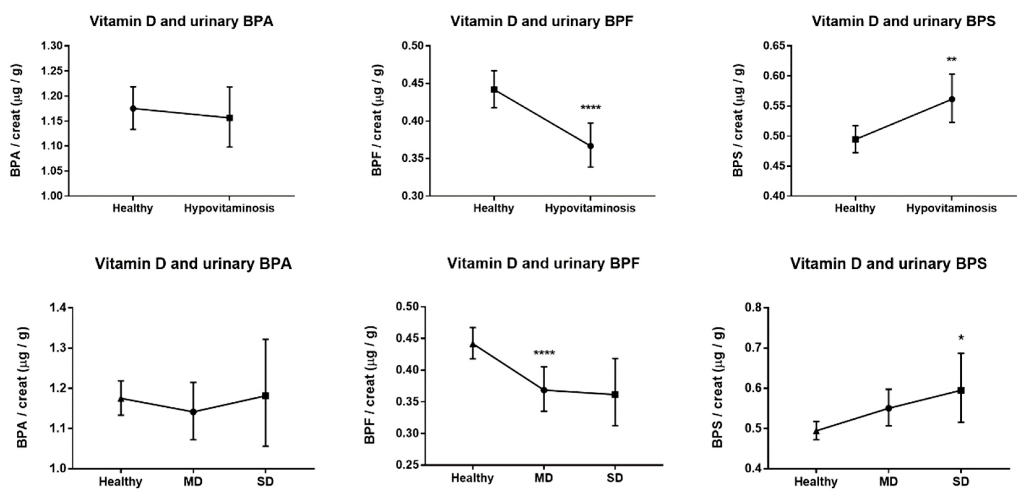
In the quantitative analysis of urinary bisphenols, we observed distinct and divergent patterns of association with vitamin D deficiency for each compound. Specifically, urinary BPA levels showed no statistically significant differences between individuals with or without vitamin D deficiency, suggesting a neutral association in our sample. Conversely, BPF concentrations were significantly lower in individuals with vitamin D deficiency, while BPS levels were significantly higher in the same group. These opposite trends—reduction in BPF and elevation in BPS—suggest potential differences in the biological behavior, metabolism, or tissue affinity of these BPA substitutes, despite their structural similarities.
As illustrated in Figure 2, the relationship between each urinary bisphenol and serum 25(OH)D appears compound-specific. The unchanged levels of BPA contrast with the divergent directions observed for BPF and BPS, supporting the hypothesis that these substitutes may not be interchangeable in terms of biological activity or health impact. This differential behavior underscores the importance of independently evaluating the health effects of each bisphenol analog, rather than assuming class-wide similarities.

3.1.2. Binomial and Multinomial Logistic Regression

Logistic regression analyses confirmed the trends described in the previous analyses, showing a significant relationship between urinary BPS and the risk of vitamin D deficiency. The logistic regression models indicate that a one-unit logarithmic increase in urinary BPS concentration is associated with a significantly increased risk of hypovitaminosis D. Thus, for each increase of one log unit of urinary BPS, an OR (95% CI) of 1.104 (1.038–1.175) was observed for hypovitaminosis D, 1.094 (1.022–1.171) for moderate deficiency, and 1.133 (1.021–1.257) for severe deficiency (Table 2 and Table 3). Consequently, the results showed that urinary BPS is an independent risk factor for vitamin D deficiency. On the contrary, urinary BPF showed an inverse relationship, confirming the existence of potential differences in the mechanism of action associated with the new BPA substitute compounds (Table 2 and Table 3). BPA, however, has not shown significant results in any of the statistical analyses carried out.
The binomial logistic regression model also showed interesting results in the covariates used. Thus, it was observed that increased BMI, diabetes, and albuminuria are risk factors related to hypovitaminosis D, with an OR (95% CI) of 1.03 (1.02–1.05), 1.29 (1.05–1.60), and 1.39 (1.11–1.75), respectively. However, significant negative relationships were also observed, such as age, kidney disease, and dyslipidemia, with an OR (95% CI) of 0.98 (0.97–0.98), 0.61 (0.44–0.84), and 0.69 (0.58–0.82), respectively.
Furthermore, the multinomial logistic regression model showed similar results with age, BMI, and dyslipidemia in both the moderate and severe deficiency groups. In the case of kidney disease, the negative relationship was only significant in the group with moderate deficiency. In the severe deficiency group, a significant OR (95% CI) of 1.57 (1.11–2.22) for diabetes, 1.8 (1.27–2.56) for albuminuria, and 1.5 (1.11–2.02) for hypertension was observed.

3.2. Supplementary Analysis of Vitamin D Metabolites

The central axis of this work has been developed on 25(OH)D (the sum of 25(OH)D2 and D3). It is accepted that it is the marker of vitamin D status due to its higher presence and half-life [31,32,33]. However, to delve into the effects of urinary BPs, their combined linear relationship with the vitamin D metabolites was explored.
As seen in Table 4, the positive relationship observed between 25(OH)D and urinary BPF only coincides with the relationship shown by 25(OH)D3, which means that changes related to vitamin D status are only linked with vitamin D3. In the case of urinary BPS, a significantly negative relationship is observed for both 25(OH)D3 and its epimer (epi-25(OH)D3).

3.3. Risk of Bias: Exclusion of Vitamin D Supplements

Finally, data from all subjects not taking any vitamin D-related vitamin supplements were reanalyzed. Firstly, descriptive statistics showed the same trends with urinary BPs, and the same patterns detected in the covariates analyzed. Furthermore, comparative analyses showed significant changes in the same parameters (Supplementary Table S1).
Similarly, Supplementary Figure S1 shows the same relationships described in the statistical model of the entire cohort. Urinary BPA shows no significant patterns or trends. The BPF and the BPS show the same inverse patterns as in the main study.
Binomial logistic regression analysis also showed the same patterns observed in the general model, with an OR (95% CI) of 0.99 (0.90–1.09), 0.90 (0.85–0.96), and 1.09 (1.01–1.17) for BPA, BPF, and BPS, respectively. Age, sex, and BMI also showed significant results, showing the same trend as the general model. However, an interesting difference with the general model was observed, a higher risk of hypovitaminosis D in women, with an OR (95% CI) of 1.26 (1.05–1.50).
Multinomial logistic regression analysis of moderate deficiency only showed significant results with BPF, age, BMI, and kidney disease, analogous to the general model. However, the p-value for sex was 0.058, with an OR (95% CI) of 1.21 (0.99–1.47) for women. In the case of severe deficiency, sex was significant, with an OR (95% CI) of 1.41 (1.07–1.87). Finally, smokers had a higher risk of severe vitamin D deficiency, with an OR (95% CI) of 1.44 (1.09–1.90).

4. Discussion

In the present study, the existence of significant relationships between the new BPA substitute compounds and vitamin D deficiency has been evidenced for the first time. Despite their high structural homology, the inverse relationship patterns observed between BPS and BPF have confirmed their potential differential risks on human health.
The BPs analyzed in this work are similar in their two phenolic rings at a structural level. However, significant differences between these compounds could help explain the risks observed in the present retrospective study. Firstly, Danzl et al. [60] showed that BPS could not be degraded in the marine environment, contrary to BPA and BPF. Secondly, Gayrard et al. [61] found that the bioavailability of BPS was 250 times greater than that of BPA in a porcine model. In addition, in the same work, they showed that the plasma clearance of BPS was 3.5 times lower than BPA, analogous to the results described by Grandin et al. [62] in a bovine model. Hercog et al. [63] observed that BPA and BPF, but not BPS, were genotoxic in a liver cell line. Kaimal et al. [64] found that the administration of relatively low doses of BPF could cause abortions in rats, an effect that was not seen after treatment with BPS. It has also been reported that BPS has a lower cytotoxic capacity than BPA in human tubular cells [65] and BPA and BPF in the cardiovascular context [66].
Although in vitro and in vivo studies have suggested that BPS may exhibit lower cytotoxicity than BPA and other analogs, emerging evidence points to potential endocrine-disrupting and genotoxic effects of BPS. For instance, Frenzilli et al. [67] demonstrated that both BPA and BPS can induce endocrine and chromosomal alterations in brown trout, raising concerns about genotoxicity. Moreover, Marroquí et al. [18] showed that BPS and BPF can disrupt pancreatic β-cell ion channel expression, activity, and insulin release in mice via an estrogen receptor β-mediated pathway. These findings underscore the need for further investigation into the endocrine and metabolic consequences of BPS exposure, especially in sensitive populations.
Previous work by our team has shown that BPA can affect the renal and vascular systems, promoting podocyte damage in mice [68], affecting the structural integrity of the human podocyte [69], and inducing kidney damage analogous to that observed in diabetic nephropathy [70], promoting increased blood pressure [12], and generating endothelial dysfunction through mechanisms of cell death by necroptosis [71] and cell senescence [72]. During the development of the experimental models, we found interesting indications that pointed to angiotensin II (Ang II) as one of the mediators of the nephro-vascular pathology induced by BPA. There is an essential negative interaction between Ang II and the nephroprotective protein Klotho, whose downregulation is associated with increased circulating levels of fibroblast growth factor 23 (FGF23) [73]. In CKD, the increase in FGF-23 is associated with a compensatory mechanism for phosphate retention and with the inhibition of the renal enzyme 1α-hydroxylase, which consequently reduces the levels of active vitamin D (calcitriol) [32]. Finally, the alteration in the circulating calcitriol levels will induce the reduction in the intestinal capacity for calcium absorption [74]. Interestingly, there is evidence in the literature that the administration of BPA can affect the intestinal capacity for calcium absorption [75,76].
In this way, it shows the possible relationship between bisphenol and calcitriol. Epidemiological publications on the BPA–vitamin D paradigm have determined some interesting associations. Erden et al. [49] observed a negative relationship between serum BPA and 25(OH)D in a group of 128 subjects, in whom there were 43 healthy and 85 with obstructive sleep apnea syndrome (OSAS). Brandie et al. [48] found a negative relationship between urinary BPA and serum levels of 25(OH)D and 1,25(OH)D in a cohort of 299 elderly Italians. Johns et al. [47] analyzed data from the 2005–2010 NHANES cohort and found that in women only, an interquartile range increase in urinary BPA was associated with −3.71% (95% CI, −6.41, −1.02) in 25(OH)D. Their subsequent study in pregnant women [50] showed a non-significant association between BPA and 25(OH)D. However, they did detect an association with the risk of vitamin D deficiency.
It is interesting to note that the results of this study have not shown any significant relationship between urinary BPA and vitamin D in any of the analyses carried out. However, significant relationships with BPA substitute molecules have been detected. Currently, no academic publications explore the possible relationship between emerging BPs and vitamin D in a large general population cohort. This shows the novelty of this study, in which a differential effect has been detected for the first time.
Descriptive statistics and linear regression analysis have shown the first indications of this differential effect, showing completely different patterns between them. Subsequently, binomial and multinomial logistic regression models have confirmed that BPS is a risk factor for vitamin D deficiency, independent of other covariates, such as age, BMI, sex, diabetes, kidney disease, hypertension, dyslipidemia, or smoking. The analysis and evidence have shown the inexistence of a significant association between BPA and 25(OH)D. However, the inverse patterns between BPA substitute BPs have shown that, despite the structural homology, there are potential differential risks on human health. This fact reaffirms the need to strengthen research and limit their use by industry.
Previous work by our group evidenced a relationship between new BPA substitute molecules excreted in the urine of the general population with various metabolic and cardiovascular pathologies, including diabetes, heart disease, hypertension, and renal dysfunction. Epidemiological analyses have shown associations between urinary concentrations of these compounds and an increased risk of diabetes in the adult population [77,78]. Additionally, combined exposure to BPA and its substitutes has been associated with a higher incidence of renal, cardiovascular diseases, and adverse effects on vascular endothelium [79,80]; these findings reinforce the idea that BPA substitutes, despite being presented as safer alternatives, may exert harmful biological effects similar to or different from those of BPA itself. The current results, showing differential associations between BPS, BPF, and vitamin D deficiency, align with this growing body of evidence and underscore the urgent need for broader studies addressing the systemic health impacts of these molecules.
The analysis of vitamin D metabolites has shown that vitamin D3 is precisely the one that is related to substitute BPs, with no significant relationships being observed with vitamin D2. D3 is synthesized through ultraviolet radiation that strikes the skin, which transforms 7-dehydrocholesterol into cholecalciferol or vitamin D3, or through the consumption of certain foods, such as egg yolks and oily fish. For its part, vitamin D2 is acquired through the diet [29,30,31]. Considering that both vitamins have a common entry into the body (diet) and a differential entry (skin), the idea that BPS could affect the synthesis of this vitamin through the skin is consistent.
On the other hand, the analysis of serum vitamin D includes the quantitative study of 25(OH)D, that is, 25(OH)D2 + 25(OH)D3, the forms hydroxylated by the liver but still inactive. The analysis of these metabolites is carried out due to their high half-life in the body (13–15 days), unlike calcitriol (hours) [33]. Another possibility suggested by the results is that BPS exposure could affect the hepatic metabolism of 25(OH)D3, thus affecting 25(OH)D status independently of renal function. Moreover, recent studies suggest that BPS may interfere directly with hepatic function, which is essential for the conversion of vitamin D into its circulating form, 25(OH)D3. Experimental models in mice have shown that BPS exposure alters hepatic mitochondrial dynamics, induces endoplasmic reticulum stress, and disrupts lipid and glucose metabolism, especially under obesogenic conditions [81]. Additionally, BPS promotes hepatic steatosis by upregulating monocarboxylate transporter 1 (MCT1) and impairing the mitochondrial respiratory system [82]. A third study has revealed that BPS blocks hepatic autophagy via a PPARα–EP300–mTORC1 signaling axis, leading to lipid accumulation and the progression of NAFLD [83]. These findings provide plausible mechanistic support for the idea that BPS may impair hepatic vitamin D hydroxylation, contributing to lower serum 25(OH)D3 levels even independently of renal function. However, further studies are needed to confirm this mechanism in humans.
The results have identified counterintuitive patterns between vitamin D status and eGFR, which this phenomenon could explain. There is evidence in the academic literature that identifies the same patterns between 25(OH)D and kidney function. The works of Teumer et al. [84] and Geng et al. [85] detected a negative association between serum 25(OH)D and kidney function in six cohorts and the NHANES cohort, respectively. Moore et al. [86] found that patients with the highest eGFR had the lowest 25(OH)D levels. Finally, Priyadarshini et al. [87] determined that most of their CKD patients had normal vitamin D levels. The authors argued that the intake of vitamin supplements was a possible cause of the phenomenon. However, the present work eliminated this possibility by excluding all those individuals who take supplements. Future epidemiological studies should incorporate PTH values due to their relationship with active vitamin D, calcium metabolism, and renal function.
Finally, the analysis of individuals who do not consume vitamin supplements has reaffirmed the relationships between urinary BPs and vitamin D. However, the supplementary analysis has shown interesting relationships between the sex of individuals and tobacco consumption associated with vitamin D status. In this case, the reanalysis of the data has shown that the consumption of vitamin supplements could mask specific associations of great relevance for the study. Sex could play an essential role in vitamin D metabolism, which would mean that the effect of BPS in this context could be more relevant for women. The percentage of women in the healthy group was 44.9%, while the severe deficiency group was 53.3% in the analysis of subjects without vitamin supplements. In the entire cohort, 52.5% of women were in the healthy group, and 55.3% were in the severe deficiency group. The consumption of vitamin supplements masked the relationship with sex in the corrected logistic models. It should be considered in future studies related to the object of study of this work.
The possible relationship between sex, smoking, and vitamin D is an interesting topic derived from the results of the present study. It is important to note that it has been documented that sex may be a differentiating element in vitamin D levels due to biological differences. For instance, sex hormones such as estrogen can modulate the activity of the hydroxylase enzymes responsible for vitamin D activation, and variations in body composition also play a role [88]. Regarding tobacco use, regular tobacco users have been reported to have lower vitamin D levels. In the recent meta-analysis conducted by Yang’s team, the authors suggest that smoking could affect vitamin D metabolism by different mechanisms, from increased skin aging to the expression of inflammatory mediators or even PTH inhibition [89].
A recent study by Brennan et al. [90] analyzed the relationship between bisphenol exposure and vitamin D levels in a small cohort of 57 women, evaluating correlations between the concentrations of BPA, BPS, and the combined measure (ƩBPs) with serum 25(OH)D. Their results did not reveal statistically significant associations. While this study is an important initial step, several methodological aspects differentiate it from our approach. Firstly, the limited sample size considerably restricts the statistical power, making it difficult to detect subtle associations. In contrast, our study leverages a large, representative population sample from NHANES, which includes both sexes and a broad age range. Moreover, rather than simple correlation analyses, we applied multivariate logistic regression models adjusted for relevant covariates to evaluate the risk of vitamin D deficiency across bisphenol exposure levels. This approach allowed us to uncover associations not previously identified, supporting the notion that BPS exposure may represent a potential risk factor for vitamin D deficiency in the general population.
In the present study, individuals under 18 years of age were excluded due to methodological considerations. Childhood and adolescence are characterized by significant physiological variability, including growth, puberty, and unique dietary patterns, which strongly influence vitamin D metabolism and endocrine responses. These age-related factors, along with the widespread use of pediatric vitamin D supplementation, may act as confounders in studies evaluating environmental exposures. Moreover, bisphenol exposure patterns may differ substantially in minors compared to adults, both in intensity and source. Although not included in this analysis, this group represents a population of high interest and vulnerability. Future studies should specifically address the potential effects of bisphenols on vitamin D status in children and adolescents, as this remains an open question in environmental health research.
The lack of a significant association between urinary BPA concentrations and vitamin D status in this study contrasts with the findings observed for BPS. This difference may be partially explained by the distinct pharmacokinetic profiles of these compounds. BPA is known to have a short half-life and is rapidly metabolized and excreted through urine within a few hours of exposure. Therefore, urinary BPA levels primarily reflect recent exposure and may not accurately represent bioavailable concentrations or a long-term internal dose. This could lead to a potential underestimation of its biological effects in cross-sectional studies such as this one. On the other hand, BPA substitutes like BPS may exhibit different absorption, metabolism, or retention patterns, potentially resulting in a greater cumulative effect or prolonged interaction with vitamin D metabolism pathways. Moreover, the vitamin D biomarker used in this study—25-hydroxyvitamin D—is a stable circulating precursor with a long half-life, and it reflects the combined influence of multiple physiological and environmental factors over time. These temporal mismatches in the kinetics of BPA and vitamin D may also explain the absence of a statistically significant relationship.
Furthermore, retrospective cohort studies do not allow us to identify causal relationships but to identify patterns of relationship or association independent of the effect of other covariates. Future longitudinal studies, accompanied by cellular and animal models, exploring the relationship between BPS and vitamin D will be necessary to confirm the results of this manuscript. There are epidemiological studies that have linked BPA substitute molecules with pathologies such as diabetes [91,92], obesity [93,94,95], hypertension [96], and depression [97]. Similarly, vitamin D deficiency has been associated with changes in bone metabolism, diabetes, obesity, hypertension, psychiatric disorders, and even cancer [39,40,41,42,43,44,45,46]. Therefore, the idea that vitamin D deficiency could be a risk factor associated with BPS is consistent, helping to explain the global associations described in the literature.

5. Conclusions

This study has identified new and relevant associations between BPS exposure and vitamin D deficiency. BPS is a molecule of growing industrial use, and human exposure is expected to increase unless regulatory frameworks are updated. Although BPS has been considered a potentially less toxic alternative to BPA, emerging evidence—including our findings—suggests that it may still exert significant biological effects, particularly as an endocrine disruptor. Considering these results, and given the precautionary principle in public health, regulatory agencies should critically assess the widespread use of BPA substitutes until their safety is clearly established through longitudinal and mechanistic studies.

Supplementary Materials

The following supporting information can be downloaded at: https://www.mdpi.com/article/10.3390/greenhealth1020010/s1, Figure S1: Variation in urinary bisphenol concentrations according to vitamin D status in participants not taking vitamin D-related supplements; Table S1: Descriptive statistics of vitamin D status among individuals not taking vitamin D-related supplements.

Funding

This research received no external funding.

Institutional Review Board Statement

Not applicable.

Informed Consent Statement

Not applicable.

Data Availability Statement

The original data can be found on the NHANES cohort web page (https://wwwn.cdc.gov/nchs/nhanes/default.aspx, accessed on 15 January 2022).

Acknowledgments

The author wants to thank Ricardo J. Bosch for suggestions and comments, Rosalía Gómez-Toledano (philologist and English teacher for 37 years) for proofreading the manuscript, and Elisa Moreno-Mizileanu for her indispensable logistical support. And especially to all the volunteers who have participated in the NHANES cohort and to all the members involved in its execution and transparent publication, because thanks to them, science can move in the right direction.

Conflicts of Interest

The author declares no conflicts of interest.

References

  1. Baralla, E.; Pasciu, V.; Varoni, M.V.; Nieddu, M.; Demuro, R.; Demontis, M.P. Bisphenols’ occurrence in bivalves as sentinel of environmental contamination. Sci. Total Environ. 2021, 785, 147263. [Google Scholar] [CrossRef]
  2. Wu, L.H.; Zhang, X.M.; Wang, F.; Gao, C.J.; Chen, D.; Palumbo, J.R.; Guo, Y.; Zeng, E.Y. Occurrence of bisphenol S in the environment and implications for human exposure: A short review. Sci. Total Environ. 2018, 615, 87–98. [Google Scholar] [CrossRef] [PubMed]
  3. Vasiljevic, T.; Harner, T. Bisphenol A and its analogues in outdoor and indoor air: Properties, sources and global levels. Sci. Total Environ. 2021, 789, 148013. [Google Scholar] [CrossRef] [PubMed]
  4. Vandenberg, L.N.; Chahoud, I.; Heindel, J.J.; Padmanabhan, V.; Paumgartten, F.J.R.R.; Schoenfelder, G. Urinary, circulating, and tissue biomonitoring studies indicate widespread exposure to bisphenol A. Environ. Health Perspect. 2010, 118, 1055–1070. [Google Scholar] [CrossRef]
  5. Freire, C.; Molina-Molina, J.M.J.-M.; Iribarne-Duran, L.M.; Jimenez-Diaz, I.; Vela-Soria, F.; Mustieles, V.; Arrebola, J.P.; Fernández, M.F.; Artacho-Cordón, F.; Olea, N. Concentrations of bisphenol A and parabens in socks for infants and young children in Spain and their hormone-like activities. Environ. Int. 2019, 127, 592–600. [Google Scholar] [CrossRef] [PubMed]
  6. Moreno-Gómez-Toledano, R.; Arenas, M.I.; Sánchez-Esteban, S.; Cook, A.; Saura, M.; Bosch, R.J. Critical Analysis of Human Exposure to Bisphenol a and its Novel Implications on Renal, Cardiovascular and Hypertensive Diseases. In Hot Topics in Endocrinology and Metabolism, 1st ed.; Heshmati, H.M., Ed.; IntechOpen: London, UK, 2021; pp. 1–20. [Google Scholar]
  7. Xu, L.C.; Sun, H.; Chen, J.F.; Bian, Q.; Qian, J.; Song, L.; Wang, X.R. Evaluation of androgen receptor transcriptional activities of bisphenol A, octylphenol and nonylphenol in vitro. Toxicology 2005, 216, 197–203. [Google Scholar] [CrossRef]
  8. Teng, C.; Goodwin, B.; Shockley, K.; Xia, M.; Huang, R.; Norris, J.; Merrick, B.A.; Jetten, A.M.; Austin, C.P.; Tice, R.R. Bisphenol A affects androgen receptor function via multiple mechanisms. Chem. Biol. Interact. 2013, 203, 556–564. [Google Scholar] [CrossRef]
  9. Carwile, J.L.; Michels, K.B. Urinary bisphenol A and obesity: NHANES 2003–2006. Environ. Res. 2011, 111, 825–830. [Google Scholar] [CrossRef]
  10. Lang, I.A.; Galloway, T.S.; Scarlett, A.; Henley, W.E.; Depledge, M.; Wallace, R.B.; Melzer, D. Association of urinary bisphenol A concentration with medical disorders and laboratory abnormalities in adults. JAMA 2008, 300, 1303–1310. [Google Scholar] [CrossRef]
  11. Moreno-Gómez-Toledano, R.; Arenas, M.I.; Vélez-Vélez, E.; Coll, E.; Quiroga, B.; Bover, J.; Bosch, R.J. Bisphenol A exposure and kidney diseases: Systematic review, meta-analysis and NHANES 03–16 study. Biomolecules 2021, 11, 1046. [Google Scholar] [CrossRef]
  12. Saura, M.; Marquez, S.; Reventun, P.; Olea-Herrero, N.; Isabel Arenas, M.; Moreno-Gomez-Toledano, R.; Gómez-Parrizas, M.; Muñóz-Moreno, C.; González-Santander, M.; Zaragoza, C.; et al. Oral administration of bisphenol A induces high blood pressure through angiotensin II/CaMKII-dependent uncoupling of eNOS. FASEB J. 2014, 28, 4719–4728. [Google Scholar] [CrossRef]
  13. Nunez, P.; Fernandez, T.; Garcia-Arevalo, M.; Alonso-Magdalena, P.; Nadal, A.; Perillan, C.; Arguelles, J. Effects of bisphenol A treatment during pregnancy on kidney development in mice: A stereological and histopathological study. J. Dev. Orig. Health Dis. 2018, 9, 208–214. [Google Scholar] [CrossRef]
  14. Bano, U.; Memon, S.; Shahani, M.Y.; Shaikh, P.; Gul, S. Epigenetic effects of in utero bisphenol A administration: Diabetogenic and atherogenic changes in mice offspring. Iran. J. Basic Med. Sci. 2019, 22, 521–528. [Google Scholar]
  15. Rochester, J.R.; Bolden, A.L. Bisphenol S and F: A systematic review and comparison of the hormonal activity of bisphenol a substitutes. Environ. Health Perspect. 2015, 123, 643–650. [Google Scholar] [CrossRef]
  16. Skledar, D.G.; Schmidt, J.; Fic, A.; Klopčič, I.; Trontelj, J.; Dolenc, M.S.; Finel, M.; Mašič, L.P. Influence of metabolism on endocrine activities of bisphenol S. Chemosphere 2016, 157, 152–159. [Google Scholar] [CrossRef]
  17. Conroy-Ben, O.; Garcia, I.; Teske, S.S. In silico binding of 4,4′-bisphenols predicts in vitro estrogenic and antiandrogenic activity. Environ. Toxicol. 2018, 33, 569–578. [Google Scholar] [CrossRef]
  18. Marroqui, L.; Martinez-Pinna, J.; Castellano-Muñoz, M.; dos Santos, R.S.; Medina-Gali, R.M.; Soriano, S.; Quesada, I.; Gustafsson, J.A.; Encinar, J.A.; Nadal, A. Bisphenol-S and Bisphenol-F alter mouse pancreatic β-cell ion channel expression and activity and insulin release through an estrogen receptor ERβ mediated pathway. Chemosphere 2021, 265, 129051. [Google Scholar] [CrossRef] [PubMed]
  19. Qiu, W.; Shao, H.; Lei, P.; Zheng, C.; Qiu, C.; Yang, M.; Zheng, Y. Immunotoxicity of bisphenol S and F are similar to that of bisphenol A during zebrafish early development. Chemosphere 2018, 194, 1–8. [Google Scholar] [CrossRef]
  20. Ramskov Tetzlaff, C.N.; Svingen, T.; Vinggaard, A.M.; Rosenmai, A.K.; Taxvig, C. Bisphenols B, E, F, and S and 4-cumylphenol induce lipid accumulation in mouse adipocytes similarly to bisphenol A. Environ. Toxicol. 2020, 35, 543–552. [Google Scholar] [CrossRef] [PubMed]
  21. Sidorkiewicz, I.; Czerniecki, J.; Jarzabek, K.; Zbucka-Kretowska, M.; Wolczynski, S. Cellular, transcriptomic and methylome effects of individual and combined exposure to BPA, BPF, BPS on mouse spermatocyte GC-2 cell line. Toxicol. Appl. Pharmacol. 2018, 359, 1–11. [Google Scholar] [CrossRef] [PubMed]
  22. dos Santos, B.; Ivantsova, E.; Guzman, A.P.; Martyniuk, C.J. Critical review of the toxicity mechanisms of bisphenol F in zebrafish (Danio rerio): Knowledge gaps and future directions. Chemosphere 2022, 297, 134132. [Google Scholar] [CrossRef]
  23. Catenza, C.J.; Farooq, A.; Shubear, N.S.; Donkor, K.K. A targeted review on fate, occurrence, risk and health implications of bisphenol analogues. Chemosphere 2021, 268, 129273. [Google Scholar] [CrossRef] [PubMed]
  24. Lehmler, H.-J.J.; Liu, B.Y.; Gadogbe, M.; Bao, W. Exposure to Bisphenol A, Bisphenol F, and Bisphenol S in U.S. Adults and Children: The National Health and Nutrition Examination Survey 2013-2014. ACS Omega 2018, 3, 6523–6532. [Google Scholar] [CrossRef]
  25. Sol, C.M.; Van Zwol-Janssens, C.; Philips, E.M.; Asimakopoulos, A.G.; Martinez-Moral, M.P.; Kannan, K.; Jaddoe, V.W.V.; Trasande, L.; Santos, S. Maternal bisphenol urine concentrations, fetal growth and adverse birth outcomes: A population-based prospective cohort. Environ. Health 2021, 20, 60. [Google Scholar] [CrossRef] [PubMed]
  26. Wang, Y.-X.; Liu, C.; Shen, Y.; Wang, Q.; Pan, A.; Yang, P.; Chen, Y.J.; Deng, Y.L.; Lu, Q.; Cheng, L.M.; et al. Urinary levels of bisphenol A, F and S and markers of oxidative stress among healthy adult men: Variability and association analysis. Environ. Int. 2019, 123, 301–309. [Google Scholar] [CrossRef]
  27. Jin, H.B.; Xie, J.H.; Mao, L.L.; Zhao, M.R.; Bai, X.X.; Wen, J.; Shen, T.; Wu, P. Bisphenol analogue concentrations in human breast milk and their associations with postnatal infant growth. Environ. Pollut. 2020, 259, 113779. [Google Scholar] [CrossRef]
  28. Li, A.J.; Zhuang, T.F.; Shi, W.; Liang, Y.; Liao, C.Y.; Song, M.Y.; Jiang, G. Serum concentration of bisphenol analogues in pregnant women in China. Sci. Total Environ. 2020, 707, 136100. [Google Scholar] [CrossRef]
  29. Ross, C.; Taylor, C.L.; Yaktine, A.L.; Del Valle, H.B. (Eds.) Dietary Reference Intakes for Calcium Vitamin, D. Dietary Reference Intakes for Calcium and Vitamin D; National Academies Press: Washington, DC, USA, 2011. [Google Scholar]
  30. Cesari, M.; Incalzi, R.A.; Zamboni, V.; Pahor, M. Vitamin D hormone: A multitude of actions potentially influencing the physical function decline in older persons. Geriatr. Gerontol. Int. 2011, 11, 133–142. [Google Scholar] [CrossRef]
  31. Tripkovic, L.; Lambert, H.; Hart, K.; Smith, C.P.; Bucca, G.; Penson, S.; Chope, G.; Hyppönen, E.; Berry, J.; Vieth, R.; et al. Comparison of vitamin D2and vitamin D3 supplementation in raising serum 25-hydroxyvitamin D status: A systematic review and meta-analysis. Am. J. Clin. Nutr. 2012, 95, 1357–1364. [Google Scholar] [CrossRef]
  32. Jean, G.; Souberbielle, J.C.; Chazot, C. Vitamin D in chronic kidney disease and dialysis patients. Nutrients 2017, 9, 328. [Google Scholar] [CrossRef] [PubMed]
  33. Bresson, J.L.; Burlingame, B.; Dean, T.; Fairweather-Tait, S.; Heinonen, M.; Hirsch-Ernst, K.I.; Mangelsdorf, I.; McArdle, H.; Naska, A.; Neuhäuser-Berthold, M.; et al. Dietary reference values for vitamin D. EFSA J. 2016, 14, 4547. [Google Scholar] [CrossRef]
  34. Cashman, K.D. Vitamin D Deficiency: Defining, Prevalence, Causes, and Strategies of Addressing. Calcif. Tissue Int. 2020, 106, 14–29. [Google Scholar] [CrossRef]
  35. Carswell, A.T.; Oliver, S.J.; Wentz, L.M.; Kashi, D.S.; Roberts, R.; Tang, J.C.Y.; Izard, R.M.; Jackson, S.; Allan, D.; Rhodes, L.E.; et al. Influence of Vitamin D supplementation by sunlight or oral D3 on exercise performance. Med. Sci. Sports Exerc. 2018, 50, 2555–2564. [Google Scholar] [CrossRef] [PubMed]
  36. Amrein, K.; Scherkl, M.; Hoffmann, M.; Neuwersch-Sommeregger, S.; Köstenberger, M.; Tmava Berisha, A.; Martucci, G.; Pilz, S.; Malle, O. Vitamin D deficiency 2.0: An update on the current status worldwide. Eur. J. Clin. Nutr. 2020, 74, 1498–1513. [Google Scholar] [CrossRef]
  37. Palacios, C.; Gonzalez, L. Is vitamin D deficiency a major global public health problem? J. Steroid Biochem. Mol. Biol. 2014, 144, 138–145. [Google Scholar] [CrossRef]
  38. Lips, P.; Cashman, K.D.; Lamberg-Allardt, C.; Bischoff-Ferrari, H.A.; Obermayer-Pietsch, B.; Bianchi, M.L.; tepan, J.; El-Hajj Fuleihan, G.; Bouillon, R. Current Vitamin D status in European and Middle East countries and strategies to prevent Vitamin D deficiency: A position statement of the European Calcified Tissue Society. Eur. J. Endocrinol. 2019, 180, P23–P54. [Google Scholar] [CrossRef]
  39. Holick, M.F.; Binkley, N.C.; Bischoff-Ferrari, H.A.; Gordon, C.M.; Hanley, D.A.; Heaney, R.P.; Murad, M.H.; Weaver, C.M. Evaluation, Treatment, and Prevention of Vitamin D Deficiency: An Endocrine Society Clinical Practice Guideline. J. Clin. Endocrinol. Metab. 2011, 96, 1911–1930. [Google Scholar] [CrossRef]
  40. Szymczak-Pajor, I.; Śliwińska, A. Analysis of Association between Vitamin D Deficiency and Insulin Resistance. Nutrients 2019, 11, 794. [Google Scholar] [CrossRef]
  41. Tao, S.; Yuan, Q.; Mao, L.; Chen, F.L.; Ji, F.; Cui, Z.H. Vitamin D deficiency causes insulin resistance by provoking oxidative stress in hepatocytes. Oncotarget 2017, 8, 67605–67613. [Google Scholar] [CrossRef] [PubMed]
  42. Fiamenghi VI, Mello ED de. Vitamin D deficiency in children and adolescents with obesity: A meta-analysis. J. Pediatr. 2021, 97, 273–279. [Google Scholar] [CrossRef] [PubMed]
  43. Pereira-Santos, M.; Costa, P.R.F.; Assis, A.M.O.; Santos, C.A.S.T.; Santos, D.B. Obesity and vitamin D deficiency: A systematic review and meta-analysis. Obes. Rev. 2015, 16, 341–349. [Google Scholar] [PubMed]
  44. Walsh, J.S.; Bowles, S.; Evans, A.L. Vitamin D in obesity. Curr. Opin. Endocrinol. Diabetes Obes. 2017, 24, 389–394. [Google Scholar] [CrossRef] [PubMed]
  45. Chang, S.W.; Lee, H.C. Vitamin D and health—The missing vitamin in humans. Pediatr. Neonatol. 2019, 60, 237–244. [Google Scholar] [CrossRef]
  46. Wang, H.; Chen, W.; Li, D.; Yin, X.; Zhang, X.; Olsen, N.; Zheng, S.G. Vitamin D and Chronic Diseases. Aging Dis. 2017, 8, 346–353. [Google Scholar] [CrossRef] [PubMed]
  47. Johns, L.E.; Ferguson, K.K.; Meeker, J.D. Relationships Between Urinary Phthalate Metabolite and Bisphenol A Concentrations and Vitamin D Levels in U.S. Adults: National Health and Nutrition Examination Survey (NHANES), 2005–2010. J. Clin. Endocrinol. Metab. 2016, 101, 4062–4069. [Google Scholar] [CrossRef]
  48. Brandi, M.L.; Bandinelli, S.; Iantomasi, T.; Giusti, F.; Talluri, E.; Sini, G.; Nannipieri, F.; Battaglia, S.; Giusti, R.; Egan, C.G.; et al. Association between vitamin D and bisphenol A levels in an elderly Italian population: Results from the InCHIANTI study. Endocr. Connect. 2022, 11, e210571. [Google Scholar] [CrossRef]
  49. Erden, E.S.; Genc, S.; Motor, S.; Ustun, I.; Ulutas, K.T.; Bilgic, H.K.; Oktar, S.; Sungur, S.; Erem, C.; Gokce, C. Investigation of serum bisphenol A, vitamin D, and parathyroid hormone levels in patients with obstructive sleep apnea syndrome. Endocrine 2014, 45, 311–318. [Google Scholar] [CrossRef]
  50. Johns, L.E.; Ferguson, K.K.; Cantonwine, D.E.; McElrath, T.F.; Mukherjee, B.; Meeker, J.D. Urinary BPA and phthalate metabolite concentrations and plasma vitamin D levels in pregnant women: A repeated measures analysis. Environ. Health Perspect. 2017, 125, 87026. [Google Scholar] [CrossRef]
  51. Centers for Disease Control and Prevention (CDC). National Health and Nutrition Examination Survey Data. National Center for Health Statistics (NCHS). 2016. Available online: https://wwwn.cdc.gov/nchs/nhanes/continuousnhanes/default.aspx?BeginYear=2013 (accessed on 15 January 2022).
  52. Centers for Disease Control and Prevention (CDC). NHANES Dietary Data. National Center for Health Statistics (NCHS). 2016. Available online: https://wwwn.cdc.gov/nchs/nhanes/search/datapage.aspx?Component=Dietary&Cycle=2013-2014 (accessed on 15 January 2022).
  53. Centers for Disease Control and Prevention (CDC). NHANES Questionnaire Data. National Center for Health Statistics (NCHS). 2016. Available online: https://wwwn.cdc.gov/nchs/nhanes/search/datapage.aspx?Component=Questionnaire&Cycle=2013-2014 (accessed on 15 January 2022).
  54. Michels, W.M.; Grootendorst, D.C.; Verduijn, M.; Elliott, E.G.; Dekker, F.W.; Krediet, R.T. Performance of the Cockcroft-Gault, MDRD, and new CKD-EPI formulas in relation to GFR, age, and body size. Clin. J. Am. Soc. Nephrol. 2010, 5, 1003–1009. [Google Scholar] [CrossRef]
  55. Poggio, E.D.; Wang, X.; Greene, T.; Van Lente, F.; Hall, P.M. Performance of the modification of diet in renal disease and Cockcroft-Gault equations in the estimation of GFR in health and in chronic kidney disease. J. Am. Soc. Nephrol. 2005, 16, 459–466. [Google Scholar] [CrossRef]
  56. Levey, A.S.; Stevens, L.A.; Schmid, C.H.; Zhang, Y.; Castro, A.F.; Feldman, H.I.; Kusek, J.W.; Eggers, P.; Van Lente, F.; Greene, T.; et al. A new equation to estimate glomerular filtration rate. Ann. Intern. Med. 2009, 150, 604–612. [Google Scholar] [CrossRef]
  57. Vega, G.L.; Wang, J.; Grundy, S.M. Chronic kidney disease and statin eligibility. J. Clin. Lipidol. 2021, 15, 173–180. [Google Scholar] [CrossRef]
  58. Joo, Y.S.; Kim, H.W.; Lee, S.; Nam, K.H.; Yun, H.R.; Jhee, J.H.; Han, S.H.; Yoo, T.H.; Kang, S.W.; Park, J.T. Dietary zinc intake and incident chronic kidney disease. Clin. Nutr. 2021, 40, 1039–1045. [Google Scholar] [CrossRef]
  59. Pirkle, J.L. Exposure of the US Population to Environmental Tobacco Smoke. JAMA 1996, 275, 1233. [Google Scholar] [CrossRef] [PubMed]
  60. Danzl, E.; Sei, K.; Soda, S.; Ike, M.; Fujita, M. Biodegradation of bisphenol A, bisphenol F and bisphenol S in seawater. Int. J. Environ. Res. Public Health 2009, 6, 1472–1484. [Google Scholar] [CrossRef]
  61. Gayrard, V.; Lacroix, M.Z.; Grandin, F.C.; Collet, S.H.; Mila, H.; Viguie, C.; Gély, C.A.; Rabozzi, B.; Bouchard, M.; Léandri, R.; et al. Oral systemic bioavailability of bisphenol A and bisphenol S in pigs. Environ. Health Perspect. 2019, 127, 77005. [Google Scholar] [CrossRef] [PubMed]
  62. Grandin, F.; Picard-Hagen, N.; Gayrard, V.; Puel, S.; Viguie, C.; Toutain, P.-L.; Debrauwer, L.; Lacroix, M.Z. Development of an on-line solid phase extraction ultra-high-performance liquid chromatography technique coupled to tandem mass spectrometry for quantification of bisphenol S and bisphenol S glucuronide: Applicability to toxicokinetic investigations. J. Chromatogr. A 2017, 1526, 39–46. [Google Scholar] [CrossRef] [PubMed]
  63. Hercog, K.; Maisanaba, S.; Filipič, M.; Sollner-Dolenc, M.; Kač, L.; Žegura, B. Genotoxic activity of bisphenol A and its analogues bisphenol S, bisphenol F and bisphenol AF and their mixtures in human hepatocellular carcinoma (HepG2) cells. Sci. Total Environ. 2019, 687, 267–276. [Google Scholar] [CrossRef]
  64. Kaimal, A.; Al Mansi, M.H.; Dagher, J.B.; Pope, C.; Varghese, M.G.; Rudi, T.B.; Almond, A.E.; Cagle, L.A.; Beyene, H.K.; Bradford, W.T.; et al. Prenatal exposure to bisphenols affects pregnancy outcomes and offspring development in rats. Chemosphere 2021, 276, 130118. [Google Scholar] [CrossRef]
  65. Mas, S.; Ruiz-Priego, A.; Abaigar, P.; Santos, J.; Camarero, V.; Egido, J.; Ortiz, A.; Gonzalez-Parra, E. Bisphenol S is a haemodialysis-associated xenobiotic that is less toxic than bisphenol A. Clin. Kidney J. 2021, 14, 1147–1155. [Google Scholar] [CrossRef]
  66. Ji, G.; Gu, J.; Guo, M.; Zhou, L.; Wang, Z.; Shi, L.; Gu, A. A systematic comparison of the developmental vascular toxicity of bisphenol A and its alternatives in vivo and in vitro. Chemosphere 2022, 291, 132936. [Google Scholar] [CrossRef] [PubMed]
  67. Frenzilli, G.; Martorell-Ribera, J.; Bernardeschi, M.; Scarcelli, V.; Jonsson, E.; Diano, N.; Moggio, M.; Guidi, P.; Sturve, J.; Asker, N. Bisphenol A and Bisphenol S Induce Endocrine and Chromosomal Alterations in Brown Trout. Front. Endocrinol. 2021, 12, 645519. [Google Scholar] [CrossRef]
  68. Olea-Herrero, N.; Arenas, M.I.; Munoz-Moreno, C.; Moreno-Gómez-Toledano, R.; Gonzalez-Santander, M.; Arribas, I.; Bosch, R.J. Bisphenol-A induces podocytopathy with proteinuria in mice. J. Cell. Physiol. 2014, 229, 2057–2066. [Google Scholar] [CrossRef]
  69. Moreno-Gómez-Toledano, R.; Arenas, M.I.; González-Martínez, C.; Olea-Herrero, N.; Reventún, P.; Di Nunzio, M.; Sánchez-Esteban, S.; Arilla-Ferreiro, E.; Saura, M.; Bosch, R.J. Bisphenol A impaired cell adhesion by altering the expression of adhesion and cytoskeleton proteins on human podocytes. Sci. Rep. 2020, 10, 16638. [Google Scholar] [CrossRef]
  70. Moreno-Gómez-Toledano, R.; Arenas, M.I.; Muñoz-Moreno, C.; Olea-Herrero, N.; Reventun, P.; Izquierdo-Lahuerta, A.; Antón-Cornejo, A.; González-Santander, M.; Zaragoza, C.; Saura, M.; et al. Comparison of the renal effects of bisphenol A in mice with and without experimental diabetes. Role of sexual dimorphism. Biochim. Biophys. Acta (BBA)-Mol. Basis Dis. 2022, 1868, 166296. [Google Scholar] [CrossRef]
  71. Reventun, P.; Sanchez-Esteban, S.; Cook, A.; Cuadrado, I.; Roza, C.; Moreno-Gómez-Toledano, R.; Muñoz, C.; Zaragoza, C.; Bosch, R.J.; Saura, M. Bisphenol A induces coronary endothelial cell necroptosis by activating RIP3/CamKII dependent pathway. Sci. Rep. 2020, 10, 4190. [Google Scholar] [CrossRef]
  72. Moreno-Gómez-Toledano, R.; Sanchez-Esteban, S.; Cook, A.; Mínguez-Moratinos, M.; Ramírez-Carracedo, R.; Reventun, P.; Delgado-Marín, M.; Bosch, R.J.; Saura, M. Bisphenol A induces accelerated cell aging in murine endothelium. Biomolecules 2021, 11, 1429. [Google Scholar] [CrossRef]
  73. Cantero-Navarro, E.; Fernández-Fernández, B.; Ramos, A.M.; Rayego-Mateos, S.; Rodrigues-Diez, R.R.; Sánchez-Niño, M.D.; Sanz, A.B.; Ruiz-Ortega, M.; Ortiz, A. Renin-angiotensin system and inflammation update. Mol. Cell. Endocrinol. 2021, 529, 111254. [Google Scholar] [CrossRef]
  74. Christakos, S.; Dhawan, P.; Porta, A.; Mady, L.J.; Seth, T. Vitamin D and intestinal calcium absorption. Mol. Cell. Endocrinol. 2011, 347, 25–29. [Google Scholar] [CrossRef] [PubMed]
  75. Otsuka, H.; Sugimoto, M.; Ikeda, S.; Kume, S. Effects of bisphenol A administration to pregnant mice on serum Ca and intestinal Ca absorption. Anim. Sci. J. 2012, 83, 232–237. [Google Scholar] [CrossRef] [PubMed]
  76. Kim, S.; An, B.-S.S.; Yang, H.; Jeung, E.-B.B. Effects of octylphenol and bisphenol A on the expression of calcium transport genes in the mouse duodenum and kidney during pregnancy. Toxicology 2013, 303, 99–106. [Google Scholar] [CrossRef]
  77. Moreno-Gomez-Toledano, R.; Velez-Velez, E.; Arenas, I.M.; Saura, M.; Bosch, R.J. Association between urinary concentrations of bisphenol A substitutes and diabetes in adults. World J. Diabetes 2022, 13, 521–531. [Google Scholar] [CrossRef] [PubMed]
  78. Moreno-Gómez-Toledano, R.; Delgado-Marín, M.; Cook-Calvete, A.; González-Cucharero, C.; Alcharani, N.; Jiménez-Guirado, B.; Hernandez, I.; Ramirez-Carracedo, R.; Tesoro, L.; Botana, L.; et al. New environmental factors related to diabetes risk in humans: Emerging bisphenols used in synthesis of plastics. World J. Diabetes 2023, 14, 1301–1313. Available online: https://pubmed.ncbi.nlm.nih.gov/37664470/ (accessed on 19 December 2023). [CrossRef] [PubMed]
  79. Moreno-Gómez-Toledano, R.; Delgado-Marín, M.; Sánchez-Esteban, S.; Cook-Calvete, A.; Ortiz, S.; Bosch, R.J.; Saura, M. Combination of Bisphenol A and Its Emergent Substitute Molecules Is Related to Heart Disease and Exerts a Differential Effect on Vascular Endothelium. Int. J. Mol. Sci. 2023, 24, 12188. Available online: https://pubmed.ncbi.nlm.nih.gov/37569562/ (accessed on 31 July 2025). [CrossRef]
  80. Moreno-Gómez-Toledano, R. Relationship between emergent BPA-substitutes and renal and cardiovascular diseases in adult population. Environ. Pollut. 2022, 313, 120106. [Google Scholar] [CrossRef] [PubMed]
  81. Barreto-Reis, E.; Sepúlveda-Fragoso, V.; Diniz, L.G.; Soares, D.J.S.; de Souza Carvalho-Laureano, T.; de Paula Alves, A.P.; de Luca, B.G.; Alexandre-Santos, B.; Stockler-Pinto, M.B.; de Carvalho, D.P.; et al. Chronic exposure to bisphenol S impairs hepatic mitochondrial dynamics, induces endoplasmic reticulum stress, and worsens metabolism in high-fat diet fed mice. Environ. Pollut. 2025, 382, 126780. Available online: https://pubmed.ncbi.nlm.nih.gov/40623585/ (accessed on 31 July 2025). [CrossRef]
  82. Mornagui, B.; Rezg, R.; Repond, C.; Pellerin, L. Bisphenol S favors hepatic steatosis development via an upregulation of liver MCT1 expression and an impairment of the mitochondrial respiratory system. J. Cell. Physiol. 2022, 237, 3057–3068. [Google Scholar] [CrossRef]
  83. Liang, J.; Xu, C.; Xu, J.; Yang, C.; Kong, W.; Xiao, Z.; Chen, X.; Liu, Q.; Weng, Z.; Wang, J.; et al. PPARα Senses Bisphenol S to Trigger EP300-Mediated Autophagy Blockage and Hepatic Steatosis. Environ. Sci. Technol. 2023, 57, 21581–21592. [Google Scholar] [CrossRef]
  84. Teumer, A.; Gambaro, G.; Corre, T.; Bochud, M.; Vollenweider, P.; Guessous, I.; Kleber, M.E.; Delgado, G.E.; Pilz, S.; März, W.; et al. Negative effect of vitamin D on kidney function: A Mendelian randomization study. Nephrol. Dial. Transplant. 2018, 33, 2139–2145. [Google Scholar] [CrossRef]
  85. Geng, J.; Qiu, Y.; Li, Y.; Li, J.; Liao, R.; Du, H.; Jiang, L.; Wang, L.; Qin, Z.; Yang, Q.; et al. Associations Between 25-Hydroxyvitamin D, Kidney Function, and Insulin Resistance Among Adults in the United States of America. Front. Nutr. 2022, 8, 1334. [Google Scholar] [CrossRef]
  86. Moore, L.W.; Suki, W.N.; Lunsford, K.E.; Sabek, O.M.; Knight, R.J.; Gaber, A.O. Cross-sectional evaluation of the relationship between vitamin D status and supplement use across levels of kidney function in adults. BMJ Open 2019, 9, 22471. [Google Scholar] [CrossRef]
  87. Priyadarshini, G.; Parameswaran, S.; Sahoo, J.; Selvarajan, S.; Rajappa, M. Vitamin D deficiency in chronic kidney disease: Myth or reality? Clin. Chim. Acta 2021, 523, 35–37. [Google Scholar] [CrossRef] [PubMed]
  88. Lerchbaum, E.; Rabe, T. Vitamin D and female fertility. Curr. Opin. Obstet. Gynecol. 2014, 26, 145–150. Available online: https://pubmed.ncbi.nlm.nih.gov/24717915/ (accessed on 31 July 2025). [CrossRef]
  89. Yang, L.; Zhao, H.; Liu, K.; Wang, Y.; Liu, Q.; Sun, T.; Chen, S.; Ren, L. Smoking behavior and circulating vitamin D levels in adults: A meta-analysis. Food Sci. Nutr. 2021, 9, 5820–5832. Available online: https://pubmed.ncbi.nlm.nih.gov/34646549/ (accessed on 31 July 2025). [CrossRef]
  90. Brennan, E.; Butler, A.E.; Nandakumar, M.; Thompson, K.; Sathyapalan, T.; Atkin, S.L. Relationship between endocrine disrupting chemicals (phthalate metabolites, triclosan and bisphenols) and vitamin D in female subjects: An exploratory pilot study. Chemosphere 2024, 349, 140894. Available online: https://pubmed.ncbi.nlm.nih.gov/38070612/ (accessed on 31 July 2025). [CrossRef]
  91. Duan, Y.; Yao, Y.; Wang, B.; Han, L.; Wang, L.; Sun, H.; Chen, L. Association of urinary concentrations of bisphenols with type 2 diabetes mellitus: A case-control study. Environ. Pollut. 2018, 243, 1719–1726. [Google Scholar] [CrossRef] [PubMed]
  92. Moreno-Gómez-Toledano, R.; Vélez-Vélez, E.; Arenas, M.I.; Saura, M.; Bosch, R.J. Association between urinary concentrations of bisphenol A substitutes and diabetes in adults. World J. Diabetes 2022, 13, 521–531. [Google Scholar] [CrossRef]
  93. Liu, B.; Lehmler, H.J.; Sun, Y.; Xu, G.; Sun, Q.; Snetselaar, L.G.; Wallace, R.B.; Bao, W. Association of bisphenol A and its substitutes, bisphenol F and bisphenol S, with obesity in united states children and adolescents. Diabetes Metab. J. 2019, 43, 59–75. [Google Scholar] [CrossRef]
  94. Liu, B.; Lehmler, H.-J.; Sun, Y.; Xu, G.; Liu, Y.; Zong, G.; Sun, Q.; Hu, F.B.; Wallace, R.B.; Bao, W. Bisphenol A substitutes and obesity in US adults: Analysis of a population-based, cross-sectional study. Lancet Planet. Health 2017, 1, e114–e122. [Google Scholar] [CrossRef] [PubMed]
  95. Jacobson, M.H.; Woodward, M.; Bao, W.; Liu, B.; Trasande, L. Urinary bisphenols and obesity prevalence among U.S. children and adolescents. J. Endocr. Soc. 2019, 3, 1715–1726. [Google Scholar] [CrossRef] [PubMed]
  96. Jiang, S.L.; Liu, H.M.; Zhou, S.; Zhang, X.; Peng, C.; Zhou, H.; Tong, Y.; Lu, Q. Association of bisphenol A and its alternatives bisphenol S and F exposure with hypertension and blood pressure: A cross-sectional study in China. Environ. Pollut. 2020, 257, 113639. [Google Scholar] [CrossRef] [PubMed]
  97. Hao, K.Y.; Luo, J.; Sun, J.; Ge, H.H.; Wang, Z.G. Associations of urinary bisphenol A and its alternatives bisphenol S and F concentrations with depressive symptoms among adults. Chemosphere 2021, 279, 130573. [Google Scholar] [CrossRef] [PubMed]
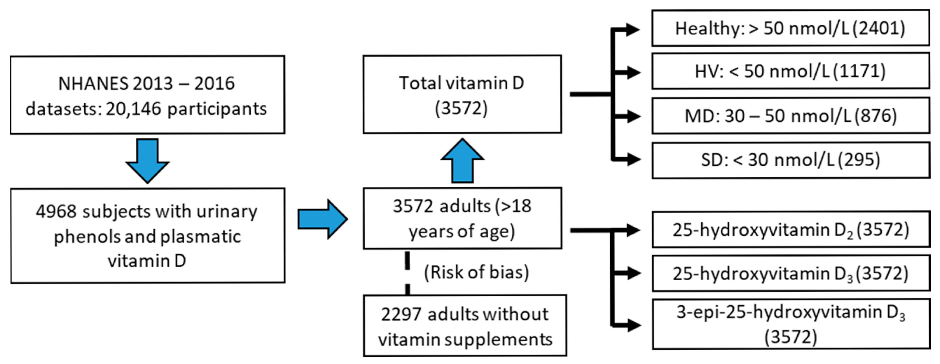
Figure 1. Schematic representation of the selection and grouping process of the data extracted from the NHANES 13–16 cohort. Abbreviations: HV, hypovitaminosis; MD, moderate deficiency; SD, severe deficiency. Note that the number of subjects analyzed in the main vitamin D analysis was 3572 subjects, and that all had quantified levels of vitamin D2, D3, and epi-D3. However, the second subgroup analyzed, the subjects who did not take vitamin supplements, corresponds to 2297 subjects.
Greenhealth 01 00010 g001
Figure 2. Variation in urinary bisphenol concentrations according to vitamin D status. Each compound shows a distinct pattern. Data are presented as geometric means (95% CI) due to non-parametric distribution. Mann–Whitney test was used for comparisons between healthy and HV groups. Kruskal–Wallis test followed by Dunn’s post hoc test was used for comparisons among healthy, MD, and SD groups. * p ≤ 0.05; ** p ≤ 0.01; **** p ≤ 0.0001. Abbreviations: MD, moderate deficiency (30–50 nmol/L); SD, severe deficiency (<30 nmol/L).
Greenhealth 01 00010 g002
Table 1. Descriptive statistics according to vitamin D status.
HealthyHVMDSD
N2401 (67.22%)1171 (32.78%)876 (24.52%)295 (8.26%)
Age46.27 (45.49–47.06)39.45 (38.53–40.4) d39.4 (38.32–40.51) d39.61 (37.84–41.47) d
Gender, % of men47.546.747.444.7
BMI, kg/m227.92 (27.68–28.16)29.54 (29.12–29.96) d29.33 (28.85–29.81) d30.18 (29.29–31.1) d
CKD, %10.94.94.26.8
DM, %17.817.616.421
Dyslipidemia, %45.831.732.928.1
Hypertension, %44.638.736.944.1
Smokers, %44.143.240.451.5
Albuminuria, %11.613.211.618
ACR, mg/g9.65 (9.23–10.1)10.37 (9.67–11.13)9.63 (8.93–10.39)12.92 (10.99–15.2) b,#
BPA, µg/g creat.1.17 (1.13–1.22)1.16 (1.1–1.22)1.15 (1.08–1.22)1.18 (1.06–1.32)
BPF, µg/g creat.0.44 (0.42–0.47)0.37 (0.34–0.4) d0.37 (0.33–0.4) d0.36 (0.31–0.42)
BPS, µg/g creat.0.49 (0.47–0.52)0.56 (0.52–0.6) b0.55 (0.51–0.6)0.59 (0.52–0.69) a
Results are expressed as a percentage or geometric mean (95% confidence interval). Note that the HV group includes both the MD and SD categories. Comparisons between healthy and HV were performed using the Mann–Whitney test. For comparisons among healthy, MD, and SD groups, the Kruskal–Wallis test followed by Dunn’s post hoc test was used. a p ≤ 0.05, b p ≤ 0.01, d p ≤ 0.0001 compared to the healthy group. # p ≤ 0.05 compared to the MD group. Abbreviations: HV, hypovitaminosis (<50 nmol/L); MD, moderate deficiency (30–50 nmol/L); SD, severe deficiency (<30 nmol/L); BMI, body mass index; CKD, chronic kidney disease; DM, diabetes mellitus; ACR, albumin-to-creatinine ratio; BPA, bisphenol A; BPF, bisphenol F; BPS, bisphenol S; Creat., creatinine.
Table 2. Binomial logistic regression with vitamin D status as the dichotomous dependent variable (healthy–hypovitaminosis).
AnalysisOR (95% CI)p-Value
BPA, µg/g creat. 110.98 (0.91–1.06)0.602
20.99 (0.91–1.07)0.747
30.98 (0.91–1.07)0.699
BPF, µg/g creat. 110.91 (0.86–0.95)0.000
20.92 (0.87–0.97)0.001
30.92 (0.87–0.97)0.001
BPS, µg/g creat. 111.09 (1.03–1.16)0.002
21.11 (1.05–1.18)0.001
31.10 (1.04–1.17)0.002
Three models were applied as follows: (1) unadjusted, (2) adjusted for age, sex, and BMI, and (3) fully adjusted for age, sex, BMI, diabetes, CKD, albuminuria, hypertension, dyslipidemia, and smoking. Abbreviations: OR, odds ratio; CI, confidence interval; BPA, bisphenol A; BPF, bisphenol F; BPS, bisphenol S; Creat., creatinine. 1 log-transformed.
Table 3. Multinomial logistic regression with vitamin D as the dependent variable (healthy–moderate deficiency–severe deficiency).
AnalysisHealthyModerate DeficiencySevere Deficiency
OR (95% CI)p-ValueOR (95% CI)p-ValueOR (95% CI)p-Value
BPA, µg/g creat. 11REF-0.97 (0.89–1.06)0.4981.01 (0.88–1.15)0.928
2REF-0.98 (0.90–1.07)0.6451.01 (0.88–1.16)0.895
3REF-0.98 (0.90–1.08)0.7190.99 (0.86–1.13)0.839
BPF, µg/g creat. 11REF-0.91 (0.86–0.96)0.0010.90 (0.82–0.98)0.021
2REF-0.92 (0.87–0.98)0.0050.91 (0.83–0.99)0.039
3REF-0.92 (0.87–0.98)0.0090.89 (0.81–0.98)0.015
BPS, µg/g creat. 11REF-1.08 (1.01–1.15)0.0201.14 (1.03–1.26)0.010
2REF-1.10 (1.03–1.18)0.0061.15 (1.04–1.28)0.006
3REF-1.09 (1.02–1.17)0.0091.13 (1.02–1.26)0.019
Three models were applied as follows: (1) unadjusted, (2) adjusted for age, sex, and BMI, and (3) fully adjusted for age, sex, BMI, diabetes, CKD, albuminuria, hypertension, dyslipidemia, and smoking. Abbreviations: OR, odds ratio; CI, confidence interval; BPA, bisphenol A; BPF, bisphenol F; BPS, bisphenol S; Creat., creatinine. 1 log-transformed.
Table 4. Linear regression between vitamin D metabolites and urinary bisphenol levels.
BPA, µg/g Creat. 1BPF, µg/g Creat. 1BPS, µg/g Creat. 1
Dependent Variableβp-Valueβp-Valueβp-Value
Total (25(OH)D2 + 25(OH)D3), nmol/L0.0000.9780.0600.000−0.0510.000
25(OH)D2, nmol/L0.0070.6830.0090.6100.0060.730
25(OH)D3, nmol/L−0.0040.8090.0570.001−0.0550.001
epi-25(OH)D3, nmol/L0.0050.7780.0300.076−0.0380.023
Abbreviations: BPA, bisphenol A; BPF, bisphenol F; BPS, bisphenol S; Creat., creatinine; 25(OH)D, 25-hydroxyvitamin D. 1 log-transformed.
Disclaimer/Publisher’s Note: The statements, opinions and data contained in all publications are solely those of the individual author(s) and contributor(s) and not of MDPI and/or the editor(s). MDPI and/or the editor(s) disclaim responsibility for any injury to people or property resulting from any ideas, methods, instructions or products referred to in the content.