Review

Mitochondrial Dysfunction and Glycolytic Shift in the Tumor Microenvironment: Impact on Paclitaxel Efficacy in Cancer Therapy

1
Department of Pharmaceutics, Smt. Kishoritai Bhoyar College of Pharmacy, Nagpur 440012, Maharashtra, India
2
Department of Pharmaceutical Sciences, Rashtrasant Tukdoji Maharaj Nagpur University, Nagpur 440033, Maharashtra, India
3
Department of Pharmaceutics, School of Pharmaceutical Education & Research (SPER), Jamia Hamdard, New Delhi 110062, India
*
Authors to whom correspondence should be addressed.
Clin. Bioenerg. 2025, 1(1), 5; https://doi.org/10.3390/clinbioenerg1010005
Submission received: 19 May 2025 / Revised: 26 June 2025 / Accepted: 3 July 2025 / Published: 9 July 2025

Abstract

Tumor cells often exhibit mitochondrial dysfunction and a pronounced glycolytic shift (the “Warburg effect”) that alters the tumor microenvironment. These metabolic changes, including mitochondrial DNA mutations and impaired oxidative phosphorylation, confer survival advantages and can reduce sensitivity to chemotherapeutics such as paclitaxel. In hypoxic environments, cancer cells upregulate glycolysis via HIF-1α, consequently lowering the extracellular pH through lactate secretion, which is associated with resistance to paclitaxel. Likewise, cancer-associated fibroblasts and immune cells undergo metabolic reprogramming in the tumor microenvironment. Glycolytic CAFs produce lactate and pyruvate that fuel tumor cells, reinforcing drug resistance, and tumor-driven polarization of macrophages toward an immunosuppressive M2 phenotype further impairs the anti-tumor response. Here, we review recent findings on how these metabolic adaptations attenuate paclitaxel efficacy and discuss strategies to overcome resistance. We highlight 15 key studies that reported cancer types, metabolic alterations, molecular targets, and outcomes related to paclitaxel response. Overall, the data suggest that targeting metabolic vulnerabilities, for example, by inhibiting glycolysis (HK2, PGAM1, and PDK) or modulating mitochondrial function, may restore paclitaxel sensitivity. Understanding metabolic crosstalk in the tumor microenvironment provides a basis for combined therapies that improve outcomes in paclitaxel-resistant cancers.

1. Introduction

Paclitaxel (PTX) (Taxol®), a microtubule-stabilizing taxane, induces mitotic arrest by binding β-tubulin subunits, disrupting spindle dynamics and triggering apoptosis in rapidly proliferating tumor cells [1]. Chemically defined as C47H51NO14, this hydrophobic diterpenoid requires Cremophor EL/ethanol solubilization, contributing to non-linear pharmacokinetics and hypersensitivity reactions in 30% of previously unmedicated patients. Dose-limiting toxicities include cumulative sensory neuropathy via axonal mitochondrial toxicity and neutropenia from bone marrow suppression. Critically, toxicity profiles exhibit age/gender stratification, with patients >65 years showing a higher neurotoxicity risk requiring dose reduction, while women experience disproportionately severe neuropathy linked to CYP3A4 metabolic dimorphism.
In addition, its clinical efficacy is further constrained by tumor metabolic reprogramming, a hallmark adaptation characterized by mitochondrial dysfunction (e.g., mtDNA mutations, electron transport chain impairment) and aerobic glycolysis (the Warburg effect) [2,3,4]. These alterations support biosynthetic precursor generation (i.e., ribose-5-phosphate, NADPH) and redox homeostasis (enhanced GSH), simultaneously desensitizing cancer cells to paclitaxel-induced mitochondrial apoptosis [5,6]. Critically, metabolic rewiring extends beyond tumor cells to shape the tumor microenvironment (TME), establishing chemoprotective niches.
The TME orchestrates multilayered resistance through hypoxia-inducible factor 1α (HIF-1α) activation in poorly vascularized regions. It transcriptionally upregulates glycolytic enzymes (e.g., HK2, LDHA) and inhibits pyruvate dehydrogenase via PDK1, enforcing glycolytic dependency. Concurrently, cancer-associated fibroblasts (CAFs) adopt a “reverse Warburg” phenotype, undergoing aerobic glycolysis to secrete lactate and pyruvate that fuel tumor oxidative phosphorylation (OXPHOS). This metabolic symbiosis acidifies the TME (pH 6.5–6.9), impairing paclitaxel uptake while promoting M2-like tumor-associated macrophage (TAM) polarization through lactate-mediated histone lactylation. Immune cell dysfunction follows as lactate-induced PD-L1 upregulation and Treg expansion further compromise cytotoxic responses, creating a multifaceted resistant ecosystem [7].
This review synthesizes advances from recent studies elucidating how metabolic adaptations across tumor, stromal, and immune compartments attenuate paclitaxel efficacy. We analyzed mitochondrial defects disrupting microtubule integrity and apoptosis, hypoxia-driven HIF-1α/PDK axis activation in glycolytic enforcement, CAF-mediated metabolic coupling and acidification, and immuno-metabolic suppression via TAM polarization. Finally, we evaluated emerging strategies to overcome resistance by targeting nodal vulnerabilities (e.g., HK2 inhibition, OXPHOS disruption, TME pH normalization), emphasizing biomarker-guided combinatorial approaches.
While existing reviews describe general links between metabolism and chemoresistance, few comprehensively integrate the roles of stromal components like CAFs and TAMs in paclitaxel-specific resistance. This review uniquely synthesizes recent evidence on how mitochondrial dysfunction, tumor glycolysis, and crucially, metabolic crosstalk with CAFs and TAMs, collectively drive paclitaxel resistance. We provide a targeted analysis of metabolic vulnerabilities exploitable for combination therapies, filling a gap in understanding the TME’s holistic impact on paclitaxel efficacy. Notably, clinical translation must address pharmacological constraints (i.e., solubility, toxicity) and patient factors (age/gender variability) and the fact that the current evidence derives predominantly from preclinical models lacking human TME complexity. Future clinical validation must address metabolic heterogeneity and compensatory pathway activation in human tumors.

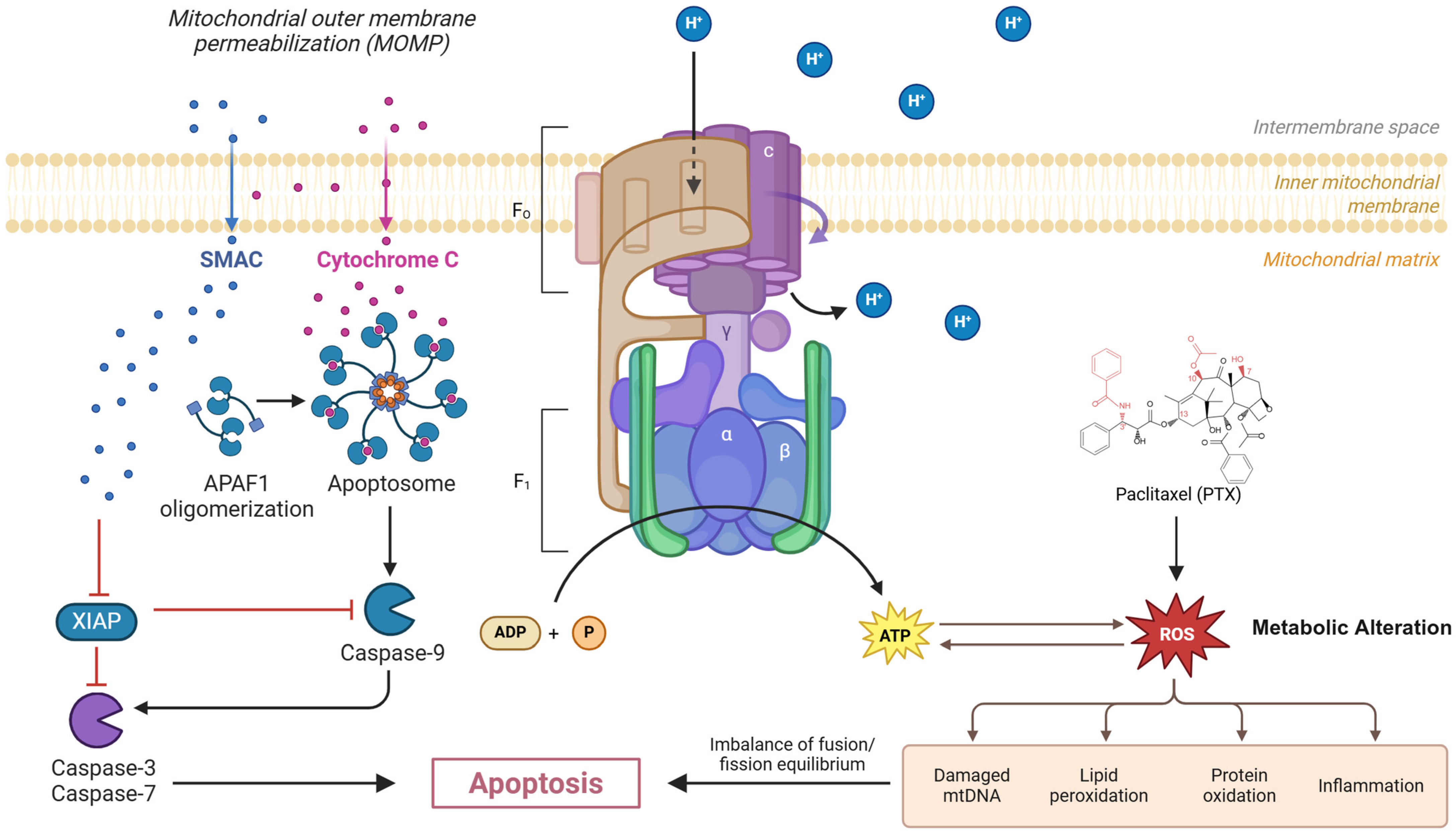
2. Mechanistic Insights

Paclitaxel’s cytotoxicity requires not only microtubule stabilization but also intact apoptotic pathways, which are influenced by cellular metabolism. In oxygenated (OXPHOS-active) cancer cells, paclitaxel induces mitochondrial stress (Figure 1): it increases reactive oxygen species (ROS) generation and releases cytochrome c, leading to apoptosis [8]. Penjweini et al. (2025) showed that even low-dose paclitaxel suppresses OXPHOS and ATP production in oxidative tumor cells, causing mitochondrial fragmentation and ROS accumulation [9]. Notably, glycolytic tumor cells (already relying on glycolysis) were less affected by these mitochondrial insults. Thus, a glycolytic phenotype may inherently protect cancer cells from paclitaxel-induced mitochondrial apoptosis. Conversely, paclitaxel itself can drive metabolic reprogramming: in breast cancer, prolonged paclitaxel treatment induces a shift from oxidative metabolism toward glycolysis, with reduced mitochondrial respiration [10,11]. This suggests a feed-forward resistance mechanism in which surviving cells increase glycolysis to evade drug toxicity.
A key factor in this metabolic switch is the regulation of glycolytic enzymes and their modulators. Upregulation of hexokinase 2 (HK2), phosphoglycerate mutase 1 (PGAM1), and pyruvate dehydrogenase kinases (PDK) has been linked to chemo-resistance [12]. For example, Tsai-Lin et al. found that paclitaxel-resistant ovarian clear cell carcinoma cells exhibit high HK2 expression and lactate production; targeted degradation of HK2 with a PROTAC partly restored paclitaxel sensitivity [13]. Similarly, PGAM1 promotes glycolysis in ovarian carcinoma: PGAM1-overexpressing cells produced excess pyruvate and lactate and were highly paclitaxel-resistant, whereas PGAM1 knockdown re-sensitized cells to the drug [14]. In lung cancer, paclitaxel-resistant cells upregulate PDK2, which inhibits pyruvate dehydrogenase (PDH) and forces glycolysis. Suppressing PDK2 via siRNA or dichloroacetate (a PDK inhibitor) reduced glycolysis and markedly re-sensitized resistant cells to paclitaxel [15]. These examples illustrate how overactive glycolysis is often triggered by upstream signals (e.g., HIF-1α, oncogenes) and fuels resistance.
Mitochondrial dysfunction also arises from genomic alterations. Deleterious mtDNA mutations (often induced by prior therapies) can reduce the efficiency of the electron transport chain and lower ATP output [16,17]. In a remarkable study, Girolimetti et al. showed that cisplatin-induced mtDNA mutations in an ovarian cancer model led to loss of filamentous (paclitaxel-target) microtubules and increased paclitaxel resistance [18]. This suggests that mtDNA lesions can indirectly promote chemo-resistance by altering both metabolism and cytoskeletal integrity. Impaired OXPHOS due to either mtDNA mutations or altered expression of mitochondrial fusion/fission proteins has been observed in chemo-resistant cells. Zhou et al. reported that paclitaxel-resistant A549 lung cancer cells have increased mitochondrial fusion (high Mfn1/2, low Fis1) and reduced OXPHOS capacity. These mitochondrial changes corresponded to diminished drug sensitivity [4].
Hypoxia is an important environmental driver of these metabolic shifts. Under low oxygen, HIF-1α stabilization transcriptionally upregulates GLUT1, HK2, LDHA, and PDK1, pushing cells toward glycolysis [19]. In breast cancer, hypoxia-induced HIF-1α activity has been directly linked to paclitaxel resistance via enhanced autophagy [20]. Recently, Liu et al. showed that the Nogo-B receptor (NgBR) augments HIF-1α–mediated glycolysis in ER-positive breast cancer; NgBR knockdown lowered HIF-1α, reduced glycolysis, and enhanced paclitaxel-induced apoptosis [21]. These studies underscore how hypoxic and signaling networks converge on metabolic pathways to modulate the drug response.
The net effect of these metabolic adaptations is a tumor cell less vulnerable to paclitaxel’s actions. Glycolytic cells generate high NADPH and GSH, buffering ROS and reducing apoptosis. Acidic lactate-rich microenvironments can impair drug uptake and promote drug efflux transporters. Additionally, a high glycolytic flux means more substrates for anabolic repair and survival pathways [22]. In summary, mitochondrial dysfunction (lowered OXPHOS) and a glycolytic shift create a chemo-resistant phenotype by altering redox balance, energy supply, and signaling pathways critical for paclitaxel’s cytotoxicity.
Paclitaxel (PTX) disrupts mitochondrial homeostasis by inducing excessive reactive oxygen species (ROS) production, leading to oxidative damage, lipid peroxidation, protein oxidation, inflammation, and mitochondrial DNA (mtDNA) mutations. These events impair ATP production through disruption of the mitochondrial electron transport chain and ATP synthase (F0F1 complex). ROS accumulation triggers mitochondrial outer membrane permeabilization (MOMP), causing the release of cytochrome c and SMAC into the cytosol. Cytochrome c promotes apoptosome formation through APAF1 oligomerization, which activates caspase-9 and downstream effector caspases-3 and -7, culminating in apoptosis. SMAC inhibits XIAP, removing its suppression of caspases. Metabolic alterations, including mtDNA damage and fusion/fission imbalance, further sensitize cells to paclitaxel-induced cytotoxicity.

3. Metabolic Drivers of Paclitaxel Resistance in Major Cancers

Recent experimental evidence highlights that metabolic reprogramming significantly influences PTX response across cancer types. Table 1 outlines shared metabolic pathways like glycolysis, OXPHOS, and lipid metabolism across ovarian, breast, and NSCLC cancers. Table 2 details glycolysis-associated metabolic alterations and their impact on PTX resistance, while Table 3 focuses on mitochondrial and lipid metabolism-driven resistance mechanisms. Together, these findings emphasize the therapeutic potential of targeting metabolic vulnerabilities to improve PTX efficacy.
Table 1. Similar metabolic pathways in different cancer types.
Metabolic PathwayOvarianBreast (TNBC)NSCLCCross-Cancer Relevance
Glycolytic fluxHK2 ↑, PGAM1 ↑Baseline glycolysis ↑PDK2 ↑Universal resistance driver
OXPHOS dependenceResidual disease ↑Contextual vulnerability
Lipid metabolismCPT1A ↑, FASN ↑FASN ↑, CPT1 ↓Hypoxia-associated adaptation
Hypoxia responseIndirectIndirectHIF-1α directNSCLC > ovarian/breast
Abbreviation: ↑: Increase; ↓: Decrease.
Table 2. Glycolysis-Related Metabolic Alterations Impacting Paclitaxel (PTX) Response.
Cancer TypeModel SystemMetabolic AlterationImpact on PTX EfficacyKey Molecular Target/PathwayStrategy TestedOutcomeKey FindingRef
Ovarian clear cell carcinoma (OCCC)OCCC cell lines (resistant vs. parental) [in vitro]Increased HK2-driven glycolysis (decreased OCR/ATP, increased NADH/NAD+)PTX resistance (reduced apoptosis)HK2HK2-PROTAC degraderReduced glycolysis, partial restoration of PTX sensitivityHK2 degradation partially restores PTX sensitivity[13]
Triple-negative breast cancer (TNBC)TNBC cell lines, patient organoids [in vitro/ex vivo]PTX-induced shift to enhanced glycolysis (decreased OXPHOS)Enhanced glycolysis correlates with residual disease; chemoresistanceGlycolytic enzymes (general)Glycolysis inhibitors (e.g., 2-DG)Markedly improved PTX response in organoidsGlycolysis inhibition improves PTX response[3]
Ovarian carcinomaSKOV3/SKOV3-TR30 cells [in vitro]Increased PGAM1 → increased pyruvate/lactate productionSKOV3-TR30 has higher PTX resistancePGAM1 (glycolysis)PGAM1 siRNA/inhibitorDecreased glycolysis, decreased PTX resistancePGAM1 inhibition decreases PTX resistance[14]
Epithelial ovarian cancer (EOC)SKOV3-R, A2780-R cells and xenografts [in vitro/vivo]Increased KHDRBS3 → increased glycolysis (via MIR17HG/CLDN6)KHDRBS3 upregulated in PTX-R cells; drives resistanceKHDRBS3/CLDN6KHDRBS3 siRNADecreased glycolysis, restored PTX sensitivity in vitro and in vivoKHDRBS3 knockdown restores PTX sensitivity[23]
ER-positive breast cancerMCF7 cells (NgBR high vs. knockdown) [in vitro]NgBR increased → increased HIF-1α, increased glycolysisNgBR expression promotes PTX resistanceNgBR/HIF-1αNgBR knockdownDecreased glycolysis, increased PTX-induced apoptosisNgBR knockdown increases PTX sensitivity[21]
Various (osteosarcoma, NSCLC)Mouse xenografts (MV522, MG63) [in vivo]Hypoxia → increased glycolysis (Warburg phenotype)Glycolytic tumors resistant; combination neededGlycolysis pathway2-Deoxy-D-glucose (2-DG) + PTXSignificantly slower tumor growth vs. PTX aloneGlycolysis inhibitor (2-DG) enhances PTX efficacy in vivo[22]
NSCLC (A549-R)A549-PTX cells [in vitro]Increased PDK2 → enhanced glycolysis, inhibited PDHPDK2 overexpression in PTX-R cellsPDK2PDK2 siRNA; DCA (PDK inhibitor)Decreased glycolysis, increased apoptosis; DCA + PTX synergyPDK2 inhibition/DCA restores PTX sensitivity[15]
Abbreviations: PTX, Paclitaxel; OCR, Oxygen Consumption Rate; ATP, Adenosine Triphosphate; NADH, Nicotinamide Adenine Dinucleotide (Reduced); NAD+, Nicotinamide Adenine Dinucleotide (Oxidized); OXPHOS, Oxidative Phosphorylation; HK2, Hexokinase 2; PROTAC, PROteolysis TArgeting Chimera; 2-DG, 2-Deoxy-D-glucose; PGAM1, Phosphoglycerate Mutase 1; siRNA, small interfering RNA; KHDRBS3, KH RNA Binding Domain Containing, Signal Transduction Associated 3; CLDN6, Claudin 6; HIF-1α, Hypoxia-Inducible Factor 1-alpha; NgBR, Nogo-B Receptor; PDK2, Pyruvate Dehydrogenase Kinase 2; PDH, Pyruvate Dehydrogenase; DCA, Dichloroacetate; NSCLC, Non-Small Cell Lung Cancer; TNBC, Triple-Negative Breast Cancer; EOC, Epithelial Ovarian Cancer; OCCC, Ovarian Clear Cell Carcinoma.

3.1. Ovarian Cancer: Glycolytic and Lipid Metabolism Dominance

In ovarian cancer, multiple studies highlight glycolytic enzymes as resistance drivers. The PGAM1 study showed that SKOV3-TR30 (paclitaxel-resistant) cells had elevated glycolytic flux and lowered mitochondrial function compared to parental SKOV3i. PGAM1 knockdown or inhibition lowered pyruvate/lactate production and significantly decreased resistance (improving paclitaxel efficacy) [14]. Similarly, Wu et al. found that an RNA-binding protein, KHDRBS3, is upregulated in paclitaxel-resistant SKOV3 and A2780 ovarian cancer cells. KHDRBS3 promoted glycolysis via a MIR17HG/CLDN6 axis; its knockdown reduced glycolysis and restored paclitaxel sensitivity both in vitro and in xenografts [23]. These results link oncogenic signals to metabolic remodeling and chemo-resistance. In line with this, Ma et al. analyzed paclitaxel-resistant A2780 cells and identified key lipid metabolism enzymes—CPT1A (fatty acid oxidation), SCD (monounsaturated FA synthesis), and FASN (lipid synthesis)—as overexpressed in resistant cells. Pharmacologic inhibition of CPT1A or SCD decreased viability and increased apoptosis in A2780/PTX cells, markedly sensitizing them to paclitaxel [24]. Together, these studies suggest that targeting metabolic enzymes (glycolytic or lipid-related) can reverse drug resistance.

3.2. Breast Cancer: Metabolic Heterogeneity Across Subtypes

In breast cancer, the metabolic phenotype also predicts the paclitaxel response. Derouane et al. used triple-negative breast cancer (TNBC) lines and patient-derived organoids to show that paclitaxel treatment induces a metabolic switch toward glycolysis, with concomitant reduction in mitochondrial OXPHOS. Importantly, TNBC tumors from patients who did not respond to neoadjuvant taxane-based chemotherapy had higher glycolysis signatures than responders, implying that preexisting glycolytic reliance confers resistance. Pharmacologic blockade of glycolysis in TNBC organoids greatly improved the response to paclitaxel + epirubicin [3]. Correspondingly, Evans et al. profiled TNBC patient biopsies and PDX models and found that tumors with high OXPHOS gene expression had worse long-term outcomes after taxane therapy, whereas OXPHOS inhibition (with IACS-10759) stabilized growth of TNBC PDX tumors in vivo [25]. These findings indicate that even in TNBC—often considered highly glycolytic—residual disease cells may depend on mitochondrial respiration, and blocking OXPHOS can enhance chemotherapy efficacy.

3.3. NSCLC: Hypoxia Induced Metabolic Rewiring

In lung cancer, hypoxia and lipid metabolism are prominent. Under chronic hypoxia, NSCLC cells upregulate HIF-1α-dependent lipogenic enzymes (e.g., FASN, ADRP) and downregulate CPT1 (involved in fatty acid oxidation), reducing FA utilization [26,27]. Guo et al. showed that a natural compound, FV-429, can reverse this metabolic state. FV-429 activated fatty acid oxidation and suppressed lipid accumulation in hypoxic NSCLC cells, thereby restoring paclitaxel-induced G2/M arrest and enhancing drug sensitivity. This demonstrates that reprogramming lipid metabolism (via the HIF-1α pathway) can overcome hypoxia-driven paclitaxel resistance [28]. In NSCLC also, Sun et al. noted that paclitaxel-resistant A549 cells express high levels of pyruvate dehydrogenase kinase-2 (PDK2), enforcing glycolysis. PDK2 knockdown reduced glycolysis and re-sensitized cells to paclitaxel; similarly, co-treatment with the PDK inhibitor dichloroacetate (DCA) synergistically killed resistant cells. Thus, blockers of glycolysis regulation can potentiate paclitaxel [15].

3.4. TME Metabolic Crosstalk

Beyond tumor cells, components of the TME also influence paclitaxel action. CAFs frequently adopt a glycolytic program and secrete metabolites that shape the drug response. In breast cancer, CAFs can be induced by tumor-derived signals (e.g., estrogen/GPER) to increase glycolysis, producing lactate and pyruvate that feed cancer cells [29]. The resulting “metabolic coupling” increases mitochondrial activity in cancer cells and contributes to multidrug resistance [30]. Notably, lactate from CAFs acidifies the TME, and this acidic microenvironment has been directly associated with resistance to paclitaxel (and other drugs) in breast cancer cells [31]. Experimentally, CAF–cancer co-cultures or conditioned media often show that CAFs promote epithelial–mesenchymal transition (EMT) and survival of cancer cells through cytokine signaling (e.g., IL-6/STAT3) that ultimately leads to chemotherapy resistance. Indeed, Wang et al. demonstrated that ovarian CAFs secrete IL-6, which activates STAT3 and EMT in nearby cancer cells, inhibiting apoptosis and conferring paclitaxel resistance [32]. In prostate and pancreatic cancers, interactions between tumor-associated macrophages (TAMs) and CAFs further drive resistance. For example, CAF-derived IGF-1/2 was shown to promote paclitaxel resistance in pancreatic/breast models [33,34,35].
Immune cells are similarly subject to TME metabolic reprogramming. Hypoxia and high lactate bias macrophages toward an immunosuppressive M2-like state (relying on fatty acid oxidation and oxidative metabolism) [36]. M2-like TAMs produce IL-10 and upregulate Bcl-2 via STAT3, actions that have been shown to blunt paclitaxel efficacy in breast cancer [37]. Indeed, paclitaxel-resistant ovarian cancer cells secrete CCL2, recruiting M2-TAMs in the TME and creating a feedback loop of resistance [38]. Conversely, shifting macrophages to an M1 phenotype (or blocking M2 polarization) can reduce paclitaxel resistance. Collectively, these studies indicate that metabolic interplay between cancer, fibroblasts, and immune cells in the hypoxic TME creates an environment that protects tumors from paclitaxel.
Table 3. Mitochondrial/Lipid Metabolism-Related Alterations Impacting Paclitaxel (PTX) Response.
Cancer TypeModel SystemMetabolic AlterationImpact on PTX EfficacyKey Molecular Target/PathwayStrategy TestedOutcomeKey FindingRef
NSCLC (A549)A549 vs. A549-PTX cells [in vitro]Increased mitochondrial fusion (Mfn ↑, Fis1 ↓), Decreased membrane potential, impaired OXPHOSCorrelated with PTX resistanceMito. fusion/fission (Mfn, Fis1)ObservationalCorrelated with reduced PTX sensitivityMitochondrial dysfunction correlates with PTX resistance[4]
TNBC PDX modelsPatient-derived xenografts (BL1 subtype) [in vivo]High OXPHOS gene signature in resistant tumorsHigh OXPHOS signature → worse PTX outcomeMito. ETC complexesOXPHOS inhibitor (IACS-10759) ± combos (e.g., CDK4i)Tumor growth stabilized; combos enhance responseOXPHOS inhibition stabilizes tumors, combos enhance PTX efficacy[25]
General cancer (incl. TNBC)Human cancer cell lines [in vitro]Paclitaxel → mitochondrial dysfunction in OXPHOS cells (increased ROS, cytochrome c release)OXPHOS cells sensitive; glycolytic cells less affectedMitochondrial complex I/IIIObservationalOxidative cells sensitive to Taxol; glycolytic cells less affectedOXPHOS dependency correlates with PTX sensitivity[9]
Ovarian cancerBr22i cell clones [in vitro]Platinum-induced mtDNA mutations → decreased OXPHOS, disrupted tubulinmtDNA mutations confer PTX resistancemtDNA-encoded ETC proteinsObservationalmtDNA mutations confer PTX resistanceAcquired mtDNA defects cause PTX resistance[18]
TNBC persistent cellsMDA-MB-231 persistent post-chemo [in vitro]Increased reliance on pyruvate-driven OXPHOS (TCA upregulated)Chemo-persistent cells less PTX-sensitiveMito. pyruvate carrier (MPC)MPC inhibitor (UK-5099)Decreased OXPHOS, re-sensitized cells to chemotherapyInhibiting pyruvate import re-sensitizes persistent cells[39]
NSCLC (hypoxia-resistant)A549 hypoxia-selected cells [in vitro]Hypoxia: Increased FASN/ADRP, decreased CPT1 (increased lipid uptake, decreased oxidation)Hypoxia-induced cells resist PTX (G2/M arrest hampered)HIF-1α/FASN/CPT1 (FA metabolism)FV-429 (wogonin analog)Reprogrammed FA metabolism, restored PTX sensitivityTargeting hypoxia-induced lipid metabolism restores PTX sensitivity[28]
Breast cancer (4T1 in mice)4T1 tumor with PTX-albumin [in vivo]Targeting TAM mitochondrial metabolism in lung metastasis microenvironmentChemo-resistant lung metastases microenvironmentMitochondrial complex I (via TAM)TPP-TAM (mito-targeted AMPK activator) + PTX@AlbEnhanced PTX uptake, T cell infiltration, and tumor killingTargeting TAM mitochondrial metabolism enhances PTX efficacy in metastasis[40]
Ovarian cancer (A2780)A2780-PTX resistant line [in vitro]Increased CPT1A, increased FASN, increased SCD (enhanced lipid synthesis/β-oxidation)Lipid-rich metabolism drives PTX resistanceCPT1A, FASN, SCD (Lipid metabolism)Inhibitors of CPT1A, SCD, FASNDecreased viability, increased apoptosis; sensitized to PTXInhibiting lipid metabolism sensitizes to PTX[41]
Abbreviations: PTX, Paclitaxel; OXPHOS, Oxidative Phosphorylation; ETC, Electron Transport Chain; Mfn, Mitofusin; Fis1, Mitochondrial Fission 1 Protein; mtDNA, Mitochondrial DNA; ROS, Reactive Oxygen Species; MPC, Mitochondrial Pyruvate Carrier; TCA, Tricarboxylic Acid Cycle; HIF-1α, Hypoxia-Inducible Factor 1-alpha; FASN, Fatty Acid Synthase; ADRP, Adipose Differentiation-Related Protein; CPT1, Carnitine Palmitoyltransferase 1; FA, Fatty Acid; SCD, Stearoyl-CoA Desaturase; TAM, Tumor-Associated Macrophage; AMPK, AMP-activated Protein Kinase; TPP, Triphenylphosphonium; PDX, Patient-Derived Xenograft; CDK4i, CDK4/6 Inhibitor; TNBC, Triple-Negative Breast Cancer; NSCLC, Non-Small Cell Lung Cancer.
While glycolytic/lipid targets show efficacy in vitro (e.g., PGAM1 siRNA, SCD inhibitors), only 33% of interventions have progressed to in vivo validation (i.e., KHDRBS3 siRNA, IACS-10759, FV-429). Notably, no clinical trials yet have tested these combinations, highlighting a critical translational gap. Common resistance nodes emerge: (1) glycolytic enzyme upregulation (i.e., HK2/PGAM1/PDK2) across carcinomas, (2) hypoxia-induced metabolic shifts, and (3) TME-mediated chemoprotection. Paradoxically, OXPHOS dependence in TNBC residual disease reveals cancer-specific metabolic heterogeneity requiring distinct therapeutic approaches

4. Integrated Resistance Mechanisms in the TME

The data reinforce that a glycolytic phenotype with impaired mitochondrial function underpins paclitaxel resistance. Mechanistically, this occurs because paclitaxel primarily induces apoptosis by stabilizing microtubules, triggering mitochondrial outer membrane permeabilization (MOMP) and cytochrome c release. Reduced OXPHOS capacity stemming from mtDNA mutations or fission/fusion imbalance directly compromises this apoptotic pathway, rendering cancer cells insensitive to paclitaxel’s microtubule-stabilizing action. Conversely, a high glycolytic flux provides survival advantages: it generates NADPH for antioxidant defense, countering paclitaxel-induced reactive oxygen species, and supplies biosynthetic precursors for damage repair. Interventions restoring mitochondrial function or inhibiting glycolysis can reverse resistance by re-enabling apoptosis execution [3,42,43,44,45].
Fibroblast-mediated metabolic crosstalk significantly amplifies resistance. CAFs undergoing a “reverse Warburg effect” secrete lactate and pyruvate [41,46]. This lactate fuels adjacent cancer cell OXPHOS (Figure 2) under nutrient stress but also has direct detrimental effects. Extracellular lactate, transported into cancer cells via monocarboxylate transporters (MCTs), contributes to intracellular acidification. Furthermore, the resulting extracellular acidosis impairs paclitaxel uptake and can directly disrupt microtubule polymerization dynamics, hindering the drug’s target engagement [47]. CAF-derived lactate and the acidic microenvironment also promote survival signaling. Critically, CAF-secreted cytokines like IL-6 induce STAT3-driven EMT and inhibit pro-apoptotic proteins, further suppressing paclitaxel-induced cell death in ovarian cancer models [32]. This illustrates the intricate link between CAF-cancer metabolic exchange (lactate shuttle) and pro-survival signaling pathways.
Hypoxia within the TME exerts a dual effect, directly forcing tumor cells toward glycolysis via HIF-1α stabilization and altering stromal behavior [7,48,49,50]. The resulting lactate production and acidosis, as noted, impair drug efficacy. Hypoxia and lactate also cripple anti-tumor immunity by impairing glucose-dependent effector T cells and promoting the polarization of tumor-associated macrophages (TAMs) toward an M2-like, immunosuppressive phenotype reliant on fatty acid oxidation (FAO) [51,52]. These M2-TAMs secrete IL-10, further suppressing immunity and creating a protective niche that shields tumor cells from paclitaxel. Thus, metabolic competition where cancer cells avidly consume glucose, CAFs generate lactate, and immune cells are starved collectively fosters a resistant TME [53].
Beyond chemotherapy resistance, mitochondrial dysfunction and glycolytic shifts also shape immunotherapy sensitivity. Tumor cells with impaired OXPHOS often upregulate immune checkpoint proteins (e.g., PD-L1) via HIF-1α/STAT3 pathways, while lactate from glycolytic tumors suppresses cytotoxic T-cell function and promotes regulatory T-cell infiltration. This metabolic–immune crosstalk may explain why highly glycolytic tumors resist checkpoint blockade.
Importantly, metabolic reprogramming is highly dynamic and context-dependent, enabling tumors to evade therapy. Cancer cells possess significant plasticity to switch between dominant metabolic pathways (e.g., glycolysis, FAO, glutaminolysis) when challenged. For instance, persistent triple-negative breast cancer (TNBC) cells surviving combination chemotherapy were found to increase OXPHOS dependence on mitochondrial pyruvate; blocking mitochondrial pyruvate uptake reversed this adaptation and sensitized the cells. This demonstrates that even initially glycolytic cells can shift to oxidative metabolism under pressure. Similarly, inhibiting one pathway (e.g., glycolysis) can induce compensatory reliance on another (e.g., FAO or glutaminolysis) [25].
Consequently, tumors characterized by mitochondrial dysfunction and a glycolytic, acidic TME exhibit reduced paclitaxel responsiveness [54]. Strategies to overcome this include improving perfusion (reoxygenation), buffering acidosis, or directly targeting critical metabolic pathways or their regulators (e.g., HIF-1α, PDK). As demonstrated in Table 1, combining paclitaxel with metabolic inhibitors (e.g., 2-deoxy-D-glucose, dichloroacetate) often yields synergistic effects by forcing cells out of the resistant glycolytic state or blocking compensatory pathways [15,22].
Glucose uptake via GLUT transporters enhances mitochondrial OxPhos, leading to an elevated intracellular ATP/ADP ratio. This increase inhibits KATP channels and regulates calcium influx through voltage-gated calcium channels, supporting mitochondrial activity and cell survival. During anticancer therapy, these metabolic adaptations help a subset of tumor cells survive. Disseminating tumor cells (DTCs) may intravasate into blood vessels, extravasate into distant tissues, and initiate secondary tumors. Following therapy withdrawal, metabolic reprogramming, particularly increased reliance on mitochondrial metabolism and OxPhos, facilitates tumor relapse.

5. Metabolic Targeting Strategies

Given these insights, novel therapeutic approaches aim to target metabolic vulnerabilities in tandem with paclitaxel. One strategy is direct inhibition of glycolytic enzymes or regulators. HK2 inhibitors (or degraders) are under investigation; the HK2-PROTAC study indicates this could restore paclitaxel sensitivity [55,56]. Similarly, inhibiting PGAM1 or PDK2 (as in the ovarian and lung studies) interrupts the glycolytic flux that supports resistance [57]. The use of clinically available drugs like 2-DG or analogs of pyruvate kinase inhibitors could be explored in combination with taxanes. Targeting mitochondrial function is another avenue. OXPHOS inhibitors (e.g., IACS-10759, BAY 87-2243) have shown promise in TNBC and might be combined with paclitaxel to attack residual oxidative cells [58]. Mitochondria-targeted compounds (like triphenylphosphonium-conjugated agents) are being developed. The Acta Pharm Sin B study used TPP-liganded tamoxifen nanoparticles to block complex I in tumor cells, which enhanced albumin-bound paclitaxel accumulation and T-cell infiltration in a breast cancer model [40]. Such “nanoadjuvants” that disrupt tumor mitochondrial metabolism have the potential to transform the TME into one more permissive for chemo- and immunotherapy.
Modulation of lipid metabolism is also promising. The results of the CPT1A/FASN/SCD study suggest that adding fatty acid oxidation inhibitors (e.g., etomoxir for CPT1A) or FASN inhibitors could resensitize resistant ovarian tumors to paclitaxel [24]. In NSCLC, agents like FV-429 that reverse hypoxia-driven lipogenesis restored the drug response. Statins or other lipid-lowering drugs could similarly be repurposed as chemo-sensitizers [28,59]. Finally, metabolic interventions can boost immune-mediated effects of paclitaxel. Since glycolysis supports regulatory immune cells, combining paclitaxel with immuno-metabolic therapies may help. For example, inhibiting IDO (tryptophan metabolism) or adenosine signaling in the TME could relieve metabolic suppression of T cells. Targeting TAM metabolism (e.g., with CSF-1R inhibitors or PI3Kγ inhibitors) is under investigation in combination with taxanes. Overall, there is a trend toward combining paclitaxel with agents that modulate hypoxia (e.g., VEGF inhibitors), normalize pH, or inhibit CAF-secreted factors (e.g., IL-6 antibodies) to disrupt the pro-resistance niche. Each strategy must be precisely matched to the tumor’s metabolic profile. Biomarkers such as PET imaging of glucose uptake, measurement of lactate in the TME, or gene signatures of metabolic enzymes could guide the use of metabolic inhibitors alongside paclitaxel [60]. Encouragingly, preclinical studies (Table 1) have shown that metabolic reprogramming agents can safely amplify paclitaxel’s efficacy, providing a rationale for clinical trials.

6. Conclusions

Mitochondrial dysfunction and a glycolytic shift in the tumor microenvironment act in concert to diminish paclitaxel efficacy. Importantly, several of these findings are supported by data (Table 1). Future cancer therapy may integrate metabolic reprogramming interventions with conventional chemotherapy by restoring mitochondrial function or starving tumor cells of glycolytic fuel to overcome paclitaxel resistance. Targeting metabolic vulnerabilities shows significant clinical promise. Tumors with high glycolytic signatures (e.g., basal-like TNBC, hypoxic ovarian carcinomas) may benefit most from glycolysis inhibitors (i.e., 2-DG, HK2/PGAM1/PDK blockers) combined with paclitaxel. Conversely, OXPHOS-dependent tumors (e.g., BL1 TNBC PDX models) or those with lipid-rich metabolism (e.g., CPT1A/FASN-high ovarian cancers) may respond better to OXPHOS inhibitors (i.e., IACS-10759) or lipid metabolism modulators. High-risk subgroups include patients with mtDNA mutations (e.g., platinum-pre-treated ovarian cancer), HIF-1α overexpression, or TAM-rich microenvironments. Metabolic profiling (i.e., PET imaging, lactate levels, gene signatures) should guide personalized combined treatment. Such combination strategies, guided by tumor metabolic profiling, hold promise for improving outcomes in chemo-resistant cancers.

Author Contributions

Conceptualization: T.P., J.T., A.T., M.U. and M.Q. Literature Review and Writing Original Draft: T.P., J.T., A.T. and M.Q. Illustrations and Visualization: M.Q. Methodology and Data Curation: T.P., J.T., A.T. and M.Q. Writing Review and Editing: T.P., J.T., A.T., S.S., M.Q., U.M.H., R.K. and M.U. Supervision: M.U., A.T. and J.T. All authors have read and agreed to the published version of the manuscript.

Funding

This research received no external funding.

Institutional Review Board Statement

Not applicable.

Informed Consent Statement

All authors have read and approved the final manuscript and consent to its publication.

Data Availability Statement

Not applicable.

Conflicts of Interest

The authors have no competing interests to declare that are relevant to the content of this article.

Abbreviations

2-DG: 2-Deoxy-D-glucose; ADP: Adenosine Diphosphate; AMPK: AMP-Activated Protein Kinase; APAF1: Apoptotic Protease Activating Factor 1; ATP: Adenosine Triphosphate; CAFs: Cancer-Associated Fibroblasts; CaVGIC: Voltage-Gated Calcium Channel; CDK4i: Cyclin-Dependent Kinase 4/6 Inhibitor; CLDN6: Claudin 6; CPT1/CPT1A: Carnitine Palmitoyltransferase 1/Isoform A; DCA: Dichloroacetate; DTC: Disseminating Tumor Cell; EOC: Epithelial Ovarian Cancer; ETC: Electron Transport Chain; FA: Fatty Acid; FAO: Fatty Acid Oxidation; FASN: Fatty Acid Synthase; Fis1: Mitochondrial Fission 1 Protein; GSH: Glutathione; GLUT: Glucose Transporter; HIF-1α: Hypoxia-Inducible Factor 1 Alpha; HK2: Hexokinase 2; KATP: ATP-Sensitive Potassium Channel; KHDRBS3: KH RNA Binding Domain Containing, Signal Transduction Associated 3; LDHA: Lactate Dehydrogenase A; MCT: Monocarboxylate Transporter; Mfn: Mitofusin (e.g., Mfn1, Mfn2); MOMP: Mitochondrial Outer Membrane Permeabilization; MPC: Mitochondrial Pyruvate Carrier; mtDNA: Mitochondrial DNA; NADH: Nicotinamide Adenine Dinucleotide (Reduced); NAD+: Nicotinamide Adenine Dinucleotide (Oxidized); NgBR: Nogo-B Receptor; NSCLC: Non-Small Cell Lung Cancer; OXPHOS: Oxidative Phosphorylation; PDH: Pyruvate Dehydrogenase; PDK/PDK1/PDK2: Pyruvate Dehydrogenase Kinase/Isoforms 1 and 2; PGAM1: Phosphoglycerate Mutase 1; PTX: Paclitaxel; PDX: Patient-Derived Xenograft; PROTAC: PROteolysis TArgeting Chimera; ROS: Reactive Oxygen Species; SCD: Stearoyl-CoA Desaturase; siRNA: Small Interfering RNA; SMAC: Second Mitochondria-Derived Activator of Caspases; STAT3: Signal Transducer and Activator of Transcription 3; TAMs: Tumor-Associated Macrophages; TCA: Tricarboxylic Acid Cycle; TME: Tumor Microenvironment; TNBC: Triple-Negative Breast Cancer; TPP: Triphenylphosphonium; XIAP: X-linked Inhibitor of Apoptosis Protein.

References

  1. Smith, E.R.; Huang, M.; Schlumbrecht, M.P.; George, S.H.L.; Xu, X.X. Rationale for combination of paclitaxel and CDK4/6 inhibitor in ovarian cancer therapy—Non-mitotic mechanisms of paclitaxel. Front. Oncol. 2022, 12, 907520. [Google Scholar] [CrossRef] [PubMed]
  2. Heiden, M.G.V.; Cantley, L.C.; Thompson, C.B. Understanding the Warburg Effect: The Metabolic Requirements of Cell Proliferation. Science 2009, 324, 1029. [Google Scholar] [CrossRef] [PubMed]
  3. Derouane, F.; Desgres, M.; Moroni, C.; Ambroise, J.; Berlière, M.; Van Bockstal, M.R.; Galant, C.; van Marcke, C.; Vara-Messler, M.; Hutten, S.J.; et al. Metabolic adaptation towards glycolysis supports resistance to neoadjuvant chemotherapy in early triple negative breast cancers. Breast Cancer Res. 2024, 26, 29. [Google Scholar] [CrossRef] [PubMed]
  4. Zhou, X.; Li, R.; Chen, R.; Liu, J. Altered mitochondrial dynamics, biogenesis, and functions in the paclitaxel-resistant lung adenocarcinoma cell line A549/Taxol. Med. Sci. Monit. 2020, 26, e918216-1. [Google Scholar] [CrossRef]
  5. Rong, G.; Kang, H.; Wang, Y.; Hai, T.; Sun, H. Candidate Markers That Associate with Chemotherapy Resistance in Breast Cancer through the Study on Taxotere-Induced Damage to Tumor Microenvironment and Gene Expression Profiling of Carcinoma-Associated Fibroblasts (CAFs). PLoS ONE 2013, 8, e70960. [Google Scholar] [CrossRef]
  6. Feng, B.; Wu, J.; Shen, B.; Jiang, F.; Feng, J. Cancer-associated fibroblasts and resistance to anticancer therapies: Status, mechanisms, and countermeasures. Cancer Cell Int. 2022, 22, 166. [Google Scholar] [CrossRef]
  7. Peng, X.; Gong, F.; Chen, Y.; Jiang, Y.; Liu, J.; Yu, M.; Zhang, S.; Wang, M.; Xiao, G.; Liao, H. Autophagy promotes paclitaxel resistance of cervical cancer cells: Involvement of Warburg effect activated hypoxia-induced factor 1-α-mediated signaling. Cell Death Dis. 2014, 5, e1367. [Google Scholar] [CrossRef]
  8. Zhou, X.; An, B.; Lin, Y.; Ni, Y.; Zhao, X.; Liang, X. Molecular mechanisms of ROS-modulated cancer chemoresistance and therapeutic strategies. Biomed. Pharmacother. 2023, 165, 115036. [Google Scholar] [CrossRef]
  9. Penjweini, R.; Link, K.A.; Qazi, S.; Mattu, N.; Zuchowski, A.; Vasta, A.; Sackett, D.L.; Knutson, J.R. Low dose Taxol causes mitochondrial dysfunction in actively respiring cancer cells. J. Biol. Chem. 2025, 301, 108450. [Google Scholar] [CrossRef]
  10. Avolio, R.; Matassa, D.S.; Criscuolo, D.; Landriscina, M.; Esposito, F. Modulation of Mitochondrial Metabolic Reprogramming and Oxidative Stress to Overcome Chemoresistance in Cancer. Biomolecules 2020, 10, 135. [Google Scholar] [CrossRef]
  11. Lv, L.; Yang, S.; Zhu, Y.; Zhai, X.; Li, S.; Tao, X.; Dong, D. Relationship between metabolic reprogramming and drug resistance in breast cancer. Front. Oncol. 2022, 12, 942064. [Google Scholar] [CrossRef] [PubMed]
  12. Peng, J.; Cui, Y.; Xu, S.; Wu, X.; Huang, Y.; Zhou, W.; Wang, S.; Fu, Z.; Xie, H. Altered glycolysis results in drug-resistant in clinical tumor therapy. Oncol. Lett. 2021, 21, 369. [Google Scholar] [CrossRef] [PubMed]
  13. Lin, T.Y.; Gu, S.Y.; Lin, Y.H.; Shih, J.H.; Lin, J.H.; Chou, T.Y.; Lee, Y.C.; Chang, S.F.; Lang, Y.D. Paclitaxel-resistance facilitates glycolytic metabolism via Hexokinase-2-regulated ABC and SLC transporter genes in ovarian clear cell carcinoma. Biomed. Pharmacother. 2024, 180, 117452. [Google Scholar] [CrossRef] [PubMed]
  14. Feng, Y.; Zhang, X.; Zhang, S.; Xu, S.; Chen, X.; Zhou, C.; Xi, Y.; Xie, X.; Lu, W. PGAM1 Promotes Glycolytic Metabolism and Paclitaxel Resistance via Pyruvic Acid Production in Ovarian Cancer Cells. Front. Biosci.—Landmark 2022, 27, 262. [Google Scholar] [CrossRef]
  15. Sun, H.; Zhu, A.; Zhou, X.; Wang, F. Suppression of pyruvate dehydrogenase kinase-2 re-sensitizes paclitaxel-resistant human lung cancer cells to paclitaxel. Oncotarget 2017, 8, 52642–52650. [Google Scholar] [CrossRef]
  16. Zong, Y.; Li, H.; Liao, P.; Chen, L.; Pan, Y.; Zheng, Y.; Zhang, C.; Liu, D.; Zheng, M.; Gao, J. Mitochondrial dysfunction: Mechanisms and advances in therapy. Signal Transduct. Target. Ther. 2024, 9, 124. [Google Scholar] [CrossRef]
  17. van Gisbergen, M.W.; Voets, A.M.; Starmans, M.H.W.; de Coo, I.F.M.; Yadak, R.; Hoffmann, R.F.; Boutros, P.C.; Smeets, H.J.M.; Dubois, L.; Lambin, P. How do changes in the mtDNA and mitochondrial dysfunction influence cancer and cancer therapy? Challenges, opportunities and models. Mutat. Res. Rev. Mutat. Res. 2015, 764, 16–30. [Google Scholar] [CrossRef]
  18. Girolimetti, G.; Guerra, F.; Iommarini, L.; Kurelac, I.; Vergara, D.; Maffia, M.; Vidone, M.; Amato, L.B.; Leone, G.; Dusi, S.; et al. Platinum-induced mitochondrial DNA mutations confer lower sensitivity to paclitaxel by impairing tubulin cytoskeletal organization. Hum. Mol. Genet. 2017, 26, 2961–2974. [Google Scholar] [CrossRef]
  19. Basheeruddin, M.; Qausain, S. Hypoxia-Inducible Factor 1-Alpha (HIF-1α): An Essential Regulator in Cellular Metabolic Control. Cureus 2024, 16, e63852. [Google Scholar] [CrossRef]
  20. Škubník, J.; Pavlíčková, V.S.; Ruml, T.; Rimpelová, S. Autophagy in cancer resistance to paclitaxel: Development of combination strategies. Biomed. Pharmacother. 2023, 161, 114458. [Google Scholar] [CrossRef]
  21. Liu, C.; Li, S.; Zhang, X.; Jin, C.; Zhao, B.; Li, L.; Miao, Q.R.; Jin, Y.; Fan, Z. Nogo-B receptor increases glycolysis and the paclitaxel resistance of estrogen receptor-positive breast cancer via the HIF-1α-dependent pathway. Cancer Gene Ther. 2023, 30, 647–658. [Google Scholar] [CrossRef] [PubMed]
  22. Maschek, G.; Savaraj, N.; Priebe, W.; Braunschweiger, P.; Hamilton, K.; Tidmarsh, G.F.; De Young, L.R.; Lampidis, T.J. 2-Deoxy-D-glucose Increases the Efficacy of Adriamycin and Paclitaxel in Human Osteosarcoma and Non-Small Cell Lung Cancers in Vivo. Cancer Res. 2004, 64, 31–34. [Google Scholar] [CrossRef] [PubMed]
  23. Wu, X.; Qiu, L.; Feng, H.; Zhang, H.; Yu, H.; Du, Y.; Wu, H.; Zhu, S.; Ruan, Y.; Jiang, H. KHDRBS3 promotes paclitaxel resistance and induces glycolysis through modulated MIR17HG/CLDN6 signaling in epithelial ovarian cancer. Life Sci. 2022, 293, 120328. [Google Scholar] [CrossRef] [PubMed]
  24. Ma, Q.; Liu, Z.; Wang, T.; Zhao, P.; Liu, M.; Wang, Y.; Zhao, W.; Yuan, Y.; Li, S. Resensitizing Paclitaxel-Resistant Ovarian Cancer via Targeting Lipid Metabolism Key Enzymes CPT1A, SCD and FASN. Int. J. Mol. Sci. 2023, 24, 16503. [Google Scholar] [CrossRef]
  25. Evans, K.W.; Yuca, E.; Scott, S.S.; Zhao, M.; Arango, N.P.; Pico, C.X.C.; Saridogan, T.; Shariati, M.; Class, C.A.; Bristow, C.A.; et al. Oxidative phosphorylation is a metabolic vulnerability in chemotherapy-resistant triple-negative breast cancer. Cancer Res. 2021, 81, 5572–5581. [Google Scholar] [CrossRef]
  26. Bakleh, M.Z.; Zen, A.H. The Distinct Role of HIF-1α and HIF-2α in Hypoxia and Angiogenesis. Cells 2025, 14, 673. [Google Scholar] [CrossRef]
  27. Valli, A.; Rodriguez, M.; Moutsianas, L.; Fischer, R.; Fedele, V.; Huang, H.L.; Van Stiphout, R.; Jones, D.; Mccarthy, M.; Vinaxia, M.; et al. Hypoxia induces a lipogenic cancer cell phenotype via HIF1α-dependent and -independent pathways. Oncotarget 2014, 6, 1920. [Google Scholar] [CrossRef]
  28. Guo, Y.; Yang, L.; Guo, W.; Wei, L.; Zhou, Y. FV-429 enhances the efficacy of paclitaxel in NSCLC by reprogramming HIF-1α-modulated FattyAcid metabolism. Chem. Biol. Interact. 2021, 350, 109702. [Google Scholar] [CrossRef]
  29. Yu, T.; Yang, G.; Hou, Y.; Tang, X.; Wu, C.; Wu, X.A.; Guo, L.; Zhu, Q.; Luo, H.; Du, Y.E.; et al. Cytoplasmic GPER translocation in cancer-associated fibroblasts mediates cAMP/PKA/CREB/glycolytic axis to confer tumor cells with multidrug resistance. Oncogene 2017, 36, 2131–2145. [Google Scholar] [CrossRef]
  30. Tavares-Valente, D.; Baltazar, F.; Moreira, R.; Queirós, O. Cancer cell bioenergetics and pH regulation influence breast cancer cell resistance to paclitaxel and doxorubicin. J. Bioenerg. Biomembr. 2013, 45, 467–475. [Google Scholar] [CrossRef]
  31. Qiao, Y.; Liu, Y.; Ran, R.; Zhou, Y.; Gong, J.; Liu, L.; Zhang, Y.; Wang, H.; Fan, Y.; Fan, Y.; et al. Lactate metabolism and lactylation in breast cancer: Mechanisms and implications. Cancer Metastasis Rev. 2025, 44, 48. [Google Scholar] [CrossRef] [PubMed]
  32. Wang, L.; Zhang, F.; Cui, J.Y.; Chen, L.; Chen, Y.T.; Liu, B.W. CAFs enhance paclitaxel resistance by inducing EMT through the IL-6/JAK2/STAT3 pathway. Oncol. Rep. 2018, 39, 2081–2090. [Google Scholar] [CrossRef] [PubMed]
  33. Qin, Q.; Yu, R.; Eriksson, J.E.; Tsai, H.I.; Zhu, H. Cancer-associated fibroblasts in pancreatic ductal adenocarcinoma therapy: Challenges and opportunities. Cancer Lett. 2024, 591, 216859. [Google Scholar] [CrossRef] [PubMed]
  34. Gunaydin, G. CAFs Interacting with TAMs in Tumor Microenvironment to Enhance Tumorigenesis and Immune Evasion. Front. Oncol. 2021, 11, 668349. [Google Scholar] [CrossRef]
  35. Wang, W.; Cheng, B.; Yu, Q. Cancer-associated fibroblasts as accomplices to confer therapeutic resistance in cancer. Cancer Drug Resist. 2022, 5, 889. [Google Scholar] [CrossRef]
  36. Xiao, L.; Wang, Q.; Peng, H. Tumor-associated macrophages: New insights on their metabolic regulation and their influence in cancer immunotherapy. Front. Immunol. 2023, 14, 1157291. [Google Scholar] [CrossRef]
  37. He, Z.; Chen, D.; Wu, J.; Sui, C.; Deng, X.; Zhang, P.; Chen, Z.; Liu, D.; Yu, J.; Shi, J.; et al. Yes associated protein 1 promotes resistance to 5-fluorouracil in gastric cancer by regulating GLUT3-dependent glycometabolism reprogramming of tumor-associated macrophages. Arch. Biochem. Biophys. 2021, 702, 108838. [Google Scholar] [CrossRef]
  38. Wang, S.; Wang, J.; Chen, Z.; Luo, J.; Guo, W.; Sun, L.; Lin, L. Targeting M2-like tumor-associated macrophages is a potential therapeutic approach to overcome antitumor drug resistance. npj Precis. Oncol. 2024, 8, 31. [Google Scholar] [CrossRef]
  39. Winter, M.; Eldjoudi, A.N.; Guette, C.; Hondermarck, H.; Bourette, R.P.; Fovez, Q.; Laine, W.; Ghesquiere, B.; Adriaenssens, E.; Kluza, J.; et al. Mitochondrial adaptation decreases drug sensitivity of persistent triple negative breast cancer cells surviving combinatory and sequential chemotherapy. Neoplasia 2023, 46, 100949. [Google Scholar] [CrossRef]
  40. Zhou, Z.; Luo, W.; Zheng, C.; Wang, H.; Hu, R.; Deng, H.; Shen, J. Mitochondrial metabolism blockade nanoadjuvant reversed immune-resistance microenvironment to sensitize albumin-bound paclitaxel-based chemo-immunotherapy. Acta Pharm. Sin. B 2024, 14, 4087–4101. [Google Scholar] [CrossRef]
  41. Pavlides, S.; Whitaker-Menezes, D.; Castello-Cros, R.; Flomenberg, N.; Witkiewicz, A.K.; Frank, P.G.; Casimiro, M.C.; Wang, C.; Fortina, P.; Addya, S.; et al. The reverse Warburg effect: Aerobic glycolysis in cancer associated fibroblasts and the tumor stroma. Cell Cycle 2009, 8, 3984–4001. [Google Scholar] [CrossRef] [PubMed]
  42. Ganapathy-Kanniappan, S.; Geschwind, J.F.H. Tumor glycolysis as a target for cancer therapy: Progress and prospects. Mol. Cancer 2013, 12, 152. [Google Scholar] [CrossRef] [PubMed]
  43. Tatode, A.; Agrawal, P.R.; Taksande, J.; Qutub, M.; Premchandani, T.; Umekar, M.; Danao, K. Role of folate receptor and CD44 in targeting of docetaxel and paclitaxel fabricated conjugates for efficient cancer therapy. J. Med. Surg. Public Health 2025, 5, 100163. [Google Scholar] [CrossRef]
  44. Zheng, C.; Luo, W.; Liu, Y.; Chen, J.; Deng, H.; Zhou, Z.; Shen, J. Killing three birds with one stone: Multi-stage metabolic regulation mediated by clinically usable berberine liposome to overcome photodynamic immunotherapy resistance. Chem. Eng. J. 2023, 454, 140164. [Google Scholar] [CrossRef]
  45. Zhou, Z.; Liu, Y.; Jiang, X.; Zheng, C.; Luo, W.; Xiang, X.; Qi, X.; Shen, J. Metformin modified chitosan as a multi-functional adjuvant to enhance cisplatin-based tumor chemotherapy efficacy. Int. J. Biol. Macromol. 2023, 224, 797–809. [Google Scholar] [CrossRef]
  46. Akter, R.; Awais, M.; Boopathi, V.; Ahn, J.C.; Yang, D.C.; Kang, S.C.; Yang, D.U.; Jung, S.K. Inversion of the Warburg Effect: Unraveling the Metabolic Nexus between Obesity and Cancer. ACS Pharmacol. Transl. Sci. 2024, 7, 560. [Google Scholar] [CrossRef]
  47. Mishra, R.; Haldar, S.; Placencio, V.; Madhav, A.; Rohena-Rivera, K.; Agarwal, P.; Duong, F.; Angara, B.; Tripathi, M.; Liu, Z.; et al. Stromal epigenetic alterations drive metabolic and neuroendocrine prostate cancer reprogramming. J. Clin. Investig. 2018, 128, 4472–4484. [Google Scholar] [CrossRef]
  48. Zhang, D.; Wang, Y.; Shi, Z.; Liu, J.; Sun, P.; Hou, X.; Zhang, J.; Zhao, S.; Zhou, B.P.; Mi, J. Metabolic Reprogramming of Cancer-Associated Fibroblasts by IDH3α Downregulation. Cell Rep. 2015, 10, 1335–1348. [Google Scholar] [CrossRef]
  49. LaGory, E.L.; Giaccia, A.J. The Ever Expanding Role of HIF in Tumour and Stromal Biology. Nat. Cell Biol. 2016, 18, 356. [Google Scholar] [CrossRef]
  50. Kim, I.; Choi, S.; Yoo, S.; Lee, M.; Kim, I.S. Cancer-Associated Fibroblasts in the Hypoxic Tumor Microenvironment. Cancers 2022, 14, 3321. [Google Scholar] [CrossRef]
  51. Karimova, A.F.; Khalitova, A.R.; Suezov, R.; Markov, N.; Mukhamedshina, Y.; Rizvanov, A.A.; Huber, M.; Simon, H.U.; Brichkina, A. Immunometabolism of tumor-associated macrophages: A therapeutic perspective. Eur. J. Cancer 2025, 220, 115332. [Google Scholar] [CrossRef] [PubMed]
  52. Jin, X.; Zhang, N.; Yan, T.; Wei, J.; Hao, L.; Sun, C.; Zhao, H.; Jiang, S. Lactate-mediated metabolic reprogramming of tumor-associated macrophages: Implications for tumor progression and therapeutic potential. Front. Immunol. 2025, 16, 1573039. [Google Scholar] [CrossRef] [PubMed]
  53. Jiang, G.; Hong, J.; Sun, L.; Wei, H.; Gong, W.; Wang, S.; Zhu, J. Glycolysis regulation in tumor-associated macrophages: Its role in tumor development and cancer treatment. Int. J. Cancer 2024, 154, 412–424. [Google Scholar] [CrossRef] [PubMed]
  54. Cunha, A.; Rocha, A.C.; Barbosa, F.; Baião, A.; Silva, P.; Sarmento, B.; Queirós, O. Glycolytic Inhibitors Potentiated the Activity of Paclitaxel and Their Nanoencapsulation Increased Their Delivery in a Lung Cancer Model. Pharmaceutics 2022, 14, 2021. [Google Scholar] [CrossRef]
  55. Sang, R.; Fan, R.; Deng, A.; Gou, J.; Lin, R.; Zhao, T.; Hai, Y.; Song, J.; Liu, Y.; Qi, B.; et al. Degradation of Hexokinase 2 Blocks Glycolysis and Induces GSDME-Dependent Pyroptosis to Amplify Immunogenic Cell Death for Breast Cancer Therapy. J. Med. Chem. 2023, 66, 8464–8483. [Google Scholar] [CrossRef]
  56. He, K.; Tao, F.; Lu, Y.; Fang, M.; Huang, H.; Zhou, Y. The Role of HK2 in Tumorigenesis and Development: Potential for Targeted Therapy with Natural Products. Int. J. Med. Sci. 2025, 22, 790. [Google Scholar] [CrossRef]
  57. Aa, X.; Li, T.; Chen, N.; Wang, H.; Wang, X.; Ma, Y. PGAM1 regulates the glycolytic metabolism of SCs in tibetan sheep and its influence on the development of SCs. Gene 2021, 804, 145897. [Google Scholar] [CrossRef]
  58. Yap, T.A.; Ahnert, J.R.; Piha-Paul, S.A.; Fu, S.; Janku, F.; Karp, D.D.; Naing, A.; Dumbrava, E.E.I.; Pant, S.; Subbiah, V.; et al. Phase I trial of IACS-010759 (IACS), a potent, selective inhibitor of complex I of the mitochondrial electron transport chain, in patients (pts) with advanced solid tumors. J. Clin. Oncol. 2019, 37, 3014. [Google Scholar] [CrossRef]
  59. Jiang, W.; Hu, J.W.; He, X.R.; Jin, W.L.; He, X.Y. Statins: A repurposed drug to fight cancer. J. Exp. Clin. Cancer Res. 2021, 40, 241. [Google Scholar] [CrossRef]
  60. Lemberg, K.M.; Gori, S.S.; Tsukamoto, T.; Rais, R.; Slusher, B.S. Clinical development of metabolic inhibitors for oncology. J. Clin. Investig. 2022, 132, e148550. [Google Scholar] [CrossRef]
Figure 1. Paclitaxel-induced mitochondrial dysfunction promotes apoptosis via metabolic and oxidative stress pathways.
Clinbioenerg 01 00005 g001
Figure 2. Metabolic regulation of mitochondrial oxidative phosphorylation (OxPhos) in therapy response, dissemination, and relapse of cancer cells.
Clinbioenerg 01 00005 g002
Disclaimer/Publisher’s Note: The statements, opinions and data contained in all publications are solely those of the individual author(s) and contributor(s) and not of MDPI and/or the editor(s). MDPI and/or the editor(s) disclaim responsibility for any injury to people or property resulting from any ideas, methods, instructions or products referred to in the content.

