Article

Functional Profiling of Antimicrobial Resistance in Rabbit Gut Microbiome

by
Pitakthai Chamtim
1,*,
Nachon Raethong
2 and
Roypim Thananusak
3,4
1
Academic Service Unit, National Laboratory Animal Center, Mahidol University, Nakhon Pathom 73170, Thailand
2
Institute of Nutrition, Mahidol University, Nakhon Pathom 73170, Thailand
3
Omics Center for Agriculture, Bioresource, Food and Health Kasetsart University (OmiKU), Faculty of Science, Kasetsart University, Bangkok 10900, Thailand
4
Duckweed Holobiont Resource & Research Center (DHbRC), Faculty of Science, Kasetsart University, Bangkok 10900, Thailand
*
Author to whom correspondence should be addressed.
Acta Microbiol. Hell. 2025, 70(2), 21; https://doi.org/10.3390/amh70020021
Submission received: 8 April 2025 / Revised: 7 May 2025 / Accepted: 14 May 2025 / Published: 27 May 2025

Abstract

:
Antimicrobial resistance (AMR) is a growing concern in laboratory animals, where antibiotic use can disrupt the gut microbiome and promote resistant strains. Rabbits, commonly used in biomedical research, are particularly susceptible to gut dysbiosis, increasing the risk of infection and subsequent antibiotic use. This study used 16S rRNA amplicon sequencing and PICRUSt2 to predict resistance-related functions in rabbits under standard laboratory conditions. Among 2427 amplicon sequence variants, 42 functional classes were identified, with AMR comprising 1.55% of the predicted functions, including beta-lactam, vancomycin, and cationic antimicrobial peptide resistance. These AMR functions were associated with commensal genera such as Bacteroides and Ruminococcus, while no associations were observed with pathogens such as Escherichia or Salmonella. The study provides functional evidence of intrinsic AMR in the rabbit gut microbiome and underscores the importance of antimicrobial stewardship in laboratory animal research.

1. Introduction

Antimicrobial resistance (AMR) has emerged as one of the most critical global health threats, responsible for an estimated 1.27 million deaths worldwide in 2019 [1] and projected to cause up to 10 million deaths annually by 2050 [2]. While AMR is widely recognized for its impact on human health and food production, it also facilitates the animal-to-human transfer of resistant zoonotic pathogens, compounding the global burden. The overuse of antibiotics in livestock, both for treating infections and promoting growth, has accelerated the emergence of resistance reservoirs within animal populations [3]. Beyond livestock, the spread of AMR has become a significant issue impacting the health and welfare of animals, including both laboratory animals and livestock species raised for food production [4]. In intensive farming systems, AMR complicates the treatment and management of infectious diseases, making therapeutic approaches more challenging, complex, and costly, leading to increased morbidity and mortality, and causing substantial economic losses [5], as recently demonstrated in fattening bulls affected by Mycoplasma bovis respiratory infections [6]. Laboratory animal facilities and biomedical research laboratories are specialized environments that are susceptible to the accumulation and dissemination of resistant microorganisms [7]. This vulnerability arises from the frequent use of antibiotics, both to prevent and treat infections, as well as to minimize pathogen contamination. Additional factors include direct microbial transmission among laboratory animals, personnel, and equipment within contained animal housing facilities. Therefore, surveillance and monitoring of AMR functions in laboratory animals are vital for mitigating risks and preventing the spread of resistant organisms in these specialized environments [8,9]. In laboratory animals, the microbiome serves as an ecosystem of microorganisms residing within the body, particularly in the gastrointestinal tract, commonly referred to as the gut microbiome. The gut microbiome plays a crucial role in animal health, facilitating the digestion and absorption of nutrients while also modulating immune responses [10,11]. Changes in microbiome composition or diversity, such as a decline in beneficial microbes or an increase in antimicrobial-resistant bacteria, can lead to dysbiosis, which increases susceptibility to infectious diseases and impacts experimental outcomes [12,13].
New Zealand White (NZW) rabbits (Oryctolagus cuniculus) are among the most commonly used laboratory animals in biomedical research due to their appropriate size, rapid reproduction, gentle temperament, and ease of management [14]. However, rabbits are highly sensitive to environmental stress, and poor housing conditions can harm their welfare, leading to increased disease risk and behavioral issues. Key stressors include inadequate enrichment, overcrowding, and improper feeding, which can also affect gut microbiota and immune responses, impacting research results [15]. Dietary interventions, such as oregano supplementation, can enhance gut barrier function and immune status in rabbits [16], but the gut microbiome of rabbits is susceptible to microbial contamination from external environments, which can lead to gut dysbiosis, resulting in a weakened health status and increased susceptibility to infection [17]. Consequently, the therapeutic use of antibiotics may become necessary. However, the inappropriate or excessive use of antibiotics is a key factor that may select for resistant commensal bacteria in the gut microbiome [18,19]. These resistant commensal bacteria can then transfer resistance functions to pathogenic bacteria via horizontal gene transfer mechanisms [20,21]. Such a transfer can result in pathogenic bacteria acquiring resistance functions, permanently transforming them into resistant pathogens, thereby complicating future treatment of infections. Currently, limited information exists regarding the prevalence of AMR functions in the gut microbiome of rabbits.
Conventional techniques, such as culture-based methods or polymerase chain reaction (PCR) based assays, have inherent limitations in thoroughly characterizing microbial communities. Consequently, high-throughput sequencing technologies, particularly 16S rRNA amplicon sequencing, combined with advanced bioinformatics tools such as PICRUSt2 (Phylogenetic Investigation of Communities by Reconstruction of Unobserved States 2), have gained significant importance [22,23]. PICRUSt2 enables precise predictions of the functional potential and metabolic capabilities of microbial communities based on 16S rRNA sequencing data [24]. PICRUSt2-based functional prediction has been used to investigate gut microbial alterations following dietary interventions in humans, revealing shifts in pathways related to short-chain fatty acid production, amino acid biosynthesis, and inflammatory regulation under diets such as the Mediterranean and high-fiber regimens [25]. Additionally, PICRUSt2 has been employed to predict nitrogen and carbon cycling functions in soil microbiomes under different agricultural practices, highlighting enhanced nitrogen fixation, denitrification, and carbon sequestration in systems like no-till farming and crop rotation [26]. PICRUSt2 has also been applied to profile genes associated with osmoadaptation and seasonal variation in Tuz Lake, a hypersaline environment, uncovering shifts between potassium uptake and compatible solute accumulation strategies [27], and to explore the functional potential of bacterial communities in alfalfa silage harvested at different growth stages, where later-stage harvests improved fermentation quality and promoted carbohydrate and amino acid metabolism [28].
Given its broad utility in functional microbiome profiling, the present study aimed to predict and quantify AMR functions in the gut microbiome of NZW rabbits bred and maintained under standard laboratory conditions, using 16S rRNA amplicon sequencing coupled with PICRUSt2 analysis. Advancing predictive functional profiling in animal models will not only improve animal health and livestock productivity but also support the development of integrated AMR surveillance frameworks for humans, animals, and the environment, thereby supporting the One Health approach to safeguard global health and planetary sustainability.

2. Materials and Methods

2.1. Animal and Sample Collection

This study received approval from the Institutional Animal Care and Use Committee (IACUC) at the National Laboratory Animal Center (NLAC) under No. RA2023-21. Three male NZW rabbits were bred and reared from birth to 12 weeks (weighing 2.5–2.8 kg) at the production unit of NLAC. All animals were accompanied by an Animal Health Certificate verifying their genetic quality and health status. Then, all rabbits were raised for quarantine purposes after being transferred to the Quarantine room using aseptic methods. Briefly, during transfer, the rabbits were handled by personnel wearing sterile gloves, masks, and gowns; sterilized transport cages were used, direct contact with external environmental surfaces was minimized to prevent microbial contamination, and the animals were transported using a dedicated, pathogen-free experimental animal transport vehicle that met standard hygiene and cleanliness requirements. The rabbits were housed under controlled environmental conditions with a temperature of 20 ± 3 °C and a relative humidity of 30–70%, maintained by a Humidity Ventilation Air Conditioning system. A 12 h light/12 h dark cycle was implemented to regulate daily illumination. All rabbits had ad libitum access to sterilized pelleted feed and water. After a 24 h quarantine period, fecal pellets were collected from each rabbit that was healthy and showed no signs of disease at the time of sampling. All fecal samples were stored at −80 °C until further processing.

2.2. DNA Extraction and 16S rRNA Amplicon Sequencing

Fecal materials collected from individual rabbits were retrieved from frozen storage and homogenized into fine powder using sterile mortars and pestles inside aseptic containers. A subsample of approximately 0.25 g was accurately weighed and placed into tubes designated for DNA extraction. The DNeasy PowerSoil Pro Kit (Qiagen, Hilden, Germany) was utilized to extract total microbial genomic DNA from the fecal samples of each rabbit, following the manufacturer’s protocols. The DNA yield was quantified by measuring absorbance at 260 nm using a NanoDrop 2000 spectrophotometer (Thermo Fisher Scientific, Wilmington, DE, USA). To evaluate sample purity, absorbance ratios at 260/280 nm and 260/230 nm were calculated. A ratio close to 1.8–2.0 indicates low protein contamination, whereas a 260/230 ratio near 2.0–2.2 suggests minimal contamination from organic solvents or other inhibitors. DNA integrity was further assessed by running the extracts on a 1% agarose gel under an electric field of 100 volts for 30 min. DNA bands were visualized under UV light, and their migration patterns were compared with a molecular weight standard using a Gel Documentation System (Bio-Rad, Hercules, CA, USA). Only high-quality DNA samples showing distinct, intact bands were selected for downstream sequencing.
Library construction and sequencing for the 16S rRNA amplicon of the V3 and V4 regions were performed at Biomarker Technologies Co., Ltd. (Beijing, China). Briefly, genomic DNA extracted from each sample was amplified at the V3–V4 region of the 16S rRNA gene using the universal primers 338F (5′-ACTCCTACGGGAGGCAGCA-3′) and 806R (5′-GGACTACHVGGGTWTCTAAT-3′), which target conserved regions within bacterial genomes. Both the forward and reverse 16S primers were ligated with sample-specific Illumina index sequences. After amplification, the PCR products are analyzed by LabChip GX (Perkin Elmer, Waltham, MA, USA) for fragment analysis and integrity assessment. The qualified library was sequenced on the Illumina NovaSeq 6000 platform (Illumina, San Diego, CA, USA) with paired-end (PE) 250 bp mode. Raw imaging outputs from the high-throughput sequencing platform underwent base calling to convert signal intensities into nucleotide reads. The resulting raw reads, along with their corresponding quality scores, were stored in FASTQ files.

2.3. Bioinformatic Analysis

Raw reads were first filtered by Trimmomatic v0.33, with the following parameters: ILLUMINACLIP:TruSeq3-PE.fa:2:30:10:2:True, LEADING:3, TRAILING:3, and MINLEN:36, to remove adapter sequences, trim low-quality bases, and discard short reads [29]. Then, adapter sequences, primers, poly-A tails, and other types of unwanted sequences were identified and removed by cutadapt 1.9.1, with default parameters [30]. Amplicon sequence variants (ASVs) represent exact biological sequences and enable single-nucleotide resolution, allowing for precise identification of microbial species or even strains within samples [31]. DADA2, implemented in QIIME2, was applied to denoise the sequences and generate ASVs [32]. The ASVs were taxonomically assigned using a Bayesian classifier and BLAST (version 2.9.0) search against the SILVA reference database [33]. Bacterial community composition was summarized across taxonomic ranks, including phylum, class, order, family, genus, and species, and abundance profiles were generated using QIIME [34]. To enhance data quality, taxa with very low abundance (fewer than two reads) in the original feature table were excluded. In addition to taxonomic assignment, the ASVs were subsequently used for functional analysis of reads by the PICRUSt2 pipeline [24]. The predicted functional characteristics of the bacterial community were inferred from the Kyoto Encyclopedia of Genes and Genomes (KEGG) database [35], which predicts the function of a bacterial community according to the proportion of marker gene sequences in samples.

3. Results

3.1. Summary of Sequencing Data and Taxonomic Assignment

The V3–V4 regions of the bacterial 16S rRNA gene were sequenced using the Illumina NovaSeq 6000 platform, yielding a total of 240,405 PE reads, with an average of 80,135 ± 72 PE reads per sample. After quality filtering (91.23% retention), clean reads averaged 73,103 ± 295 PE reads per sample (Table 1). Denoising via the DADA2 pipeline reduced the average to 72,648 ± 316 PE reads, and subsequent chimera removal yielded an average of 43,473 ± 3069 high-quality, non-chimeric PE reads. Non-chimeric sequencing data with 130,419 PE reads in total were then assigned to 2427 ASVs. Taxonomic assignment of the ASVs revealed that the gut microbiome of laboratory rabbits was dominated by four major bacterial phyla including Firmicutes (75.446 ± 2.369%), Bacteroidota (17.114 ± 7.175%), Actinobacteriota (5.485 ± 3.969%), and Verrucomicrobiota (1.241 ± 0.856%), which collectively formed the core microbial community as detailed in Table 2. At the family level, Firmicutes were largely composed of Eubacteriaceae (24.806 ± 13.583%), Ruminococcaceae (17.244 ± 5.909%), Lachnospiraceae (16.639 ± 4.871%), and Oscillospiraceae (6.212 ± 2.285%). Minor families in Firmicutes included Christensenellaceae, Erysipelotrichaceae, Monoglobaceae, Butyricicoccaceae, and several unclassified Clostridia taxa. Within Bacteroidota, the dominant families were Bacteroidaceae (13.386 ± 3.744%) and Muribaculaceae (2.435 ± 3.59%), with lower abundances of Rikenellaceae, Tannerellaceae, Marinifilaceae, and unclassified Bacteroidales. Actinobacteriota consisted of Atopobiaceae (4.418 ± 3.395%) and Eggerthellaceae (0.963 ± 0.532%), while minor members included Bifidobacteriaceae, Corynebacteriaceae, and Streptomycetaceae. Verrucomicrobiota was represented by Akkermansiaceae (1.241 ± 0.856%), known for mucin degradation and supporting gut barrier integrity. Additionally, several low-abundance or uncultured families, such as Saccharimonadaceae, Desulfovibrionaceae, and Burkholderiales, were also detected, indicating the presence of diverse microbial constituents in the rabbit gut ecosystem.

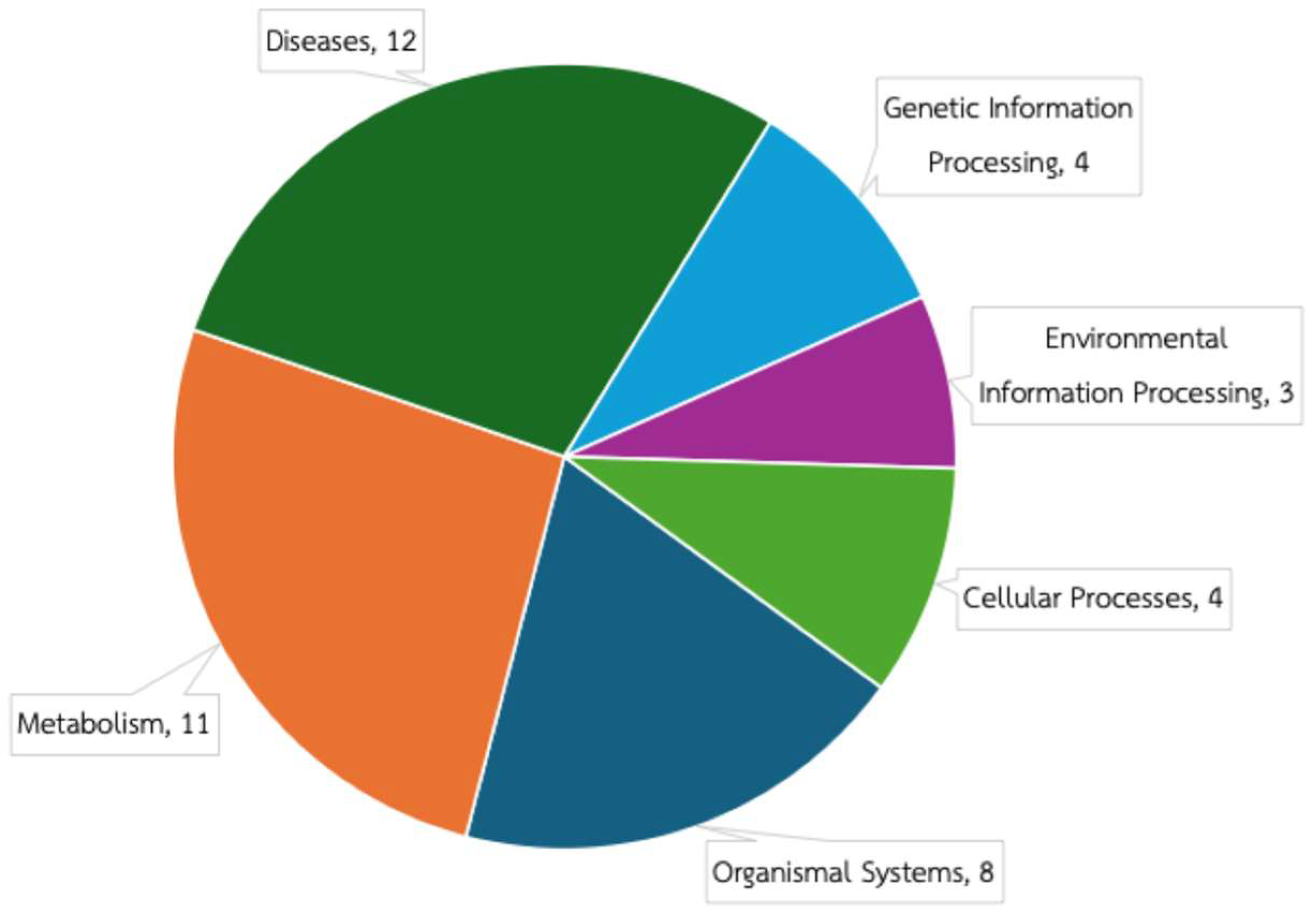
3.2. Predicted Functional Composition of Rabbit Gut Microbiome

The predicted functional composition of the rabbit gut microbiome was assessed using PICRUSt2 based on a total of 2427 ASVs. The predicted microbial functions were classified into 42 functional classes across six major KEGG pathways, as shown in Figure 1. The largest number of functional classes was associated with diseases, followed by metabolism, organismal systems, genetic information processing, cellular processes, and environmental information processing.
However, when considering the cumulative relative abundance at the functional class level, functions related to metabolism were the most predominant, exceeding those associated with disease-related pathways, as shown in Figure 2. Notably, the predominant predicted functions were associated with carbohydrate metabolism, which exhibited the highest relative abundance (17.03 ± 0.14%). This suggests that the rabbit gut microbiome plays a central role in the breakdown and utilization of dietary carbohydrates, consistent with their herbivorous diet. Other abundant functional classes included amino acid metabolism, membrane transport, metabolism of cofactors and vitamins, and energy metabolism.
Importantly, AMR was also identified with 1.55 ± 0.00%. This indicates the presence of gut microbial populations that harbor resistance mechanisms, potentially conferring resilience against a broad range of antimicrobial agents.

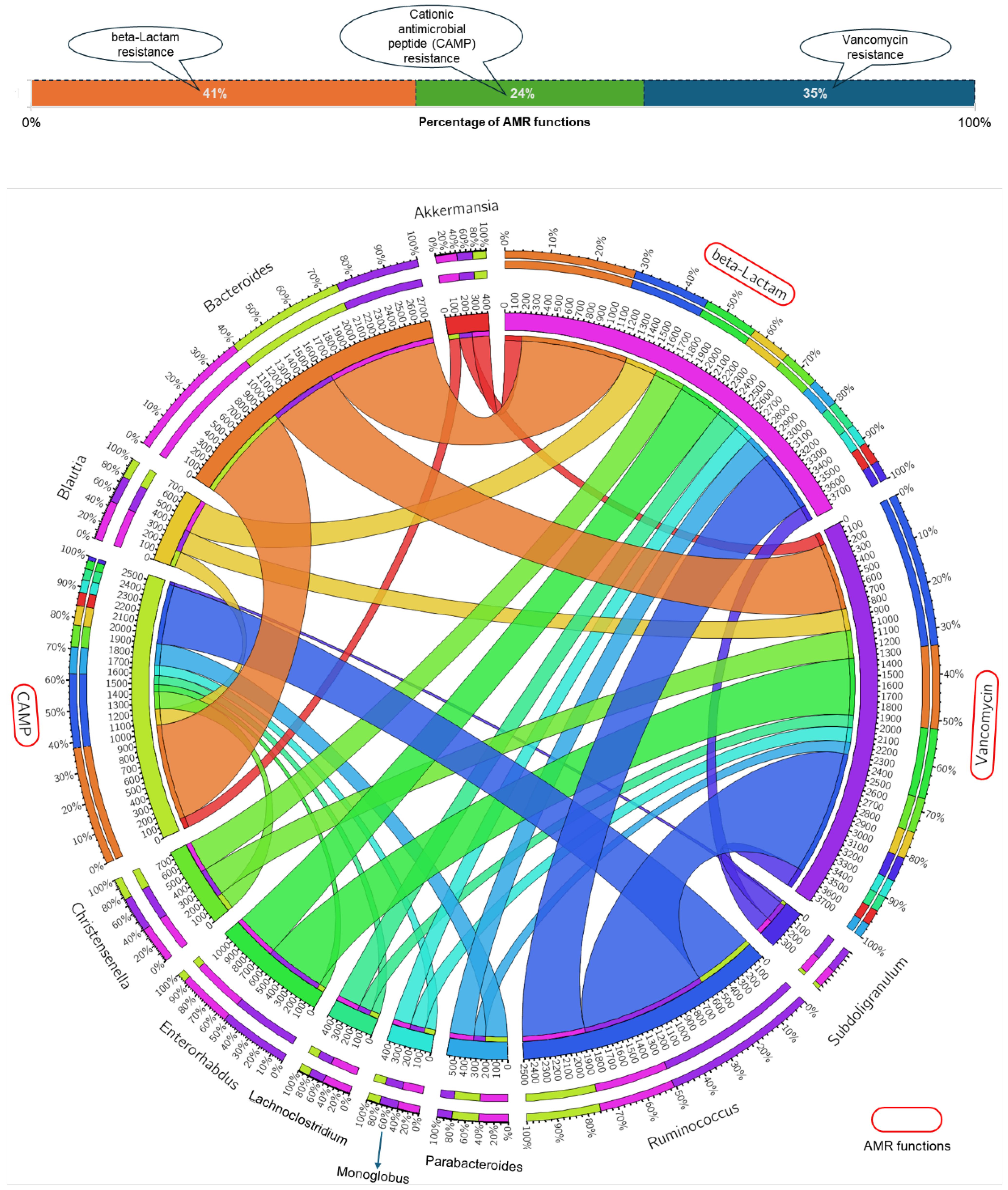
3.3. Gut Microbial Genera and AMR Function Relationship

Among the identified AMR functions, beta-lactam resistance accounted for 41%, Vancomycin resistance 35%, and cationic antimicrobial peptide (CAMP) resistance 24% (Figure 3). The top ten genera associated with these AMR functions were Bacteroides, Ruminococcus, Enterorhabdus, Christensenella, Blautia, Parabacteroides, Lachnoclostridium, Monoglobus, Akkermansia, and Subdoligranulum. Notably, Bacteroides and Ruminococcus exhibited broad contributions across multiple AMR functions, while Christensenella and Blautia were more specifically associated with beta-lactam and Vancomycin resistance. In contrast, Akkermansia and Subdoligranulum were primarily linked to beta-lactam and Vancomycin resistance. Interestingly, no associations were observed between AMR functions and common pathogenic genera such as Escherichia or Salmonella. This finding underscores that in laboratory rabbits, AMR potential is more strongly linked to non-pathogenic, resident gut microbes rather than pathogens.

4. Discussion

AMR is an emerging concern in laboratory animal facilities, particularly due to antibiotic use and microbial transmission that may disrupt the microbiome and promote resistant strains. Rabbits, widely used in biomedical research, are susceptible to gut dysbiosis and AMR development. This study used 16S rRNA amplicon sequencing and PICRUSt2 to predict AMR functions in the rabbit gut microbiome. Three AMR-related functions, including CAMP resistance, beta-lactam resistance, and vancomycin resistance, were identified. The detection of CAMP resistance reflects a natural defense mechanism in bacteria. This resistance is associated with the presence of genes within the dltABCD operon, which functions to incorporate D-alanine residues into lipoteichoic acids in the cell wall of Gram-positive bacteria. This modification helps alter the molecular structure and reduce the net negative charge of the bacterial cell membrane, thereby decreasing the toxicity of cationic peptides [37]. Additionally, some bacteria possess the bifunctional lysylphosphatidylglycerol flippase/synthetase protein MprF, which contributes to the synthesis of lysyl-phosphatidylglycerol, further reducing the interaction with cationic peptides. Although the mprF gene has been reported to be involved in resistance to gentamicin [38], genomic analyses of common gut bacteria suggest that resistance to CAMPs is widespread and likely represents an intrinsic feature of these microorganisms. For instance, a previous study has shown that Bacteroides possess genes involved in lipid A modification and surface charge alteration, which contribute to innate resistance against host-derived CAMPs [39]. Regarding beta-lactam resistance, this mechanism is commonly mediated by the presence of beta-lactamase and chloramphenicol O-acetyltransferase genes, which contribute to resistance against ampicillin, chloramphenicol, and tetracycline in various lactic acid bacteria (LAB) strains, as well as common gut microbes. For example, a study on Lactiplantibacillus plantarum PMO 08 reported ampicillin resistance linked to non-transferable genetic elements, suggesting the presence of intrinsic resistance mechanisms [40]. Similarly, genomic analysis of Limosilactobacillus fermentum ATCC 23271, originally isolated from the human intestine, identified genes associated with beta-lactam resistance, further supporting the notion of inherent resistance traits in commensal gut bacteria [41]. Consistently, genes associated with vancomycin resistance have been shown to occur naturally and appear to be intrinsic to gut microbial taxa, including non-pathogenic commensals [42].
Although AMR-related functions were observed in the rabbit gut microbiome, these functions were associated with commensal genera such as Ruminococcus, Blautia, Parabacteroides, Akkermansia, and Bacteroides. No associations were observed between AMR functions and common pathogenic genera such as Escherichia or Salmonella. These findings suggest that adherence to antimicrobial stewardship practices in laboratory settings is effective, and particularly suggest that the gut microbiome of laboratory animals, notably rabbits, is not a significant reservoir for AMR pathogens. This effectiveness can be attributed to several key stewardship practices implemented in laboratory animal facilities. The judicious use of antibiotics, specifically restricting their application to confirmed therapeutic needs rather than prophylactic or growth-promotion purposes, plays a crucial role in mitigating the development of AMR. Additionally, stringent infection surveillance, early diagnosis protocols, and biosecurity measures aimed at minimizing animal-to-animal transmission of resistant microorganisms are fundamental components of effective antimicrobial stewardship. Regular microbiological monitoring of animal colonies to detect emerging resistance patterns further supports responsible antibiotic management. Together, these proactive practices help maintain a balanced gut microbiome, reduce the selective pressure for resistant strains, and prevent the horizontal transfer of antimicrobial resistance genes among laboratory animals, thereby safeguarding both animal welfare and experimental integrity [8,9]. However, even under standardized and controlled conditions, it is important to recognize that AMR mechanisms of gut microbiome in response to beta-lactam, CAMPs, and vancomycin are a natural trait of laboratory rabbits. Therefore, inappropriate or excessive use of beta-lactam, CAMPs, and vancomycin may promote the selection and potential horizontal transfer of resistance genes from the commensals to pathogens. While the results could be used as therapeutic reference for laboratory rabbits, it is also important to note that this study assessed AMR functions using 16S rRNA sequencing data, which limits the resolution for identifying the localization of AMR genes within mobile genetic elements. To overcome this limitation, future studies should integrate whole metagenome shotgun sequencing (WMGS), which enables the direct detection and quantification of antimicrobial resistance genes (ARGs) and their genomic context, including linkage to mobile genetic elements such as plasmids and transposons. Unlike 16S rRNA-based inference, WMGS can accurately resolve the bacterial taxa carrying specific resistance genes and detect co-localization with virulence factors or horizontal gene transfer markers. This approach allows researchers to assess the potential for ARG dissemination across microbial communities and better understand the risk of resistance transmission in laboratory animals [23,43].
Beyond laboratory settings, effective dissemination of research findings to the broader public is essential to enhance awareness, counter misinformation, and promote evidence-based practices regarding AMR. Social media platforms such as Twitter, Facebook, and Instagram offer valuable tools for reaching audiences beyond the scientific community. Transparent communication through accessible digital channels not only enhances research credibility but also addresses the growing challenge of misinformation in animal science. In a parallel vein, a study using Instagram showed social media’s effectiveness in conveying complex scientific topics to a broad audience [44]. Notably, this study demonstrated that Instagram is an effective platform for delivering information related to animal health and nutrition, fostering both community engagement and public awareness. Incorporating similar digital dissemination strategies could extend the impact of findings on rabbit gut microbiome AMR, ensuring that key messages on antimicrobial stewardship and animal welfare are communicated effectively across diverse societal sectors.
Overall, this research provides valuable and essential functional insights into the intrinsic AMR profiles present within the gut microbiome of laboratory rabbits. These findings offer practical guidelines for the care and antimicrobial stewardship of laboratory rabbits, improved animal welfare, while also contributing to the optimization of AMR risk assessment and disease management strategies in laboratory animal facilities. Future studies should adopt WMGS for more comprehensive functional profiling and to further support AMR surveillance, microbiome management, and animal welfare.

Author Contributions

Conceptualization, P.C. and N.R.; methodology, P.C., N.R. and R.T.; software, N.R. and R.T.; validation, N.R.; formal analysis, N.R.; investigation, P.C. and N.R.; resources, P.C.; data curation, P.C., N.R. and R.T.; writing—original draft preparation, P.C. and N.R.; writing—review and editing, P.C., N.R. and R.T.; visualization, P.C. and N.R.; supervision, P.C.; project administration, P.C.; funding acquisition, P.C. All authors have read and agreed to the published version of the manuscript.

Funding

This research project is supported by a grant for professional development of support staff from Mahidol University.

Institutional Review Board Statement

The animal study protocol was approved by the National Laboratory Animal Center Animal Care and Use Committee (NLAC-ACUC) (protocol code: RA2023-01; date of approval: 29 March 2024).

Informed Consent Statement

Not applicable.

Data Availability Statement

The data generated/analyzed during the current study are available upon request.

Acknowledgments

This research project is supported by Mahidol University. The authors are grateful to the National Laboratory Animal Center, Mahidol University, for providing the laboratory facilities and resources.

Conflicts of Interest

The authors declare no conflicts of interest.

Abbreviations

The following abbreviations are used in this manuscript:
AMRantimicrobial resistance
PICRUSt2Phylogenetic Investigation of Communities by Reconstruction of Unobserved States 2
NLACNational Laboratory Animal Center
IACUCInstitutional Animal Care and Use Committee
NZWNew Zealand White
PCRpolymerase chain reaction
PEpaired-end
KEGGKyoto Encyclopedia of Genes and Genomes
ASVamplicon sequence variant
CAMPcationic antimicrobial peptide
ARGantimicrobial resistance gene

References

  1. Murray, C.J.L.; Ikuta, K.S.; Sharara, F.; Swetschinski, L.; Robles Aguilar, G.; Gray, A.; Han, C.; Bisignano, C.; Rao, P.; Wool, E.; et al. Global burden of bacterial antimicrobial resistance in 2019: A systematic analysis. Lancet 2022, 399, 629–655. [Google Scholar] [CrossRef] [PubMed]
  2. O’Neill, J. Tackling Drug-Resistant Infections Globally: Final Report and Recommendations. Wellcome Trust: London, UK, 2016; 84p. [Google Scholar]
  3. Pandey, S.; Doo, H.; Keum, G.B.; Kim, E.S.; Kwak, J.; Ryu, S.; Choi, Y.; Kang, J.; Kim, S.; Lee, N.R.; et al. Antibiotic resistance in livestock, environment and humans: One Health perspective. J. Anim. Sci. Technol. 2024, 66, 266–278. [Google Scholar] [CrossRef] [PubMed]
  4. Ahmed, S.K.; Hussein, S.; Qurbani, K.; Ibrahim, R.H.; Fareeq, A.; Mahmood, K.A.; Mohamed, M.G. Antimicrobial resistance: Impacts, challenges, and future prospects. J. Med. Surg. Public Health 2024, 2, 100081. [Google Scholar] [CrossRef]
  5. Carson, C.A.; Weese, J.S. Advances in addressing antimicrobial resistance. Rev. Sci. Tech. 2024, Special Edition, 113–123. [Google Scholar] [CrossRef]
  6. Masebo, N.T.; Giovanna, M.; Flavia, S.D.R.; Laura, A.; Damiano, C.; Marco, D.P.; Andrea, B.; Eliana, S.; Marilena, B.; Joaquin, H.B.; et al. Evaluation of antimicrobial and non-steroidal anti-inflammatory treatments for BRD on health and welfare in fattening bulls: A cross-sectional study. Vet. Q. 2024, 44, 1–11. [Google Scholar] [CrossRef]
  7. Samreen; Ahmad, I.; Malak, H.A.; Abulreesh, H.H. Environmental antimicrobial resistance and its drivers: A potential threat to public health. J. Glob. Antimicrob. Resist. 2021, 27, 101–111. [Google Scholar] [CrossRef]
  8. Wilcox, R.S.; Marenda, M.S.; Devlin, J.M.; Wilks, C.R. Antimicrobial use in laboratory rodent facilities in Australia and New Zealand—A cross-sectional survey of veterinarians and facility managers. PLoS ONE 2024, 19, e0292908. [Google Scholar] [CrossRef]
  9. Narver, H.L. Antimicrobial stewardship in laboratory animal facilities. J. Am. Assoc. Lab. Anim. Sci. 2017, 56, 6–10. [Google Scholar]
  10. Desselberge, U. The mammalian gut microbiome, immune responses and disease: From observational to causal relationships. J. Cell. Immunol. 2020, 2, 294–300. [Google Scholar]
  11. Shi, Y.; Peng, G.; Gebremariam, A.A.; Iqbal, M.M.; Daemi, H.B.; Khan, M.A.; Ullah, R.; Wang, D. Analytical insights, modulation and compositional dynamics of the feline gut microbiota: A review. Animal Diseases 2024, 4, 36. [Google Scholar] [CrossRef]
  12. Blackmer-Raynolds, L.; Sampson, T.R. Overview of the Gut Microbiome. Semin. Neurol. 2023, 43, 518–529. [Google Scholar] [CrossRef] [PubMed]
  13. Pierre, J.F.; Leone, V.A. Chapter 37—The microbiome and health. In Present Knowledge in Nutrition, 11th ed.; Marriott, B.P., Birt, D.F., Stallings, V.A., Yates, A.A., Eds.; Academic Press: Cambridge, MA, USA, 2020; pp. 605–624. [Google Scholar]
  14. Mapara, M.; Thomas, B.S.; Bhat, K.M. Rabbit as an animal model for experimental research. Dent. Res. J. (Isfahan) 2012, 9, 111–118. [Google Scholar] [CrossRef]
  15. El-Sabrout, K.; Sherasiya, A.; Ahmad, S.; Aggag, S.; Nannoni, E.; Cavallini, D.; Buonaiuto, G. Environmental Enrichment in Rabbit Husbandry: Comparative Impacts on Performance and Welfare. Animals 2024, 14, 2367. [Google Scholar] [CrossRef] [PubMed]
  16. Abd El-Aziz, A.; Elfadadny, A.; Abo Ghanima, M.; Cavallini, D.; Fusaro, I.; Giammarco, M.; Buonaiuto, G.; El-Sabrout, K. Nutritional Value of Oregano-Based Products and Its Effect on Rabbit Performance and Health. Animals 2024, 14, 3021. [Google Scholar] [CrossRef] [PubMed]
  17. Chamtim, P. Fecal Microbiome Analysis of New Zealand White Rabbits. In Proceedings of the the 2nd Thailand Bioinformatics Research Network Conference (TBRN 2025), Centara Riverside Hotel, Chiang Mai, Thailand, 22–24 January 2025; pp. 38–47. [Google Scholar]
  18. Ramirez, J.; Guarner, F.; Fernandez, L.B.; Maruy, A.; Sdepanian, V.L.; Cohen, H. Antibiotics as Major Disruptors of Gut Microbiota. Front. Cell. Infect. Microbiol. 2020, 10, 572912. [Google Scholar] [CrossRef]
  19. Chowdhury, G.; Ramamurthy, T. Influence of Antimicrobials on the Gut Microbiota. In Antimicrobial Resistance: Global Challenges and Future Interventions; Thomas, S., Ed.; Springer Singapore: Singapore, 2020; pp. 53–79. [Google Scholar]
  20. Paul, D.; Das, B. Chapter One—Gut microbiome in the emergence of antibiotic-resistant bacterial pathogens. In Progress in Molecular Biology and Translational Science; Das, B., Singh, V., Eds.; Academic Press: Cambridge, MA, USA, 2022; Volume 192, pp. 1–31. [Google Scholar]
  21. McInnes, R.S.; McCallum, G.E.; Lamberte, L.E.; van Schaik, W. Horizontal transfer of antibiotic resistance genes in the human gut microbiome. Curr. Opin. Microbiol. 2020, 53, 35–43. [Google Scholar] [CrossRef]
  22. Hu, X.; Wang, F.; Yang, S.; Yuan, X.; Yang, T.; Zhou, Y.; Li, Y. Rabbit microbiota across the whole body revealed by 16S rRNA gene amplicon sequencing. BMC Microbiol 2021, 21, 312. [Google Scholar] [CrossRef]
  23. Do, T.T.; Tamames, J.; Stedtfeld, R.D.; Guo, X.; Murphy, S.; Tiedje, J.M.; Walsh, F. Antibiotic Resistance Gene Detection in the Microbiome Context. Microb. Drug Resist. 2017, 24, 542–546. [Google Scholar] [CrossRef]
  24. Douglas, G.M.; Maffei, V.J.; Zaneveld, J.R.; Yurgel, S.N.; Brown, J.R.; Taylor, C.M.; Huttenhower, C.; Langille, M.G.I. PICRUSt2 for prediction of metagenome functions. Nat. Biotechnol. 2020, 38, 685–688. [Google Scholar] [CrossRef]
  25. Malinowska, A.M.; Kok, D.E.; Steegenga, W.T.; Hooiveld, G.J.E.J.; Chmurzynska, A. Human gut microbiota composition and its predicted functional properties in people with western and healthy dietary patterns. Eur. J. Nutr. 2022, 61, 3887–3903. [Google Scholar] [CrossRef]
  26. Shi, Y.; Gahagan, A.C.; Morrison, M.J.; Gregorich, E.; Lapen, D.R.; Chen, W. Stratified Effects of Tillage and Crop Rotations on Soil Microbes in Carbon and Nitrogen Cycles at Different Soil Depths in Long-Term Corn, Soybean, and Wheat Cultivation. Microorganisms 2024, 12, 1635. [Google Scholar] [CrossRef] [PubMed]
  27. Sahin Dogan, S.; Kocabaş, A. Profiling the genes associated with osmoadaptation and their variation by seasonally in Tuz Lake. Commun. Fac. Sci. Univ. Ank. Ser. C Biol. 2023, 32, 174–191. [Google Scholar] [CrossRef]
  28. Wang, S.; Wang, Y.; Liu, H.; Li, X.; Zhao, J.; Dong, Z.; Li, J.; Kaka, N.A.; Nazar, M.; Shao, T. Using PICRUSt2 to explore the functional potential of bacterial community in alfalfa silage harvested at different growth stages. Chem. Biol. Technol. Agric. 2022, 9, 98. [Google Scholar] [CrossRef]
  29. Bolger, A.M.; Lohse, M.; Usadel, B. Trimmomatic: A flexible trimmer for Illumina sequence data. Bioinformatics 2014, 30, 2114–2120. [Google Scholar] [CrossRef]
  30. Martin, M. Cutadapt removes adapter sequences from high-throughput sequencing reads. EMBnet J. 2011, 17, 3. [Google Scholar] [CrossRef]
  31. Callahan, B.J.; McMurdie, P.J.; Holmes, S.P. Exact sequence variants should replace operational taxonomic units in marker-gene data analysis. ISME J. 2017, 11, 2639–2643. [Google Scholar] [CrossRef]
  32. Callahan, B.J.; McMurdie, P.J.; Rosen, M.J.; Han, A.W.; Johnson, A.J.A.; Holmes, S.P. DADA2: High-resolution sample inference from Illumina amplicon data. Nat. Methods 2016, 13, 581–583. [Google Scholar] [CrossRef]
  33. Quast, C.; Pruesse, E.; Yilmaz, P.; Gerken, J.; Schweer, T.; Yarza, P.; Peplies, J.; Glöckner, F.O. The SILVA ribosomal RNA gene database project: Improved data processing and web-based tools. Nucleic Acids Res. 2013, 41, D590–D596. [Google Scholar] [CrossRef]
  34. Caporaso, J.G.; Kuczynski, J.; Stombaugh, J.; Bittinger, K.; Bushman, F.D.; Costello, E.K.; Fierer, N.; Peña, A.G.; Goodrich, J.K.; Gordon, J.I.; et al. QIIME allows analysis of high-throughput community sequencing data. Nat. Methods 2010, 7, 335–336. [Google Scholar] [CrossRef]
  35. Kanehisa, M.; Goto, S. KEGG: Kyoto encyclopedia of genes and genomes. Nucleic Acids Res. 2000, 28, 27–30. [Google Scholar] [CrossRef]
  36. Krzywinski, M.; Schein, J.; Birol, I.; Connors, J.; Gascoyne, R.; Horsman, D.; Jones, S.J.; Marra, M.A. Circos: An information aesthetic for comparative genomics. Genome Res. 2009, 19, 1639–1645. [Google Scholar] [CrossRef]
  37. Abachin, E.; Poyart, C.; Pellegrini, E.; Milohanic, E.; Fiedler, F.; Berche, P.; Trieu-Cuot, P. Formation of D-alanyl-lipoteichoic acid is required for adhesion and virulence of Listeria monocytogenes. Mol. Microbiol. 2002, 43, 1–14. [Google Scholar] [CrossRef] [PubMed]
  38. Nishi, H.; Komatsuzawa, H.; Fujiwara, T.; McCallum, N.; Sugai, M. Reduced content of lysyl-phosphatidylglycerol in the cytoplasmic membrane affects susceptibility to moenomycin, as well as vancomycin, gentamicin, and antimicrobial peptides, in Staphylococcus aureus. Antimicrob. Agents Chemother. 2004, 48, 4800–4807. [Google Scholar] [CrossRef]
  39. Cullen, T.W.; Schofield, W.B.; Barry, N.A.; Putnam, E.E.; Rundell, E.A.; Trent, M.S.; Degnan, P.H.; Booth, C.J.; Yu, H.; Goodman, A.L. Gut microbiota. Antimicrobial peptide resistance mediates resilience of prominent gut commensals during inflammation. Science 2015, 347, 170–175. [Google Scholar] [CrossRef] [PubMed]
  40. Oh, Y.J.; Kim, S.A.; Yang, S.H.; Kim, D.H.; Cheng, Y.Y.; Kang, J.I.; Lee, S.Y.; Han, N.S. Integrated genome-based assessment of safety and probiotic characteristics of Lactiplantibacillus plantarum PMO 08 isolated from kimchi. PLoS ONE 2022, 17, e0273986. [Google Scholar] [CrossRef] [PubMed]
  41. dos Santos, C.I.; Campos, C.D.L.; Nunes-Neto, W.R.; do Carmo, M.S.; Nogueira, F.A.B.; Ferreira, R.M.; Costa, E.P.S.; Gonzaga, L.F.; Araújo, J.M.M.; Monteiro, J.M.; et al. Genomic Analysis of Limosilactobacillus fermentum ATCC 23271, a Potential Probiotic Strain with Anti-Candida Activity. J. Fungi 2021, 7, 794. [Google Scholar] [CrossRef]
  42. Ahmed, M.O.; Baptiste, K.E. Vancomycin-Resistant Enterococci: A Review of Antimicrobial Resistance Mechanisms and Perspectives of Human and Animal Health. Microb. Drug Resist. 2017, 24, 590–606. [Google Scholar] [CrossRef]
  43. Boolchandani, M.; D’Souza, A.W.; Dantas, G. Sequencing-based methods and resources to study antimicrobial resistance. Nat. Rev. Genet. 2019, 20, 356–370. [Google Scholar] [CrossRef]
  44. Lamanna, M.; Muca, E.; Buonaiuto, G.; Formigoni, A.; Cavallini, D. From posts to practice: Instagram’s role in veterinary dairy cow nutrition education—How does the audience interact and apply knowledge? A survey study. J. Dairy Sci. 2025, 108, 1659–1671. [Google Scholar] [CrossRef]
Figure 1. KEGG pathway classification of predicted microbial functions based on PICRUSt2 analysis.
Amh 70 00021 g001
Figure 2. Predicted functional composition of the rabbit gut microbiome. The red frame highlights the predicted antimicrobial resistance (AMR) functions, which accounted for 1.55 ± 0.00% of the total functional composition.
Amh 70 00021 g002
Figure 3. Circular chord diagram illustrating the relationship between AMR functions and microbial genera. The outer ring segments represent either AMR functions or microbial genera. The colored ribbons (chords) inside the circle denote the contribution of each genus to specific AMR functions, with the width of each ribbon reflecting the magnitude of association. The figure was generated using Circos [36] through the Interactive Online Version of Circos (available at http://mkweb.bcgsc.ca/tableviewer/, accessed on 7 April 2025).
Amh 70 00021 g003
Table 1. Summary of sequencing data and ASV assignment.
Sequencing FeatureAverageTotal
Raw sequencing data (PE reads)80,135 ± 72240,405
Clean sequencing data (PE reads)73,103 ± 295219,310
Denoised sequencing data (PE reads)72,648 ± 316217,945
Non-chimeric sequencing data (PE reads)43,473 ± 3069130,419
Number of ASVs905 ± 1102427
Table 2. Taxonomic assignment of the ASVs identified in the gut microbiome of laboratory rabbits.
PhylumFamilyAverage Relative Abundance
Acidobacteriota 0.001 ± 0.001%
unclassified_Subgroup_170.001 ± 0.001%
Actinobacteriota 5.485 ± 3.969%
Atopobiaceae4.418 ± 3.395%
Bifidobacteriaceae0.001 ± 0.002%
Corynebacteriaceae0.004 ± 0.004%
Eggerthellaceae0.963 ± 0.532%
Frankiaceae0.001 ± 0.001%
Streptomycetaceae0.003 ± 0.002%
unclassified_Actinobacteria0.002 ± 0.003%
unclassified_Actinobacteriota0.044 ± 0.057%
unclassified_Coriobacteriales0.05 ± 0.024%
Bacteroidota 17.114 ± 7.175%
Bacteroidaceae13.386 ± 3.744%
Marinifilaceae0.199 ± 0.167%
Muribaculaceae2.435 ± 3.59%
Prevotellaceae0.003 ± 0.003%
Rikenellaceae0.527 ± 0.12%
Tannerellaceae0.556 ± 0.411%
unclassified_Bacteroidales0.004 ± 0.007%
unclassified_Bacteroidia0.003 ± 0.002%
Chloroflexi 0.001 ± 0.001%
uncultured_Chloroflexi_bacterium0.001 ± 0.001%
Cyanobacteria 0.02 ± 0.017%
unclassified_Gastranaerophilales0.011 ± 0.019%
uncultured_rumen_bacterium0.009 ± 0.016%
Desulfobacterota 0.007 ± 0.009%
Desulfovibrionaceae0.007 ± 0.009%
Firmicutes 75.446 ± 2.369%
[Clostridium]_methylpentosum_group0.09 ± 0.06%
[Eubacterium]_coprostanoligenes_group0.719 ± 0.226%
Anaerofustaceae0.016 ± 0.014%
Anaerovoracaceae0.179 ± 0.177%
Butyricicoccaceae0.025 ± 0.023%
Christensenellaceae1.15 ± 0.22%
Defluviitaleaceae0.018 ± 0.027%
Desulfurisporaceae0.001 ± 0.001%
Erysipelatoclostridiaceae0.005 ± 0.007%
Erysipelotrichaceae1.295 ± 0.859%
Eubacteriaceae24.806 ± 13.583%
Exiguobacteraceae0.002 ± 0.001%
Hungateiclostridiaceae0.054 ± 0.068%
Lachnospiraceae16.639 ± 4.871%
Lactobacillaceae0.001 ± 0.001%
Limnochordaceae0.001 ± 0.001%
Monoglobaceae1.846 ± 0.879%
Oscillospiraceae6.212 ± 2.285%
Peptococcaceae0.2 ± 0.084%
Ruminococcaceae17.244 ± 5.909%
Syntrophomonadaceae0.001 ± 0.002%
UCG_0100.927 ± 0.981%
UCG_0110.005 ± 0.005%
unclassified_Bacilli0.001 ± 0.002%
unclassified_Clostridia1.21 ± 0.358%
unclassified_Clostridia_UCG_0141.989 ± 1.446%
unclassified_Clostridia_vadinBB60_group0.208 ± 0.165%
unclassified_Erysipelotrichales0.003 ± 0.005%
unclassified_Firmicutes0.08 ± 0.058%
unclassified_Lachnospirales0.003 ± 0.003%
unclassified_Oscillospirales0.073 ± 0.041%
unclassified_RF390.126 ± 0.044%
uncultured_Clostridiales_bacterium0.005 ± 0.002%
uncultured_Erysipelotrichaceae_bacterium0.021 ± 0.024%
uncultured_Lachnospiraceae_bacterium0.017 ± 0.03%
uncultured_rumen_bacterium0.276 ± 0.297%
Gemmatimonadota 0.002 ± 0.001%
Gemmatimonadaceae0.002 ± 0.001%
Patescibacteria 0.269 ± 0.135%
Saccharimonadaceae0.266 ± 0.137%
unclassified_Candidatus_Campbellbacteria0.002 ± 0.002%
unclassified_Saccharimonadales0.001 ± 0.001%
Planctomycetota 0.001 ± 0.001%
Pirellulaceae0.001 ± 0.001%
Proteobacteria 0.416 ± 0.211%
Acetobacteraceae0.001 ± 0.001%
Alteromonadaceae0.001 ± 0.001%
Caulobacteraceae0.001 ± 0.001%
Enterobacteriaceae0.001 ± 0.002%
Oxalobacteraceae0.013 ± 0.008%
Rhizobiaceae0.001 ± 0.002%
Solimonadaceae0.001 ± 0.001%
Succinivibrionaceae0.001 ± 0.001%
TRA3_200.001 ± 0.001%
unclassified_Burkholderiales0.162 ± 0.049%
unclassified_Proteobacteria0.002 ± 0.002%
unclassified_Rhodospirillales0.231 ± 0.264%
Verrucomicrobiota 1.241 ± 0.856%
Akkermansiaceae1.241 ± 0.856%
Bold text indicates phyla names and their average relative abundances.
Disclaimer/Publisher’s Note: The statements, opinions and data contained in all publications are solely those of the individual author(s) and contributor(s) and not of MDPI and/or the editor(s). MDPI and/or the editor(s) disclaim responsibility for any injury to people or property resulting from any ideas, methods, instructions or products referred to in the content.

