Review

The Invisible Threat That Leaves You Breathless—A Literature Review on Pneumothorax in the Emergency Department

by
Silvia Fattori
*,
Gabriele Bellio
,
Matteo Maria Cimino
and
Hayato Kurihara
Emergency Surgery Unit, Foundation IRCCS Ca’ Granda Ospedale Maggiore Policlinico, 20122 Milano, Italy
*
Author to whom correspondence should be addressed.
Emerg. Care Med. 2025, 2(2), 24; https://doi.org/10.3390/ecm2020024
Submission received: 22 March 2025 / Revised: 24 April 2025 / Accepted: 28 April 2025 / Published: 2 May 2025

Abstract

:
Pneumothoraces are a common and potentially severe condition in the emergency setting. Various pathophysiological mechanisms (spontaneous and traumatic) could be involved, consequently defining the diagnostic–therapeutic pathway. Understanding these underlying etiologies is essential for advancing diagnostic strategies and guiding therapeutic measures. Recent insights into diagnostic and therapeutic strategies focus on the role of ultrasound and the effectiveness of small-bore chest tubes in managing all types of pneumothoraces with a reduced risk of complications. Moreover, observation is emerging as a possible conservative approach in hemodynamically stable patients regardless of the etiology of the pneumothorax. This review aims to provide a valuable resource to improve diagnostic and therapeutic management, comparing traditional methods and promising, less invasive therapeutic interventions.

1. Introduction

Pneumothorax is defined as the presence of air within the pleural space, resulting in partial or complete lung collapse due to the disruption of the pressure gradient required for lung expansion. It is classified into spontaneous and traumatic pneumothorax, each with distinct pathophysiological mechanisms and clinical presentations.
Primary spontaneous pneumothorax (PSP) is conventionally defined as a pneumothorax that occurs without an external triggering event and in the absence of clinically apparent lung disease. PSP is one of the most common thoracic conditions affecting adolescents and young adults, with an annual incidence of 15.5 to 22.7 cases per 100,000 people and a female-to-male ratio of 1:3.3 to 1:5. Its clinical course is variable, with recurrence rates ranging from 25% to 54% [1]. Significant variability exists in the management of PSP despite its high incidence and the availability of international guidelines. Secondary spontaneous pneumothorax (SSP) is characterized as a pneumothorax that occurs as a complication of an underlying lung disease [2]. SSP is more common in males and, unlike PSP, typically affects older individuals over 55 years old [3]. While nearly any lung disease can lead to SSP, it is most frequently associated with chronic obstructive pulmonary disease (COPD) [4].
Traumatic pneumothorax is a common consequence of chest trauma. Thoracic injuries cause 25% of trauma-related deaths, with pneumothorax occurring in 40–50% of these cases [5]. Thoracic trauma etiology is broadly classified into blunt and penetrating mechanisms, while traumatic pneumothorax is usually categorized as open and tension pneumothorax. In cases of major trauma, up to 80% of patients present concomitant trauma in other corporeal districts, while there are often associated chest injuries, such as rib fractures, pulmonary contusion, and hemothorax, which may negatively contribute to patient clinical status [6]. Additionally, concomitant hemothorax is present in 20% of patients with traumatic pneumothorax and often requires chest tube placement regardless of the presence of air in the pleural space. In up to 51% of trauma patients, occult pneumothorax, undetectable on initial chest X-ray but identifiable on advanced imaging, can complicate timely diagnosis and management [7].
The classification of pneumothorax size and its management is still debated. Currently, the most commonly used guidelines are those of the American College of Chest Physicians, which set a threshold of 3 cm based on the distance between the apex and the thoracic cupola [7]. The British Thoracic Society, on the other hand, classifies pneumothorax size by measuring the intrapleural distance at the level of the hilum, with a threshold of 2 cm [8].
Pneumothorax treatment is often provided by various healthcare professionals, including emergency physicians and surgeons. This review aims to provide a comprehensive analysis of the current literature on the diagnosis and management of spontaneous and traumatic pneumothorax. Given the fundamental importance of prompt recognition and appropriate treatment to prevent serious complications, this document aims to provide practical insights and indications to standardize clinical practice and simplify diagnostic and therapeutic strategies.
For this narrative review, a thorough investigation of the existing literature on pneumothorax was conducted (Figure 1). Exclusion criteria included studies regarding the pediatric population, case reports and series, and languages other than English. The search was performed using keywords and combinations of terms, and articles were assessed for relevance by reading titles and abstracts. Studies that met the selection criteria were included in the full analysis, during which they were organized and classified according to main themes to facilitate critical synthesis and coherent discussion. Two team members independently conducted the research and selection process, while the remaining two authors supervised and validated the study methods.

2. Pathogenesis

2.1. Primary Spontaneous Pneumothorax

The exact pathogenesis of spontaneous pneumothorax remains incompletely understood, with several hypothetical mechanisms and contributing factors. Although PSP was initially believed to occur in individuals with no underlying lung abnormalities, recent evidence suggests an interplay of structural lung changes, environmental exposures, and genetic predisposition as key contributors to its development.

2.1.1. Structural Lung Changes

Subpleural blebs and bullae have long been associated with PSP, but they are only identified in approximately 20% of cases [1]. The formation of these lesions remains unclear. Theories suggest that increasing negative pressure or enhanced mechanical stretch at the lung apex during growth may play a role. This is supported by the observation that PSP commonly occurs in tall, thin males between 10 and 30 years of age. Another hypothesis suggests that subpleural blebs result from a congenital phenomenon where apical lung tissue grows faster than its vascular supply, leading to ischemia and structural weakness [3]. Pathologic evaluations of resected lung specimens reveal disrupted mesothelial cells, inflammatory changes, and small pores (10–20 microns) in the visceral pleura, which may explain air leakage without a complete breach in the pleural membrane [9]. Fluorescein leakage observed on autofluorescence thoracoscopy provides further support for the existence of visceral pleural porosity as a mechanism of air escape [10].
In addition to blebs and bullae, emphysema-like changes (ELCs), chronic small airway inflammation, and abnormal matrix metalloproteinase (MMP) activity are implicated in spontaneous pneumothorax pathogenesis. These factors weaken the lung parenchyma and predispose it to rupture. Peripheral airway obstruction, resulting in air trapping, has also been proposed as a contributing mechanism [1].

2.1.2. Environmental Factors

Smoking, whether current or past, is a significant risk factor for PSP, with the risk being directly proportional to the amount of smoking. Cigarette smoke-induced airway inflammation and respiratory bronchiolitis contribute to structural changes in the lung, increasing susceptibility to pneumothorax [11].

2.1.3. Genetic Predisposition

A genetic component is thought to play a role in PSP, as clustering of cases within certain families has been reported [12,13]. Although specific genetic mutations or hereditary factors remain under investigation, they may contribute to pleural porosity, apical pulmonary ischemia, or other structural vulnerabilities.

2.2. Secondary Spontaneous Pneumothorax

SSP occurs as a complication of underlying lung diseases, with COPD being the most common cause. A total of 50–70% of SSP cases are attributed to COPD, where the rupture of apical bullae or blebs is the usual trigger [4,14]. Other diseases linked to SSP include cystic fibrosis, lung cancer, tuberculosis, and other infections [15].
In SSP, it is believed that inflammation leads to the replacement of normal pleural mesothelial cells with a fibroelastic layer, creating a porous visceral pleura prone to rupture. Contributing mechanisms include apical lung ischemia, genetic predisposition, and varying negative pressure gradients [16].

2.3. Traumatic Pneumothorax

Traumatic pneumothorax occurs when air accumulates in the pleural space due to an external injury, disrupting the normal pressure gradient required for lung expansion. Traumatic pneumothorax can be classified as iatrogenic or non-iatrogenic. Non-iatrogenic is further divided into penetrating and blunt trauma-induced pneumothoraces [4]. The condition is a significant concern in trauma settings, with thoracic injuries accounting for approximately 25% of trauma-related mortality [5]. Up to 40–50% of these patients are found to have a pneumothorax, highlighting the importance of prompt recognition and treatment [4,17].
The mechanism of injury plays a critical role in understanding the development of traumatic pneumothorax.

2.3.1. Penetrating Chest Trauma

Penetrating injuries, such as stab wounds or gunshot wounds, cause pneumothorax by establishing direct communication between the pleural space and the external environment or by damaging lung parenchyma, causing air leakages. Approximately 80% of penetrating chest injuries result in pneumothorax [18]. In cases where the wound size approaches two-thirds of the tracheal diameter, an open pneumothorax may develop, allowing free communication between the atmosphere and the pleural cavity. This results in progressive lung collapse and potential hemodynamic instability [19].

2.3.2. Blunt Chest Trauma

Blunt trauma-induced pneumothorax often arises from two primary mechanisms:
  • Rib Fractures: Fractured ribs may lacerate the visceral pleura and lung parenchyma, allowing air to escape into the pleural cavity. The likelihood of pneumothorax increases with the number of rib fractures sustained [17,20];
  • Sudden Lung Compression: High-impact forces, such as those from motor vehicle collisions or falls, may compress the lung tissue, leading to alveolar rupture and subsequent air leakage into the pleural space [19].
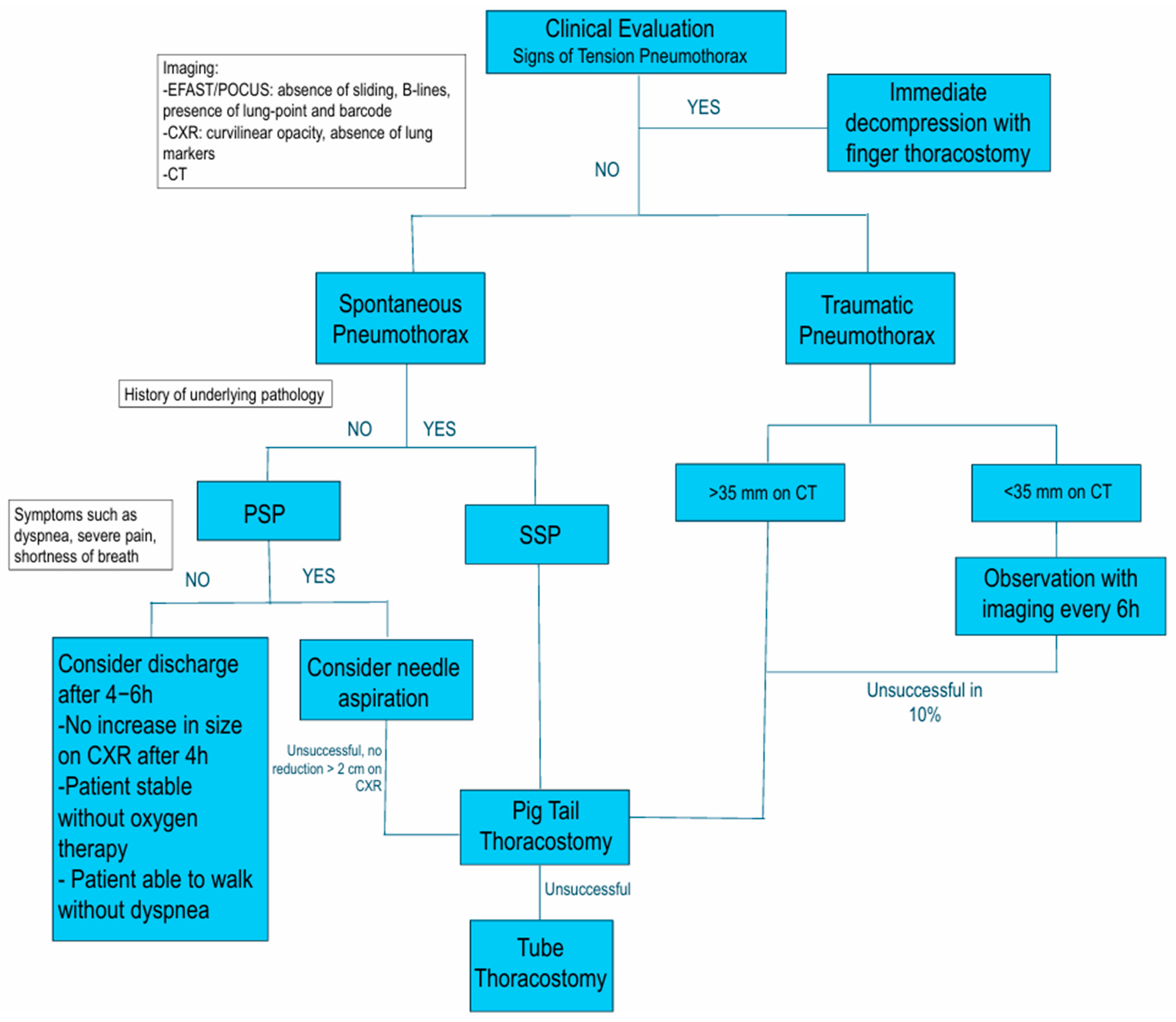
Regardless of the injury mechanism, any type or size of pneumothorax has the potential to progress into a tension pneumothorax, a life-threatening complication. In tension pneumothorax, air enters the pleural space through a one-way valve mechanism. This leads to progressive air accumulation, causing ipsilateral lung collapse, mediastinal shift, compression of the contralateral lung, and obstruction of venous return. These events culminate in cardiovascular compromise and, if untreated, death [4,19].

3. Diagnosis

The accurate and rapid diagnosis of pneumothorax is essential for planning subsequent treatment to prevent potentially life-threatening complications.

3.1. Clinical Presentation
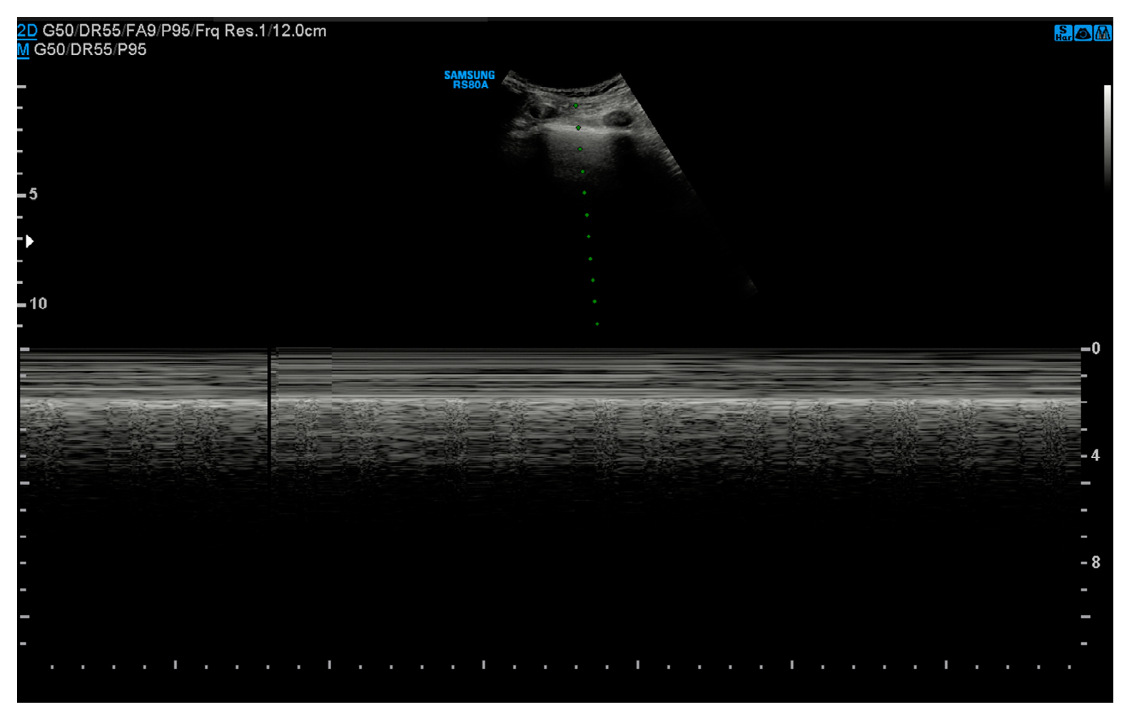
Imaging studies are fundamental in the diagnostic workup of pneumothorax, providing crucial confirmation and guiding management decisions. However, early recognition through physical examination remains essential to rapidly detect life-threatening complications that necessitate immediate intervention.
Common symptoms and signs of pneumothorax include decreased breath sounds, reduced tactile fremitus, hypoxia, and reported chest pain and shortness of breath. An essential tool for evaluation is arterial blood gas analysis, providing key insights into oxygenation, ventilation, and the presence and severity of shock.
Tension pneumothorax is a form of obstructive shock requiring prompt treatment even in the absence of radiographic confirmation, as this may cause delays. As intrapleural air accumulates, increasing the pressure can lead to a mediastinal shift and vena cava compression, reducing venous return. Warning signs include tracheal deviation, jugular venous distension, subcutaneous emphysema, decreased or absent breath sounds on the affected side, tachycardia, cyanosis, hypoxia, and hypotension [19].
Although history, signs, and symptoms can guide the diagnosis of pneumothoraces, they are often normal in small pneumothoraces requiring further imaging. As described by Barton et al. in their study of swine models, despite initial vital signs, hemodynamic instability is observed when the pneumothorax reaches approximately 57% of the lung capacity [21]. This highlights the importance of early detection and intervention to prevent progression to a critical state.

3.2. Ultrasound

Over the past two decades, ultrasound (US) has emerged as a highly effective diagnostic tool for pulmonary pathology. US imaging provides a real-time, two-dimensional reconstruction of anatomical planes, displaying tissues and organs in varying shades of gray based on the interaction with ultrasonic waves (Figure 2). Different probes may be used to assess the chest parietal plane effectively. Linear high-frequency probes offer high resolution and are ideal for examining superficial pleural structures, such as the presence of pneumothorax. Convex and low-frequency probes provide moderate resolution and are suited for evaluating deep structures such as pulmonary consolidations, pleural effusions, and the diaphragm. Sector probes, primarily used for cardiac imaging, may also be employed for thoracic assessments in specific cases, such as pleural effusion thanks to being able to image between the ribs and high penetration depth [22].
In emergency settings, Extended Focused Assessment with Sonography for Trauma (E-FAST) is used to evaluate the chest and abdomen, offering the advantage of rapid assessment of injuries without radiation exposure. In recent years, the use of point-of-care ultrasound (POCUS) by healthcare providers has become a valuable diagnostic tool for detecting pathologies, even in non-traumatic settings. Studies have demonstrated that US is superior to Chest X-ray (CXR) in detecting pneumothorax, with sensitivity reaching 91%, predictive values of 98–100%, and in the case of detection of lung point, a specificity of 100% [23,24]. Principal sonographic signs include (Table 1):
  • Absence of pleural sliding: Normally, the visceral pleura glides against the parietal pleura with respiration. In pneumothorax, this movement is absent;
  • Absence of B-lines: Comet-tail artifacts (B-lines) are reverberation anomalies that disappear with the presence of air in the pleural space;
  • Lung point sign: The point where normal lung sliding meets the absent sliding of the pneumothorax; this is highly specific for pneumothoraces;
  • Barcode or stratosphere sign: A static, uniform appearance on M-mode ultrasound indicating loss of lung movement (Figure 3).
The advantages of US include its portability, real-time assessment capability, low costs, and avoidance of radiation exposure, making it a potentially useful tool for follow-up. It is already widely used in the assessment of certain conditions, such as respiratory failure in non-critical patients [22]. However, its application in the follow-up of pneumothorax remains debated, as its accuracy is highly operator-dependent and may be limited by factors such as patient body habitus or the presence of subcutaneous emphysema.

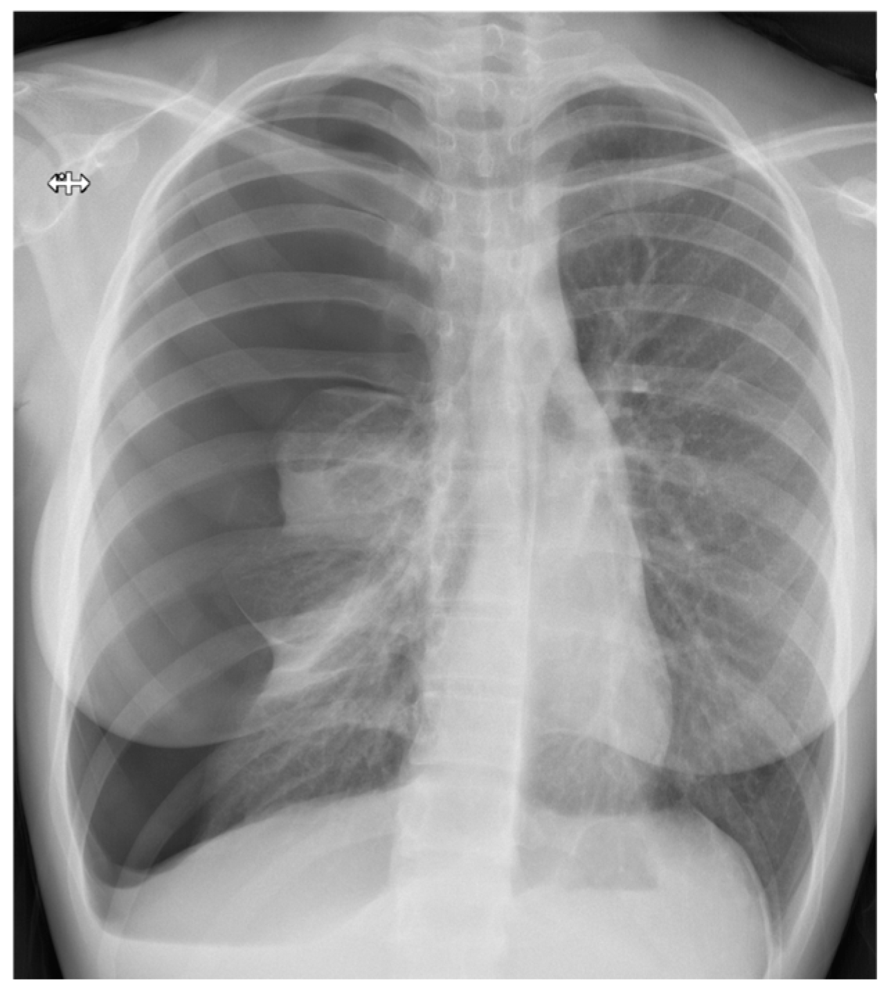
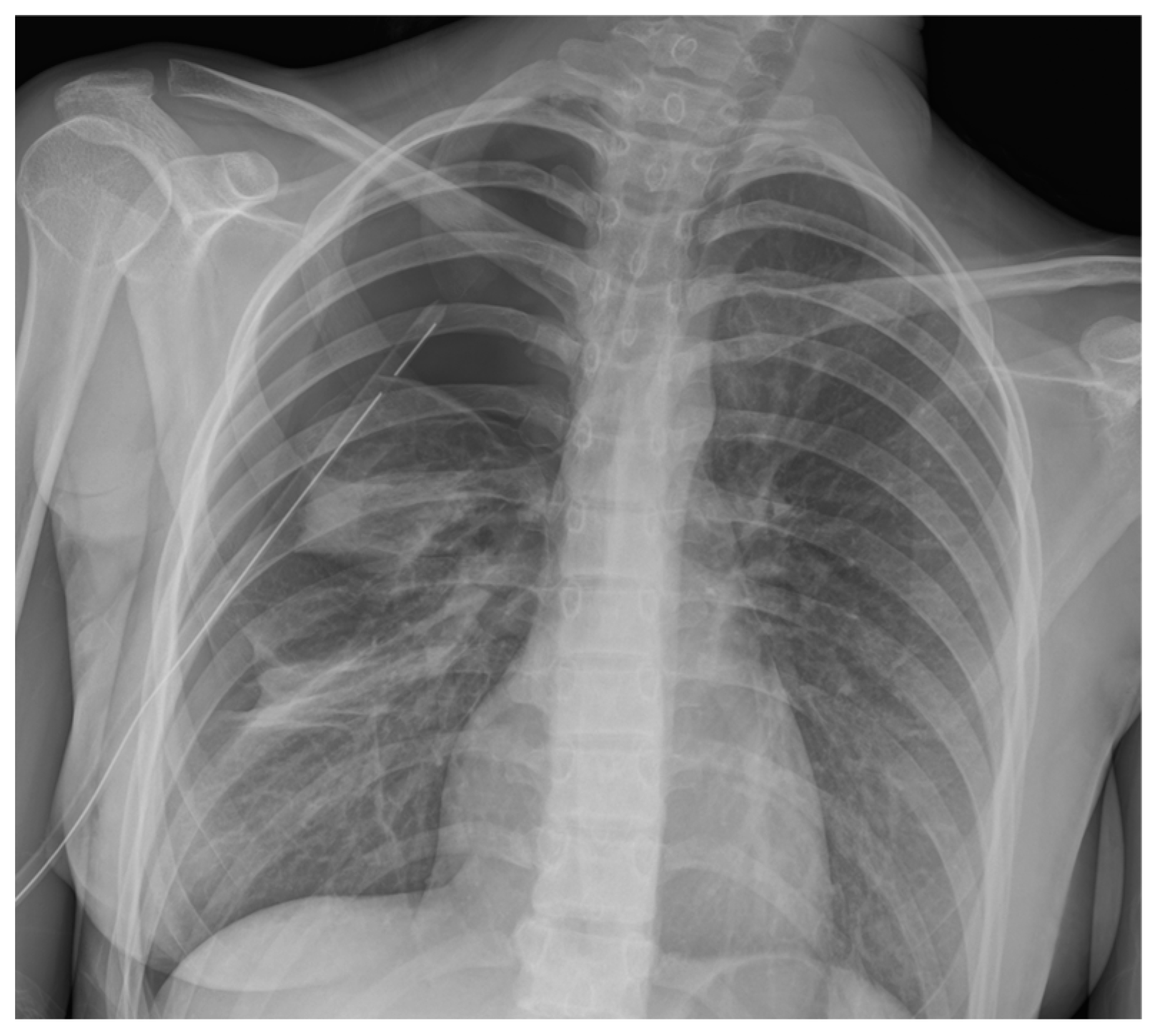
3.3. Chest Radiography

Conventional chest radiography remains the primary first-line, most common imaging modality for evaluating thoracic injuries, recommended by most trauma guidelines, including ATLS (Advanced Trauma Life Support) (Figure 4 and Figure 5) [25]. In hemodynamically unstable patients, CXR combined with E-FAST is the only imaging approach performed prior to surgical intervention. Numerous studies and meta-analyses have found that AP CXR has specificity (99–100%) but poor sensitivity (35–72%) in identifying pneumothoraces [23]. Aswin et al. found the sensitivity and specificity of CXR in detecting pneumothorax were 71.4% and 100%, respectively [26], while Salama et al.’s study showed the sensitivity and specificity as 75% and 88.9%, respectively [27].
To maximize the detection of both the direct and indirect signs of chest wall injuries and pulmonary pathologies, chest radiographs should ideally be obtained with posteroanterior (PA) and laterolateral (LL) views with the patient in an upright position and full expiration [28]. However, in trauma settings, this is rarely feasible. Instead, most trauma patients undergo anteroposterior (AP) radiographs in a supine position. In these scenarios, bedside CXR may fail to detect small pneumothoraces, as free air mainly distributes anteriorly or anteromedially near the diaphragm rather than at the apex. Studies report that up to 76% of traumatic pneumothoraces are missed on initial radiographs [17]. In the upright patient, air in the pleural space separates the lung from the chest wall, making the visceral pleural line visible as a thin curvilinear opacity. Despite CXR’s low sensitivity, it provides important information regarding the ribs, mediastinum, and trachea, and, moreover, it is particularly helpful in detecting tension pneumothorax. Radiologic signs of a tension pneumothorax include contralateral mediastinal shift, inferior diaphragm displacement, hyperlucency of the hemithorax, and ipsilateral lung collapse [28].

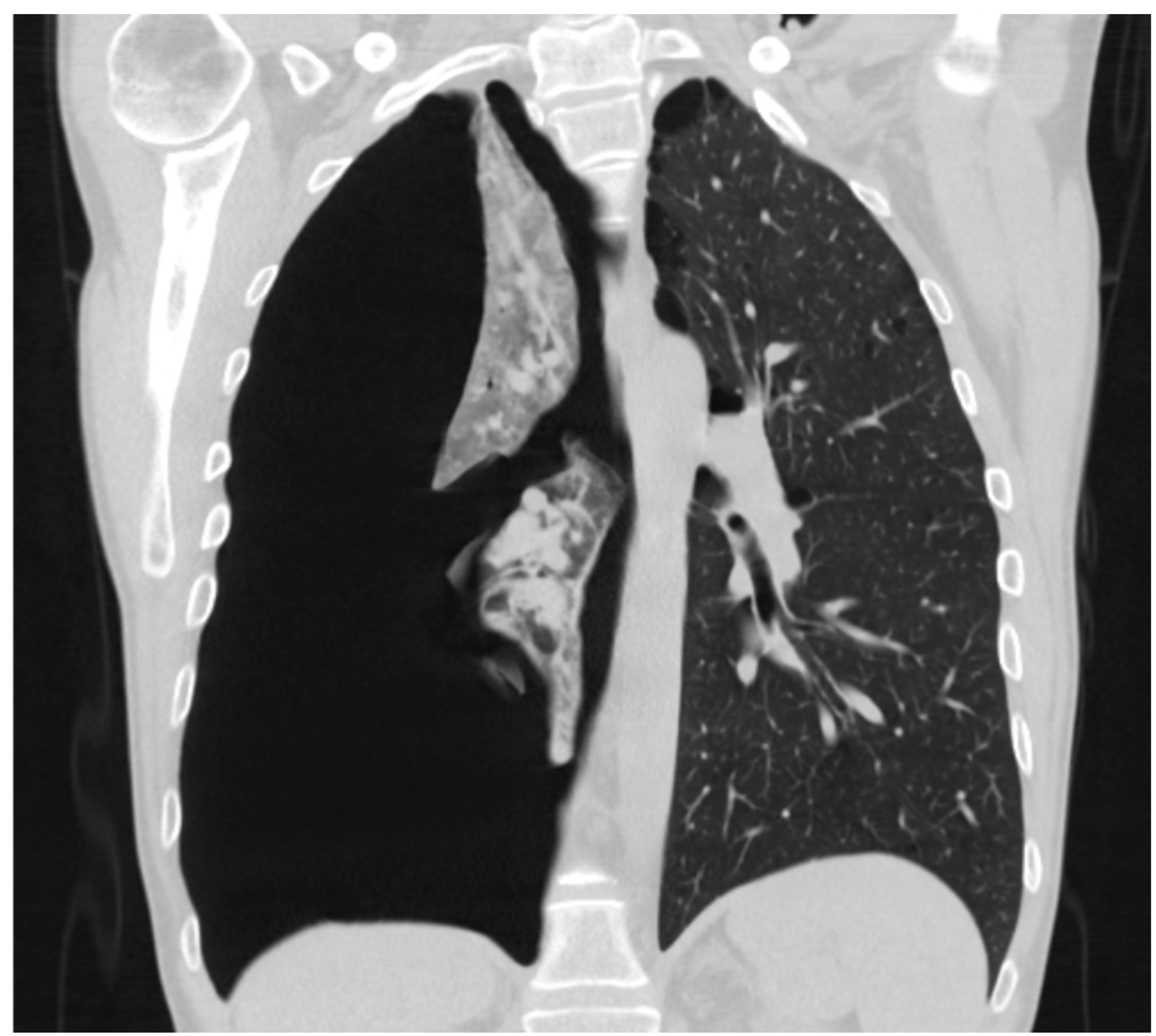
3.4. Computed Tomography Scan

The Computed Tomography Scan (CT) remains the gold standard for detecting pneumothorax (Figure 6), offering superior sensitivity in identifying even small amounts of air in the pleural space. Its introduction has led to increased recognition of occult pneumothorax, which occurs in up to 22% of blunt trauma patients [29]. Undiagnosed occult pneumothorax can progress to a life-threatening tension pneumothorax, particularly in mechanically ventilated patients. However, the use of CT is limited by factors such as hemodynamic stability, availability, cost, and radiation exposure.
In the context of PSP, CT is commonly requested to detect preoperatively pulmonary abnormalities after pneumothorax resolution, including blebs and bullae. High-resolution CT with thin slices and multi-planar reconstruction enhances its sensitivity by up to 95.7% to identify surgically resectable lesions [30]. Despite its diagnostic advantages, current guidelines restrict routine CT use only to complicated PSP cases, as its specificity remains low, potentially leading to overdiagnosis [31]. Studies suggest that surgical exploration often reveals more blebs than CT scans, with detection rates of 76–100% during surgery compared to 36–79% on imaging [32]. The role of preoperative CT in PSP remains controversial, as neither the American College of Chest Physicians nor the British Thoracic Society provides specific recommendations for its routine use [7,8].

3.5. Size of Pneumothorax

Pneumothorax size classification is still highly debated. In the literature, multiple studies have attempted to quantify dimensions. The British Thoracic Society defines size based on the interpleural distance at the hilum, with a small pneumothorax (<2 cm) occupying less than 50% of the hemithorax [8]. The American College of Chest Physicians classifies size based on the apex-to-cupola distance, considering a pneumothorax large if it exceeds 3 cm [7]. To establish standardized volumetric criteria, Cai et al. developed an animal model injecting known air volumes into the pleural space, correlating them with CT scans, and creating software to measure pneumothorax volume, proving highly accurate. This method was later applied to trauma patients, leading to the 35 mm rule, which has been validated in multiple studies [33,34]. These criteria guide management decisions, influencing whether observation or intervention is required.

4. Treatment

4.1. Needle Decompression and Aspiration

Needle decompression was previously recommended by ATLS as the primary emergency intervention for traumatic pneumothorax. However, recent evidence shows it has a high failure rate (38%) when performed on the anterior chest wall. Instead, decompression at the fifth intercostal space on the anterior axillary line reduces the failure rate to 13% [17,35]. Finger thoracostomy is now preferred to increase the success rate of this intervention.
In the case of PSP, needle aspiration can be considered for hemodynamically stable symptomatic patients as a first-line option [23,36]. It is recommended to use either a 14- or 16-gauge intravenous catheter or an introducer needle to prevent lung damage upon re-expansion and allow conversion to a pigtail catheter. Aspiration with a syringe should be performed until resistance is felt, the patient begins to cough, or a maximum of 2.5 L is aspirated [5]. The procedure is considered successful if the pneumothorax is reduced by more than 2 cm on CRX. If it fails, a chest drain should be inserted [23]. Moreover, the addition of Heimlich valves has shown improvement, facilitating mobilization and outpatient care and reducing hospitalization [8,23]. While the Joint ERS/EACTS/ESTS Clinical Practice Guidelines recommend needle aspiration as the initial treatment for PSP, there is insufficient evidence to support this treatment for SSP, though early data suggest potential effectiveness [36].

4.2. Chest Drainage

Chest tube placement is the primary treatment for pneumothorax. Recent guidelines recommend the placement of a chest drain if a traumatic pneumothorax exceeds 20% of chest volume or is greater than 35 mm on a CT scan [35]. Traditionally, large-bore chest tubes were preferred, but recent studies suggest that smaller-caliber tubes may be equally effective. Inaba et al. have shown that smaller chest tubes (28–32 Fr) perform similarly to larger tubes (36–40 Fr) in terms of drainage effectiveness [37]. These findings were then confirmed by a newer study confronting 20–22 Fr with large-bore in emergent thoracostomy, confirming their similar effectiveness [38]. Pigtail catheters are a valuable option, offering the benefit of being smaller, less rigid, and requiring a smaller incision than tube thoracostomy. Kulvatunyou et al. conducted a randomized trial comparing 14-Fr pigtail catheters to standard chest tubes (28 Fr) and found no difference in pneumothorax resolution [39]. The 2022 Western Trauma Association Guidelines recommend pigtail catheters or small-bore chest tubes for traumatic pneumothorax. However, small-bore chest tubes may kink or clot, so the smallest available thick-walled chest tube is currently recommended. In trauma patients, a concomitant hemothorax larger than 2 cm typically warrants the placement of a chest tube. The current literature also supports the use of small-bore chest tubes for the management of hemothorax [5,35]. Moreover, these guidelines recommend the use of prophylactic antibiotics to reduce the risk of pneumonia and empyema [35].

4.2.1. Insertion and Removal Techniques

Small-caliber chest drains are usually inserted using the Seldinger technique, which relies on the use of a guidewire or an atraumatic stylet [40]. Regarding large-caliber drains, two techniques can be used: the trocar technique and the curved clamp technique. The former is no longer recommended due to the risk of significant organ injuries, while the latter is the technique recommended by ATLS [25].
Regardless of the chosen caliber, ensuring a sterile field and administering a local anesthetic before insertion is essential. Suitable sites for drain placement are the fifth intercostal space slightly anterior to the midaxillary line (pigtail catheter or chest tube) or the second intercostal space in the midclavicular line (pigtail only). It is strongly recommended to use ultrasound prior to insertion [23,41].
The insertion technique is described with the following steps:
Insertion technique for large-bore chest tube and finger thoracostomy [19,25].
1.
Assemble the supplies and prepare the underwater seal collection device;
2.
Place the patient in a Fowler position, with the upper body tilted around 30–45°, and, when feasible, the ipsilateral patient’s arm should be extended over the head and flexed at the elbow;
3.
Prepare the sterile field and locate the 4th–5th intercostal space between the anterior and midaxillary lines for the insertion site. If there is no time to locate precisely the correct intercostal space or if the patient’s habitus prevents that, remember to place the tube inside the “triangle of safety”, delimited by the following:
a.
Inferiorly: the mammary fold in women or the inter-nipple line in men;
b.
Superiorly: the base of the axilla;
c.
Anteriorly: the lateral–posterior border of the pectoralis major muscle;
d.
Posteriorly: lateral–anterior border of the latissimus dorsi muscle.
Pre-measure the estimated depth of the chest tube using the distance between the clavicle and the insertion site;
4.
Administer local anesthesia to the site, including the skin, subcutaneous tissue, rib periosteum, and parietal pleura. When deep, identify the pleural space aspirating air in the syringe;
5.
Incise the skin 2–3 cm parallel to the ribs and blunt-dissect the subcutaneous tissue just above the superior margin of the inferior rib to avoid the neurovascular intercostal bundle;
6.
Pierce the parietal pleura with the tip of the clamp, advance the clamp over the rib, and spread it to enlarge the pleural opening. Insert a sterile gloved finger into the breach and perform a 360° sweep to clear all the possible adhesions and confirm the correct location;
7.
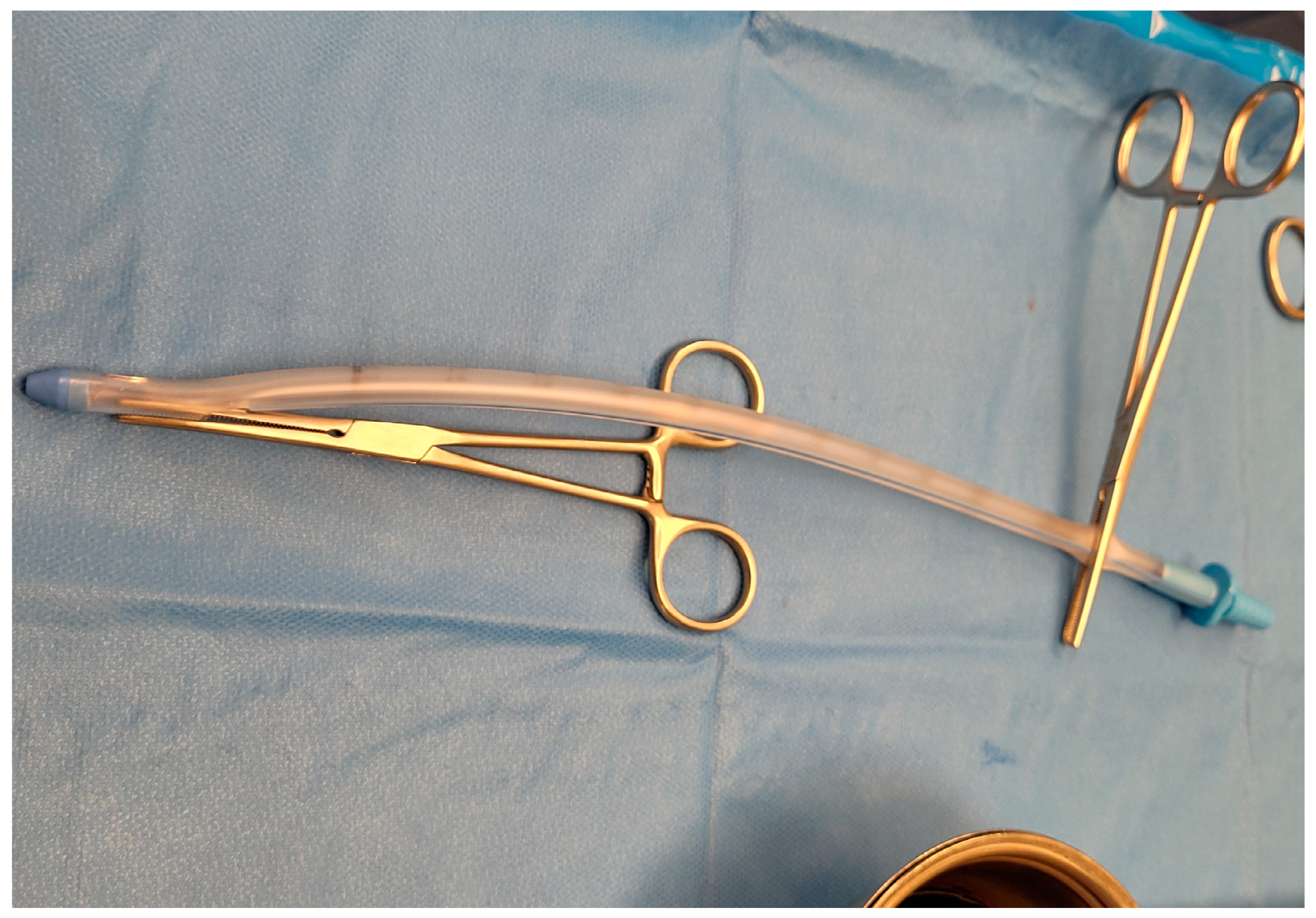
Secure the distal end of the tube with a clamp, and clamp the proximal end to use the instrument as a guide to advance the drain into the pleural space. Advance the tube aiming for the supraclavicular fossa, removing the proximal clamp once the tube is inside the pleural cavity (Figure 7). Signs such as fogging of the chest tube and the presence of drainage material may indicate the correct position;
8.
Remove the distal clamp and connect the tube to the seal apparatus. Secure the tube with a non-absorbable suture using the shoelace technique. Apply dressing.
The Seldinger technique for small-bore chest tube and pig-tail catheters [23,42].
1.
Assemble the supplies and the drainage system;
2.
Identify the site of insertion based on diagnostic imaging and the location of the pneumothorax: 4–5th intercostal space between the anterior and midaxillary lines or 2nd–3rd intercostal space in the mid-clavicular line. If possible, the patient should be positioned with a 45–50° torso elevation, with the ipsilateral arm flexed and extended over the head. Mark the insertion site with a pen;
3.
Administer local anesthesia as previously described;
4.
Insert the introducer needle attached to a 5 mL syringe, aspirating while moving through the tissue on the superior margin of the inferior rib. Aspiration of air confirms the appropriate location for catheter insertion and ensures accurate access to the targeted space;
5.
Insert the guidewire through the needle, advance it into the chest cavity, and then remove the introducer needle;
6.
Make a small incision in the skin to enlarge the breach. To create an adequate pathway for the catheter insertion, a dilator may be used over the guidewire to widen the path;
7.
Introduce the pigtail catheter into the pleural space by advancing it over the guidewire, and ensure all the holes are within the space;
8.
Withdraw the trocar and the guidewire. As the catheter is released, its coiled or “pigtail” shape forms, securing it within the pleural cavity and reducing the risk of dislodgement;
9.
Connect the catheter to the drainage system, secure the drain with sutures, and apply a dressing.
Following chest drainage placement, patients should be monitored daily for air leaks and assessed for respiratory variation in the fluid column. Stable clinical conditions, the absence of air leaks for 24 to 48 h, and full lung expansion are common criteria to determine chest tube removal. A clamping trial may be performed prior to removal to assess for recurrence of the pneumothorax. It consists of a temporary tube clamping for some time, usually at least 4–6 h, to mimic tube removal. This trial allows for the identification of a possible reaccumulation of intrapleural air [43]. If the patient tolerates the clamping trial and no recurrent pneumothorax occurs, the chest tube can be safely removed. A chest tube with an air leak should not be clamped, as this may cause a tension pneumothorax [41].

4.2.2. Complications of Chest Drainage

Approximately 10% of chest tube insertions result in complications [23]. These complications can be early (within 24 h) or late (after 24 h). Early complications include organ injury (lung, liver, spleen, or diaphragm), malposition, equipment malfunction, re-expansion pulmonary edema (REPE), and bleeding. Late complications include infection (cellulitis, pneumonia, or empyema), persistent pneumothorax, and tube malposition [17,40,41]. REPE is a rare but potentially fatal complication, occurring in less than 1% of cases with a mortality rate of up to 20% [41]. Arterial injury is the most common serious complication following chest tube insertion and can lead to life-threatening hypovolemic shock or death. The intercostal neurovascular bundle runs along each rib, specifically located in the costal groove on the inferior surface of each rib and between the internal intercostal muscle and the innermost intercostal muscle. For patients with suspected intercostal artery injury, contrast-enhanced CT is useful for localizing the active bleeding site. Transarterial embolization (TAE) is a minimally invasive option for rapid diagnosis and treatment. Further surgical options are exploratory thoracotomy and video-assisted thoracoscopic surgery (VATS). US real-time assessment capability helps minimize extrathoracic placement risks, lung injuries, and malpositioning [41]. Despite being a less invasive intervention, small-caliber drains can present the same complications as larger tubes [42].

4.2.3. Role of Negative Pressure in Chest Drainage

In traumatic pneumothorax, the role of negative pleural suction is still debated and lacking since research consensus on this topic mostly pertains to elective surgeries where its use is routinely not advised, it is recommended to apply low pressure (between −10 and −20 cm H2O) when necessary [44]. One recent prospective randomized study found no notable difference in LOS and complication rate [45]. In contrast, Arora et al. demonstrated the beneficial effect of negative pressure suction (maintained at −20 cm of H2O) in promoting lung re-expansion in patients with thoracic trauma and improving drainage efficiency compared to conventional intercostal drainage [46]. However, in spontaneous pneumothorax, the use of negative pressure is still debated as it may keep a bronchopleural or alveolar–pleural fistula open and delay healing. Currently, there is no significant evidence for the benefits or downsides of suction in spontaneous pneumothorax [36].

4.3. Surgical Management

Surgical intervention is usually reserved for recurrent pneumothorax or cases with a persistent air leak (PAL). A PAL is usually the result of the presence of an alveolar–pleural fistula establishing communication between the alveoli and the pleural space. The persistence of the connection leads to the worsening of the pneumothorax [47]. A PAL can be classified according to its severity level, as described by Cerfolio [48]:
1.
During forced expiration;
2.
Expiration only;
3.
Inspiration only;
4.
Continuous bubbling.
Patients with an air leak lasting beyond 5 to 7 days should be evaluated for further surgical options, including pleurectomy or pleurodesis [8]. VATS or open thoracotomy are the preferred approaches, with VATS offering reduced morbidity and faster recovery. The British Thoracic Society Guidelines suggest that surgical pleurodesis is more effective in preventing recurrence than chemical pleurodesis alone [8]. However, medical chemical pleurodesis may be considered for patients who are too frail or unwilling to undergo surgical treatment. The Joint ERS/EACTS/ESTS Clinical Practice Guidelines accept both treatments, assessing them to have comparable recurrence rates and stating that there is no difference between VATS with pleurodesis and VATS alone [36].

4.4. Conservative Management

The management of traumatic pneumothorax has evolved. Over the past decade, multiple studies have questioned the necessity of thoracostomy for all traumatic pneumothoraces. Cropano et al. demonstrated that a 35 mm cutoff size of pneumothorax correlates successfully with observation in hemodynamically stable patients, whether or not they were mechanically ventilated [33]. Banks et al. found that patients with a small traumatic pneumothorax on initial CXR, treated with observation, had an average length of stay (LOS) two days shorter than similar patients treated with a thoracostomy tube [49]. Western Trauma Association Guidelines recommend observation if the pneumothorax is less than 35 mm or 20% in volume, with follow-up imaging within 6 h, knowing that 10% will fail observation [35]. Conservative management for spontaneous pneumothorax is indicated for patients who are minimally symptomatic and clinically and radiologically stable, showing reduced hospital stay length, better quality of life, and no increase in risk of recurrence [23,36]. The less contemporary guidelines of the British Thoracic Society and the American College of Chest Physicians, respectively, suggest needle decompression and insertion of a small-bore chest tube for large pneumothorax (over two cm for the British Thoracic Society and over three cm for the American College of Chest Physicians) even in paucisymptomatic patients [7,8]. However, the most recent Joint ERS/EACTS/ESTS Clinical Practice Guidelines recommend an observation period of 4 h, regardless of the size of the pneumothorax. Discharge can be considered if the pneumothorax is radiologically stable, and the patient must be able to walk without dyspnoea [36]. In any case, the treatment selection should consider the patient’s preferences; however, it is essential to clearly explain the potential risks and complications, particularly the risk of failure associated with conservative management. In contrast, secondary spontaneous pneumothorax often requires hospitalization and the placement of a small-bore chest tube due to higher failure rates. In these cases, the use of a Heimlich valve may represent an option for ambulatory management [8,23].
The current literature suggests the use of an incentive spirometer in patients with traumatic rib fractures to reduce pulmonary complications [50]. Additionally, a systematic review of PSP highlights its potential benefits in shortening both LOS and the duration of chest tube usage [51].
Oxygen therapy should be administered to hypoxemic patients with a target peripheral oxygen saturation of at least 92%. The use of oxygen therapy in non-hypoxemic patients for pneumothorax management remains debated, stressing the potential harmful effects of hyperoxemia. However, the British Thoracic Society Guidelines recommend the administration of oxygen with a target saturation of 100% to improve the clearance of pneumothorax [8]. Likewise, Park et al. demonstrated a beneficial effect of oxygen supplementation in PSP resolution [52]. This practice is based on the so-called “nitrogen wash-out” theory, sustaining that by administering oxygen, its concentration in the blood increases, thus creating a concentration gradient of nitrogen between the pleural space and the blood, consequently promoting air absorption from the pleural cavity [52].
Figure 8 and Table 2 include a management algorithm and an overview of pneumothoraces.

5. Conclusions

Pneumothorax is a common pathology in the emergency department, with its management evolving in recent years toward more conservative and minimally invasive techniques whenever safe. CT is considered the gold standard for pneumothorax detection and assessment. However, in trauma contexts and emergency situations, CXR and ultrasound represent a valuable tool. CXR can provide a broad view of the lungs, trachea, and tissues, despite its low sensitivity. US is highly sensitive, portable, and rapid, being a promising tool for pneumothorax detection, potentially becoming the new gold standard. Management of pneumothorax may be stratified on clinical status, imaging, and pathophysiology. Observation and decompression can be viable alternatives in paucisymptomatic patients, while smaller thoracic catheters and pigtail catheters are effective alternatives to large-bore thoracic catheters, reducing the risk of post-procedural pain and LOS.

Author Contributions

Conceptualization, S.F. and H.K.; methodology, G.B.; investigation, S.F.; resources, S.F. and G.B.; writing—original draft preparation, S.F. and G.B.; writing—review and editing, M.M.C. and H.K.; visualization, S.F. and G.B.; supervision M.M.C. and H.K.; project administration, H.K. All authors have read and agreed to the published version of the manuscript.

Funding

This research received no external funding.

Data Availability Statement

No new data were created or analyzed in this study. Data sharing is not applicable to this article.

Conflicts of Interest

The authors declare no conflicts of interest.

Abbreviations

The following abbreviations are used in this manuscript:
PSPPrimary Spontaneous Pneumothorax
SSPSecondary Spontaneous Pneumothorax
COPDChronic Obstructive Pulmonary Disease
ELCEmphysema-like Change
MMPMatrix Metalloproteinase
USUltrasound
E-FASTExtended Focused Assessment with Sonography for Trauma
POCUSPoint-of-care Ultrasound
CXRChest X-ray
ATLSAdvanced Trauma Life Support
PAPosteroanterior
LLLaterolateral
APAnteroposterior
CTComputed Tomography Scan
REPERe-expansion Pulmonary Edema
TAETransarterial Embolization
VATSVideo-assisted Thoracic Surgery
PALPersistent Air Leak
LOSLength of Stay

References

  1. Mendogni, P.; Vannucci, J.; Ghisalberti, M.; Anile, M.; Aramini, B.; Congedo, M.T.; Nosotti, M.; Bertolaccini, L. Epidemiology and management of primary spontaneous pneumothorax: A systematic review. Interact. Cardiovasc. Thorac. Surg. 2020, 30, 337–345. [Google Scholar] [CrossRef] [PubMed]
  2. Sahn, S.A.; Heffner, J.E. Spontaneous Pneumothorax. N. Engl. J. Med. 2000, 342, 868–874. [Google Scholar] [CrossRef] [PubMed]
  3. Gupta, D. Epidemiology of pneumothorax in England. Thorax 2000, 55, 666–671. [Google Scholar] [CrossRef]
  4. Noppen, M.; De Keukeleire, T. Pneumothora. Respiration 2008, 76, 121–127. [Google Scholar] [CrossRef] [PubMed]
  5. Bettoni, G.; Gheda, S.; Altomare, M.; Cioffi, S.P.B.; Ferrazzi, D.; Cazzaniga, M.; Bonacchini, L.; Cimbanassi, S.; Aseni, P. Successful Needle Aspiration of a Traumatic Pneumothorax: A Case Report and Literature Review. Medicina 2024, 60, 548. [Google Scholar] [CrossRef]
  6. Fattori, S.; Reitano, E.; Chiara, O.; Cimbanassi, S. Predictive Factors of Ventilatory Support in Chest Trauma. Life 2021, 11, 1154. [Google Scholar] [CrossRef]
  7. Baumann, M.H.; Strange, C.; Heffner, J.E.; Light, R.; Kirby, T.J.; Klein, J.; Luketich, J.D.; Panacek, E.A.; Sahn, S.A.; ACCP Pneumothorax Consensus Group. Management of Spontaneous Pneumothorax: An American College of Chest Physicians Delphi consensus statement. Chest 2021, 119, 590–602. [Google Scholar] [CrossRef]
  8. MacDuff, A.; Arnold, A.; Harvey, J.; BTS Pleural Disease Guideline Group. Management of spontaneous pneumothorax: British Thoracic Society pleural disease guideline 2010. Thorax 2010, 65 (Suppl. S2), ii18–ii31. [Google Scholar] [CrossRef]
  9. Ohata, M.; Suzuki, H. Pathogenesis of Spontaneous Pneumothorax. Chest 1980, 77, 771–776. [Google Scholar] [CrossRef]
  10. Noppen, M.; Dekeukeleire, T.; Hanon, S.; Stratakos, G.; Amjadi, K.; Madsen, P.; Meysman, M.; D’Haese, J.; Vincken, W. Fluorescein-enhanced Autofluorescence Thoracoscopy in Patients with Primary Spontaneous Pneumothorax and Normal Subjects. Am. J. Respir. Crit. Care Med. 2006, 174, 26–30. [Google Scholar] [CrossRef]
  11. Huan, N.-C.; Sidhu, C.; Thomas, R. Pneumothorax. Clin. Chest Med. 2021, 42, 711–727. [Google Scholar] [CrossRef] [PubMed]
  12. Gupta, N.; Seyama, K.; McCormack, F.X. Pulmonary manifestations of Birt-Hogg-Dubé syndrome. Fam. Cancer 2013, 12, 387–396. [Google Scholar] [CrossRef] [PubMed]
  13. Graham, R.B.; Nolasco, M.; Peterlin, B.; Garcia, C.K. Nonsense Mutations in Folliculin Presenting as Isolated Familial Spontaneous Pneumothorax in Adults. Am. J. Respir. Crit. Care Med. 2005, 172, 39–44. [Google Scholar] [CrossRef]
  14. Guo, Y.; Xie, C.; Rodriguez, R.M.; Light, R.W. Factors related to recurrence of spontaneous pneumothorax. Respirology 2005, 10, 378–384. [Google Scholar] [CrossRef] [PubMed]
  15. Freixinet, J.L.; Caminero, J.A.; Marchena, J.; Rodríguez, P.M.; Casimiro, J.A.; Hussein, M. Spontaneous pneumothorax and tuberculosis: Long-term follow-up. Eur. Respir. J. 2011, 38, 126–131. [Google Scholar] [CrossRef]
  16. Iqbal, B.; Hallifax, R. Pneumothorax. Medicine 2023, 51, 888–892. [Google Scholar] [CrossRef]
  17. Tran, J.; Haussner, W.; Shah, K. Traumatic Pneumothorax: A Review of Current Diagnostic Practices And Evolving Management. J. Emerg. Med. 2021, 61, 517–528. [Google Scholar] [CrossRef]
  18. Liman, S. Chest injury due to blunt trauma. Eur. J. Cardiothorac. Surg. 2003, 23, 374–378. [Google Scholar] [CrossRef]
  19. Moore, E.E.; Feliciano, D.V.; Mattox, K.L. Trauma, 8th ed.; McGraw-Hill Education: New York, NY, USA, 2017. [Google Scholar]
  20. Sharma, A.; Jindal, P. Principles of diagnosis and management of traumatic pneumothorax. J. Emerg. Trauma Shock 2008, 1, 34. [Google Scholar] [CrossRef]
  21. Barton, E.D.; Rhee, P.; Hutton, K.C.; Rosen, P. The pathophysiology of tension pneumothorax in ventilated swine. J. Emerg. Med. 1997, 15, 147–153. [Google Scholar] [CrossRef]
  22. Gargani, L.; Volpicelli, G. How I do it: Lung ultrasound. Cardiovasc. Ultrasound 2014, 12, 25. [Google Scholar] [CrossRef]
  23. Gottlieb, M.; Long, B. Managing Spontaneous Pneumothorax. Ann. Emerg. Med. 2023, 81, 568–576. [Google Scholar] [CrossRef] [PubMed]
  24. Chan, K.K.; Joo, D.A.; McRae, A.D.; Takwoingi, Y.; Premji, Z.A.; Lang, E.; Wakai, A. Chest ultrasonography versus supine chest radiography for diagnosis of pneumothorax in trauma patients in the emergency department. Cochrane Database Syst. Rev. 2020, 2020, CD013031. [Google Scholar] [CrossRef]
  25. ATLS—Advanced Trauma Life Support, 10th ed.; American College of Surgeons: Chicago, IL, USA, 2018.
  26. Aswin, K.; Balamurugan, S.; Govindarajalou, R.; Saya, G.K.; Elamurugan, T.P.; Rajendran, G.; Murugan, B.; Elamurugan, T. Comparing Sensitivity and Specificity of Ultrasonography With Chest Radiography in Detecting Pneumothorax and Hemothorax in Chest Trauma Patients: A Cross-Sectional Diagnostic Study. Cureus 2023, 15, e44456. [Google Scholar] [CrossRef]
  27. Salama, K.M.; Elshaboury, I.M.; Huissen, W.M.; Eldomiaty, H.A.; Elghoboshy, K.I. Role of bedside sonography in the assessment of patients with chest trauma in the emergency department of Suez Canal University Hospital. Int. Surg. J. 2017, 4, 465. [Google Scholar] [CrossRef]
  28. Silva, D.; Schuch; Dalcin, P.D.T.R. Posteroanterior chest X-ray for the diagnosis of pneumothorax: Methods, usage, and resolution. Rep. Med. Imaging 2010, 3, 29–34. [Google Scholar] [CrossRef]
  29. Polireddy, K.; Hoff, C.; Kinger, N.P.; Tran, A.; Maddu, K. Blunt thoracic trauma: Role of chest radiography and comparison with CT—Findings and literature review. Emerg. Radiol. 2022, 29, 743–755. [Google Scholar] [CrossRef] [PubMed]
  30. Almajid, F.M.; Aljehani, Y.M.; Alabkary, S.; Alsaif, H.S. The accuracy of computed tomography in detecting surgically resectable blebs or bullae in primary spontaneous pneumothorax. Radiol. Med. 2019, 124, 833–837. [Google Scholar] [CrossRef]
  31. Tschopp, J.-M.; Bintcliffe, O.; Astoul, P.; Canalis, E.; Driesen, P.; Janssen, J.; Krasnik, M.; Maskell, N.; Van Schil, P.; Tonia, T.; et al. ERS task force statement: Diagnosis and treatment of primary spontaneous pneumothorax. Eur. Respir. J. 2015, 46, 321–335. [Google Scholar] [CrossRef]
  32. Tsou, K.-C.; Huang, P.-M.; Hsu, H.-H.; Chen, K.-C.; Kuo, S.-W.; Lee, J.-M.; Chang, Y.-C.; Chen, J.-S.; Lai, H.-S. Role of computed tomographic scanning prior to thoracoscopic surgery for primary spontaneous pneumothorax. J. Formos. Med. Assoc. 2014, 113, 606–611. [Google Scholar] [CrossRef]
  33. Cropano, C.; Mesar, T.; Turay, D.; King, D.; Yeh, D.; Fagenholz, P.; Velmahos, G.; de Moya, M.A. Pneumothoraces on Computed Tomography Scan: Observation using the 35 Millimeter Rule is Safe. Panam. J. Trauma Crit. Care Emerg. Surg. 2015, 4, 48–53. [Google Scholar] [CrossRef]
  34. Eddine, S.B.Z.; Boyle, K.A.; Dodgion, C.M.; Davis, C.S.; Webb, T.P.; Juern, J.S.; Milia, D.J.; Carver, T.W.; Beckman, M.A.; Codner, P.A.; et al. Observing pneumothoraces: The 35-millimeter rule is safe for both blunt and penetrating chest trauma. J. Trauma Acute Care Surg. 2019, 86, 557–564. [Google Scholar] [CrossRef]
  35. De Moya, M.; Brasel, K.J.; Brown, C.V.; Hartwell, J.L.; Inaba, K.; Ley, E.J.; Moore, E.E.; Peck, K.A.; Rizzo, A.G.; Rosen, N.G.; et al. Evaluation and management of traumatic pneumothorax: A Western Trauma Association critical decisions algorithm. J. Trauma Acute Care Surg. 2022, 92, 103–107. [Google Scholar] [CrossRef] [PubMed]
  36. Walker, S.; Hallifax, R.; Ricciardi, S.; Fitzgerald, D.; Keijzers, M.; Lauk, O.; Petersen, J.; Bertolaccini, L.; Bodtger, U.; Clive, A.; et al. Joint ERS/EACTS/ESTS clinical practice guidelines on adults with spontaneous pneumothorax. Eur. Respir. J. 2024, 63, 2300797. [Google Scholar] [CrossRef]
  37. Inaba, K.; Lustenberger, T.; Recinos, G.; Georgiou, C.; Velmahos, G.C.; Brown, C.; Salim, A.; Demetriades, D.; Rhee, P. Does size matter? A prospective analysis of 28–32 versus 36–40 French chest tube size in trauma. J. Trauma Acute Care Surg 2012, 72, 422–427. [Google Scholar] [CrossRef]
  38. Tanizaki, S.; Maeda, S.; Sera, M.; Nagai, H.; Hayashi, M.; Azuma, H.; Kano, K.-I.; Watanabe, H.; Ishida, H. Small tube thoracostomy (20–22 Fr) in emergent management of chest trauma. Injury 2017, 48, 1884–1887. [Google Scholar] [CrossRef]
  39. Kulvatunyou, N.; Erickson, L.; Vijayasekaran, A.; Gries, L.; Joseph, B.; Friese, R.F.; O’Keeffe, T.; Tang, A.L.; Wynne, J.L.; Rhee, P. Randomized clinical trial of pigtail catheter versus chest tube in injured patients with uncomplicated traumatic pneumothorax. J. Br. Surg. 2014, 101, 17–22. [Google Scholar] [CrossRef] [PubMed]
  40. Sorino, C.; Feller-Kopman, D.; Mei, F.; Mondoni, M.; Agati, S.; Marchetti, G.; Rahman, N.M. Chest Tubes and Pleural Drainage: History and Current Status in Pleural Disease Management. J. Clin. Med. 2024, 13, 6331. [Google Scholar] [CrossRef]
  41. Ukeh, I.; Fang, A.; Patel, S.; Opoku, K.; Nezami, N. Percutaneous Chest Tube for Pleural Effusion and Pneumothorax. Semin. Interv. Radiol. 2022, 39, 234–247. [Google Scholar] [CrossRef]
  42. Lewis, M.R.; Georgoff, P. Minimally invasive management of thoracic trauma: Current evidence and guidelines. Trauma Surg. Acute Care Open 2024, 9 (Suppl. S2), e001372. [Google Scholar] [CrossRef]
  43. Becker, J.C.; Zakaluzny, S.A.; Keller, B.A.; Galante, J.M.; Utter, G.H. Clamping trials prior to thoracostomy tube removal and the need for subsequent invasive pleural drainage. Am. J. Surg. 2020, 220, 476–481. [Google Scholar] [CrossRef]
  44. Asciak, R.; Bedawi, E.O.; Bhatnagar, R.; Clive, A.O.; Hassan, M.; Lloyd, H.; Reddy, R.; Roberts, H.; Rahman, N.M. British Thoracic Society Clinical Statement on pleural procedures. Thorax 2023, 78 (Suppl. S3), s43–s68. [Google Scholar] [CrossRef] [PubMed]
  45. Priyadarshi, A.; Gupta, S.; Priyadarshini, P.; Kumar, A.; Alam, J.; Bagaria, D.; Choudhary, N.; Sagar, S.; Gupta, A.; Mishra, B.; et al. Role of low-pressure negative pleural suction in patients with thoracic trauma—a randomized controlled trial. Eur. J. Trauma Emerg. Surg. 2024, 50, 2105–2111. [Google Scholar] [CrossRef] [PubMed]
  46. Arora, D.; Choudhary, I.S.; Dutt, A.; Banerjee, N.; Chauhan, A.S.; Rodha, M.S.; Sharma, N.; Puranik, A.K.; Chauhan, N.K.; Gupta, M.K.; et al. Efficacy of slow negative pleural suction in thoracic trauma patients undergoing tube thoracostomy—A randomised clinical trial. Injury 2025, 56, 111928. [Google Scholar] [CrossRef] [PubMed]
  47. Dugan, K.C.; Laxmanan, B.; Murgu, S.; Hogarth, D.K. Management of Persistent Air Leaks. Chest 2017, 152, 417–423. [Google Scholar] [CrossRef]
  48. Cerfolio, R.J. Advances in thoracostomy tube management. Surg. Clin. N. Am. 2002, 82, 833–848. [Google Scholar] [CrossRef]
  49. Banks, K.C.; Mooney, C.M.; Mazzolini, K.; Browder, T.D.; Victorino, G.P. Comparison of outcomes between observation and tube thoracostomy for small traumatic pneumothoraces. Am. J. Emerg. Med. 2023, 66, 36–39. [Google Scholar] [CrossRef]
  50. Sum, S.-K.; Peng, Y.-C.; Yin, S.-Y.; Huang, P.-F.; Wang, Y.-C.; Chen, T.-P.; Tung, H.-H.; Yeh, C.-H. Using an incentive spirometer reduces pulmonary complications in patients with traumatic rib fractures: A randomized controlled trial. Trials 2019, 20, 797. [Google Scholar] [CrossRef]
  51. Namwaing, P.; Ngamjarus, C.; Sakaew, W.; Sawunyavisuth, B.; Sawanyawisuth, K.; Khamsai, S.; Srichaphan, T. Chest physical therapy and outcomes in primary spontaneous pneumothorax: A systematic review. J. Med. Assoc. Thail. 2021, 104, S165–S168. [Google Scholar] [CrossRef]
  52. Park, C.B.; Moon, M.H.; Jeon, H.W.; Cho, D.G.; Song, S.W.; Won, Y.D.; Kim, Y.H.; Kim, Y.-D.; Jeong, S.C.; Kim, K.S.; et al. Does oxygen therapy increase the resolution rate of primary spontaneous pneumothorax? J. Thorac. Dis. 2017, 9, 5239–5243. [Google Scholar] [CrossRef]
Figure 1. Methodology flowchart.
Ecm 02 00024 g001
Figure 2. Normal lung on US.
Ecm 02 00024 g002
Figure 3. Barcode sign of pneumothorax on US.
Ecm 02 00024 g003
Figure 4. CXR of right pneumothorax.
Ecm 02 00024 g004
Figure 5. CXR of right pneumothorax with chest drain.
Ecm 02 00024 g005
Figure 6. CT of right pneumothorax.
Ecm 02 00024 g006
Figure 7. Clamp’s position on chest tube.
Ecm 02 00024 g007
Figure 8. Pneumothorax management algorithm.
Ecm 02 00024 g008
Table 1. Overview of US signs.
Ultrasound Signs
Normal LungPneumothorax
Lung slidingAbsence of lung sliding
Lung pulseStratosphere sign on M-mode
B-linesLung point
Seashore sign on M-modeVisible air in pleural cavity
Table 2. Overview of pneumothorax types.
Type of PneumothoraxClinical FeaturesDiagnostic CriteriaTherapeutic IndicationsPossible Complications
Primary SpontaneousYoung, healthy patients in absence of clinically apparent lung diseasePOCUS, Chest X-ray, CT in complicated PSPObservation in minimally symptomatic; oxygen therapy, aspiration, and small-bore chest tube if symptomatic Recurrence, infection, PAL
Secondary SpontaneousPatients over 55 y.o. with underlying lung disease (e.g., COPD)POCUS, Chest X-ray, CT often requiredSmall-bore chest tube; if unsuccessful, consider a larger caliberPAL, recurrence, infection
TraumaticPenetrating or blunt chest traumaE-FAST, Chest X-ray, CTObservation if <35 mm on CT, chest tube if >35 mm. Treatment of the concomitant injuriesTension pneumothorax, infection, pain
TensionHemodynamic instability, acute dyspnea, tracheal deviationClinical diagnosisEmergency decompression with finger thoracostomy Cardiac arrest, death
Disclaimer/Publisher’s Note: The statements, opinions and data contained in all publications are solely those of the individual author(s) and contributor(s) and not of MDPI and/or the editor(s). MDPI and/or the editor(s) disclaim responsibility for any injury to people or property resulting from any ideas, methods, instructions or products referred to in the content.

