Review

Techniques for Evaluating Airborne Biocrust Diaspores: From Fundamentals to Advanced Approaches

by
Mateus Fernandes Oliveira
1,*,† and
Adaíses Simone Maciel-Silva
1,2,*,†
1
Programa de Pós-Graduação em Biologia Vegetal, Instituto de Ciências Biológicas, Universidade Federal de Minas Gerais, Av. Antônio Carlos, 6627, Pampulha, Belo Horizonte 31270-901, Minas Gerais, Brazil
2
Departamento de Botânica, Instituto de Ciências Biológicas, Universidade Federal de Minas Gerais, Av. Antônio Carlos, 6627, Pampulha, Belo Horizonte 31270-901, Minas Gerais, Brazil
*
Authors to whom correspondence should be addressed.
These authors contributed equally to this work.
Aerobiology 2025, 3(1), 1; https://doi.org/10.3390/aerobiology3010001
Submission received: 30 September 2024 / Revised: 17 December 2024 / Accepted: 6 January 2025 / Published: 15 January 2025

Abstract

:
Biological soil crusts (biocrusts) are communities which thrive primarily in the upper soil layers of arid and semi-arid environments. Biocrusts produce soil-binding compounds, tolerate extreme conditions, and disperse through both sexual and asexual diaspores via wind, water, or animals. Despite their significance, dispersal mechanisms involving airborne diaspores in biocrusts remain largely unexplored and poorly understood. This review provides an overview of techniques, from basic to advanced, to help researchers investigate these often-overlooked aspects of biocrust ecology. We discuss both passive and active methods for sampling airborne organisms, highlighting their potential in studies of biocrust organisms. We present traditional techniques, such as microscope glass slides coated with adhesive substances, as well as more advanced equipment like Rotorods. For organism identification, we explore traditional morphological methods, but also introduce more modern approaches, such as metabarcoding, which allow for the simultaneous study of multiple organism groups. This review underscores the potential of these methods to enhance our understanding of the aerobiology of biocrusts. By shedding light on these dispersal dynamics, this review aims to support future research and foster advancements in biogeography, ecosystem restoration, and conservation strategies.

1. Introduction

Biological soil crusts (hereafter referred to as biocrusts; Figure 1) are typically composed of poikilohydric organisms, including fungi (which can be free-living, lichenized, or mycorrhizal), heterotrophic bacteria and archaea, cyanobacteria, and algae from various lineages (e.g., Chlorophyta, Heterokontophyta, Charophyta), along with bryophytes, specifically mosses and liverworts [1,2,3,4,5]. These communities inhabit the uppermost millimeters of soil, either within or directly on the surface [1], producing a variety of organic compounds that bind soil particles together [6,7,8]. Depending on the dominant photosynthetic group present, biocrusts can be categorized as cyanobacteria-dominated (Figure 1A–C), lichen-dominated (Figure 1D–F), or moss-dominated (Figure 1G–I), each playing distinct ecological roles [1].
Biocrust species exhibit limited competitive ability for light, thriving particularly in ecosystems characterized by open spaces or interstitial areas between vascular plants, where sunlight can reach the soil surface [9,10,11]. Indeed, biocrusts are recognized as significant components of vegetation in arid and semi-arid ecosystems [12], such as hot and cold deserts [13,14], savannas [15,16], and dry forests [17,18]. However, they can also thrive in unique ecosystems found in tropical climates, such as rocky outcrops [19]. Additionally, biocrusts are significant in disturbed ecosystems, as disturbances like tree falls, mechanical clearing, and fires expose bare soil, allowing for rapid colonization by moss-dominated biocrusts [20,21]. Similarly, in coastal dune areas, factors such as coastal storms, sand movement, and salt spray stress vegetation, creating ideal conditions for cyanobacteria-dominated biocrusts to establish themselves [22,23].
Covering approximately 12% of the Earth’s terrestrial surface [24], biocrusts play an important role in the biogeochemical dynamics of the ecosystems’ soils [9,12], contributing to nitrogen and carbon fixation [25,26,27,28] and affecting the phosphorus cycle [29,30]. Additionally, they facilitate soil aggregation [31,32], enhance water infiltration, and improve soil moisture retention [33,34]. Biocrusts are also crucial for the success of other species, as they directly interact with seed establishment [35,36] and offer both food and habitat for various microanimals [37,38]. Precisely because of these diverse functions, biocrusts are recognized as ecosystem engineers [39,40], actively shaping ecological processes and becoming of great scientific interest.
In addition to their critical ecological functions, biocrusts are recognized for their resilience to extreme conditions, including water scarcity, elevated temperatures, and excessive light [9,12]. Many species found in biocrusts exhibit a range of morphological and physiological adaptations that facilitate their survival and proliferation in these harsh environments [41,42]. In fact, many biocrust organisms are poikilohydric and can tolerate prolonged desiccation [43]. Additionally, some species of cyanobacteria, algae, and lichens acquire photoprotection through the presence of sunscreen pigments [44,45,46]. Notably, bryophytes exhibit various morphological adaptations which enhance water retention and distribution, including hyaline hair points, lamellae, papillae, and alar cells in their leaves [47,48].
Biocrust species not only exhibit a range of morphological and physiological adaptations but also possesses unique reproductive strategies and dispersal mechanisms that enhance their persistence and distribution in harsh environments. Indeed, biocrust organisms disperse through diaspores, which are reproductive or propagative structures, via both sexual and asexual propagation through wind, water, or animals [49]. Cyanobacteria and algae are recognized for their wind dispersal capabilities, making them particularly intriguing due to their ability to colonize new environments [50,51]. Lichens and bryophytes typically disperse short distances through vegetative propagules or fragmentation [48,52,53], while these structures can also be transported over relatively long distances by the wind [54,55]. On the other hand, the spores of many bryophyte and lichen species can travel long distances through the air [56,57,58,59]. Despite their importance, dispersal mechanisms involving airborne diaspores in biocrusts remain largely unexplored and poorly understood.
While many studies on airborne organisms do not explicitly reference biocrusts, many species identified in these airborne analyses have been previously documented within biocrust communities. As an example, the alga Chlorella vulgaris Beijerinck, which is commonly found in biocrusts globally [2,60], has also been recorded in airborne samples [61]. This overlap highlights the ecological connections between airborne propagules and biocrusts, suggesting that understanding the dynamics of airborne organisms can provide crucial insights into the composition, distribution, and resilience of biocrust species. To date, only two recent revision studies have emphasized aerobiology (the study of airborne organisms and particles) as a crucial area for improving our understanding of biocrusts [62,63].
Within this framework, our review encourages further research in aerobiology, with a focus on photosynthetic biocrust organisms (cyanobacteria, algae, lichens, and bryophytes). We begin by detailing how these organisms reproduce and the processes by which they become viable airborne biocrusts. Following that, we provide an overview of essential techniques, ranging from basic to advanced, to assist researchers in investigating these often-overlooked aspects of biocrust ecology. This includes methods for sampling, culturing, and identifying biocrust organisms. By integrating aerobiology into biocrust research, we gain a deeper understanding of their dispersal mechanisms and ability to colonize new areas, which is crucial for biogeography and fundamental for ecosystem recovery and restoration.

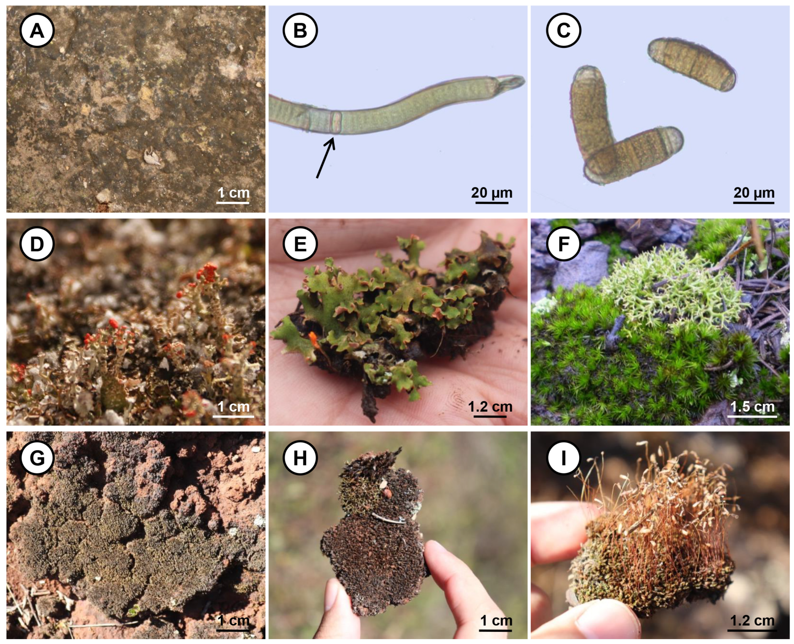
2. From Soil to Air: The Emergence and Viability of Airborne Biocrusts

To understand how the species present in biocrusts can be transported through the air, it is important to comprehend aspects of morphology and how each taxonomic group reproduces (Figure 2). Different dispersal strategies, such as the production of spores and vegetative fragments, influence each group’s ability to become airborne [62,63]. Additionally, tolerance to adverse atmospheric conditions, such as desiccation, UV radiation, and temperature variations, is crucial for the survival of these organisms during transport [64,65]. Environmental factors, such as wind strength and direction, also play an important role in the aerial transport of biocrusts, varying across different climates and regions [62,63,64,65]. In summary, understanding these aspects is fundamental to grasp how biocrust organisms disperse through the air and colonize new habitats.
Cyanobacteria are prokaryotic, photosynthetic organisms characterized by simple morphologies, such as coccoid, filamentous, or colonial forms [66,67]. In biocrusts, filamentous cyanobacteria serve as early colonizers, using their filaments and exuded organic material to stabilize the soil and create favorable microclimates for the establishment of other organisms [2,6]. Reproduction in cyanobacteria is strictly asexual, occurring through methods such as budding, binary fission, or specialized structures like akinetes and hormogonia, which act as effective propagules [66]. These structures, along with filaments and cell clusters from biocrusts, can be dispersed through the air, facilitating the spread of cyanobacteria over different environments [49,62].
Unlike cyanobacteria, algae are eukaryotic organisms [68]. The term “algae” refers to a broad group of organisms that are not necessarily phylogenetically related [69]. Chlorophyta, Charophyta, and Heterokontophyta (diatoms) are the most representative phyla in biocrusts [2]. Green algae, from Chlorophyta and Charophyta, share similar traits, displaying simple morphologies such as coccoid, filamentous, or colonial forms, and propagate through zygotes, asexual spores, or filament fragments [70]. In contrast, diatoms are unicellular or colonial algae characterized by frustules, which are silica-based two-part cell walls [71]. Diatoms can produce resistant structures known as auxospores to withstand unfavorable conditions, and they can reproduce both sexually and asexually [72]. Both green algae and diatoms presenting in biocrusts are capable of dispersing their propagules via air, including through resistant cells and fragments [49,62].
Lichens are complex organisms formed by a symbiotic relationship between fungi and photosynthetic partners, typically green algae or cyanobacteria [5,73,74]. Lichen thalli exhibit remarkable diversity in morphology, categorized into crustose (flat and adherent), foliose (leaf-like and lobed), fruticose (shrub-like and branched), gelatinous (moisture-responsive), and squamulose (scale-like overlapping lobes), allowing them to thrive in various habitats and ecological roles [75]. Some lichens reproduce asexually by utilizing unspecialized thallus fragments or by forming specialized asexual structures, such as soredia and isidia, which contain cells from both the fungal and photosynthetic partners [76]. These specialized structures can be readily dislodged from the surface of the thallus by environmental factors like wind and rain [62,63,77]. Lichens can also produce pycnidia, specialized flask-shaped structures that generate asexual spores known as conidia [78]. Sexual reproduction in lichens is limited to the fungal component, which produces sexual spores called ascospores [74,76] that can be readily dispersed over long distances through the air [79].
Within biocrusts, mosses and liverworts are the most representative organisms of bryophytes, with mosses being even more dominant due to their adaptability to a wide range of environmental conditions [4,39]. Mosses are characterized by leafy gametophytes, while liverworts can have either leafy or thallose gametophytes with a dorsiventral orientation [48]. Both mosses and liverworts exhibit various propagation methods, including regeneration from specialized caducous structures such as leaves, leaf apices, shoots, branches, and bulbils, and the production of specialized propagules like gemmae, protonemal brood cells, and tubers [59,80,81,82]. They can also reproduce through fragmentation, where parts of the plant break into unspecialized fragments, and through clonal reproduction [59,82]. Sexual reproduction in bryophytes occurs through the formation of gametes in specialized structures, resulting in the production of sporophytes that develop spores germinating into new individuals [80]. Spores are the structures most readily dispersed by wind, facilitating their colonization of new environments [59,80,81,82,83,84]. Despite their effective dispersal, however, the distribution of bryophytes is influenced by multiple factors, including habitat specificity, substrate availability, and microclimatic conditions, which can limit their widespread occurrence and contribute to their often patchy distribution [83,84,85,86,87].
All diaspores identified in biocrust organisms possess significant potential to enter the atmosphere, becoming dislodged from the biocrusts and lifted into the air by the wind [62,63]. Furthermore, biocrusts can be eroded and fragmented into fine dust particles, which contribute organic material to dust transported over long distances during storms [65,88,89]. As a result, these organisms can be transported over long distances, aiding in their dispersal [62,63]. However, it is important to note that structures present in the air may not necessarily remain viable, as they encounter multiple stressors in the atmosphere [64,65]. These stressors can include fluctuations in temperature, humidity, and exposure to UV radiation, all of which can adversely affect the survival of these airborne reproductive units. Indeed, experiments reveal that certain species are more sensitive to these extreme conditions. For instance, gametophytes of the moss Sphagnum girgensohnii Russow could not withstand two hours of stratospheric exposure, showing no signs of regeneration [90]. Spores of four species belongs to the moss genus Orthotrichum showed decreased spore germination and increased ultrastructural damage with higher UV doses [91].
Although some species may not tolerate the adverse conditions of the atmospheric environment, most studies indicate the presence of viable particles of cyanobacteria, algae, lichens, and bryophytes in the air. Additionally, some experiments assess the tolerance of various organisms and their reproductive structures to atmospheric stressors. Species of cyanobacteria such as Phormidium angustissimum West & G.S.West and Chroococcus minor (Kützing) Nägeli have been successfully cultured and identified in airborne samples collected from an office building in Malaysia [61]. The filamentous algae Planktonema lauterbornii Schmidle was recorded from clouds above Antarctica [92]. Ascospores from lichen-forming fungi in the Teloschistaceae family represented 0.04% of the total spores recorded during an aerobiological monitoring study conducted in Italy [56]. Among the mosses, Sphagnum magellanicum Brid., S. fallax (H. Klinggr.) H. Klinggr., and Atrichum angustatum (Brid.) Bruch & Schimp. demonstrated resilience to stratospheric exposure, showing initial stages of regeneration [90]. Furthermore, the moss spores of Funaria hygrometrica Hedw. and Pogonatum inflexum (Lindb.) Sande Lac. exhibited tolerance to thermal fluctuations (ranging from 80 °C to −80 °C) and UV treatments, with spores germinating successfully [93]. Remarkably, F. hygrometrica not only germinated but also grew into mature gametophytes that produced sporophytes and viable new spores [93].

3. Techniques for Sampling Airborne Biocrusts

To study biological particles in the air, they must be captured using a sampling device specifically designed to enhance visibility under a microscope, enable cultivation, and allow for processing of samples for genomic analyses. Various methodologies are available for studying airborne organisms, and we aim to compile techniques to encourage further research in aerobiology with a focus on photosynthetic biocrust organisms. For further detail or refinement of these techniques, we recommend consulting the referenced works in this section for a more comprehensive understanding, particularly those that specifically address organisms that are potentially part of biocrusts (Table 1). While existing studies may not focus directly on biocrusts, they investigate taxonomic groups commonly found within these communities. As such, they provide valuable insights into how airborne biocrust organisms can be studied in relation to specific issues like geographic distribution, dispersal mechanisms, and ecological dynamics.
When collecting airborne biological particles, it is essential to understand their behavioral characteristics. Particle size, especially for those under 100 μm in diameter, is the critical factor in selecting an appropriate sampling methodology [94]. Inadequately designed sampling equipment can lead to substantial loss of particles before analysis, ultimately undermining the integrity of both quantitative and qualitative data [95]. Most cyanobacteria and algae typically have a size range of 1 to 150 μm for unicellular organisms and from 2 to 300 μm for clusters of filamentous and colonial forms [96]. Lichen ascospores have an average diameter of 30 μm [97], while soredia generally range in size from 30 to 100 μm [54]. It has been estimated that a 20 μm moss spore could be carried by wind over distances of 1000 km, while a 55 μm spore may only reach approximately 40 km; in contrast, spores of Archidium sp., with a diameter of about 250 μm, are unlikely to disperse more than one meter under similar environmental conditions [98]. In addition, all asexual diaspores are quite light, as the components of biocrusts are poikilohydric and lose water from their bodies [1,2,3,4,5], making these structures easily carried by the wind [62,63]. Finally, fragments of both micro- and macroorganisms within biocrusts can be dispersed [49], significantly contributing to bioaerosol loads in the atmosphere [62]. A thorough understanding of particle dispersion dynamics is crucial for accurate ecological assessments and studies of airborne biological diversity, while understanding particle sizes is essential for determining the most effective sampling methods for the target organism group.
Another important point to note is that the composition and quantity of airborne biological particles can fluctuate significantly based on various factors, including the time of day, prevailing weather conditions, seasonal changes, and specific geographical locations [65,99,100]. For instance, increased rainfall was associated with higher relative abundance of cyanobacteria in airborne samples from Hawaii [101]. Additionally, a study conducted in an inland temperate region identified the temporal variation in microalgae and cyanobacteria, revealing that, although present throughout the year, the highest abundance was observed in February and April, underscoring the influence of environmental conditions, such as wind speed and relative humidity, on the distribution of these airborne organisms [102]. Humidity can influence spore release regulation in mosses, as demonstrated in Brachythecium populeum (Hedw.) Schimp., where the outer teeth of the capsule uniquely respond to moisture by bending inward to close when wet and flicking outward to open when dry, thus dispersing the spores [103]. Moreover, the oscillation of moss sporophytes is a key factor in controlling spore dispersal, with both air turbulence and the length of the sporophyte affecting when this movement begins [104]. Consequently, careful consideration must be given to the selection of sampling devices, sampling sites, timing, and duration of operations to ensure that a representative sample is collected [99].

3.1. Passive Traps

One of the simplest methods for collecting airborne biological particles involves using microscope glass slides [105]. In this technique, one side of the slide is coated with an adhesive substance, such as glycerin, and positioned horizontally with the sticky surface facing upward [81,105]. The slide can then be easily transported to the laboratory for observation under a microscope [105]. As an example, exposed sticky slides have been employed to assess moss and lichen dispersal in Antarctica [106]. This technique is also effective for studying spore release in mosses, having been utilized specifically on Polytrichum commune Hedw. [107] and Atrichum angustatum (Brid.) Bruch & Schimp. [108].
An enhancement over this basic method is the Durham sampler, often referred to as the gravity slide sampler [109,110]. This improved technique features a more sophisticated setup that may include a support structure designed to maintain the slide’s optimal orientation relative to the wind and turbulence, effectively influencing the sedimentation process [105,109,110]. While the Durham sampler is primarily recommended for qualitative assessments, its limitation lies in the unknown exact volume of air sampled, which restricts its reliability for detailed quantitative analysis [105]. Nonetheless, it remains valuable in ecological studies, particularly in understanding airborne propagules’ dispersal and dynamics [105,109]. This technique has been successfully employed to capture airborne pollen [111,112], but studies applying this method for evaluating cryptogamic airborne organisms, such as cyanobacteria [113], are limited.
Another specialized device designed for collecting airborne biological particles is the Tauber trap [114]. The Tauber trap is a cylindrical device, typically made of plastic or glass, that captures airborne spores and particles by drawing them into a collecting chamber with a sticky surface coated in adhesive [81,105,109,114]. The primary use of the Tauber trap is in ecological and environmental studies, where it helps monitor the dispersal of pollen [115,116]. However, it was already used for studies with bryophytes [117,118]. The Tauber trap has limitations, as it primarily yields qualitative data, making it difficult to accurately quantify captured biological particles [105]. Its performance can also be affected by environmental factors like wind speed, humidity, and temperature, and it may collect contaminants if not maintained properly, which can compromise the results [105,109].
The most commonly used method for sampling spores and other diaspores in the atmosphere, known as culture-plate sampling, involves placing open Petri dishes containing nutrient agar at various heights and distances from a known source [81,105,109]. This setup allows airborne particles to settle on the medium, facilitating the capture and subsequent cultivation of microorganisms and diaspores for identification and analysis [105,109]. The typical exposure time for these samples ranges from 10 to 30 min, although adjustments may be made for shorter or longer durations based on specific environmental conditions [105]. By varying the placement of the dishes, researchers can assess the dispersal patterns of different species across a given area [81]. However, similar to the Durham sampler and Tauber trap, this approach has its limitations, primarily in its use for qualitative rather than quantitative assessments [105]. This method has been successfully employed for the collection of airborne cyanobacteria and microalgae [116,119], as well as for the dispersal studies of bryophytes and lichens [106,120,121]. While solid media are commonly utilized in this method, adaptations can be made by using liquid media, which facilitate material transfer and also limit the growth of bacteria and fungi [122].
Another innovative method for the passive capture of airborne organisms involves the use of acetate wool and cloths in specialized traps [119,123]. Acetate wool is utilized in a trap designed with a flowerpot, where the wool is placed at the opening, allowing airborne particles, such as pollen, to settle passively onto the material [123]. To analyze the captured airborne biological particles, the acetate wool is dried and dissolved in acetone, and the resulting samples are dehydrated and mounted in silicone oil for observation [123]. In contrast, cloth traps consist of a square aluminum frame covered with a white flannel cloth, supported by a stainless steel mesh to prevent contamination from the ground, and anchored securely to the soil for stability [117]. To extract spores from the cloth for microscopic observation, the cloth is cut into pieces, and the cellulose is dissolved while preserving the spores [117]. The mixture is then centrifuged to separate the spores, which are washed with distilled water and finally suspended in silicone oil for examination under a microscope [117]. For instance, acetate wool traps have been evaluated for their effectiveness in capturing pollen [123], while cloth traps are used for collecting bryophyte spores [117].
Diaspore traps can be constructed using mats of artificial turf attached to 1-inch-thick plywood sheets [55]. Designed for bryophyte collection, each trap includes a filter paper-lined tray at the bottom and is anchored with two 50 cm metal stakes for added stability [55]. When positioned vertically on a summit, the traps capture wind-dispersed diaspores, which accumulate in the trays through gravity and precipitation [55]. A total of 6130 gametophytic fragments, representing 26 species of mosses and liverworts, were collected. The majority of the samples were individual leaves (65%) and leafy branch fragments (32%), with spore production being uncommon to rare [55]. These results are promising for collecting asexual diaspores of both mosses and liverworts, and the diaspore traps could be readily adapted to collect lichen soredia, isidia, and thallus fragments.

3.2. Active Traps

Active traps are devices designed to capture airborne biological particles by actively drawing in air or utilizing a mechanism to trap organisms [109]. Unlike passive traps, which rely on natural airflow for particles to settle, active traps often employ methods such as suction fans, mechanical devices, or specific sampling protocols to enhance the capture efficiency [105,109]. These traps can provide a more controlled sampling environment, allowing researchers to collect and analyze airborne organisms in a targeted manner [109]. The mechanisms involved in active trapping for airborne biological particles include fans or blowers to direct airflow, vacuum systems to capture particles in filters, centrifugation to separate particles using centrifugal force, filtration with varying pore sizes, impact traps with adhesive surfaces, and condensation traps that cool air to collect particles [105,109]. These mechanisms can be used individually or in combination for enhanced efficiency.
A common active air sampling device is the Rotorod, also known as the whirling arm trap, which features a rotating rod coated with a sticky substance that captures airborne particles as air is drawn through the sampler [105,109,124,125,126]. The trap consists of a pair of vertical arms that spin rapidly powered by a small electric motor [124]. Typically shaped like a ‘U’ or ‘H’, the arms’ rotation causes particles to adhere to the sticky tape on their leading edges [105,109,125]. This spinning action generates centrifugal force, enhancing particle capture efficiency and facilitating effective air quality monitoring for further sample analysis [105]. Compact and lightweight, the device is designed for brief operation (a few minutes), making it ideal for exploratory sampling in various environments [109]. The use of the Rotorod sampler includes investigating the dispersal of algae, cyanobacteria, lichens, and bryophytes across various ecosystems, encompassing both natural environments [54,127,128,129], and urban settings [51,130,131]. This adaptability enables the study of different ecological conditions and dispersal dynamics in locations with distinct characteristics.
The Hirst impact sampler, developed in 1952, is a device designed to measure the atmospheric concentration of pollen, spores, and other biological particles over time through morphological identification [132]. It features a single-stage impactor with a 2 × 14 mm slit, allowing sampled air to flow smoothly and impact a collection surface, such as a microscope slide or plastic tape, moving at 2 mm per hour [99,105,109,132]. This device can operate continuously for up to a week, providing hourly deposition data while being wind-oriented for optimal sampling efficiency [132]. Particle identification is conducted using a light microscope in conjunction with the relevant literature for reference [99], while DNA-based methods can also be employed for more precise identification of airborne biological particles [133]. The Hirst impact sampler has already been employed to investigate airborne diaspores of cyanobacteria and algae [103,133], lichens [56], and bryophytes [103,133,134]. This equipment is also used for general air monitoring, detecting pollen grains, fungal spores, and cyanobacterial diaspores [135].
The Burkard trap was developed in the mid-20th century as an evolution of the original Hirst impact sampler to enhance the monitoring of airborne biological particles [109,136]. Structurally, it consists of a motor-driven system that pulls air through a slit, where particles are trapped on an adhesive surface like plastic tape or a microscope slide [81,136]. This adhesive surface rotates slowly, allowing continuous collection of particles [136]. The tape or slides are later analyzed under a microscope to identify and quantify the biological particles, providing data on particle concentrations over time [136]. The Burkard trap provides detailed temporal data on pollen, fungal spores, and other particles, and is widely used for air quality and biological monitoring [105,109,136].
Cascade impactors are active air sampling devices specifically designed for collecting particulate matter across multiple stages [105,109]. Initially introduced in 1945 [137], these instruments utilize a series of increasingly smaller nozzles that guide air onto various deposition surfaces [105,109]. Larger particles are captured in the initial stages, while smaller ones are sorted and collected in subsequent stages, allowing for effective size-based separation [105,109,137]. The Andersen sampler, widely employed in aerobiological studies, features plates with 400 holes at each stage, placed above Petri dishes containing solid agar culture medium [138]. Modifications, such as the incorporation of liquid media in Petri dishes, can enhance the efficiency of particle capture, particularly for cyanobacteria and algae [139]. This adaptation allows for better retention and cultivation of these microorganisms, improving the overall effectiveness of the sampling process.
In addition to the primary devices used for airborne biological particle capture, advanced techniques are being developed to address evolving research demands. A notable example is a device designed specifically for the direct detection of airborne dispersal in lichens [140]. This device is optimized for trapping hydrophobic fungal propagules and airborne particles on impact, while consistently processing high air volumes [140]. It operates continuously for up to 24 h or intermittently for 72 h, offering durability, portability, and low cost, and supports DNA-based diagnostics with minimal transfer loss, allowing extensive field deployment [140]. Additionally, this was the first attempt to use mechanized propagule traps in combination with DNA diagnostics to detect lichen dispersal [140]. Theresults offer promising insights for future studies on the spread of lichen epiphytes and other passively dispersed microscopic organisms, including fungi and bryophytes [140], potentially opening new avenues for ecological research on microscopic dispersal processes.
Table 1. References applying aerobiological techniques for sampling airborne organisms and their diaspores.
Aerobiological TechniquesOrganismsReferences
Microscope glass slidesBryophytes[106,107,108]
Durham samplerCyanobacteria[113]
Tauber trapBryophytes[117,118]
Culture-plate samplingCyanobacteria, algae,
bryophytes, lichens
[106,116,119,120,121]
Specialized trap with clothsBryophytes[117]
Diaspore trapBryophytes[55]
RotorodCyanobacteria, algae,
bryophytes, lichens
[51,54,127,128,129,130,131]
Hirst impact samplerCyanobacteria, algae,
bryophytes, lichens
[56,133,134,135]

4. From Air to Laboratory: Sifting Through Biological Airborne Samples

Most of the techniques presented for collecting diaspores of cyanobacteria, algae, lichens, and bryophytes from the air involve a straightforward laboratory sorting process. Microscope glass slides can be taken directly from the field to the laboratory, enabling nearly immediate observation under the microscope [81,105]. The same applies to samples collected using Durham samplers [109,110] and Tauber traps [109,114]. In contrast, some sampling methods necessitate prior preparation of culture media, whether solid or liquid [99,105,109]. These media can serve as the adhesive substrate for the device or be used to cultivate the samples for future identification. Specifically, the culture-plate sampling technique always requires the production of a culture medium tailored to the specific group of organisms being studied [81,105,109], whether cyanobacteria, algae, fungi, lichens, or bryophytes. In other methods, such as the Rotorod, Hirst impact sampler, Burkard trap, and cascade impactors, you can choose to observe the samples immediately by scraping the material and preparing a slide, or you can opt to place the sample in a culture medium to allow the organisms to grow for proper identification [99,105,109,124,125,126,132,136,137]. In the following paragraphs, we will provide tips and references regarding culture media and identification keys specific to each group of organisms found both in airborne samples and in biocrusts.
Cultivating cyanobacteria and algae necessitates careful consideration of various specificities that differ among groups. The selection of culture medium is crucial, as distinct species may have unique nutritional requirements. For cyanobacteria, liquid media such as BG-11 [141,142,143], Z8 [144], and Gromov No. 6 [145] are commonly employed. In contrast, for algae, liquid media like Guillard [146] and Walne [147] may be more suitable, depending on the specific species being cultivated. It is important to highlight that specific media are available for soil [148], freshwater [149], and marine organisms [150], each with its own unique requirements. These specificities in culture media can be leveraged to enhance both the quality and quantity of species grown for subsequent identification. Additionally, monitoring growth and regularly replacing the medium are essential to maintain a favorable environment for the healthy development of cyanobacteria and algae. The photoperiod and temperature conditions in the cultivation room can also significantly influence the success of the culture [151,152]. Accurate identification relies on the use of identification keys [153,154,155,156,157], though seeking region-specific keys is recommended for greater precision, as they provide insights tailored to local species, enhancing identification accuracy. We also recommend consulting the AlgaeBase database (https://www.algaebase.org/), which serves as a valuable resource for confirming species identifications.
Experimental studies on lichens, particularly those focused on cultivation, often encounter significant challenges, leading to limited success [158]. Known for their difficulty in laboratory growth [159], lichens face complications primarily because fungi and their photosynthetic partners do not always thrive in the same culture medium [158]. Additionally, the diaspores of these organisms frequently lack distinct morphological characteristics, making traditional identification methods less effective and, in some cases, impossible [140]. Consequently, there is a notable scarcity of dispersal data concerning lichens, despite promising advancements in genetic identification techniques [140]. To address these challenges, it is essential to utilize available resources strategically. Conducting a survey of the species present in the area can enhance the accuracy of species determination by allowing for comparisons between the diaspores collected from airborne sampling and those identified [56]. Lichenologists commonly employ several cultivation media, including malt–yeast medium [160], Bold’s mineral medium [161], and MY10 [162], for the fungal component, as well as Detmer medium for the photosynthetic partners [163]. These strategies and resources can help bridge the significant gap in both the cultivation and identification of airborne diaspores of lichens, ultimately contributing to a better understanding of their ecology and distribution.
Moss and liverwort diaspores can be readily cultivated in the laboratory, leading to the development of adult individuals that can be taxonomically identified. The most commonly used cultivation medium is Knop’s solution [164,165], which has successfully supported the germination of both spores and asexual diaspores, such as gemmae and gametophyte fragments [166,167,168,169,170,171]. Another widely used medium for cultivating bryophytes is the Murashige & Skoog medium, known for its balanced nutrient composition that supports the healthy growth of these plants [172]. Spore identification can be quite complex, often requiring high-resolution microscopy, and in some cases, acetolysis techniques [173] and Scanning Electron Microscopy (SEM) are necessary for accurate identification. However, there are some studies with excellent photographic documentation that can assist in spore identification, particularly for genera such as Plagiochila [174] and Amphidium [175]. These resources provide valuable visual references for researchers working with bryophyte spores. For the identification of adult plants, we recommend consulting local bryofloristic surveys or taxonomic revisions, especially for families such as Dicranaceae [176], Pottiaceae [177], and Bryaceae [178], which are among the most prevalent in biocrust communities [4].
Alternatively, if the aim is to understand the overall diversity of organisms, advanced techniques such as DNA barcoding and metabarcoding might be considered. DNA barcoding involves sequencing a specific, standardized region of the genome from a single organism, with this region serving as a “barcode” that can be compared to a reference database to accurately identify the species [179]. This method is highly effective for identifying individual species, especially when visual identification is difficult. In contrast, metabarcoding is used to identify multiple species from complex environmental samples, such as soil, water, or aerobiological samples [180]. Rather than sequencing the DNA from a single organism, metabarcoding extracts and sequences DNA from an entire community of organisms by targeting short regions of the genome shared by many species, enabling the identification of all organisms present in the sample and providing a powerful tool for biodiversity assessment and ecosystem monitoring [180,181]. In fact, it has been widely used in aerobiology for identifying pollen [182,183,184,185] and fungi [185,186,187]. Cyanobacteria, algae, lichen, and bryophytes were also identified with this approach in aerobiological studies [101,133,140,188,189]. Another significant advantage is that this approach allows for the simultaneous processing of a diverse range of organisms during sampling [133,189], thereby facilitating species identification based on their genetic material. Both DNA barcoding and metabarcoding require primers, which play a crucial role in initiating the amplification of specific DNA regions [190,191]. To assist with this, Table 2 lists the most commonly used primers for each organism group.

5. Making Your Own Research Design

Conducting research in aerobiology requires a systematic approach to ensure thorough investigation and reliable results. By following a structured methodology, you can effectively address key questions regarding the dispersal of airborne organisms and their interactions within biocrust environments. Based on the information presented so far, we can outline a logical sequence to facilitate the design of your experiment on airborne biocrusts (Figure 3):
I.
Define the objective of the experiment, clearly establishing what you wish to investigate, such as the dispersal of airborne organisms within biocrust environments.
II.
Review the literature, consulting previous works to understand the methodologies employed in studies related to airborne organisms also found within biocrusts, and identify those that can be applied to your research. We have compiled many of these references in Table 1, where we categorize them by organism group and the techniques employed, providing a valuable starting point for your bibliographic research.
III.
Select the methodologies by deciding which approaches will be used. Options may include low-cost passive techniques (e.g., Durham sampler, Tauber trap, culture-plate sampling) or active techniques that require more elaborate devices (e.g., Rotorod, Hirst impact sampler, Burkard trap), followed by traditional cultivation and classical identification of organisms. Remember that to accurately identify organisms, it is often necessary to collaborate with specialist taxonomists and utilize equipment such as stereomicroscopes and microscopes, in addition to having access to identification keys. Alternatively, you can choose advanced approaches for species identification, such as DNA metabarcoding, which provides a more comprehensive analysis of community composition. In this case, Table 2 provides a useful resource for learning more about these techniques as applied to biocrust organisms, especially by presenting data on the most commonly used primers in barcoding methods for biocrust organisms.
IV.
If you plan to cultivate organisms, make sure to select growth media that are suitable for their specific requirements. Always consult the literature to identify the most appropriate conditions. Additionally, ensure that the cultivation rooms have standardized temperature and photoperiod settings that are tailored to the needs of your target organisms. Maintaining these controlled conditions is essential for achieving reliable and consistent results.
V.
Consider adapting existing methodologies to better suit your specific needs, such as modifying sampling protocols or analytical techniques that have proven effective in related studies. If required, develop new devices to address challenges encountered during data collection, as demonstrated in previous research tackling similar issues. Notably, airborne biocrust particles exhibit relatively unknown aerodynamic behaviors compared to typical bioaerosols like pollen, fungal spores, and bacteria, which can lead to challenges with existing sampling methods. Thus, we emphasize the importance of studies focusing on the aerodynamics of these diaspores. Research utilizing wind tunnels [105], for instance, may help in investigating these dynamics and facilitating the development of more efficient sampling devices.
VI.
Finally, studying biocrusts is inherently a multidisciplinary task. We strongly recommend seeking partnerships with professionals from various fields, including taxonomists, ecologists, geneticists, geologists, and even physicists. Collaborating with experts from different disciplines is crucial for overcoming challenges within the study of biocrust aerobiology, such as accurately identifying the vast diversity of organisms, sampling and analysis techniques, and technical issues related to aerodynamic sizes and behavior in the atmosphere. A multidisciplinary team makes it easier to tackle the complexity of biocrusts and the various variables involved in their analysis. This collaborative approach can not only accelerate the research process but also provide more effective and innovative solutions to the diverse obstacles encountered.
By following these steps, you will be wellequipped to design a robust experiment that contributes valuable insights into the understanding of airborne biocrusts. This structured approach not only enhances the reliability of your findings but also lays the groundwork for future research in this fascinating field, ultimately enriching our knowledge of biodiversity and ecosystem dynamics. The integration of aerobiology with biocrust research is essential, as it emphasizes the significance of atmospheric propagule dispersal in establishing and sustaining biocrust communities [62,63]. By examining biogeographical patterns through this lens, we gain valuable insights into the conditions necessary for biocrust dispersal, growth, and reproduction.
Additionally, aerobiology can be a crucial factor in understanding both passive and active recovery processes of biocrusts and their associated ecosystems. In fact, their restoration can be enhanced by leveraging natural ecological processes, promoting the deposition of airborne propagules, and minimizing anthropogenic disturbances, with success ultimately reliant on the timing and frequency of adequate precipitation in relation to the arrival of viable propagules [62]. Furthermore, capturing diaspores from the air could address a significant challenge in active recovery efforts, which currently rely on naturally occurring biocrusts, resulting in considerable pressure on these populations due to the high biomass requirements for effective field inoculation [192]. Thus, one potential solution to this challenge is the propagation of biocrusts in laboratory settings [193,194], as collecting airborne organisms could provide a sustainable means of cultivating biocrusts without depleting existing natural colonies. This approach not only alleviates pressure on natural populations but also fosters the development of diverse and resilient biocrust communities, which are essential for ecosystem restoration.

6. Conclusions

Biocrusts reproduce through various mechanisms, and their components can be aerosolized and carried into the air. However, studies focusing on this phenomenon remain limited. There are multiple methodologies available, some of which have already been tested on groups of organisms likely present in biocrusts, suggesting that these methods would be an excellent choice for future research. Despite this potential, much of the discussion surrounding airborne biocrusts remains theoretical. Through this review, we aim to encourage researchers to explore this promising avenue of investigation, as there is still much to be uncovered. Understanding the aerobiology of biocrusts could have significant implications for biogeography and environmental recovery processes. By advancing research in this area, we can enhance our knowledge of ecosystem dynamics and contribute to the development of effective conservation and restoration strategies.

Author Contributions

M.F.O. and A.S.M.-S. contributed equally to this study. All authors have read and agreed to the published version of the manuscript.

Funding

This research received no external funding.

Data Availability Statement

All data generated or analyzed during this study are included in this article.

Acknowledgments

M.F.O and A.S.M.-S. are grateful to Coordenação de Aperfeiçoamento de Pessoal de Nível Superior (CAPES, 8887.876207/2023-00) for a scholarship to the first author.

Conflicts of Interest

The authors declare no conflicts of interest.

References

  1. Weber, B.; Belnap, J.; Büdel, B.; Antoninka, A.J.; Barger, N.N.; Chaudhary, V.B.; Darrouzet-Nardi, A.; Eldridge, D.J.; Faist, A.M.; Ferrenberg, S.; et al. What is a biocrust? A refined, contemporary definition for a broadening research community. Biol. Rev. 2022, 97, 1768–1785. [Google Scholar] [CrossRef] [PubMed]
  2. Büdel, B.; Dulic, T.; Darienko, T.; Rybalka, N.; Friedl, T. Cyanobacteria and algae of biological soil crusts. In Biological Soil Crusts: An Organizing Principle in Drylands; Weber, B., Büdel, B., Belnap, J., Eds.; Springer: Cham, Switzerland, 2016; pp. 55–80. [Google Scholar]
  3. Maier, S.; Muggia, L.; Kuske, C.R.; Grube, M. Bacteria and non-lichenized fungi within biological soil crusts. In Biological Soil Crusts: An Organizing Principle in Drylands; Weber, B., Büdel, B., Belnap, J., Eds.; Springer: Cham, Switzerland, 2016; pp. 81–100. [Google Scholar]
  4. Seppelt, R.D.; Downing, A.J.; Deane-Coe, K.K.; Zhang, Y.; Zhang, J. Bryophytes within biological soil crusts. In Biological Soil Crusts: An Organizing Principle in Drylands; Weber, B., Büdel, B., Belnap, J., Eds.; Springer: Cham, Switzerland, 2016; pp. 101–120. [Google Scholar]
  5. Rosentreter, R.; Eldridge, D.J.; Westberg, M.; Williams, L.; Grube, M. Structure, composition, and function of biocrust lichen communities. In Biological Soil Crusts: An Organizing Principle in Drylands; Weber, B., Büdel, B., Belnap, J., Eds.; Springer: Cham, Switzerland, 2016; pp. 121–138. [Google Scholar]
  6. Mager, D.M.; Thomas, A.D. Extracellular polysaccharides from cyanobacterial soil crusts: A review of their role in dryland soil processes. J. Arid Environ. 2011, 75, 91–97. [Google Scholar] [CrossRef]
  7. Eldridge, D.J.; Greene, R.S.B. Microbiotic soil crusts: A review of their roles in soil and ecological processes in the range lands of Australia. Aust. J. Soil Res. 1994, 32, 389–415. [Google Scholar] [CrossRef]
  8. Garcia-Pichel, F.; Felde, V.J.M.N.L.; Drahorad, S.L.; Weber, B. Microstructure and weathering processes within biological soil crusts. In Biological Soil Crusts: An Organizing Principle in Drylands; Weber, B., Büdel, B., Belnap, J., Eds.; Springer: Cham, Switzerland, 2016; pp. 237–255. [Google Scholar]
  9. Benalp, J.; Büdel, B.; Lange, O.L. Biological soil crusts: Characteristics and distribution. In Biological Soil Crusts: Structure, Function, and Management; Benalp, J., Lange, O.L., Eds.; Springer: Berlin, Germany, 2001; pp. 3–30. [Google Scholar]
  10. Thompson, W.A.; Eldridge, D.J.; Bonser, S.P. Structure of biological soil crust communities in Callitris glaucophylla woodlands of South Wales, Australia. J. Veg. Sci. 2006, 17, 271–280. [Google Scholar] [CrossRef]
  11. Issa, O.M.; Trichet, J.; Défarge, C.; Couté, A.; Valentin, C. Morphology and microstructure of microbiotic soil crusts on a tiger bush sequence (Niger, Sahel). CATENA 1999, 37, 175–196. [Google Scholar] [CrossRef]
  12. Benalp, J.; Weber, B.; Büdel, B. Biological soil crusts as an organizing principle in drylands. In Biological Soil Crusts as an Organizing Principle in Drylands; Weber, B., Büdel, B., Benalp, J., Eds.; Springer: Cham, Switzerland, 2016; pp. 3–13. [Google Scholar]
  13. Lan, S.; Thomas, A.D.; Rakes, J.B.; Garcia-Pichel, F.; Wu, L.; Hu, C. Cyanobacterial community composition and their functional shifts associated with biocrust succession in the Gurbantunggut Desert. Environ. Microbiol. Rep. 2021, 13, 884–898. [Google Scholar] [CrossRef]
  14. Steven, B.; Lionard, M.; Kuske, C.R.; Vincent, W.F. High bacterial diversity of biological soil crusts in water tracks over permafrost in the high arctic polar desert. PLoS ONE 2013, 8, e71489. [Google Scholar] [CrossRef] [PubMed]
  15. Neher, D.A.; Walters, T.L.; Tramer, E.; Weicht, T.R.; Veluci, R.M.; Saiya-Cork, K.; Will-Wolf, S.; Toppin, J.; Traub, J.; Johansen, J.R. Biological soil crust and vascular plant communities in a sand savanna of northwestern Ohio. J. Torrey Bot. Soc. 2003, 130, 244–252. [Google Scholar] [CrossRef]
  16. Machado-de-Lima, N.M.; Fernandes, V.M.C.; Roush, D.; Ayuso, S.V.; Rigonato, J.; Garcia-Pichel, F.; Branco, L.H.Z. The compositionally distinct cyanobacterial biocrusts from Brazilian savanna and their environmental drivers of community diversity. Front. Microbiol. 2019, 10, 2798. [Google Scholar] [CrossRef] [PubMed]
  17. Maya, Y.; López-Cortés, A. Cyanobacterial microbiotic crusts in eroded soils of a tropical dry forest in the Baja California Peninsula, Mexico. Geomicrobiol. J. 2002, 19, 505–518. [Google Scholar] [CrossRef]
  18. Szyja, M.; Menezes, A.G.S.; Oliveira, F.D.A.; Leal, I.; Tabarelli, M.; Büdel, B.; Wirth, R. Neglected but potent dry forest players: Ecological role and ecosystem service provision of biological soil crusts in the human-modified Caatinga. Front. Ecol. Evol. 2019, 7, 1–18. [Google Scholar] [CrossRef]
  19. Oliveira, M.F.; Figueredo, C.C.; Konell, A.H.; Maciel-Silva, A.S. A first evaluation of biological soil crusts diversity in three distinctive rocky outcrops in Brazil. Flora 2024, 320, 152613. [Google Scholar] [CrossRef]
  20. Seitz, S.; Nebel, M.; Goebes, P.; Käppeler, K.; Schmidt, K.; Shi, X.; Song, Z.; Webber, C.L.; Weber, B.; Scholten, T. Bryophyte-dominated biological soil crusts mitigate soil erosion in a nearly successional Chinese subtropical forest. Biogeosciences 2017, 14, 5775–5788. [Google Scholar] [CrossRef]
  21. Grover, H.S.; Bowker, M.A.; Fulé, P.Z. Improved, scalable techniques to cultivate fire mosses for rehabilitation. Restor. Ecol. 2020, 28, S17–S24. [Google Scholar] [CrossRef]
  22. Mikhailyuk, T.; Glaser, K.; Tsarenko, P.; Demchenko, E.; Karsten, U. Composition of biological soil crusts from sand dunes of the Baltic Sea Coast in the context of an integrative approach to the taxonomy of microalgae and cyanobacteria. Eur. J. Phycol. 2019, 54, 263–290. [Google Scholar] [CrossRef]
  23. Corbin, J.D.; Thiet, R.K. Temperate biocrusts: Mesicc ounterparts to their better-known dryland cousins. Front. Ecol. Environ. 2020, 18, 456–464. [Google Scholar] [CrossRef]
  24. Rodriguez-Caballero, E.; Belnap, J.; Büdel, B.; Crutzen, P.J.; Andreae, M.O.; Pöschl, U.; Weber, B. Dryland photoautotrophic soil surface communities endangered by global change. Nat. Geosci. 2018, 11, 185–189. [Google Scholar] [CrossRef]
  25. Mager, D.M. Carbohydrates in cyanobacterial soil crusts as a source of carbon in the southwest Kalahari, Botswana. Soil Biol. Biochem. 2010, 42, 313–318. [Google Scholar] [CrossRef]
  26. Miralles, I.; Trasar-Cepeda, C.; Leirós, M.C.; Gil-Sotres, F. Labile carbon in biological soil crusts in the Tabernas Desert, SE Spain. Soil Biol. Biochem. 2013, 58, 1–8. [Google Scholar] [CrossRef]
  27. Zhou, X.; Tao, Y.; Yin, B.; Tucker, C.; Zhang, Y. Nitrogen pools in soil covered by biological soil crusts of different successional stages in a temperate desert in Central Asia. Geoderma 2020, 366, 114166. [Google Scholar] [CrossRef]
  28. Hu, P.; Zhang, W.; Xiao, L.; Yang, R.; Xiao, D.; Zhao, J.; Wang, W.; Chen, H.; Wang, K. Moss-dominated biological soil crusts modulate soil nitrogen following vegetation restoration in a subtropical karst region. Geoderma 2019, 352, 70–79. [Google Scholar] [CrossRef]
  29. Baumann, K.; Glaser, K.; Mutz, J.E. Biological soil crusts of temperate forests: Their role in P cycling. Soil Biol. Biochem. 2017, 109, 156–166. [Google Scholar] [CrossRef]
  30. Baumann, K.; Jung, P.; Samolov, E.; Lehnert, L.W.; Büdel, B.; Karsten, U.; Bendix, J.; Achilles, S.; Schermer, M.; Matus, F.; et al. Biological soil crusts along a climatic gradient in Chile: Richness and imprints of phototrophic microorganisms in phosphorus biogeochemical cycling. Soil Biol. Biochem. 2018, 127, 286–300. [Google Scholar] [CrossRef]
  31. Guo, Y.; Zhao, H.; Zuo, X.; Drake, S.; Zhao, X. Biological soil crust development and its topsoil properties in the process of dune stabilization, Inner Mongolia, China. Environ. Geol. 2008, 54, 653–662. [Google Scholar] [CrossRef]
  32. Eldridge, D.J.; Leys, J.F. Exploring some relationships between biological soil crusts, soil aggregation and wind erosion. J. Arid Environ. 2003, 53, 457–466. [Google Scholar] [CrossRef]
  33. Belnap, J. The potential roles of biological soil crusts in dryland hydrologic cycles. Hydrol. Process. 2006, 20, 3159–3178. [Google Scholar] [CrossRef]
  34. Shi, W.; Wang, X.P.; Zhang, Y.F.; Pan, Y.X.; Hu, R.; Jin, Y.X. The effect of biological soil crusts on soil moisture dynamics under different rainfall conditions in the Tengger Desert, China. Hydrol. Process. 2018, 32, 1363–1374. [Google Scholar] [CrossRef]
  35. Langhans, T.M.; Storm, C.; Schwabe, A. Biological soil crusts and their microenvironment: Impact on emergence, survival and establishment of seedlings. Flora 2009, 204, 157–168. [Google Scholar] [CrossRef]
  36. Li, X.R.; Jia, X.H.; Long, L.Q.; Zerbe, S. Effects of biological soil crusts on seed bank, germination and establishment of two annual plant species in the Tengger Desert (N China). Plant Soil 2005, 277, 375–385. [Google Scholar] [CrossRef]
  37. Darby, B.J.; Neher, D.A.; Belnap, J. Soil nematode communities are ecologically more mature beneath late- than early-successional stage biological soil crusts. Appl. Soil Ecol. 2007, 35, 203–212. [Google Scholar] [CrossRef]
  38. Neher, D.A.; Lewins, S.A.; Weicht, T.R.; Darby, B.J. Microarthropod communities associated with biological soil crusts in the Colorado Plateau and Chihuahuan deserts. J. Arid Environ. 2009, 73, 672–677. [Google Scholar] [CrossRef]
  39. Oliveira, M.F.; Maciel-Silva, A.S. Biological soil crusts and how they might colonize other worlds: Insights from these Brazilian ecosystem engineers. J. Exp. Bot. 2022, 73, 4362–4379. [Google Scholar] [CrossRef]
  40. Xiao, B.; Bowker, M.A.; Zhao, Y.; Chamizo, S.; Issa, O.M. Biocrusts: Engineers and architects of surface soil properties, functions, and processes in dryland ecosystems. Geoderma 2022, 424, 116015. [Google Scholar] [CrossRef]
  41. Green, T.A.; Proctor, M.C. Physiology of photosynthetic organisms within biological soil crusts: Their adaptation, flexibility, and plasticity. In Biological Soil Crusts: An Organizing Principle in Drylands; Weber, B., Büdel, B., Belnap, J., Eds.; Springer: Cham, Switzerland, 2016; pp. 347–381. [Google Scholar]
  42. Mackelprang, R.; Vaishampayan, P.; Fisher, K. Adaptation to environmental extremes structures functional traits in biological soil crust and hypolithic microbial communities. mSystems 2022, 7, e01419-21. [Google Scholar] [CrossRef] [PubMed]
  43. Bewley, J.D.; Krochko, J.E. Desiccation-tolerance. In Physiological Plant Ecology II: Water Relations and Carbon Assimilation; Lange, O.L., Nobel, P.S., Osmond, C.B., Ziegler, H., Eds.; Springer: NewYork, NY, USA, 1982; pp. 325–378. [Google Scholar]
  44. Dillon, J.G.; Castenholz, R.W. Scytonemin, a cyanobacterial sheath pigment, protects against UVC radiation: Implications for early photosynthetic life. J. Phycol. 1999, 35, 673–681. [Google Scholar] [CrossRef]
  45. Aigner, S.; Remias, D.; Karsten, U.; Holzinger, A. Unusual phenolic compounds contribute to ecophysiological performance in the purple-colored green alga Zygogonium ericetorum (Zygnematophyceae, Streptophyta) from a high-alpine habitat. J. Phycol. 2013, 49, 648–660. [Google Scholar] [CrossRef] [PubMed]
  46. Kappen, L. Response to extreme environments. In The Lichens; Ahmadjian, V., Hale, M.E., Eds.; Academic Press: New York, NY, USA, 1973; pp. 311–380. [Google Scholar]
  47. Frahm, J.P. Diversity, life strategies, origins and distribution of tropical inselbergs bryophytes. An. Del Inst. Biol. Ser. Botánica 1996, 67, 73–86. [Google Scholar]
  48. Vanderpoorten, A.; Goffinet, B. Introduction to Bryophytes; Cambridge University Press: Cambridge, UK, 2009. [Google Scholar]
  49. Warren, S.D.; St. Clair, L.L.; Stark, L.R.; Lewis, L.A.; Pombubpa, N.; Kurbessoian, T.; Stajich, J.E.; Aanderud, Z.T. Reproduction and dispersal of biological soil crust organisms. Front. Ecol. Evol. 2019, 7, 344. [Google Scholar] [CrossRef]
  50. Sharma, N.K.; Rai, A.K.; Singh, S.; Brown, R.M., Jr. Airborne algae: Their present status and relevance. J. Phycol. 2007, 43, 615–627. [Google Scholar] [CrossRef]
  51. Sharma, N.K.; Singh, S. Differential aerosolization of algal and cyanobacterial particles in the atmosphere. Indian J. Microbiol. 2010, 50, 468–473. [Google Scholar] [CrossRef] [PubMed]
  52. Rogers, R.W. Ecological strategies of lichens. Lichenologist 1990, 22, 149–162. [Google Scholar] [CrossRef]
  53. Laaka-Lindberg, S.; Korpelainen, H.; Pohjamo, M. Dispersal of asexual propagules in bryophytes. J. Hattori Bot. Lab. 2003, 93, 319–330. [Google Scholar] [CrossRef]
  54. Marshall, W.A. Aerial dispersal of lichen soredia in the maritime Antarctic. New Phytol. 1996, 134, 523–530. [Google Scholar] [CrossRef]
  55. Robinson, S.C.; Miller, N.G. Bryophyte diversity on Adirondack alpine summits is maintained by dissemination and establishment of vegetative fragments and spores. Bryologist 2013, 116, 382–391. [Google Scholar] [CrossRef]
  56. Favero-Longo, S.E.; Sandrone, S.; Matteucci, E.; Appolonia, L.; Piervittori, R. Spores of lichen-forming fungi in the mycoaerosol and their relationships with climate factors. Sci. Total Environ. 2014, 466, 26–33. [Google Scholar] [CrossRef]
  57. Hutsemekers, V.; Dopagne, C.; Vanderpoorten, A. How far and how fast do bryophytes travel at the landscape scale? Divers. Distrib. 2008, 14, 483–492. [Google Scholar] [CrossRef]
  58. Muñoz, J.; Felicísimo, Á.M.; Cabezas, F.; Burgaz, A.R.; Martínez, I. Wind as a long-distance dispersal vehicle in the Southern Hemisphere. Science 2004, 304, 1144–1147. [Google Scholar] [CrossRef] [PubMed]
  59. Frahm, J.P. Diversity, dispersal and biogeography of bryophytes (mosses). In Protist Diversity and Geographical Distribution; Foissner, W., Hawksworth, D.L., Eds.; Springer: Dordrecht, The Netherlands, 2007; Volume 8, pp. 43–50. [Google Scholar]
  60. Bowker, M.A.; Belnap, J.; Büdel, B.; Sannier, C.; Pietrasiak, N.; Eldridge, D.J.; Rivera-Aguilar, V. Controls on distribution patterns of biological soil crusts at micro- to global scales. In Biological Soil Crusts: An Organizing Principle in Drylands; Weber, B., Büdel, B., Belnap, J., Eds.; Springer: Cham, Switzerland, 2016; pp. 173–197. [Google Scholar]
  61. Chu, W.L.; Tneh, S.Y.; Ambu, S. A survey of airborne algae and cyanobacteria within the indoor environment of an office building in Kuala Lumpur, Malaysia. Grana 2013, 52, 207–220. [Google Scholar] [CrossRef]
  62. Warren, S.D.; St. Clair, L.L.; Leavitt, S.D. Aerobiology and passive restoration of biological soil crusts. Aerobiologia 2019, 35, 45–56. [Google Scholar] [CrossRef]
  63. Warren, S.D.; St. Clair, L.L. Atmospheric transport and mixing of biological soil crust microorganisms. AIMS Environ. Sci. 2021, 8, 498–516. [Google Scholar] [CrossRef]
  64. Sahu, N.; Tangutur, A.D. Airborne algae: Overview of the current status and its implications on the environment. Aerobiologia 2015, 31, 89–97. [Google Scholar] [CrossRef]
  65. Després, V.; Huffman, J.A.; Burrows, S.M.; Hoose, C.; Safatov, A.S.; Buryak, G.; Fröhlich-Nowoisky, J.; Elbert, W.; Andreae, M.O.; Pöschl, U.; et al. Primary biological aerosol particles in the atmosphere: A review. Tellus B Chem. Phys. Meteorol. 2012, 64, 15598. [Google Scholar] [CrossRef]
  66. Lee, R.E. Cyanobacteria. In Phycology, 4th ed.; Lee, R.E., Ed.; Cambridge University Press: New York, NY, USA, 2018; pp. 33–84. [Google Scholar]
  67. Whitton, B.A.; Potts, M. Introduction to the Cyanobacteria. In Ecology of Cyanobacteria II; Whitton, B.A., Ed.; Springer: Dordrecht, The Netherlands, 2012. [Google Scholar]
  68. Lee, R.E. Basic characteristics of the algae. In Phycology, 4th ed.; Lee, R.E., Ed.; Cambridge University Press: New York, NY, USA, 2018; pp. 3–29. [Google Scholar]
  69. Guiry, M.D. How many species of algae are there? A reprise. Four kingdoms, 14 phyla, 63 classes and still growing. J. Phycol. 2024, 60, 214–228. [Google Scholar] [CrossRef]
  70. Lee, R.E. Chlorophyta. In Phycology, 4th ed.; Lee, R.E., Ed.; Cambridge University Press: New York, NY, USA, 2018; pp. 139–226. [Google Scholar]
  71. Lee, R.E. Heterokontophyta—Bacillariophyceae. In Phycology, 4th ed.; Lee, R.E., Ed.; Cambridge University Press: New York, NY, USA, 2018; pp. 369–408. [Google Scholar]
  72. Edlund, M.B.; Stoermer, E.F. Ecological, evolutionary, and systematic significance of diatom life histories. J. Phycol. 1997, 33, 897–918. [Google Scholar] [CrossRef]
  73. Lücking, R.; Hodkinson, B.P.; Leavitt, S.D. Corrections and amendments to the 2016 classification of lichenized fungi in the Ascomycota and Basidiomycota. Bryologist 2017, 120, 58–69. [Google Scholar] [CrossRef]
  74. Lutzoni, F.; Miadlikowska, J. Lichens. Curr. Biol. 2009, 19, R502–R503. [Google Scholar] [CrossRef]
  75. Jahns, H.M.; Galun, M. The Lichen Thallus. In Handbook of Lichenology; Galun, M., Ed.; CRC Press: Boca Raton, FL, USA, 1988; Volume 1, pp. 95–143. [Google Scholar]
  76. Blackwell, M.; Alexopoulos, C.J.; Mims, C.W. Introductory Mycology; Wiley: New York, NY, USA, 1996. [Google Scholar]
  77. Heinken, T. Dispersal patterns of terricolous lichens by thallus fragments. Lichenologist 1999, 34, 603–612. [Google Scholar] [CrossRef]
  78. Vobis, G.; Hawksworth, D.L.; Cole, G.T.; Kendrick, B. Conidial lichen-forming fungi. In Biology of Conidial Fungi; Cole, G.T., Kendrick, B., Eds.; Academic Press: San Diego, CA, USA, 1981; Volume 1, pp. 245–273. [Google Scholar]
  79. Marshall, W.A. Seasonality in Antarctic airborne fungal spores. Appl. Environ. Microbiol. 1997, 63, 2240–2245. [Google Scholar] [CrossRef] [PubMed]
  80. Maciel-Silva, A.S.; Pôrto, K.C. Reproduction in Bryophytes. In Reproductive Biology of Plants; Ramawat, K.G., Merillon, J.M., Shivanna, K.R., Eds.; CRC Press: Boca Raton, FL, USA, 2014; pp. 57–81. [Google Scholar]
  81. Glime, J.M. Adaptive strategies: Vegetative vs sexual diaspores. In Bryophyte Ecology; Glime, J.M., Ed.; Michigan Technological University: Houghton, MI, USA, 2017; Volume 1, pp. 1–61. [Google Scholar]
  82. Frey, W.; Kürschner, H. Asexual reproduction, habitat colonization and habitat maintenance in bryophytes. Flora 2011, 206, 173–184. [Google Scholar] [CrossRef]
  83. Vanderpoorten, A.; Patiño, J.; Désamoré, A.; Laenen, B.; Górski, P.; Papp, B.; Holá, E.; Korpelainen, H.; Hardy, O. To what extent are bryophytes efficient dispersers? J. Ecol. 2019, 107, 2149–2154. [Google Scholar] [CrossRef]
  84. Patiño, J.; Vanderpoorten, A. Bryophyte biogeography. Crit. Rev. Plant Sci. 2018, 37, 175–209. [Google Scholar] [CrossRef]
  85. Smith, R.J.; Stark, L.R. Habitat vs. dispersal constraints on bryophyte diversity in the Mojave Desert, USA. J. Arid Environ. 2014, 102, 76–81. [Google Scholar] [CrossRef]
  86. van Zanten, B.O.; Pócs, T. Distribution and dispersal of bryophytes. Adv. Bryol. 1981, 1, 479–562. [Google Scholar]
  87. van Zanten, B.O.; Gradstein, S.R. Experimental dispersal geography of neotropical liverworts. Beih. Zur Nova Hedwig. 1988, 90, 41–94. [Google Scholar]
  88. Büdel, B.; Weber, B.; Kühl, M.; Pfanz, H.; Sültemeyer, D.; Wessels, D. Reshaping of sandstone surfaces by cryptoendolithic cyanobacteria: Bioalkalization causes chemical weathering in arid landscapes. Geobiology 2004, 2, 261–268. [Google Scholar] [CrossRef]
  89. Prospero, J.M.; Blades, E.; Mathison, G.; Naidu, R. Interhemispheric transport of viable fungi and bacteria from Africa to the Caribbean with soil dust. Aerobiologia 2005, 21, 1–19. [Google Scholar] [CrossRef]
  90. Studlar, S.M.; Eddy, C.; Spencer, J. Survival of four mosses from West Virginia after two hours in the stratosphere. Evansia 2007, 24, 17–21. [Google Scholar] [CrossRef]
  91. Estébanez, B.; Medina, N.G.; Caparrós, R.; Monforte, L.; Del-Castillo-Alonso, M.Á.; Martínez-Abaigar, J.; Núñez-Olivera, E. Spores potentially dispersed to longer distances are more tolerant to ultraviolet radiation: A case study in the moss genus Orthotrichum. Am. J. Bot. 2018, 105, 996–1008. [Google Scholar] [CrossRef]
  92. Saxena, V.K. Evidence of the biogenicnuclei involvement in Antarctic coastal clouds. J. Phys. Chem. 1983, 87, 4130–4134. [Google Scholar] [CrossRef]
  93. Takahashi, Y.; Hashimoto, H.; Nakagawa, T.; Shibata, S. Survivability of moss and fungal spores in tests simulating conditions of the ISS outer wall. Biol. Sci. Space 2011, 25, 83–92. [Google Scholar] [CrossRef]
  94. Nicholson, K.W. Physical aspects of bioaerosol sampling and deposition. In Bioaerosols Handbook; Cox, C.S., Wathes, C.M., Eds.; CRC Press: Boca Raton, FL, USA, 1995; pp. 27–53. [Google Scholar]
  95. Zimmerman, N.J.; Reist, P.C.; Turner, A.G. Comparison of two biological aerosol sampling methods. Appl. Environ. Microbiol. 1987, 53, 99–104. [Google Scholar] [CrossRef] [PubMed]
  96. Schlichting, H.E. The importance of airborne algae and protozoa. J. Air Pollut. Control Assoc. 1969, 19, 946–951. [Google Scholar] [CrossRef] [PubMed]
  97. Pentecost, A. Some observations on the size and shape of lichen ascospores in relation to ecology and taxonomy. New Phytol. 1981, 89, 667–678. [Google Scholar] [CrossRef]
  98. Mogensen, G.S. The biological significance of morphological characters in bryophytes: The spore. Bryologist 1981, 84, 187–207. [Google Scholar] [CrossRef]
  99. Lacey, M.E.; West, J.S. Introduction to aerobiology. In The Air Spora; Lacey, M.E., West, J.S., Eds.; Springer: Boston, MA, USA, 2006; pp. 1–14. [Google Scholar]
  100. Mogensen, G. The Spore. In New Manual of Bryology; Schuster, R.M., Ed.; The Hattori Botanical Laboratory: Nichinan, Japan, 1983; Volume 1, pp. 325–342. [Google Scholar]
  101. Sherwood, A.R.; Wade, R.M.; Conklin, K.Y. Seasonality of tropical airborne algae: A 16-month study based on high-through put sequencing in the Hawaiian Islands. Grana 2020, 59, 354–365. [Google Scholar] [CrossRef]
  102. Žilka, M.; Tropeková, M.; Zahradníková, E.; Kováčik, Ľ.; Ščevková, J. Temporal variation in the spectrum and concentration of airborne microalgae and cyanobacteria in the urban environments of inland temperate climate. Environ. Sci. Pollut. Res. 2023, 30, 97616–97628. [Google Scholar] [CrossRef] [PubMed]
  103. Gallenmüller, F.; Langer, M.; Poppinga, S.; Kassemeyer, H.H.; Speck, T. Spore liberation in mosses revisited. AoB Plants 2018, 10, plx075. [Google Scholar] [CrossRef] [PubMed]
  104. Johansson, V.; Lönnell, N.; Sundberg, S.; Hylander, K. Release thresholds for moss spores: The importance of turbulence and sporophyte length. J. Ecol. 2014, 102, 721–729. [Google Scholar] [CrossRef]
  105. Lacey, M.E.; West, J.S. Airsampling techniques. In The Air Spora; Lacey, M.E., West, J.S., Eds.; Springer: Boston, MA, USA, 2006; pp. 35–47. [Google Scholar]
  106. Rudolph, E.D. Local dissemination of plant propagules in Antarctica. In Antarctic Ecology; Holdgate, M.W., Ed.; Academic Press: London, UK, 1970; pp. 812–817. [Google Scholar]
  107. Zhang, Z.Q.; Min, H.X.; Wang, Z.H.; Zhang, Z.H. Study on spore release of Polytrichum commune Hedw. var. commune by synergetic effects of sub-hygroscopic movement and wind. Plant Biol. 2021, 23, 1018–1026. [Google Scholar] [CrossRef] [PubMed]
  108. Stoneburner, A.; Lane, D.M.; Anderson, L.E. Spore dispersal distances in Atrichum angustatum (Polytrichaceae). Bryologist 1992, 95, 324–328. [Google Scholar] [CrossRef]
  109. Nuntiis, P.; Maggi, O.; Mandrioli, P.; Ranalli, G.; Sorlini, C. Monitoring the biological aerosol. In Cultural Heritage and Aerobiology; Mandrioli, P., Caneva, G., Sabbioni, C., Eds.; Springer: Dordrecht, The Netherlands, 2003; pp. 1–10. [Google Scholar]
  110. Durham, O.C. The volumetric incidence of atmospheric allergens: IV. A proposed standard method of gravity sampling, counting, and volumetric interpolation of results. J. Allergy 1946, 17, 79–86. [Google Scholar] [CrossRef] [PubMed]
  111. Miki, K.; Kawashima, S.; Kobayashi, S.; Nakajima, H.; Noguchi, I.; Matsuda, M.; Nakamura, Y.; Hayashi, T. Evaluating inaccurate pollen concentrations caused by turbulence using passive sampler. Aerobiologia 2022, 38, 1–12. [Google Scholar] [CrossRef]
  112. Takahashi, Y. Current state of Japanese cedar (Cryptomeria japonica D.Don) pollen information and future directions for its airborne allergen determination and improved pollen monitoring. Aerobiologia 2024, 2, 1–17. [Google Scholar] [CrossRef]
  113. Serrano-Silva, N.; Calderon-Ezquerro, M.C. Metagenomic survey of bacterial diversity in the atmosphere of Mexico City using different sampling methods. Environ. Pollut. 2018, 235, 20–29. [Google Scholar] [CrossRef] [PubMed]
  114. Tauber, H. A static non-overload pollen collector. New Phytol. 1974, 73, 359–369. [Google Scholar] [CrossRef]
  115. Hicks, S.; Hyvarinen, V.P. Sampling modern pollen deposition by means of “Tauber Traps”: Some considerations. Pollen Spores 1986, 28, 219–242. [Google Scholar]
  116. Selkirk, P.M. Vegetative reproduction and dispersal of bryophytes on subantarctic Macquarie Islandand in Antarctica. J. Hattori Bot. Lab. 1984, 55, 105–111. [Google Scholar] [CrossRef]
  117. Sundberg, S. Spore rain in relation to regional sources and beyond. Ecography 2013, 36, 364–373. [Google Scholar] [CrossRef]
  118. Lewandowska, A.U.; Śliwińska-Wilczewska, S.; Woźniczka, D. Identification of cyanobacteria and microalgae in aerosols of various sizes in the air over the Southern Baltic Sea. Mar. Pollut. Bull. 2017, 125, 30–38. [Google Scholar] [CrossRef] [PubMed]
  119. Lee, T.F.; Eggleston, P.M. Airborne algae and cyanobacteria. Grana 1989, 28, 63–66. [Google Scholar] [CrossRef]
  120. Ross-Davis, A.L.; Frego, K.A. Propagule sources of forest floor bryophytes: Spatiotemporal compositional patterns. Bryologist 2004, 107, 88–97. [Google Scholar] [CrossRef]
  121. Santos, M.F.; Carvalho, G.M.; Sérgio, C.; Ormonde, J.; Leitão, M.T. Plantas criptogâmicas na atmosfera de Coimbra, Portugal. An. Jard. Bot. Madr. 1996, 54, 30–36. [Google Scholar]
  122. Wiśniewska, K.A.; Śliwińska-Wilczewska, S.; Lewandowska, A.U. The first characterization of airborne cyanobacteria and microalgae in the Adriatic Sea Region. PLoS ONE 2020, 15, e0238808. [Google Scholar] [CrossRef] [PubMed]
  123. Cundill, P.R. A new design of pollen trap for modern pollen studies. J. Biogeogr. 1986, 13, 83–98. [Google Scholar] [CrossRef]
  124. Perkins, W.A. The Rotorod Sampler; 2nd Semiannual Report; Aerosol Laboratory, Stanford University: Stanford, CA, USA, 1957. [Google Scholar]
  125. Webster, F.X. Collection Efficiency of the Rotorod FP Sampler; Tech. Rept. 98; Metronics Associates Inc.: Palo Alto, CA, USA, 1963. [Google Scholar]
  126. Errington, F.P.; Powell, E.O. A cyclone separator for aerosol sampling in the field. Epidemiol. Infect. 1969, 67, 387–399. [Google Scholar] [CrossRef] [PubMed]
  127. Marshall, W.A.; Chalmers, M.O. Airborne dispersal of Antarctic terrestrial algae and cyanobacteria. Ecography 1997, 20, 585–594. [Google Scholar] [CrossRef]
  128. Wynn-Williams, D.D. Photofading retardant for epifluorescence microscopy in soil micro-ecological studies. Soil Biol. Biochem. 1985, 17, 739–746. [Google Scholar] [CrossRef]
  129. Marshall, W.; Convey, P. Dispersal of moss propagules on Signy Island, Maritime Antarctic. Polar Biol. 1997, 18, 376–383. [Google Scholar] [CrossRef]
  130. Sharma, K.N.; Rai, A.K.; Singh, S. Meteorological factors affecting the diversity of airborne algae in an urban atmosphere. Ecography 2006, 29, 766–772. [Google Scholar] [CrossRef]
  131. Sharma, N.K.; Singh, S.; Rai, A.K. Diversity and seasonal variation of viable algal particles in the atmosphere of a subtropical city in India. Environ. Res. 2006, 102, 252–259. [Google Scholar] [CrossRef]
  132. Hirst, J.M. An automatic volumetric spore trap. Ann. Appl. Biol. 1952, 39, 259–265. [Google Scholar] [CrossRef]
  133. de Oliveira, S.M.; Duijm, E.; Stech, M.; Ruijgrok, J.; Polling, M.; Barbosa, C.G.G.; Cerqueira, G.R.; Nascimento, A.H.M.; Godoi, R.H.M.; Taylor, P.E.; et al. Life is in the air: An expedition into the Amazonian atmosphere. Front. Ecol. Evol. 2022, 10, 789791. [Google Scholar] [CrossRef]
  134. Ščevková, J.; Tropeková, M.; Dušička, J.; Štefániková, N.; Žilka, M.; Zahradníková, E.; Kováč, J.; Mišíková, K. Moss spores: Overlooked airborne bioparticles in an urban environment. Environ. Sci. Pollut. Res. 2024, 31, 58010–58020. [Google Scholar] [CrossRef]
  135. Magyar, D.; Eszéki, E.R.; Oros, G.; Szécsi, Á.; Kredics, L.; Hatvani, L.; Körmöczi, P. The air spora of an orchid greenhouse. Aerobiologia 2011, 27, 121–134. [Google Scholar] [CrossRef]
  136. Lacey, M.E.; West, J.S. Using a Burkard Trap. In The Air Spora; Lacey, M.E., West, J.S., Eds.; Springer: Boston, MA, USA, 2006; pp. 49–58. [Google Scholar]
  137. May, K.R. The cascade impactor: An instrument for sampling coarse aerosols. J. Sci. Instrum. 1945, 22, 187. [Google Scholar] [CrossRef]
  138. Andersen, A.A. New sampler for the collection, sizing, and enumeration of viable airborne particles. J. Bacteriol. 1958, 76, 471–484. [Google Scholar] [CrossRef] [PubMed]
  139. Wiśniewska, K.; Śliwińska-Wilczewska, S.; Savoie, M.; Lewandowska, A.U. Quantitative and qualitative variability of airborne cyanobacteria and microalgae and their toxins in the coastal zone of the Baltic Sea. Sci. Total Environ. 2022, 826, 154152. [Google Scholar] [CrossRef]
  140. Eaton, S.; Zúñiga, C.; Czyzewski, J.; Ellis, C.; Genney, D.R.; Haydon, D.; NosratMirzai, R.; Yahr, R. A method for the direct detection of airborne dispersal in lichens. Mol. Ecol. Resour. 2018, 18, 240–250. [Google Scholar] [CrossRef]
  141. Hughes, E.O.; Gorham, P.R.; Zehnder, A. Toxicity of a unialgal culture of Microcystis aeruginosa. Can. J. Microbiol. 1958, 4, 225–236. [Google Scholar] [CrossRef]
  142. Allen, M.M. Simple conditions for the growth of unicellular blue-green algae on plates. J. Phycol. 1968, 4, 1–4. [Google Scholar] [CrossRef] [PubMed]
  143. Rippka, R.; Deruelles, J.; Waterbury, J.B.; Herdman, M.; Stanier, R.Y. Generic assignments, strain histories, and properties of pure cultures of cyanobacteria. Microbiology 1979, 111, 1–61. [Google Scholar] [CrossRef]
  144. Kotai, J. Instructions for Preparation of Modified Nutrient Solution Z8 for Algae; Norwegian Institute for Water Research: Oslo, Norway, 1972. [Google Scholar]
  145. Gromov, B.V.; Titova, N.N. Algal culture collection of the laboratory of microbiology. In Kul’tivirovanie Kollektsionnykh Shtammov Vodoroslei (Cultivation of Algal Collection Strains); Leningrad University Press: Leningrad, Russia, 1983; pp. 3–57. [Google Scholar]
  146. Guillard, R.R.; Ryther, J.H. Studies on marine planktonic diatoms I. Cyclotella nana Hustedt and Detonula confervacea (Cleve). Can. J. Microbiol. 1962, 8, 229–239. [Google Scholar] [CrossRef] [PubMed]
  147. Walne, P.R. Studies on the Food Value of Nineteen Genera of Algae to Juvenile Bivalves of the Genera Ostrea, Crassostrea, Mercenaria and Mytilus; Fishery Investigations; H.M. Stationery Office: London, UK, 1970. [Google Scholar]
  148. Temraleeva, A.D.; Dronova, S.A.; Moskalenko, S.V.; Didovich, S.V. Modern methods for isolation, purification, and cultivation of soil cyanobacteria. Microbiology 2016, 85, 389–399. [Google Scholar] [CrossRef]
  149. Watanabe, M. Freshwater culture media. In Algal Culturing Techniques; Anderson, R., Ed.; Elsevier: Amsterdam, The Netherlands, 2005; pp. 13–20. [Google Scholar]
  150. Harrison, P.J.; Berges, J.A. Marine Culture Media. In Algal Culturing Techniques; Anderson, R., Ed.; Elsevier: Amsterdam, The Netherlands, 2005; pp. 21–34. [Google Scholar]
  151. Castenholz, R.W. Culturing methods for cyanobacteria. Methods Enzymol. 1988, 167, 68–93. [Google Scholar]
  152. Elisabeth, B.; Rayen, F.; Behnam, T. Microalgae culture quality indicators: A review. Crit. Rev. Biotechnol. 2021, 41, 457–473. [Google Scholar] [CrossRef] [PubMed]
  153. Komárek, J. Cyanoprokaryota 3. Teil: Heterocytous Genera. In Süßwasserflora von Mitteleuropa; Büdel, B., Gärtner, G., Krienitz, L., Schagerl, M., Eds.; Springer: Berlin, Germany, 2013; Volume 19/3, pp. 1–1130. [Google Scholar]
  154. Komárek, J.; Anagnostidis, K. Cyanoprokaryota 1: Chroococcales. In Süsswasserflora von Mitteleuropa; Ettl, H., Gärtner, G., Heynig, H., Mollenheuer, D., Eds.; Springer: Berlin, Germany, 1999; Volume 1, pp. 1–548. [Google Scholar]
  155. Komárek, J.; Anagnostidis, K. Cyanoprokaryota 1. Teil: Oscillatoriales. In Süßwasserflora von Mitteleuropa 19/2; Büdel, B., Krienitz, L., Gärtner, G., Schagerl, M., Eds.; Elsevier: Berlin, Germany, 2005. [Google Scholar]
  156. Huber-Pestalozzi, G. Das Phytoplankton des Süsswassers, 3. Teil: Cryptophyceen, Chloromonadien, Peridineen. In Die Binnengewässer; Thienemann, A., Ed.; E. Schweizerbart’sche Verlagsbuchhandlung: Stuttgart, Germany, 1950. [Google Scholar]
  157. Cox, E.J. Identification of Freshwater Diatoms From Live Material; Chapman and Hall: London, UK, 1996. [Google Scholar]
  158. Stocker-Wörgötter, E. Experimental Cultivation of Lichens and Lichen Symbionts. Can. J. Bot. 1995, 73, 579–589. [Google Scholar] [CrossRef]
  159. Ahmadjian, V. Laboratory culture of lichens and lichen symbionts. In Proceedings of the Symposium on Tissue Culture of Lichen and Bryophyte; Nippon Paint: Osaka, Japan, 1987; pp. 1–13. [Google Scholar]
  160. Lilly, V.G.; Barnett, H.L. Physiology of the Fungi; McGraw-Hill: New York, NY, USA, 1951. [Google Scholar]
  161. Ahmadjian, V. The Lichen Symbiosis; John Wiley & Sons: New York, NY, USA, 1993. [Google Scholar]
  162. Hamada, N. The effect of various culture conditions on depside production by an isolated lichen mycobiont. Bryologist 1989, 92, 310–313. [Google Scholar] [CrossRef]
  163. Accorinti, J. Cultivo Unialgaly Masivo de Scenedesmus obliquus: Técnicas de Obtención. Comun. Mus. Argent. Cien. Nat. 1960, 1, 21–29. [Google Scholar]
  164. Nehira, K. Germination and protonemata. In Methods in Bryology, 1st ed.; Glime, J.M., Ed.; The Hattori Botanical Laboratory: Nichinan, Japan, 1988; pp. 113–117. [Google Scholar]
  165. Duckett, J.G.; Burch, J.; Fletcher, P.W.; Matcham, H.W.; Read, D.J.; Russell, A.J.; Pressel, S. In Vitro Cultivation of Bryophytes: A Review of Practicalities, Problems, Progress and Promise. J. Bryol. 2004, 26, 3–20. [Google Scholar] [CrossRef]
  166. Silva-e-Costa, J.D.C.; Luizi-Ponzo, A.P.; Resende, C.F.D.; Peixoto, P.H.P. Spore germination, early development and some notes on the effects of in vitro culture medium on Frullania ericoides (Nees)Mont. (Frullaniaceae, Marchantiophyta). Acta Bot. Brasil. 2017, 31, 19–28. [Google Scholar] [CrossRef]
  167. Egunyomi, A. Comparative culture studies on the spores and gemmae of Octoblepharum albidum Hedw. J. Hattori Bot. Lab. 1978, 44, 25–30. [Google Scholar] [CrossRef]
  168. Maciel-Silva, A.S. Asexual regeneration and its implications for local bryophyte establishment in a Brazilian tropical rainforest. Botany 2017, 95, 45–52. [Google Scholar] [CrossRef]
  169. Maciel-Silva, A.S.; Porto, K.C.; Simabukuro, E.A. Effect of light and water availability on spore germination and protonemal growth of the neotropical moss Thamniopsis incurva (Pilotrichaceae). Cryptogam. Bryol. 2009, 30, 243–257. [Google Scholar]
  170. Ogbimi, A.Z.; Owoeye, Y.B.; Ibiyemi, V.O.; Bofede, A.V. Effects of pH, photoperiod, and nutrients on germination and growth of Calymperes erosum C.Muell. gemmaling. J. Bot. 2014, 2014, 159457. [Google Scholar] [CrossRef]
  171. Benson-Evans, K. Some aspects of spore formation and germination in Cryptothallus mirabilis V. Malmb. Trans. Br. Bryol. Soc. 1960, 3, 729–735. [Google Scholar] [CrossRef]
  172. Murashige, T.; Skoog, F. A revised medium for rapid growth and bioassays with tobacco tissue cultures. Physiol. Plant. 1962, 15, 473–497. [Google Scholar] [CrossRef]
  173. Erdtman, G. The acetolysis method: A revised description. Sven. Bot. Tidskr. 1960, 39, 561–564. [Google Scholar]
  174. Silva-e-Costa, J.D.C.; Luizi-Ponzo, A.P. Spores of Plagiochila (Dumort.)Dumort.: The taxonomic relevance of morphology and ultrastructure. Acta Bot. Bras. 2019, 33, 391–404. [Google Scholar] [CrossRef]
  175. Passarella, M.D.A.; Luizi-Ponzo, A.P. Palynology of Amphidium Schimp. (Amphidiaceae M.Stech): Can spore morphology circumscribe the genus? Acta Bot. Bras. 2019, 33, 135–140. [Google Scholar] [CrossRef]
  176. Frahm, J.P. Dicranaceae: Campylopodioideae, Paraleucobryoideae. Flora Neotrop. 1991, 54, 1–237. [Google Scholar]
  177. Zander, R.H. Genera of the Pottiaceae: Mosses of Harsh Environments; Buffalo Society of Natural Sciences: Buffalo, NY, USA, 1993; Volume 32, pp. 1–378. [Google Scholar]
  178. Spence, J.R. Bryaceae. In Flora of North America; Flora North America Editorial Committee, Ed.; Volume 28: Bryophyta: Mosses Part 2; Oxford University Press: New York, NY, USA, 2015; pp. 117–191. [Google Scholar]
  179. Hebert, P.D.N.; Gregory, T.R. The promise of DNA barcoding for taxonomy. Syst. Biol. 2005, 54, 852–859. [Google Scholar] [CrossRef] [PubMed]
  180. Goyat, N.; Malik, A.; Singh, S.; Duhan, J.S. DNA Metabarcoding: Simplifying Biodiversity. Adv. Zool. Bot. 2023, 11, 466–474. [Google Scholar] [CrossRef]
  181. Valentini, A.; Pompanon, F.; Taberlet, P. DNA Barcoding for Ecologists. Trends Ecol. Evol. 2009, 24, 110–117. [Google Scholar] [CrossRef]
  182. Leontidou, K.; Vernesi, C.; De Groeve, J.; Cristofolini, F.; Vokou, D.; Cristofori, A. DNA Metabarcoding of airborne p ollen: New protocols for improved taxonomic identification of environmental samples. Aerobiologia 2018, 34, 63–74. [Google Scholar] [CrossRef]
  183. Korpelainen, H.; Pietiläinen, M. Biodiversity of pollen in indoor air samples as revealed by DNA metabarcoding. Nord. J. Bot. 2017, 35, 602–608. [Google Scholar] [CrossRef]
  184. Bell, K.L.; De Vere, N.; Keller, A.; Richardson, R.T.; Gous, A.; Burgess, K.S.; Brosi, B.J. Pollen DNA barcoding: Current applications and future prospects. Genome 2016, 59, 629–640. [Google Scholar] [CrossRef] [PubMed]
  185. Banchi, E.; Ametrano, C.G.; Tordoni, E.; Stanković, D.; Ongaro, S.; Tretiach, M.; Pallavicini, A.; Muggia, L. Environmental DNA assessment of airborne plant and fungal seasonal diversity. Sci. Total Environ. 2020, 738, 140249. [Google Scholar] [CrossRef]
  186. Banchi, E.; Ametrano, C.G.; Stanković, D.; Verardo, P.; Moretti, O.; Gabrielli, F.; Lazzarin, S.; Borney, M.; Tassan, F.; Tretiach, M.; et al. DNA metabarcoding uncovers fungal diversity of mixed airborne samples in Italy. PLoS ONE 2018, 13, e0194489. [Google Scholar] [CrossRef]
  187. Abrego, N.; Furneaux, B.; Hardwick, B.; Somervuo, P.; Palorinne, I.; Aguilar-Trigueros, C.A.; Andrew, N.R.; Babiy, U.V.; Bao, T.; Bazzano, G.; et al. Airborne DNA reveals predictable spatial and seasonal dynamics of fungi. Nature 2024, 631, 835–842. [Google Scholar] [CrossRef]
  188. Singh, H.W.; Wade, R.M.; Sherwood, A.R. Diurnal patterns of airborne algae in the Hawaiian islands: A preliminary study. Aerobiologia 2018, 34, 363–373. [Google Scholar] [CrossRef]
  189. Câmara, P.E.A.S.; Stech, M.; Convey, P.; Šantl-Temkiv, T.; Pinto, O.H.B.; Bones, F.L.V.; Lopes, F.A.C.; Rodrigues, L.A.D.C.; Carvalho-Silva, M.; Rosa, L.H. Assessing aerial biodiversity over Keller Peninsula, King George Island, Maritime Antarctica, using DNA metabarcoding. Antarct. Sci. 2024, 36, 37–46. [Google Scholar] [CrossRef]
  190. Hadziavdic, K.; Lekang, K.; Lanzen, A.; Jonassen, I.; Thompson, E.M.; Troedsson, C. Characterization of the 18Sr RNA Gene for Designing Universal Eukaryote Specific Primers. PLoS ONE 2014, 9, e87624. [Google Scholar] [CrossRef] [PubMed]
  191. Antil, S.; Abraham, J.S.; Sripoorna, S.; Maurya, S.; Dagar, J.; Makhija, S.; Bhagat, P.; Gupta, R.; Sood, U.; Lal, R.; et al. DNA barcoding, an effective tool for species identification: A review. Mol. Biol. Rep. 2023, 50, 761–775. [Google Scholar] [CrossRef] [PubMed]
  192. DelCampo, E.M.; DelHoyo, A.; Royo, C.; Casano, L.M.; Álvarez, R.; Barreno, E. A single primer pair gives a specific ortholog amplicon in a wide range of Cyanobacteria and plastid-bearing organisms: Applicability in inventory of reference material from collections and phylogenetic analysis. Mol. Phylogenet. Evol. 2010, 57, 1323–1328. [Google Scholar] [CrossRef]
  193. Zhou, X.; Zhao, Y.; Belnap, J.; Zhang, B.; Bu, C.; Zhang, Y. Practices of biological soil crust rehabilitation in China: Experiences and challenges. Restor. Ecol. 2020, 28, S45–S55. [Google Scholar] [CrossRef]
  194. Zhao, Y.; Bowker, M.A.; Zhang, Y.; Zaady, E. Enhanced recovery of biological soil crusts after disturbance. In Biological Soil Crusts: An Organizing Principle in Drylands; Weber, B., Büdel, B., Belnap, J., Eds.; Springer: Cham, Switzerland, 2016; pp. 499–523. [Google Scholar]
Figure 1. Biocrusts observed in different environments of Brazil, characterized by a distinct dominance of phototrophic organisms. (A) Cyanobacteria-dominated biocrust forming a thin layer over the soilin a rural area of Mariana, Minas Gerais (MG). (B) Scytonema sp., which prominently features nitrogen-fixing cells known as heterocysts (black arrow). (C) Hormogonia of Scytonema sp., an asexual reproductive structure. (D) Lichen-dominated biocrust observed on sandy soils in a shrubland of Jequitinhonha (MG), showcasing intricate structures and vibrant colors. (E) Hydrated lichen-dominated biocrust. (F) A well-developed biocrust, composed of both lichens and mosses, observed on ironstone rocky outcrop of Brumadinho (MG). (G) Moss-dominated biocrust found in ironstone rocky outcrop, exhibiting a rough texture. (H) Sampled biocrust demonstrating soil aggregation capacity. (I) Biocrust mosses with sporophytes, structures responsible for producing sexual spores.
Aerobiology 03 00001 g001
Figure 2. Overview of diaspores produced by photosynthetic biocrust organisms (cyanobacteria, algae, lichen, and bryophytes), all with a high potential for wind transport.
Aerobiology 03 00001 g002
Figure 3. A comprehensive flowchart illustrating the steps involved in designing an experimental framework, effectively guiding researchers through the essential processes of planning airborne sampling and subsequent sample identification or cultivation.
Aerobiology 03 00001 g003
Table 2. Primers commonly used to amplify DNA from organism groups present in biocrusts, along with studies that have applied them.
OrganismsPrimersReferences
CyanobacteriaEMP; UPA marker; 23SU1/23SU2; Cya359f/Cya781r[101,188,192,193]
AlgaeEuk1391f/EukBr; ITS3 and ITS4; UPA marker; Euk528f/CHLO02r[101,133,188,189,193]
LichensPrb1F, Pcn1F, Nla1F, Npa2F, Dg1R, and Psax3F[140]
BryophytesITS3 and ITS4[133,189]
Disclaimer/Publisher’s Note: The statements, opinions and data contained in all publications are solely those of the individual author(s) and contributor(s) and not of MDPI and/or the editor(s). MDPI and/or the editor(s) disclaim responsibility for any injury to people or property resulting from any ideas, methods, instructions or products referred to in the content.

