
Enhancing Dermatological Diagnosis Through Medical Image Analysis: How Effective Is YOLO11 Compared to Leading CNN Models?


Abstract
1. Introduction
2. Metholodology
2.1. Dataset Collection and Preparation
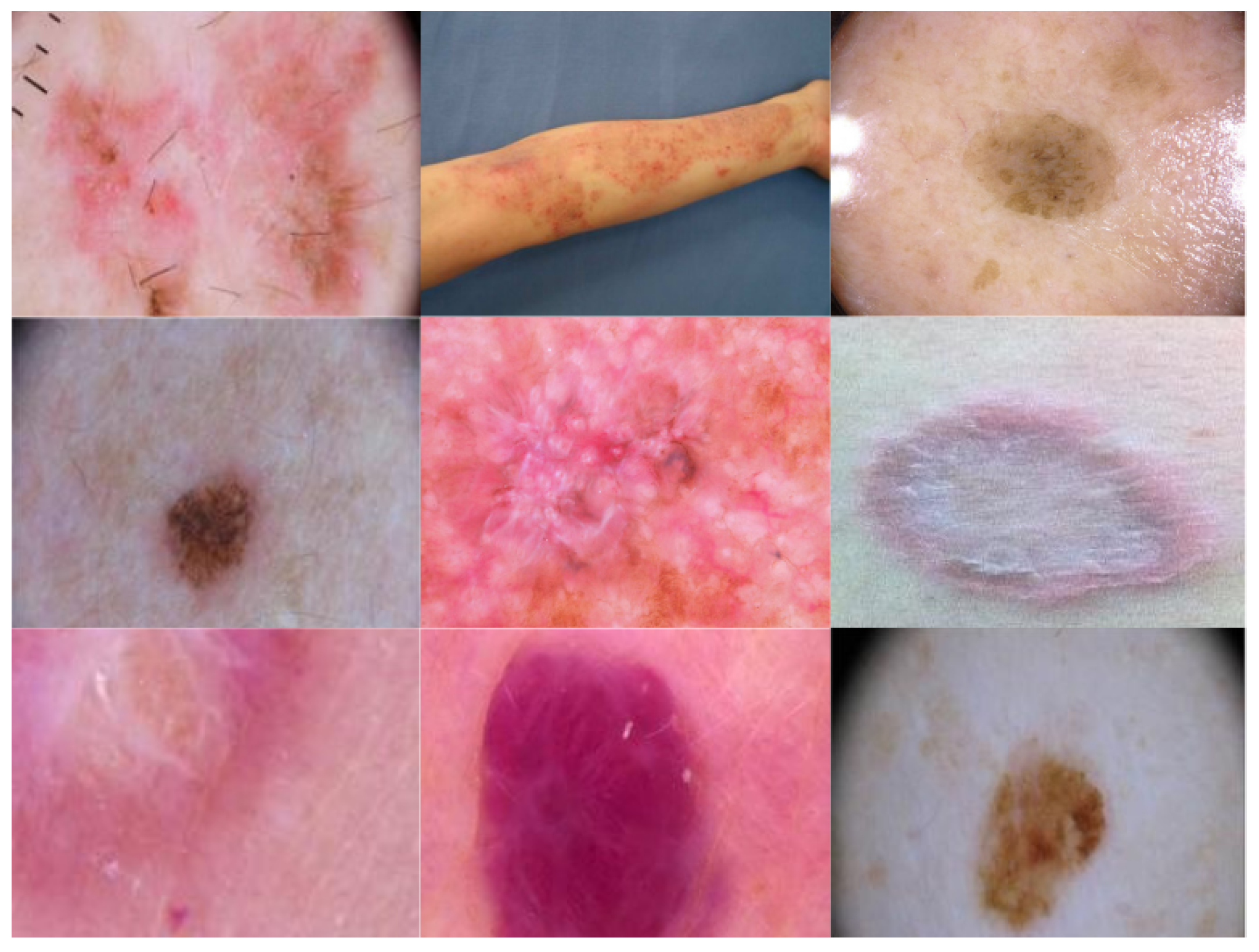
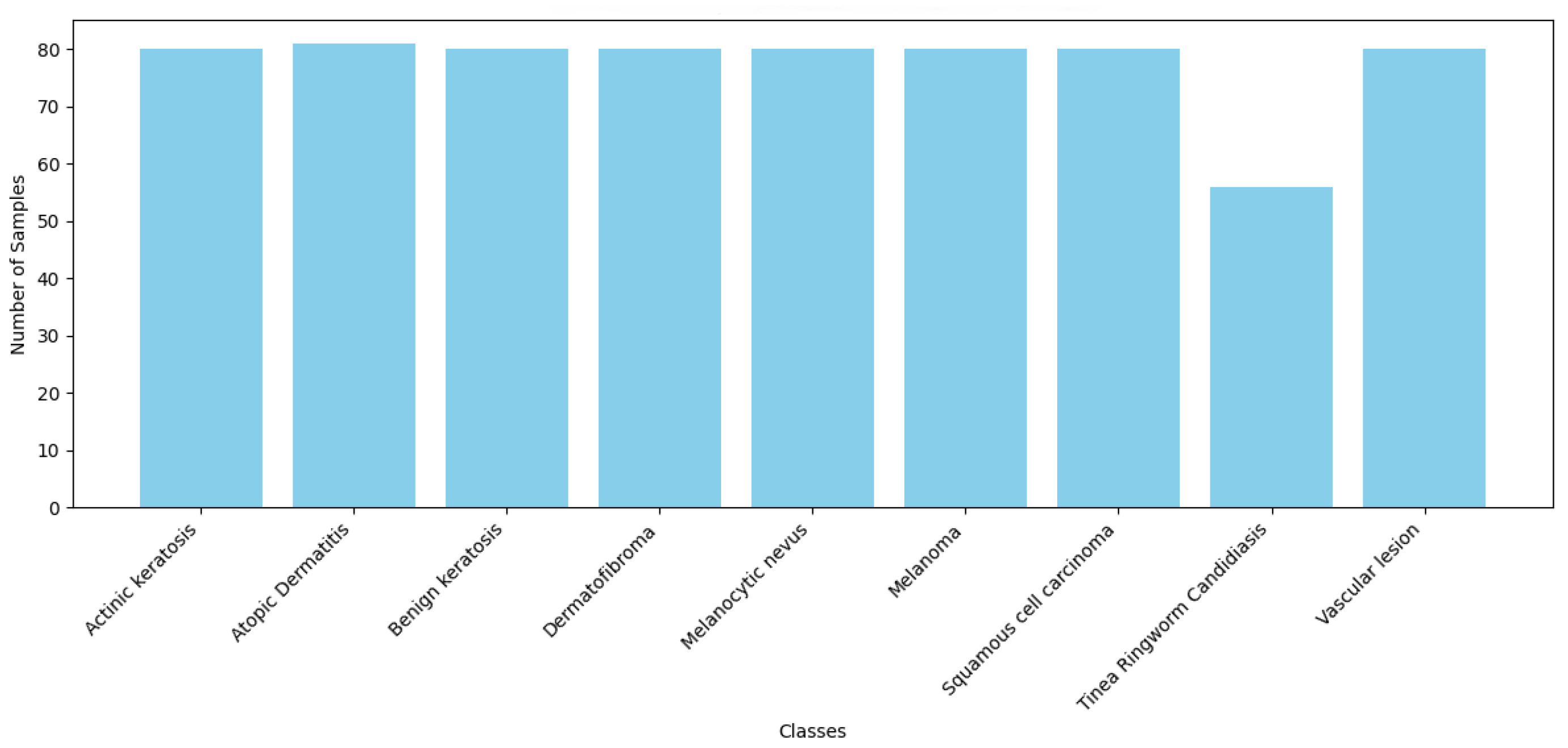
2.1.1. Dataset
- Actinic Keratosis: A type of pre-cancerous skin lesion that can develop due to prolonged exposure to ultraviolet (UV) radiation from the sun.
- Atopic Dermatitis: A persistent inflammatory skin disorder.
- Benign Keratosis: A non-cancerous skin growth.
- Dermatofibroma: A benign skin tumor.
- Melanocytic Nevus: Commonly known as a mole, a benign proliferation of melanocytes.
- Melanoma: A skin cancer originating from melanocytes.
- Squamous Cell Carcinoma: A type of skin cancer that arises from squamous epithelial cells.
- Tinea (Ringworm) Candidiasis: A fungal skin infection.
- Vascular Lesion: An abnormal clustering of blood vessels in the skin.
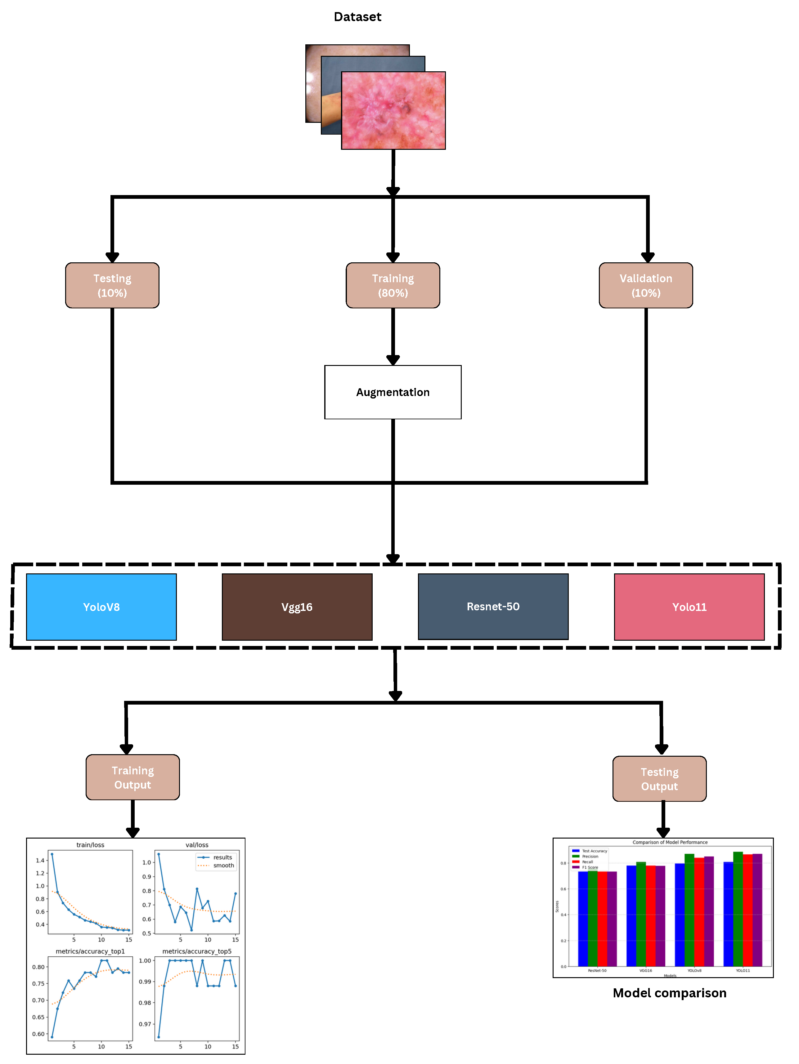
2.1.2. Dataset Augmentation
2.2. Performance Metrics
- Precision: Determines the proportion of exactly discovered favorable incidents to the total expected outcomes. Reduced false positives signify higher accuracy, which is absolutely vital in medical diagnosis to prevent needless patient anxiety.
- Recall: The ratio of positive events to the total actual outcome. In dermatological classification, a high recall is required to reduce false negatives and guarantee that cases of skin disorders are not missed.
- F1 Score: The harmonic mean of recall and precision, providing a fair assessment of a model’s performance, particularly in situations of class imbalance. It ensures that the evaluation considers both false negatives and false positives.
2.3. Proposed Model
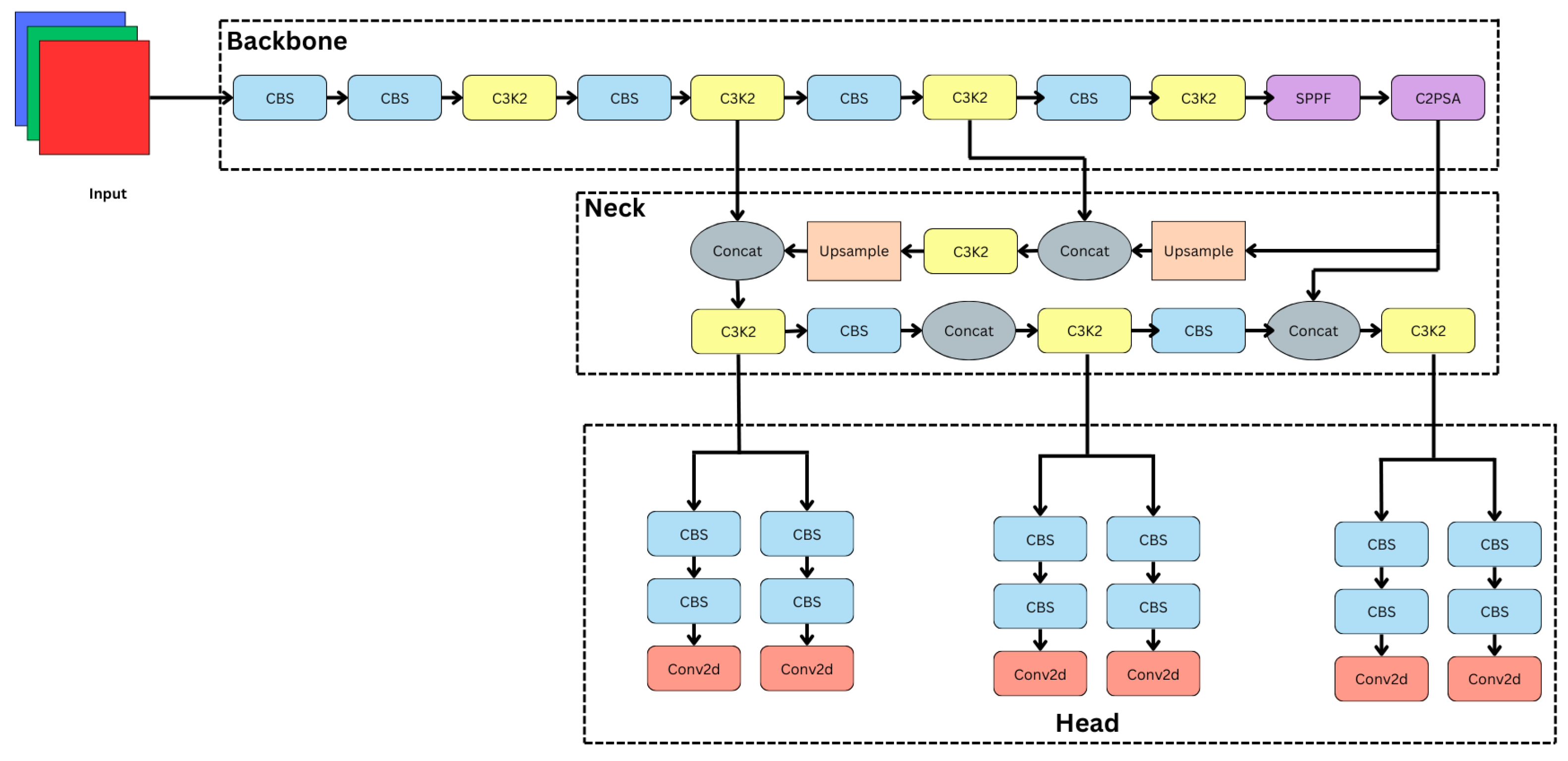
2.3.1. Backbone: Hierarchical Feature Extraction
- CBS Layers: These layers perform convolution, followed by batch normalization and the SiLU activation function, which makes sure that feature learning is strong and effective.
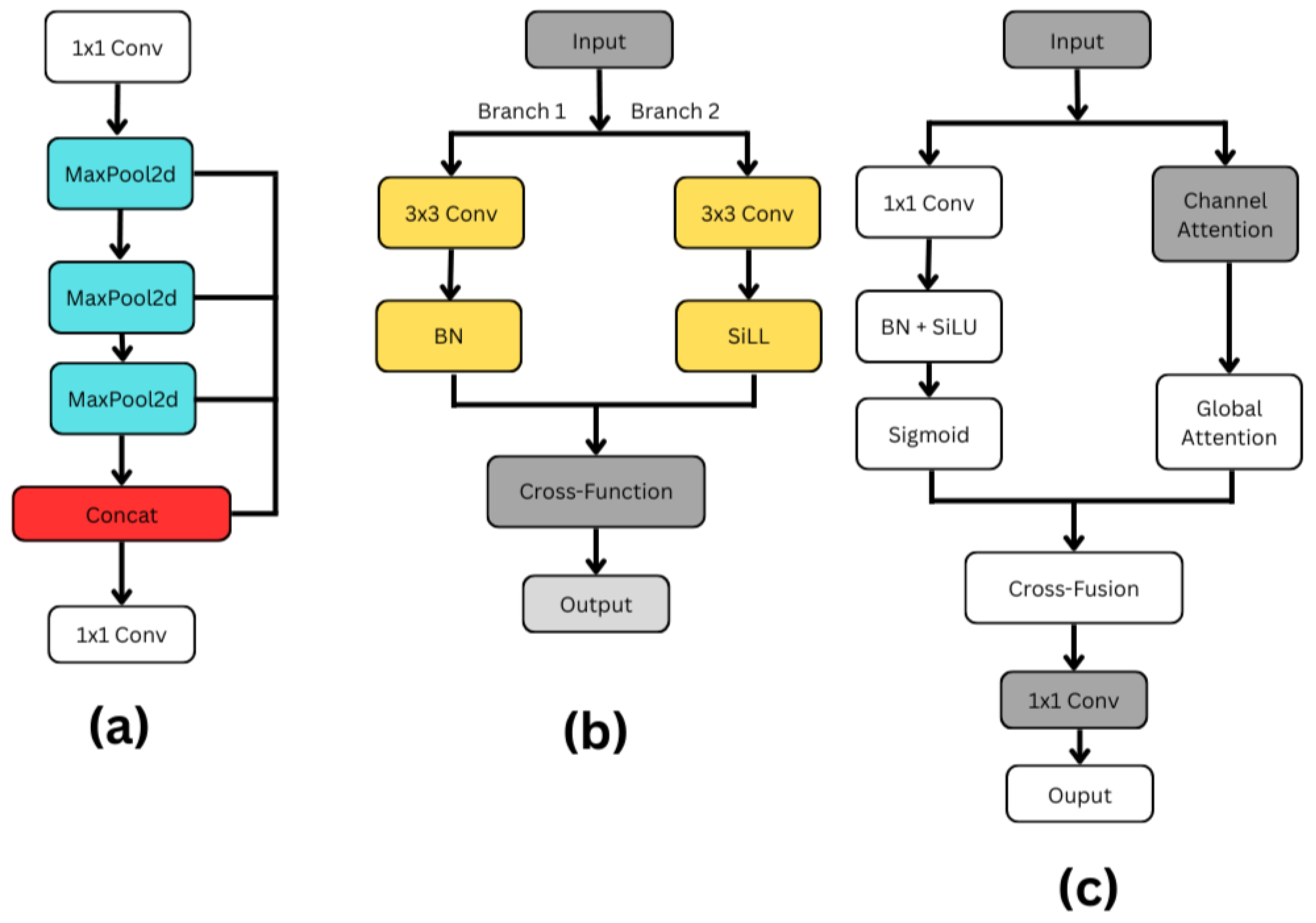
- C3k2 Blocks: As illustrated in Figure 5b, the modification of the C2f block in the neck for the C3k2 drastically changes YOLO11. The efficient feature extraction of the YOLO11 design is significantly influenced by the C3k2 block. To encourage information flow and computational efficiency, this block splits the input feature map into two distinct branches. Using a 3 × 3 convolution with a stride of 2, Branch 1 preserves significant structural information while lowering spatial dimensions. So, batch normalization (BN) normalizes feature distributions following the convolution, therefore stabilizing and speeding up training. Using SiLL (Simple Lightweight Layer) activation rather than batch normalization, Branch 2 provides a computationally efficient non-linearity by executing a 3 × 3 convolution with a stride of 2. When their changes are complete, a cross-function combines the results from both branches into a refined output using feature acquisition. This architecture provides faster processing performance, better parameter efficiency, and better multi-scale feature extraction for the C3k2 block compared to earlier designs. The c3k option improves detection performance in YOLO11 and provides network flexibility by implying that the C3k2 block can function as a conventional bottleneck (when c3k = False) or as an enhanced C3 module (when c3k = True) [35,36].
- SPPF (Spatial Pyramid Pooling Fast): As shown in Figure 5a, the SPFF (Spatial Pyramid Feature Fusion) module is designed to capture multi-scale spatial features efficiently. The process begins with a 1 × 1 convolution, which primarily serves to adjust or reduce the number of feature channels without affecting the spatial resolution of the input. After this, the feature map passes through a sequence of three MaxPool2d operations, each progressively reducing the spatial dimensions while preserving the most critical features. After every MaxPooling operation, the resulting feature maps are saved, creating multiple branches representing different levels of downsampled information.Once all pooling operations are completed, these intermediate feature maps, along with the initial 1 × 1 convolution output, are concatenated along the channel dimension. This concatenation effectively fuses information from different scales, allowing the network to have a richer understanding of both fine and coarse details in the input. To integrate and compress the multi-scale features into a cohesive representation, a final 1 × 1 convolution is applied to the concatenated output. This step refines the features and potentially reduces the number of output channels, preparing the data for the next stages of the model. Overall, the SPFF module is a lightweight yet powerful structure that captures a broad context with minimal computational overhead, making it especially useful in real-time deep learning applications like object detection [35,36].
- C2PSA (Cross-Channel Partial Self-Attention): As can be seen in Figure 5c, by including spatial and channel attention methods, the C2PSA (Cross Stage Partial with Spatial Attention) block shown in YOLO11 enhances feature extraction. Two simultaneous branches are formed from the input feature map. A 1 × 1 convolution in the main branch compresses data; batch normalization and SiLU activation follow for stability and non-linearity, finishing with a sigmoid activation generating a spatial attention map. The second branch gathers broad contextual information using global attention and emphasizes important feature channels using channel attention. A cross-fusion method combines the results from both branches, hence allowing the model to use spatial and channel-wise data at the same time. Before their move to the next stage, a final set of eleven convolutions improves the combined qualities. By combining exact spatial focus with enhanced channel attention, the C2PSA block significantly increases YOLO11’s ability to identify small, concealed, or complex items, hence improving detection accuracy in comparison to prior YOLO versions [35,36].
2.3.2. Neck: Multi-Scale Feature Fusion
- Feature Concatenation: The combination of intermediate feature maps from several backbone layers are combined to enhance the representation of dermatological patterns.
- Upsampling Operations: Low-resolution features are upsampled to retain fine details, ensuring accurate classification of small-scale lesions.
- C3k2 Blocks: C3k2 blocks help to optimize the network for strong categorization by means of further refinement and extraction of high-level representations.
2.3.3. Head: Classification and Prediction
- CBS Layers: By honing the feature maps, these layers maintain spatial information required for categorization.
- Conv2D Output Layers: The final Conv2D output layers generate categorization scores, ensuring correct diagnosis of skin disorders.
2.3.4. Advantages of YOLO11
- The new C3k2 block’s enhanced hierarchical feature extraction aids in the localization of dermatological patterns.
- By amplifying only the most important spatial features, the C2PSA module makes classification more reliable.
- Combining SPPF and upsampling techniques guarantees effective management of lesions of various sizes.
- Although architectural changes influence the excellent computational structure of YOLO11, it remains appropriate for practical dermatological applications.
2.4. The State-of-the-Art Model
2.4.1. ResNet-50
2.4.2. VGG16
2.4.3. YOLOv8
2.5. Experimental Setup
3. Results and Discussion
3.1. Evaluation
3.2. Discussion
4. Conclusions and Future Research
Author Contributions
Funding
Institutional Review Board Statement
Informed Consent Statement
Data Availability Statement
Acknowledgments
Conflicts of Interest
References
- Richard, M.; Paul, C.; Nijsten, T.; Gisondi, P.; Salavastru, C.; Taieb, C.; Trakatelli, M.; Puig, L.; Stratigos, A.; EADV Burden of Skin Diseases Project Team. Prevalence of most common skin diseases in Europe: A population-based study. J. Eur. Acad. Dermatol. Venereol. 2022, 36, 1088–1096. [Google Scholar] [CrossRef] [PubMed]
- Moustaqim-Barrette, A.; Conte, S.; Kelly, A.; Lebeau, J.; Alli, S.; Lagacé, F.; Litvinov, I.V. Evaluation of weather and environmental factors and their association with cutaneous melanoma incidence: A national ecological study. JAAD Int. 2024, 16, 264–271. [Google Scholar] [CrossRef] [PubMed]
- Arnold, M.; Singh, D.; Laversanne, M.; Vignat, J.; Vaccarella, S.; Meheus, F.; Cust, A.E.; De Vries, E.; Whiteman, D.C.; Bray, F. Global burden of cutaneous melanoma in 2020 and projections to 2040. JAMA Dermatol. 2022, 158, 495–503. [Google Scholar] [CrossRef]
- Behara, K.; Bhero, E.; Agee, J.T. AI in dermatology: A comprehensive review into skin cancer detection. PeerJ Comput. Sci. 2024, 10, e2530. [Google Scholar] [CrossRef]
- Flohr, C.; Hay, R. Putting the burden of skin diseases on the global map. Br. J. Dermatol. 2021, 184, 189–190. [Google Scholar] [CrossRef] [PubMed]
- De, A.; Mishra, N.; Chang, H.T. An approach to the dermatological classification of histopathological skin images using a hybridized CNN-DenseNet model. PeerJ Comput. Sci. 2024, 10, e1884. [Google Scholar] [CrossRef]
- Thomsen, I.M.N.; Heerfordt, I.M.; Karmisholt, K.E.; Mogensen, M. Detection of cutaneous malignant melanoma by tape stripping of pigmented skin lesions–A systematic review. Skin Res. Technol. 2023, 29, e13286. [Google Scholar] [CrossRef]
- Garrison, Z.R.; Hall, C.M.; Fey, R.M.; Clister, T.; Khan, N.; Nichols, R.; Kulkarni, R.P. Advances in early detection of melanoma and the future of at-home testing. Life 2023, 13, 974. [Google Scholar] [CrossRef]
- Aksoy, S.; Demircioglu, P.; Bogrekci, I. Advanced Artificial Intelligence Techniques for Comprehensive Dermatological Image Analysis and Diagnosis. Dermato 2024, 4, 173–186. [Google Scholar] [CrossRef]
- Luo, N.; Zhong, X.; Su, L.; Cheng, Z.; Ma, W.; Hao, P. Artificial intelligence-assisted dermatology diagnosis: From unimodal to multimodal. Comput. Biol. Med. 2023, 165, 107413. [Google Scholar] [CrossRef]
- Zhang, B.; Zhou, X.; Luo, Y.; Zhang, H.; Yang, H.; Ma, J.; Ma, L. Opportunities and challenges: Classification of skin disease based on deep learning. Chin. J. Mech. Eng. 2021, 34, 112. [Google Scholar] [CrossRef]
- Qu, H.Q.; Kao, C.; Hakonarson, H. Implications of the non-neuronal cholinergic system for therapeutic interventions of inflammatory skin diseases. Exp. Dermatol. 2024, 33, e15181. [Google Scholar] [CrossRef] [PubMed]
- Rusydiyah, E.; Novitasari, D.C.R.; Mushlihul, M.; Amin, A.N.A.; Nurrohman, H.F.; Haq, D.Z.; Putri, E.R.S. Skin cancer diagnosis system on object detection using various CNN YOLOv5 in Android mobile. J. Theor. Appl. Inf. Technol. 2024, 102, 1–11. [Google Scholar]
- Noronha, S.S.; Mehta, M.A.; Garg, D.; Kotecha, K.; Abraham, A. Deep learning-based dermatological condition detection: A systematic review with recent methods, datasets, challenges, and future directions. IEEE Access 2023, 11, 140348–140381. [Google Scholar] [CrossRef]
- Ahmad, B.; Usama, M.; Ahmad, T.; Khatoon, S.; Alam, C.M. An ensemble model of convolution and recurrent neural network for skin disease classification. Int. J. Imaging Syst. Technol. 2022, 32, 218–229. [Google Scholar] [CrossRef]
- Goswami, T.; Dabhi, V.K.; Prajapati, H.B. Skin disease classification from image-a survey. In Proceedings of the 2020 6th International Conference on Advanced Computing and Communication Systems (ICACCS), Coimbatore, India, 6–7 March 2020; pp. 599–605. [Google Scholar]
- Minarno, A.E.; Lusianti, A.; Azhar, Y.; Wibowo, H. Classification of Skin Cancer Images Using Convolutional Neural Network with ResNet50 Pre-trained Model. JOIV Int. J. Inform. Vis. 2024, 8, 2013–2019. [Google Scholar] [CrossRef]
- Akay, M.; Du, Y.; Sershen, C.L.; Wu, M.; Chen, T.Y.; Assassi, S.; Mohan, C.; Akay, Y.M. Deep learning classification of systemic sclerosis skin using the MobileNetV2 model. IEEE Open J. Eng. Med. Biol. 2021, 2, 104–110. [Google Scholar] [CrossRef]
- Aziz, F.; Saputri, D.U.E. Efficient skin lesion detection using yolov9 network. J. Med. Inform. Technol. 2024, 2, 11–15. [Google Scholar] [CrossRef]
- Srinivasu, P.N.; SivaSai, J.G.; Ijaz, M.F.; Bhoi, A.K.; Kim, W.; Kang, J.J. Classification of skin disease using deep learning neural networks with MobileNet V2 and LSTM. Sensors 2021, 21, 2852. [Google Scholar] [CrossRef]
- Sadik, R.; Majumder, A.; Biswas, A.A.; Ahammad, B.; Rahman, M.M. An in-depth analysis of Convolutional Neural Network architectures with transfer learning for skin disease diagnosis. Healthc. Anal. 2023, 3, 100143. [Google Scholar] [CrossRef]
- Agarwal, R.; Godavarthi, D. Skin disease classification using CNN algorithms. Eai Endorsed Trans. Pervasive Health Technol. 2023, 9, 1–8. [Google Scholar] [CrossRef]
- ElGhany, S.A.; Ibraheem, M.R.; Alruwaili, M.; Elmogy, M. Diagnosis of Various Skin Cancer Lesions Based on Fine-Tuned ResNet50 Deep Network. Comput. Mater. Contin. 2021, 68, 117–135. [Google Scholar] [CrossRef]
- Anand, V.; Gupta, S.; Koundal, D.; Mahajan, S.; Pandit, A.K.; Zaguia, A. Deep learning based automated diagnosis of skin diseases using dermoscopy. Comput. Mater. Contin. 2022, 71, 1–16. [Google Scholar] [CrossRef]
- Fan, J.; Kim, J.; Jung, I.; Lee, Y. A study on multiple factors affecting the accuracy of multiclass skin disease classification. Appl. Sci. 2021, 11, 7929. [Google Scholar] [CrossRef]
- Li, H.; Pan, Y.; Zhao, J.; Zhang, L. Skin disease diagnosis with deep learning: A review. Neurocomputing 2021, 464, 364–393. [Google Scholar] [CrossRef]
- Ahammed, M.; Al Mamun, M.; Uddin, M.S. A machine learning approach for skin disease detection and classification using image segmentation. Healthc. Anal. 2022, 2, 100122. [Google Scholar] [CrossRef]
- Veni, N.K.; Deepapriya, B.; Vardhini, P.H.; Kalyani, B.; Sharmila, L. A Novel Method for Prediction of Skin Diseases Using Supervised Classification Techniques. Skin 2022, 20, 21. [Google Scholar]
- Mohan, J.; Sivasubramanian, A.; Ravi, V. Enhancing skin disease classification leveraging transformer-based deep learning architectures and explainable ai. Comput. Biol. Med. 2025, 190, 110007. [Google Scholar] [CrossRef]
- Salam, A.A.; Akram, M.U.; Yousaf, M.H.; Rao, B. DermaTransNet: Where Transformer Attention Meets U-Net for Skin Image Segmentation. IEEE Access 2025, 13, 64305–64329. [Google Scholar] [CrossRef]
- Saha, D.K.; Joy, A.M.; Majumder, A. YoTransViT: A transformer and CNN method for predicting and classifying skin diseases using segmentation techniques. Inform. Med. Unlocked 2024, 47, 101495. [Google Scholar] [CrossRef]
- Sapkota, R.; Meng, Z.; Churuvija, M.; Du, X.; Ma, Z.; Karkee, M. Comprehensive performance evaluation of yolo11, yolov10, yolov9 and yolov8 on detecting and counting fruitlet in complex orchard environments. arXiv 2024, arXiv:2407.12040. [Google Scholar]
- Alruwaili, M.; Mohamed, M. An Integrated Deep Learning Model with EfficientNet and ResNet for Accurate Multi-Class Skin Disease Classification. Diagnostics 2025, 15, 551. [Google Scholar] [CrossRef]
- Daneshjou, R.; Vodrahalli, K.; Liang, W.; Novoa, R.A.; Jenkins, M.; Rotemberg, V.; Ko, J.; Swetter, S.M.; Bailey, E.E.; Gevaert, O.; et al. Disparities in dermatology ai: Assessments using diverse clinical images. arXiv 2021, arXiv:2111.08006. [Google Scholar]
- Rao, S. YOLOv11 Architecture Explained: Next-Level Object Detection with Enhanced Speed and Accuracy. Mediu. Oct 2024, 22. [Google Scholar]
- Khanam, R.; Hussain, M. Yolov11: An overview of the key architectural enhancements. arXiv 2024, arXiv:2410.17725. [Google Scholar]


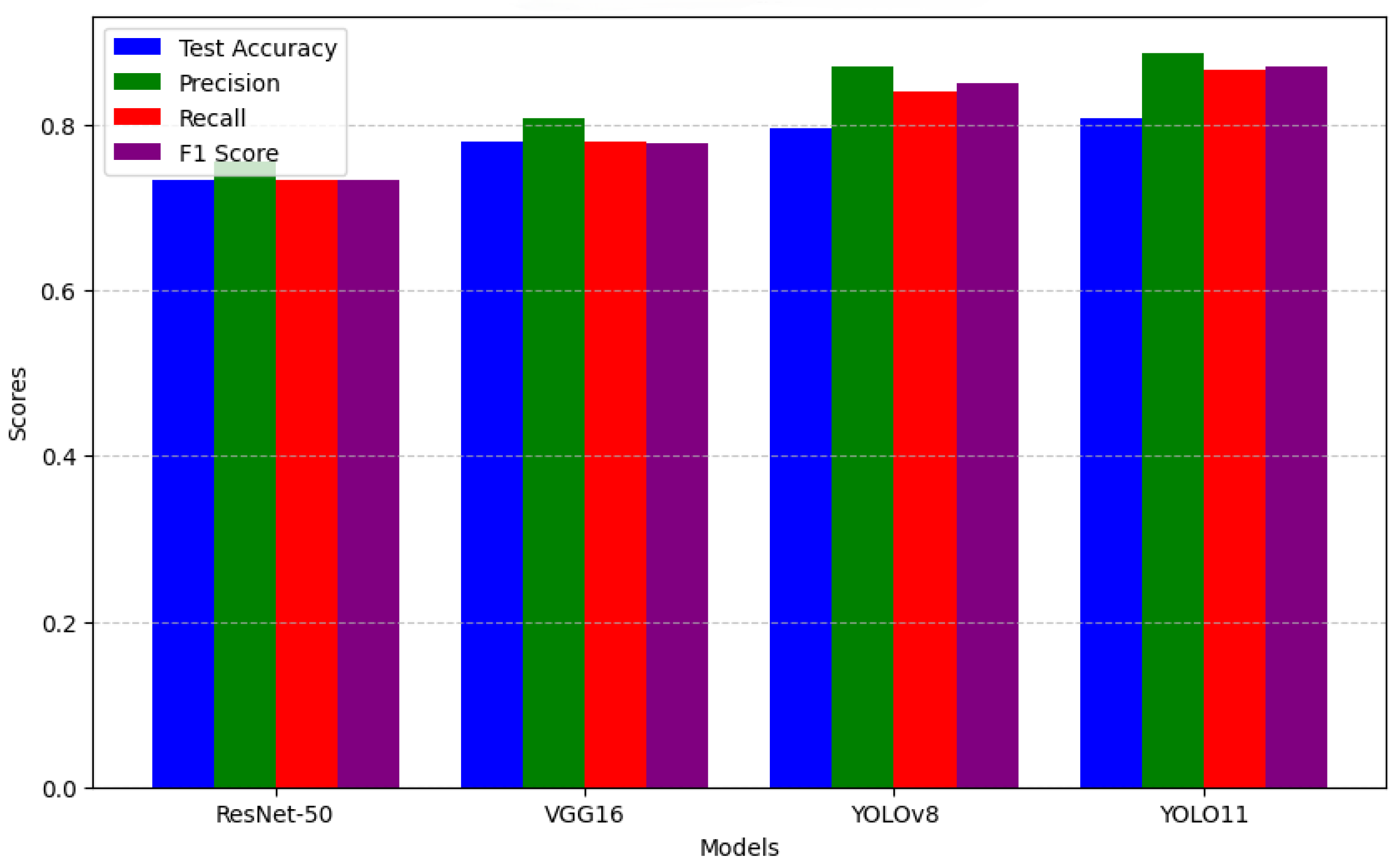
| Model | Accuracy | Precision | Recall | F1 Score |
|---|---|---|---|---|
| ResNet50 | 79.07% | 80.36% | 79.07% | 78.86% |
| VGG16 | 72.09% | 76.08% | 72.09% | 71.48% |
| YOLOv8 | 79.51% | 87.0% | 84.0% | 85.0% |
| YOLO11 | 80.72% | 88.7% | 86.7% | 87.0% |
| Hyperparameter | Value |
|---|---|
| Epochs | 25 |
| Optimizer | AdamW |
| Loss function | Cross-Entropy Loss |
| Learning rate | 1 × 10⁻⁴ |
| Weight decay | 0.0005 |
| Momentum | 0.937 |
| Batch size | 32 |
Disclaimer/Publisher’s Note: The statements, opinions and data contained in all publications are solely those of the individual author(s) and contributor(s) and not of MDPI and/or the editor(s). MDPI and/or the editor(s) disclaim responsibility for any injury to people or property resulting from any ideas, methods, instructions or products referred to in the content. |
© 2025 by the authors. Licensee MDPI, Basel, Switzerland. This article is an open access article distributed under the terms and conditions of the Creative Commons Attribution (CC BY) license (https://creativecommons.org/licenses/by/4.0/).
Share and Cite
Diptho, R.A.; Basak, S. Enhancing Dermatological Diagnosis Through Medical Image Analysis: How Effective Is YOLO11 Compared to Leading CNN Models? NDT 2025, 3, 11. https://doi.org/10.3390/ndt3020011
Diptho RA, Basak S. Enhancing Dermatological Diagnosis Through Medical Image Analysis: How Effective Is YOLO11 Compared to Leading CNN Models? NDT. 2025; 3(2):11. https://doi.org/10.3390/ndt3020011
Chicago/Turabian StyleDiptho, Rakib Ahammed, and Sarnali Basak. 2025. "Enhancing Dermatological Diagnosis Through Medical Image Analysis: How Effective Is YOLO11 Compared to Leading CNN Models?" NDT 3, no. 2: 11. https://doi.org/10.3390/ndt3020011
APA StyleDiptho, R. A., & Basak, S. (2025). Enhancing Dermatological Diagnosis Through Medical Image Analysis: How Effective Is YOLO11 Compared to Leading CNN Models? NDT, 3(2), 11. https://doi.org/10.3390/ndt3020011