Article

Locomotory Effect of Reversibly Restraining the Pectines of Scorpions

by
Douglas D. Gaffin
*,
Sofía E. Gálvez Falcón
and
Mariëlle H. Hoefnagels
School of Biological Sciences, University of Oklahoma, Norman, OK 73019, USA
*
Author to whom correspondence should be addressed.
These authors contributed equally to this work.
Arthropoda 2025, 3(3), 12; https://doi.org/10.3390/arthropoda3030012
Submission received: 20 May 2025 / Revised: 8 July 2025 / Accepted: 9 July 2025 / Published: 6 August 2025

Abstract

Scorpions possess unique, ornate mid-ventral sensory organs called pectines. The pectines are used to process chemo- and mechanosensory information acquired from the ground as the animal walks, and they are implicated in a variety of behaviors including navigation and detection of mates and prey. Many previous researchers have investigated pecten function by cutting the organs from the animals (full ablation) and comparing their behaviors with those of intact scorpions. This drastic approach is likely to not only cause enormous stress to the ablated animals but also change their behavior. Here, we have developed a method for gently and reversibly impairing the pectines by partially covering them to prevent them from lowering to the ground. Specifically, we fabricated small rectangles of a commercially available lightly adhesive foil tape that we placed across the pectines and secured to the body wall with a thin strip of a more strongly adhesive lab tape. Using a repeated measures design, we monitored the animals’ locomotory activity overnight in small behavioral arenas under three conditions: unmodified (intact) control, pectines restrained, and sham control. We found that scorpions with their pectines restrained had a significant increase in both the distance and duration of movement when compared to unmodified and sham control animals. Our method allows for temporary, reversible compromise of pecten function and should be useful in fully understanding the role of pectines in behavior.

1. Introduction

Scorpions have several specialized sensory systems [1,2,3,4,5,6,7,8], of which their pectines are the most distinctive and elaborate. Pectines are paired, comb-like structures that extend ventrally from the third mesosomal segment, just caudal to the coxae of the fourth walking legs [9]. The distal, ground-facing surfaces of each pecten are covered with thousands of peg sensilla [4,10], which transduce chemosensory and mechanosensory information from the ground as the animal walks [11,12]. The pectines appear to be involved in several activities, including pheromone detection for mating [13,14,15,16], prey detection [17], discerning the terrain’s texture [1,18,19,20,21], and navigation to their home burrows [22,23,24].
Previous studies of pecten function have used ablation (mostly in the form of complete removal) to interpret the role of pectines in scorpion behavior [1,14,25]. However, no study has safely and reversibly compromised the pectines without causing permanent damage. This is a critical limitation, as damaging the organs permanently introduces other variables that could influence the scorpion’s behavior, possibly confounding the interpretation of results. Developing a method that allows temporary, reversible compromise of the pectines is essential to further our understanding of their role in behavior, free from unintended long-term effects.
In this study, we developed a method for reversibly restraining the pectines in scorpions to examine their role in behavior. In our laboratory assay, we found that scorpions with restrained pectines showed a significant difference in locomotory behavior when compared to sham control and unmodified control animals.

2. Materials and Methods

2.1. Collection and Care of Scorpions

Desert grassland scorpions (Paruroctonus utahensis) (Williams, 1968; Family Vaejovidae) were collected from sandy regions near Monahans, TX in April and October of 2024. We housed the animals individually in glass containers with 100 mL of native sand as a substrate. Each enclosure also contained a 4.0 cm long half section of 3.4 cm diam PVC tubing for shelter. The scorpions were fed one small cricket (Acheta domesticus) every 10 days and given water twice a week. We used several animals in our pilot studies and 18 adult females in our final behavioral trials. The holding room where the animals were kept was set to a 14:10 h light-dark cycle, with lights on at 06:00 and lights off at 20:00. The ambient temperature was kept at ~25°.

2.2. Immobilizing the Pectines

We used plastic food container lids, foam, and adhesive tapes to engineer a restraint device to hold the scorpions still while covering their pectines and applying shams. The device was designed to limit movement without causing harm or undue stress to the animal (Figure 1A). To keep the pectines elevated, under the dissecting microscope we first arranged each pecten towards the midsaggital line and applied a rectangular (4 × 3 mm) piece of washi tape (Foil Washi Tape, 3 mm wide from The Paper Studio) across the organs. The washi tape was very thin and had a gentle adhesive and could therefore be easily removed without leaving a sticky residue (a simple test for residual stickiness is shown in Supplemental Video S1). We then applied a strip (6 × 2 mm) of stronger lab tape (VWR scientific) to hold the washi tape in place (Figure 1A). One end of the strip was attached to the washi tape and the other was adhered to the ventral mesosoma, between the book lung spiracles. This arrangement ensured that the pectines were partially covered and restricted from touching the substrate during behavioral trials. Electrophysiological studies show that peg sensilla can detect pure volatile substances only when the stimulant is brought within 20 microns of the peg pore [26]. We therefore believe that pecten sensilla are primarily contact chemoreceptors and play a limited or no role in olfaction. As such, we felt that restraining the pectines would not only compromise their mechanosensory ability but also their ability to detect chemicals on the substrate. Sham control animals received washi tape and lab tape on the ventral mesosoma caudal to the pectines (Figure 1A). Unmodified control animals did not receive tape.
To reduce animal stress and to make our procedures more efficient, we did not use cold exposure or other anesthesia during our manipulations. Also, we found that chilling induced condensation on the animal’s body, which compromised adherence of the washi tape to the cuticle. Instead, the restraint device illustrated in Figure 1A greatly facilitated the placement of covers and shams on the animals. We carefully held the animal’s telson between our thumb and forefinger and positioned its body over the window in the upper plastic leaf of the restraint device. The animal would typically grab the foam rubber strip at the front edge of the window with its tarsal claws so that we could quickly close the lower plastic leaf, which gently pressed the remaining foam rubber strips against the animal’s metasoma and pedipalps. The restraining procedure is demonstrated in Supplemental Video S2. After securing the “sandwich” with a small piece of lab tape, we could maneuver the ventral side of the animal under the dissecting microscope so that we could precisely place the pecten restraints or shams where we wanted. Once placed, we photographed each of the restraints and shams before releasing the animals to their storage containers. We also verified that the restraints and shams were in place prior to releasing the animals into the behavioral arenas and checked if the shams and restraints were still in place the following morning after the tests were complete.

2.3. Behavioral Testing Setup

We conducted our behavioral trials in a room isolated from the holding room to minimize external disturbances. The room was kept at the same temperature and light/dark cycle as the holding room. The testing arenas consisted of nine cylindrical PVC couplings (10.0 cm diameter, 5 cm tall) arranged in a 3 × 3 grid pattern (Figure 1B) The arenas were placed on top of nine clean filter paper squares (12.7 × 12.7 cm) cut from 46 × 57 cm filter paper sheets (grade 613, Science Kit and Boreal Laboratories). The filter paper squares and the accompanying arenas were then placed on top of a 40 × 40 × 2.5 cm spongy foam rubber square (Poly-fit foam cushion, Fairfield) to reduce vibrations and environmental noise and to isolate each testing arena. The foam rubber square was then placed on a 61 × 61 × 1.3 cm square rubber mat (Ottomanson multipurpose exercise tile mat) to further reduce interference from potential building vibrations (scorpions are extremely vibration-sensitive [2]). The rubber mat was then placed on a rigid plywood board that was leveled to reduce gravity effects, and the entire set-up was surrounded by a fence (40 cm tall) constructed from four rubber mats (same as noted above) to dampen sound and prevent the entry of pests such as cockroaches and crickets. A single infrared camera (EP 1 megapixel Day Night Vision) was positioned 170 cm above the center of the arena grid to track the animals in all nine containers simultaneously. Photos of the camera and fence setup are shown in Supplemental File S1. The camera operated in complete darkness, using infrared light for illumination and its output cable ran through the wall via grommets to the adjacent recording room and connected to a laptop via USB.

2.4. Trial Protocol

We used a repeated-measures design in which 18 female scorpions each underwent three treatments in a pseudo-randomized sequence: (1) unmodified control animals, (2) animals with pectines restrained, and (3) sham control animals (Figure 1A, lower). The 18 animals were divided into two groups of nine for testing (Figure 1C). Group 1 animals (scorpions 1–9) were tested on nights 1, 3, and 5, whereas group 2 animals (scorpions 10–18) were tested on nights 2, 4, and 6. Each scorpion was prepped 6 h before testing began during the day phase of the light/dark cycle. Restraints and shams (if applicable) were applied and the animals were placed in individual plastic containers (Ziploc, SC Johnson, Racine, WI, USA; 238 mL) near their assigned arenas. Just before 17:00, the scorpions were placed in their arenas and remained there until 07:00 the next morning with the video camera capturing 1 frame per second for the entire 14-h period. The restraints and shams were removed by 09:00, and the scorpions were returned to their home containers and given a 24-h rest period before their next treatment and trial. Between trials, the filter paper squares were replaced and the arenas were cleaned with ethanol to remove any scents or other residues left by the previous scorpions. This process continued for six successive days and nights until all scorpions completed all three treatments (Figure 1C). We noticed that animal #7 was lethargic in its first trial (during a test of the sham condition) and died a couple of days later. As such, we introduced an alternative animal #7 in the restrained condition on the third night of filming. We then tested this animal in the unmodified condition on night 5 and in the sham condition two nights later (night 7). [Note: The animals that completed the trials remained in good health at the time of submitting this manuscript (approximately 3 months after the experiments).].

2.5. Video Processing and Statistical Analysis

A Python (Spyder ver. 5.4.3) script was written to parse video footage into nine regions corresponding to the nine arenas. Then a digital mask was placed around each arena to block extraneous activity beyond the walls of the arena. Next, a frame-by-frame subtraction method, coupled with a contour-generating algorithm, was used to construct a rough outline of each moving animal. In each frame, the xy coordinates of the centroid of each animal’s outline was determined. Each centroid was mapped to the nearest point on an 8.8-cm diameter circle centered in each arena. The distance between successive adjusted centroid locations on the circle was determined using the Pythagorean theorem, and the distances were summed to calculate the total distance traveled during the 14-h filming period. Adjusting the centroid locations and calculating distances in this way helped mitigate visual distortion towards the edges of the video frame and reduce some of the centroid jiggle that arises during the contour generation process. We also calculated distances traveled using the unadjusted centroid locations and found that all statistically significant differences between treatments were the same (see Supplemental File S2 for raw vs adjusted distance comparisons). In addition, the arenas were digitally subdivided into 18 wedge-shaped sectors, and the number of times a centroid moved into or within each sector was tallied.
Six dependent variables were extracted for each trial: (1) total distance traveled during 14 h, (2) duration of movement in seconds (derived by subtracting the video frames without movement from the total number of frames (14 h at 1 fps = 50,400 frames), (3) variance in percent time of occupancy in the 18 sectors (to assess if animals moved evenly around the arena or tended to remain in particular areas), (4) mean, (5) median, and (6) mode of the distance moved per second (excluding frames without movement).
A repeated measures ANOVA model was used to analyze the data and test for statistically significant differences between the three conditions: unmodified, restrained, and sham. We first checked the data for normality (Shapiro-Wilk test) and sphericity; if either condition was violated, we used the Friedman test (nonparametric alternative). We set our significance level at p < 0.05. If any of the dependent variables met significance, we performed post-hoc analyses with Bonferroni correction to assess pairwise significance between conditions. We also tested for the possibility of the effect of trial day (1, 2, or 3) on distance and movement duration and found no effect; see Supplemental File S3.

3. Results

Photographs of the animals with their restraints and shams before and after the trials are shown in Supplemental File S4. Both the restraints and the shams were dislodged during the trials of animals #6 and #12. As such, the data for these animals were removed from the analyses. Only the restraints of animals #3 and #8 and only the shams of animals #13 and #18 were dislodged. We used within-subject imputation to fill in the missing values where we took the mean of the other values for the condition to complete the table (this essentially gives a neutral value for use in our repeated measures ANOVA test).
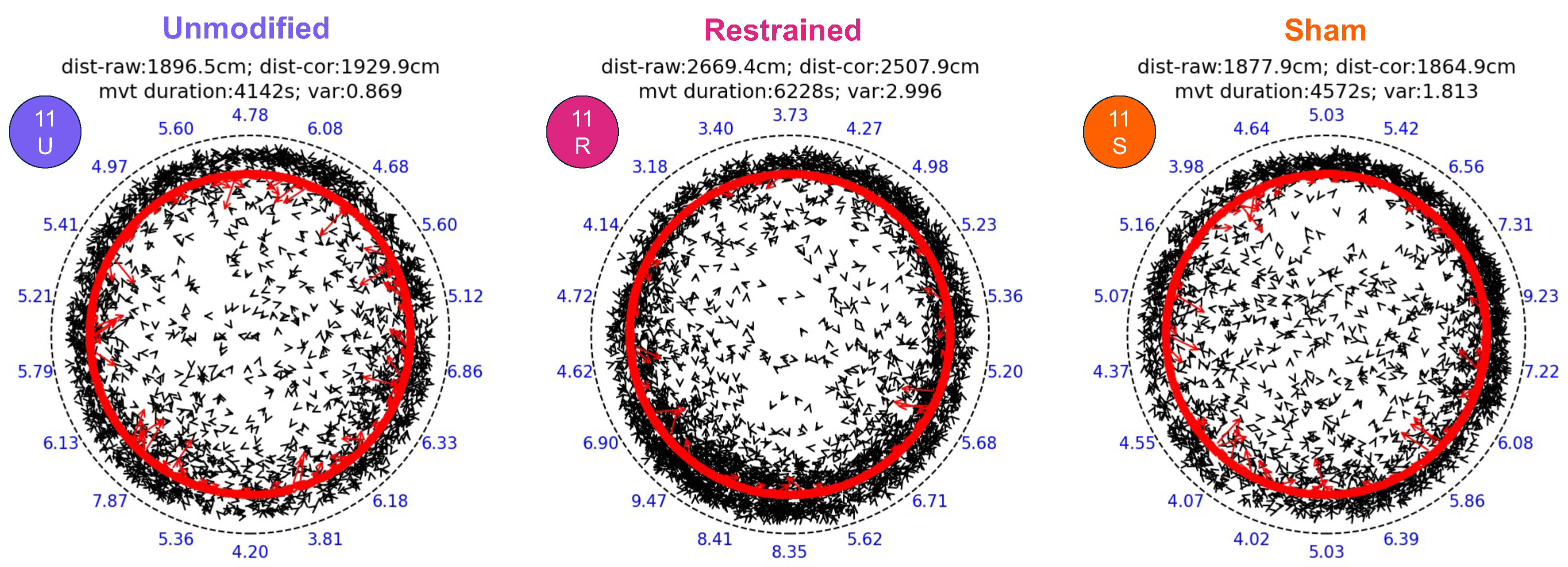
The plots of all animal movements in all trials are shown in Supplemental File S5, along with distribution histograms of the animals’ movement distances per second for the entire trials. A representative example of the movement plots of an animal (animal #11) for the three experimental conditions is shown in Figure 2. We chose this animal because its distance and duration values were closest to the mean values for all animals in our trials. The pattern of movement for animal #11 was typical for scorpions confined to arenas: the animal actively moved throughout the arena in all conditions but spent most of its time along the arena walls.
The graphical results of all the dependent variables evaluated in this study are shown in Figure 3, and the statistical tests are summarized in Table 1. We observed statistically significant differences among treatments for the two variables: “Total distance” and “Movement duration”. Post hoc analysis of these two variables showed that both the distance traveled and the duration of movement were significantly greater for the restrained condition compared to the unmodified and sham conditions; the unmodified and sham conditions were not significantly different from one another. There were no statistical differences among treatments in sector occupancy variance, mean velocity, median velocity, or mode velocity, though the p value for mean velocity was <0.1 (0.0696).

4. Discussion

To our knowledge, this study is the first attempt to reversibly impair the pectines of scorpions to evaluate behavioral effects. Our results indicate that restraining the pectines of scorpions alters their locomotory behavior. Specifically, scorpions with small pieces of foil tape partially covering their pectines walked farther and spent more time moving during our 14-h behavioral assays compared to unmodified control animals or animals with sham covers applied to their ventral mesosoma, caudal to their pectines.
We warn researchers to consider the challenges involved in developing a suitable pecten restraint that is easy to apply, reversible, and does not harm the animal. We found the method we developed to be simple and effective, but it took us a while to get there. In pilot studies, we tried many strategies including small rubber bands, silicone pipe thread tape, shrink wrap tubing, and others. For example, we initially thought we could slip a small, thin rubber band across the animal’s midsection between pairs of legs 2 and 3 to gently hold the pectines against the animal’s body. However, we found it exceedingly difficult to get the stretched band either around the animal’s pedipalps and prosoma or its telson and metasoma and then past the appropriate leg pairs to the correct position over the pectines. In another approach, we attempted to wrap a piece of non-adhesive tape (such as pipe thread tape) across the pectines, crimping the tape’s ends on the animal’s dorsum to hold it in place. These methods were also difficult and usually resulted in the animal slipping its pectines out of the restraint. In the end, the tacky washi foil tape was most useful because its adhesive is relatively light, yet it effectively restrained the pectines. A follow-up study confirming that the pectines and peg sensilla remained structurally and functionally intact after being covered could verify that this method is reversible and nondestructive. However, our randomized repeated measures design mitigates against the potential harm from the adhesive.
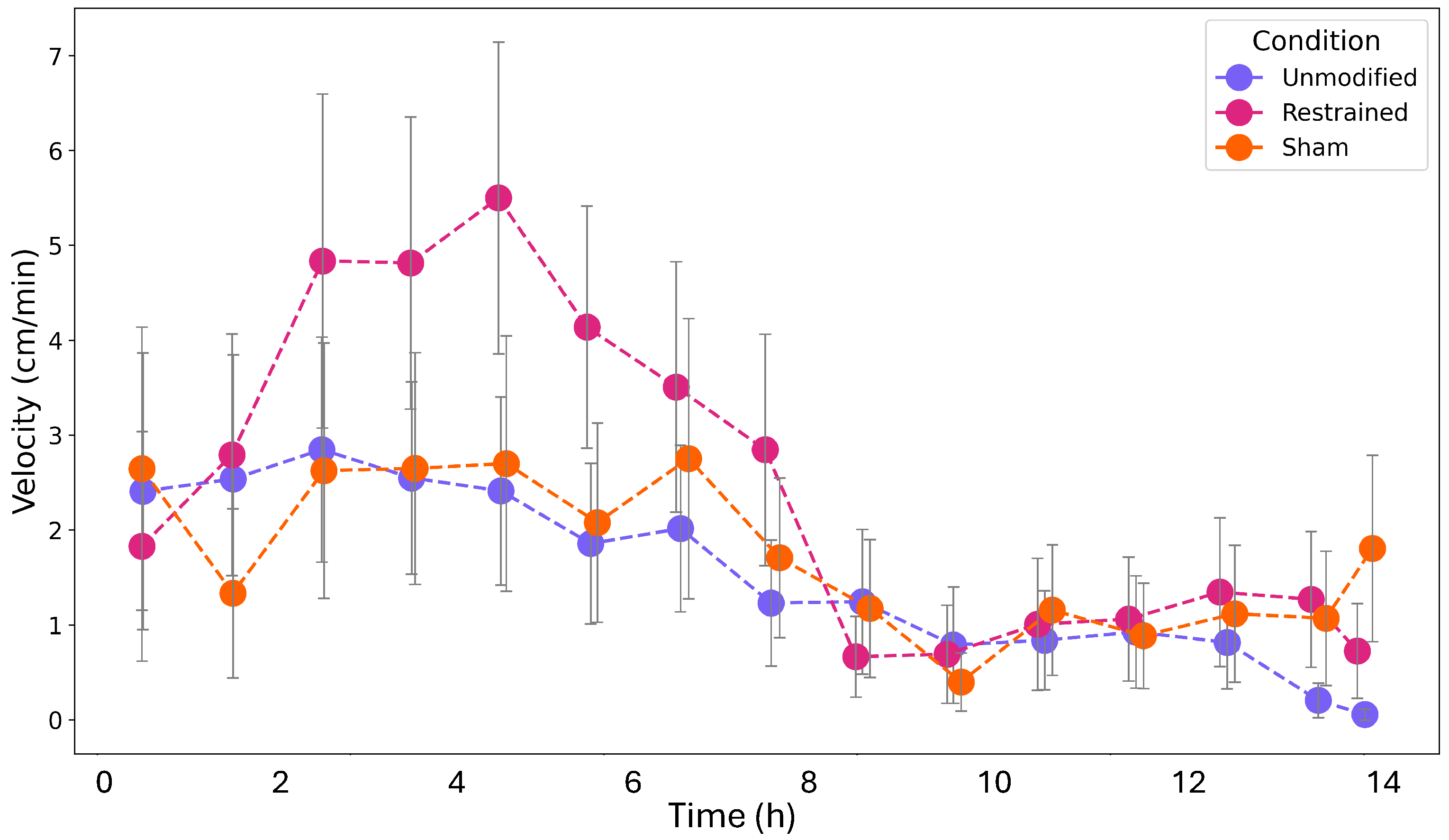
Although both distance and duration of movement in the 14-h trials were significantly greater for animals with restrained pectines, we noticed that the mean velocities of these animals were reduced compared to unmodified and sham animals (though the reduction was not statistically significant). What could explain this pattern? Perhaps the animals with restrained pectines moved longer into the night, but took shorter steps on average compared to the control trials. To investigate this possibility, we used a post hoc analysis to examine the patterns of activity in all trials. The activity plots of animals with legitimate trials are shown in Supplemental File S6, and the average velocities of the animals binned by 1-h intervals and parsed by condition type are shown in Figure 4. Note that these plots were constructed without removing rest periods (as had been done previously when examining the mean, median, and mode velocities (Figure 3)). The pattern in Figure 4 suggests that that all the scorpions in the trials were more active early, and that their velocity decreased through time, regardless of treatment. However, the animals with restrained pectines showed the greatest early activity and their movements were more sustained, with fewer periods of rest (i.e., as inferred by the low number of video frames in which the animal had not moved from its previous position).
There are several possible explanations for the increased movement of the animals with restrained pectines. These might include compensatory behavior, where animals move more to sample information using other senses; increased exploratory drive, prompted by the absence of familiar pecten input; or stress- or anxiety-like responses to the loss of sensory feedback. These possibilities (and perhaps others) offer different lenses for understanding the change in locomotory behavior and invite future investigation.
The results of this study also have important implications for future tests of pecten function and interpretations of previous ablation studies. Since the act of gently restraining the organs with a small piece of foil tape affected the movements of the animals, it is likely that the complete removal of the organs will have even more significant effects. After all, neurons projecting from the pectinal peg sensilla represent the largest sensory contribution to the central nervous system of scorpions [22]. We recommend that all future behavioral studies of pectines use reversible impairment techniques such as those developed in this paper.
We also think that future studies using similar techniques might benefit from shorter trials. Limiting tests to a two-hour window instead of a full night, for example, might reduce the influence of compensatory behavior. Our findings also reflect the animals’ behavioral flexibility in response to sensory disruption and that pectines influence more than just goal-directed navigation (for mating or getting back home). They may also modulate general movement strategies, especially when no behaviorally relevant cues are present, which opens additional questions about how scorpions integrate sensory feedback into baseline locomotor decision-making. Finally, we remind that the pectines were not completely covered in these studies but were prevented from tapping the ground. Future projects might involve experimenting with how much of the pectines can be covered reversibly to totally block sensory function.
Since the tests described in this study examined only the effect of pecten restraint on the locomotor behavior of the animals, other possible studies might investigate how restraining the pectines affect a scorpion’s response to chemical stimuli, such as extracts from prey or conspecifics [13,16,17]. In addition, evidence from unmodified animals suggests that pectines are involved in the navigation of scorpions to their home burrow [23,24,27]. To fully implicate the pectines in navigation, it is crucial to assess the animal’s ability to return home with pectines restrained as compared to both unmodified and sham control animals.

Supplementary Materials

The following supporting information can be downloaded at: https://www.mdpi.com/article/10.3390/arthropoda3030012/s1, Supplemental Video S1: Washi Tape Residue Video; Supplemental Video S2: Reretraining Procedure Video; Supplemental File S1: Photos of Camera and Fence; Supplemental File S2: Raw vs. Adjusted Distance Comparisons; Supplemental File S3: RPR Day Effect; Supplemental File S4: Cover Checks; Supplemental File S5: Plots & Dependent Variables; Supplemental File S6: Activity Monitors.

Author Contributions

Conceptualization, S.E.G.F. and D.D.G.; methodology, S.E.G.F. and D.D.G.; software, D.D.G.; formal analysis, D.D.G. and M.H.H.; investigation, D.D.G. and S.E.G.F.; resources, D.D.G.; data curation, D.D.G.; writing—original draft preparation, D.D.G.; writing—review and editing, S.E.G.F., M.H.H. and D.D.G.; visualization, D.D.G., M.H.H. and S.E.G.F.; supervision, D.D.G.; project administration, D.D.G. All authors have read and agreed to the published version of the manuscript.

Funding

This research received no external funding.

Institutional Review Board Statement

Ethical review and approval were not required for this study.

Data Availability Statement

All data supporting the results of this study are available in the Supplemental Files below. Also, the videos of the behavioral trials will be available upon publication. All python code used in our analyses is available upon request to the corresponding author.

Acknowledgments

We thank Ingo Schlupp for useful discussions concerning experimental design and statistical analysis, George Martin for engineering assistance, the staff of OU’s Laboratory Animal Resources for assistance with animal care, and the OU School of Biological Sciences for facilities support. We also thank foreman Ronnie Miller with the Sealy-Smith Foundation in Monahans, TX for allowing us to collect animals on their land.

Conflicts of Interest

The authors declare no conflict of interest.

References

  1. Abushama, F.T. On the behaviour and sensory physiology of the scorpion Leiurus quinquestriatus (H. & E.). Anim. Behav. 1964, 12, 140–153. [Google Scholar] [CrossRef]
  2. Brownell, P.H. Compressional and surface waves in sand: Used by desert scorpions to locate prey. Science 1977, 197, 479–482. [Google Scholar] [CrossRef] [PubMed]
  3. Foelix, R.F.; Schabronath, J. Fine structure of scorpion sensory organs. I. Tarsal sensilla. Bull.-Br. Arachnol. Soc. 1983, 6, 53–67. [Google Scholar]
  4. Foelix, R.F.; Müller-Vorholt, G. The fine structure of scorpion sensory organs. II.Pecten sensilla. Bull. Br. Arachnol. Soc. 1983, 6, 68–74. [Google Scholar]
  5. Fleissner, G. Intracellular recordings of light responses from spiking and nonspiking cells in the median and lateral eyes of the scorpion. Naturwissenschaften 1985, 72, 46–48. [Google Scholar] [CrossRef]
  6. Meßlinger, K. Fine structure of scorpion trichobothria (Arachnida, Scorpiones). Zoomorphology 1987, 107, 49–57. [Google Scholar] [CrossRef]
  7. Ashford, K.; Blankenship, R.; Carpenter, W.; Wheeler, I.; Gaffin, D. Response of the eastern sand scorpion, Paruroctonus utahensis, to air movement from a moth analog. J. Arachnol. 2018, 46, 226–230. [Google Scholar] [CrossRef]
  8. Fet, V.; Brewer, M.S.; Soleglad, M.E.; Neff, D.P.A. Constellation array: A new sensory structure in scorpions (Arachnida: Scorpiones). Boletín Soc. Entomológica Aragonesa 2006, 1, 269–278. [Google Scholar]
  9. Cloudsley-Thompson, J. LXVII.—On the function of the pectines of scorpions. Ann. Mag. Nat. Hist. 1955, 8, 556–560. [Google Scholar] [CrossRef]
  10. Ivanov, V.; Balashov, Y. The structural and functional organization of the pectine in a scorpion Buthus eupeus Koch (Scorpiones, Buthidae) studied by electron microscopy. In The Fauna and Ecology of Arachnida; Trudy Zoological Institute: Leningrad, Russia, 1979; Volume 85, pp. 73–87. [Google Scholar]
  11. Gaffin, D.D.; Brownell, P.H. Response properties of chemosensory peg sensilla on the pectines of scorpions. J. Comp. Physiol. 1997, 181, 291–300. [Google Scholar] [CrossRef]
  12. Knowlton, E.D.; Gaffin, D.D. Functionally redundant peg sensilla on the scorpion pecten. J. Comp. Physiol. 2011, 197, 895. [Google Scholar] [CrossRef] [PubMed]
  13. Gaffin, D.D.; Brownell, P.H. Evidence of chemical signaling in the sand scorpion, Paruroctonus mesaensis (Scorpionida: Vaejovidae). Ethology 1992, 91, 59–69. [Google Scholar] [CrossRef]
  14. Gaffin, D.; Brownell, P. Chemosensory behavior and physiology. In Scorpion Biology and Research; Oxford University Press: Oxford, UK, 2001; pp. 184–203. [Google Scholar]
  15. Melville, J.M.; Tallarovic, S.K.; Brownell, P.H. Evidence of mate trailing in the giant hairy desert scorpion, Hadrurus arizonensis (Scorpionida, Iuridae). J. Insect Behav. 2003, 16, 97–115. [Google Scholar] [CrossRef]
  16. Taylor, M.S.; Cosper, C.R.; Gaffin, D.D. Behavioral evidence of pheromonal signaling in desert grassland scorpions Paruroctonus utahensis. J. Arachnol. 2012, 40, 240–244. [Google Scholar] [CrossRef]
  17. Krapf, D. Contact chemoreception of prey in hunting scorpions (Arachnida: Scorpiones). Zool. Anz. 1986, 217, 119–129. [Google Scholar]
  18. Alexander, A.J. The Courtship and Mating of the Scorpion, Opisthophthalmus latimanus. Proc. Zool. Soc. Lond. 1957, 128, 529–544. [Google Scholar] [CrossRef]
  19. Alexander, A.J. Courtship and mating in the buthid scorpions. Proc. Zool. Soc. Lond. 1959, 133, 145–169. [Google Scholar] [CrossRef]
  20. Drozd, D.; Wolf, H.; Stemme, T. Mechanosensory pathways of scorpion pecten hair sensillae—adjustment of body height and pecten position. J. Comp. Neurol. 2022, 530, 2918–2937. [Google Scholar] [CrossRef]
  21. Peeples, H.M.; Gaffin, D.D. An assessment of the mechanosensory responses of peg sensilla on scorpion pectines. J. Arachnol. 2024, 52, 1–8. [Google Scholar] [CrossRef]
  22. Gaffin, D.D.; Curry, C.M. Arachnid navigation—A review of classic and emerging models. J. Arachnol. 2020, 48, 1–25. [Google Scholar] [CrossRef]
  23. Gaffin, D.D.; Muñoz, M.G.; Hoefnagels, M.H. Evidence of learning walks related to scorpion home burrow navigation. J. Exp. Biol. 2022, 225, jeb243947. [Google Scholar] [CrossRef] [PubMed]
  24. Ortega-Escobar, J.; Hebets, E.A.; Bingman, V.P.; Wiegmann, D.D.; Gaffin, D.D. Comparative biology of spatial navigation in three arachnid orders (Amblypygi, Araneae, and Scorpiones). J. Comp. Physiol. 2023, 209, 747–779. [Google Scholar] [CrossRef] [PubMed]
  25. Hladik, J.; Bailer, Y.; Wolf, H.; Stemme, T. Shelter selection in females of two scorpion species depends on shelter size and scent. J. Comp. Physiol. 2024, 211, 163–183. [Google Scholar] [CrossRef]
  26. Gaffin, D.D.; Walvoord, M.E. Scorpion peg sensilla: Are they the same or are they different? Euscorpius 2004, 17, 7–15. [Google Scholar]
  27. Prévost, E.D.; Stemme, T. Non-visual homing and the current status of navigation in scorpions. Anim. Cogn. 2020, 23, 1215–1234. [Google Scholar] [CrossRef]
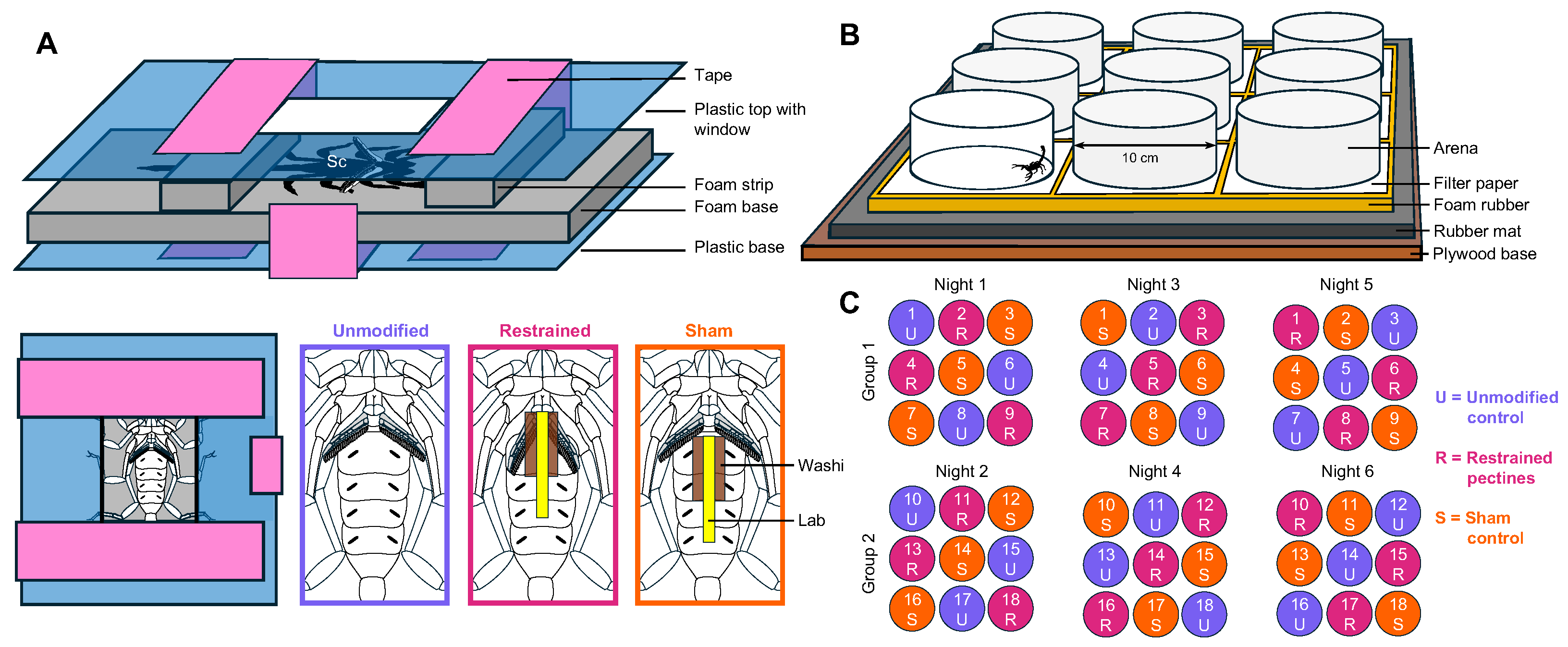
Figure 1. Restraining pectines, behavioral setup, and treatment menu. (A) Diagram of the restraint device used to restrict animal movements as we applied treatments, consisting of a plastic lid with a central window, foam strips and a foam base for protecting and holding the animal (Sc = scorpion) in position, plastic base, and tape to clamp the parts of the device together. Diagrams on the bottom right illustrate a ventral view of a scorpion receiving each treatment (unmodified control, pectines restrained, and sham control). (B) Behavioral setup showing the nine cylindrical arenas arranged on a filter paper layer atop a foam rubber base and rubber mat for vibration dampening. An IR camera was positioned 170 cm above the arenas to capture animal activity simultaneously in all nine arenas. (C) Treatment schedule for the 18 scorpions divided into two groups of nine, showing the order in which each scorpion underwent the three treatments.
Arthropoda 03 00012 g001
Figure 2. Sample plots for one animal. Data for animal #11 are shown for all three conditions. The animal’s position is plotted at 2-s intervals and its direction of travel is indicated by the direction of the arrow heads. The red arrows show the positions after transforming the points to the 8.8 cm diam circle (see Methods). The numbers at the periphery indicate the percent time of occupancy for the 18 arena sectors (based on the transformed points). Total distance moved for the raw coordinates, total distance moved for the transformed coordinates, total time of movement, and variance in percent sector occupancy are indicated above each arena.
Arthropoda 03 00012 g002
Figure 3. Dependent variable results. The small solid dots indicate the values for the six dependent variables for the 16 legitimate animals across the three experimental conditions (each animal’s trials are connected by dashed lines). The large dots with horizontal lines through their midpoints indicate the average for each condition, and the shaded boxes indicate 95% confidence intervals. Conditions with different letters are significantly different at p < 0.05.
Arthropoda 03 00012 g003
Figure 4. Activity comparisons. Shown are the averaged velocity plots (binned at 1 h intervals) for all legitimate trials parsed by condition type (whiskers are ±SE).
Arthropoda 03 00012 g004
Table 1. Summary of statistical analyses of dependent variables.
Normality and
Sphericity
Assumptions Met?
Degrees of
Freedom
Repeated Measures
ANOVA
(Parametric)
F Value
ANOVA Effect
Size Measure ( η 2)
Friedman Test
(Nonparametric)
W Value
p Value
Total distanceyes25.47920.1324---0.0094 ***
restrained vs. sham 15 0.0474 **
restrained vs. unmodified 15 0.0160 **
sham vs. unmodified 15 1.0000
Movement durationno2--- 0.54300.0002 ****
restrained vs. sham 15 0.0369 **
restrained vs. unmodified 15 0.0051 ***
sham vs. unmodified 15 1.0000
Sector occupancy varianceno2--- 0.00390.9394
Mean velocityyes22.91670.0476---0.0696 *
Median velocityyes21.24300.0217---0.3029
Mode velocityno2--- 0.02730.6456
* p < 0.1; ** p < 0.05; *** p < 0.01; **** p < 0.001.
Disclaimer/Publisher’s Note: The statements, opinions and data contained in all publications are solely those of the individual author(s) and contributor(s) and not of MDPI and/or the editor(s). MDPI and/or the editor(s) disclaim responsibility for any injury to people or property resulting from any ideas, methods, instructions or products referred to in the content.