Abstract
Weeds have acquired resistance to commonly used herbicides, and to replace them, new products, including those of natural origin, have been produced. This study evaluated the neurotoxicity, cytotoxicity, and changes in the antioxidant system caused by Natural Herbicide (NH) in SH-SY5Y neuroblastoma cells and HaCaT dermal cells. SH-SY5Y and HaCaT cells were exposed to three concentrations of NH (NH1: 0.6; NH2: 1.56; and NH3: 3.12 µL/mL) for 24 and 72 h. In the SH-SY5Y cell line, the highest concentration of NH (NH3) caused cytotoxicity at both 24 and 72 h. At 24 h, the NH3 group increased the SOD. In the NH2 and NH3 groups, there was also an increase in AChE activity after 24 h of exposure. The NH1 group led to an increase in GSH after 72 h of exposure. As for the HaCaT strain, there was cytotoxicity and an increase in SOD and GSH at all NH concentrations and for both periods analyzed (24 h and 72 h). GST was reduced after exposure to NH2 and NH3. Thus, NH showed cytotoxicity in neural and dermal cells (SH-SY5Y and HaCaT, respectively). These results show that NH altered cellular homeostasis, and the evaluation of other toxicity mechanisms is important to clarify its safety.
1. Introduction
Pesticides are chemical products, natural or synthetic, with widespread application in agriculture for the management of pests, weeds, and plant diseases. They encompass a wide range of herbicides, insecticides, fungicides, rodenticides, and nematicides [1]. However, despite the increasing use of these chemicals, they not only affect target organisms but also non-target ones that come into contact with them, including other plant species and fauna [2]. In addition, they have been reported to be harmful to human health, with studies indicating their toxicity in the nervous system [3] and epidermis [4]. Human exposure to pesticides occurs through dermal, oral, and respiratory pathways [5]. They can be directly exposed to pesticides in occupational, agricultural, or household activities, while indirect exposure may occur through environmental means, including air, water, soil, and food, with both direct and indirect exposure potentially resulting in acute toxicity and chronic diseases [6].
Among the pesticides used worldwide, herbicides are those applied to kill or suppress weeds growing in several settings, such as agricultural fields, lawns, gardens, and industrial sites [7]. Natural Herbicide (NH), marketed as a biological aqueous extract, is an organic compound comprising Annoni Grass extract (Eragrostis plana), Citronella oil (Cymbopogon sp.), Pinus extract (Pinus sp.), Platanus extract (Platanus occidentalis), and neem oil (Azadirachta indica). The cytotoxicity of this herbicide is still unknown, as well as its mechanism of action and potential health hazards. However, there are descriptions of the isolated action of each of its components in plants, which do not necessarily provide complete information about the possible risks of Natural Herbicide itself. Eragrostis plana already has its allelopathic potential described, since many of its components have phytotoxic power, and due to this, it has been used as a bioherbicide in some regions of Brazil [8]. As for Cymbopogon sp. oil, it has already been described as phytotoxic to seeds of Echinochloa crus-galli, preventing their germination [9]. Some pine species, mainly Pinus halepensis and Pinus elliottii, have phenolic compounds with phytotoxic capacity [10]. These two species cause a very efficient metabolic imbalance in weeds, which led the pine extract and its potential as a bioherbicide to be compared to the synthetic herbicide glyphosate [11]. Concerning Platanus occidentalis, it is a species for which there is little knowledge of its phytotoxicity, but it is known that it is capable of inhibiting the growth of seeds [12]. Azadirachta indica oil, known as neem, or nim, has already been used to control insects; however, there are descriptions of its ability to inhibit the growth of various plant cultures through phenolic components such as benzoic and vanillic acid [13].
Regarding the toxicity in non-target organisms, several cellular changes result from exposure to pesticides, such as mitochondrial dysfunction, inflammation, immunotoxicity, and endocrine disruption [14]. Concerning neurotoxic effects, the main mechanism occurs through the inhibition of the enzyme acetylcholinesterase (AChE), which acts in the synaptic cleft by hydrolyzing acetylcholine. When this enzyme is inhibited by a drug or toxic agent, it becomes unable to hydrolyze this neurotransmitter, leading to its permanence in the synaptic cleft and producing toxic effects [15].
Recently, the toxicity of NH was assessed using zebrafish (Danio rerio) as the experimental model. It was observed that, although NH has a lower toxicity when compared to widely used herbicides, such as glyphosate and atrazine, it still showed mutagenic potential [16]. This finding suggests that it may have toxic effects on other non-target organisms, such as human beings, which needs to be investigated. For this purpose, alternative methods to animal experimentation can be used, such as in vitro experimentation, following the principle of the 3Rs, since this type of approach leads to the replacement of animal use [17].
In this context, the human neuroblastoma cell line SH-SY5Y is widely used as an in vitro model for neuroscience studies [18]. However, it has also been established as a model for evaluating the neurological effects of pesticides and pesticide screening [19]. On the other hand, HaCaT is a non-tumorigenic monoclonal cell line, derived from adult skin keratinocytes [20]. It has been used in the toxicology field to assess the toxicity of several substances, including pesticides [21,22,23]. Therefore, this study aims to evaluate the toxicological effects of exposure to NH, using SH-SY5Y and HaCaT cell lines. For this purpose, cytotoxicity, acetylcholinesterase enzyme activity, and antioxidant system evaluation assays will be carried out to determine its risks to human health.
2. Materials and Methods
2.1. Chemicals
The natural herbicide formulation used in this study comprises ethanolic extracts (96%; v/v) obtained from five botanical sources: Cymbopogon nardus (citronella), Ateleia glazioviana (timbó), Eragrostis plana (annoni grass), Azadirachta indica (neem), and Planatus occidentalis (plantain). Characterization of natural product efficacy requires sequential analytical phases. Primary screening involves crude plant extracts followed by iterative fractionation employing chromatographic techniques to isolate bioactive constituents. As a community-developed artisanal product, the precise phytochemical ratios within this inaugural formulation remain unquantified [24].
2.2. Cell Culture and Exposure
The neuroblastoma cell line SH-SY5Y (number 0223) and keratinocyte cell line HaCaT (number 0341) were acquired from the cell bank of Rio de Janeiro (BCRJ; Rio de Janeiro, RJ, Brazil), Brazil. Cells were maintained using polypropylene bottles with an area of 75 cm2 in 15 mL of DMEM/F12 (Dulbecco’s Modified Eagle’s Medium/F12—Sigma-Aldrich, St. Louis, MO, USA) medium, supplemented with 10% Fetal Bovine Serum (SFB) (Sigma-Aldrich, St. Louis, MO, USA), 1% penicillin 10.000 U.I. mL−1, and streptomycin 10 mg/mL (Sigma-Aldrich, St. Louis, MO, USA). Cell cultures were maintained at 37 °C in a humidified atmosphere of 5% CO2 and 95% air.
Cells were exposed to NH in three concentrations (NH1: 0.6; NH2: 1.56; and NH3: 3.12 µL/mL) for 24 h to assess an acute exposure process and 72 h for more prolonged exposure. The highest concentration is equivalent to that used by farmers, and the other concentrations were determined with the aim of observing the occurrence of changes even at lower herbicide concentrations.
2.3. Cell Viability Assay
To evaluate cell viability, the trypan blue method (Sigma-Aldrich, St. Louis, MO, USA) was used. SH-SY5Y and HaCaT cells were seeded in 6-well plates at a density of 2 × 10⁵ cells per well and exposed to NH or a negative control (only enriched medium). After exposure, trypsinization and counting of cells were performed. In the trypsinization process, the cells were washed twice with phosphate-buffered saline (PBS; Sigma-Aldrich, St. Louis, MO, USA), exposed to trypsin (3 mL; Sigma-Aldrich, St. Louis, MO, USA) for 5 min, enriched medium was added (3 mL), and the cells were centrifuged at 1500 rpm for 5 min. The supernatant was discarded, and the pellet was resuspended in 1 mL of PBS. Subsequently, an aliquot of the cell solution (50 µL) was stained with trypan blue (1:1), and the cells were counted using a Neubauer Chamber in a light microscope. Non-stained cells indicate their viability since their cell membrane remains intact and there is no dye entry, whereas non-viable cells are stained blue. The viability percentage was calculated using the formula (viable cells count/total cell count) × 100 [25].
2.4. Cytotoxicity Assay
Cytotoxicity was determined using the Presto Blue™ reagent (Thermo Fisher, number A13262, USA). For this purpose, cells were cultured in 96-well plates with enriched medium at a density of 5 × 10⁴. After 24 h, the cells were exposed to the different concentrations of NH and a negative control. Exposure was carried out in periods of 24 and 72 h. Subsequently, the medium was removed, and 90 µL of medium plus 10 µL of Presto Blue™ was added per well. Then, the plates were incubated for 90 min at 37 °C and read at 570 and 600 nm in a spectrophotometer. The calculation was made from the Presto Blue™ reduction based on the manufacturer’s instructions.
2.5. Acetylcholinesterase Enzyme Activity and Antioxidant System Assay
The SH-SY5Y and HaCaT cell lines were seeded in 6-well plates, with a concentration of 2 × 10⁵ cells per well. The cells were exposed to the three NH concentrations (NH1: 0.6; NH2: 1.56; and NH3: 3.12 µL/mL) and the negative control (only enriched medium) for periods of 24 h and 72 h. Afterwards, the cells were recovered by trypsinization. To achieve this, a double wash with PBS was performed, followed by incubation with trypsin (3 mL) for 5 min, inactivation with enriched medium (3 mL), and centrifugation at 1500 rpm for 5 min. The cells obtained were submitted to three wash sequences with PBS and centrifugation, after which their membranes were ruptured by freezing (−20 °C; 30 min) and thawing. Then, analyses of the biomarkers of the antioxidant system were carried out, i.e., superoxide dismutase and glutathione S-transferase activities, and reduced glutathione concentration. A biological marker of neurotoxicity was also measured: acetylcholinesterase activity [26,27]. In addition, quantification of total proteins was performed.
The superoxide dismutase (SOD) analysis was carried out according to the method proposed by Gao et al. [28]. This method is based on the occurrence of self-oxidation of pyrogallol (Neon®; Suzano, SP, Brazil) in weakly basic solutions and on the capacity of SOD to inhibit this reaction. The reading was performed at a wavelength of 440 nm, with the results being expressed in U of SOD/mg of protein.
The evaluation of glutathione S-transferase (GST) enzyme activity was based on the method described by Keen et al. [29]. GST activity is expressed in nmol.min−1.mg protein−1.
The concentration of reduced glutathione (GSH) was analyzed by the method of Sedlak and Lindsay [30]. Readings were taken at 415 nm, and the results are expressed in µg GSH.mg protein−1.
The enzyme acetylcholinesterase (AChE), analyzed on the neuroblastoma cell line SH-SY5Y had its activity evaluated by the method described by Ellman and collaborators, based on measuring the absorbance of a yellow color that is formed from thiocholine when it reacts with 5,5′-dithiobis-(2-nitrobenzoic acid; Sigma-Aldrich, St. Louis, MO, USA) [31]. The activity is expressed in nmol.min−1.mg protein−1.
The quantification of total proteins in the samples was performed according to the Bradford method (Sigma-Aldrich, St. Louis, MO, USA) [32]. The absorbance reading was performed at 595 nm.
2.6. Statistical Analysis
Data normality was verified using the Shapiro–Wilk test, and statistical analysis was performed using the Graph Pad Prism 5.0 program. In cases where the data met the normality criteria, these were analyzed by one-way ANOVA, followed by Tukey’s post-test. Outliers were detected using the Grubbs test. The results were considered statistically significant when p < 0.05.
3. Results
3.1. Cytotoxicity of NH on SH-SY5Y Cell Line
The NH cytotoxicity assessment showed that changes occurred in the SH-SY5Y cell line for both periods of exposure to the herbicide. There was a significant difference in the percentage reduction of Presto Blue™ both at 24 h and 72 h at the highest herbicide concentration (NH3) (Figure 1).
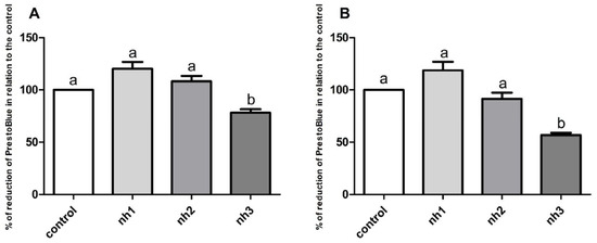
Figure 1.
Cytotoxicity analysis: Percentage of reduction of Presto Blue™ in relation to the control after exposure of the SH-SY5Y cell line to the natural herbicide (nh) for periods of 24 h (A) and 72 h (B). Data are displayed as mean ± standard error of the mean. The p-value considered significant was p < 0.05; groups with the same letters do not differ statistically, and groups with different letters present a statistically significant difference. ANOVA, Tukey’s post-hoc. n = 18.
3.2. Cytotoxicity of NH on HaCaT Cell Line
HaCaT cells showed cytotoxicity after exposure to all NH concentrations within 24 h and 72 h. A significant difference was observed in the percentage reduction of Presto Blue™ compared to the control for both exposure periods (Figure 2).
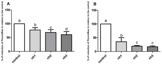
Figure 2.
Cytotoxicity analysis: Percentage of reduction of Presto Blue™ in relation to the control after exposure of the HaCaT cell line to the natural herbicide (nh) for periods of 24 h (A) and 72 h (B). Data are displayed as mean ± standard error of the mean. The p-value considered significant was p < 0.05; groups with the same letters do not differ statistically, and groups with different letters present a statistically significant difference. ANOVA, Tukey’s post-hoc. n = 18.
3.3. Acetylcholinesterase Activity on SH-SY5Y Cell Line
After 24 h, AChE activity increased in the NH2 and NH3 groups. For the 72 h exposure period, all herbicide concentrations caused a significant increase in AChE activity (Figure 3).
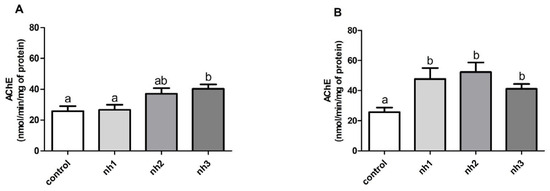
Figure 3.
Analysis of acetylcholinesterase activity (AChE) on SH-SY5Y cells exposed to natural herbicide (nh) for 24 h (A) and 72 h (B). The results are expressed as mean ± standard error of the mean. The p-value considered significant was p < 0.05; groups with the same letters do not differ statistically, and groups with different letters present a statistically significant difference. ANOVA, Tukey’s post-hoc. n = 12.
3.4. Effects of NH on Oxidative Stress Markers on SH-SY5Y Cell Line
Regarding the antioxidant system on the SH-SY5Y cell line, the NH2 group led to increased levels of GSH after 24 h of exposure. An increase in GSH was also observed in the NH1 group, but without a statistically significant difference (Figure 4E). SOD enzyme activity increased after 24 h of exposure to the two highest NH concentrations (Figure 4A), but after 72 h of exposure, it was not changed (Figure 4B). GST enzyme was not altered by NH at any concentration or period analyzed (Figure 4C,D).
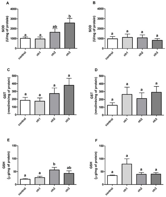
Figure 4.
Analysis of the antioxidant system and SH-SY5Y cells exposed to the natural herbicide (nh). SOD = superoxide dismutase (U/mg protein), 24 h (A) and 72 h (B); GST = glutathione S-transferase (nmol/min/mg protein), 24 h (C) and 72 h (D); and GSH = reduced glutathione (μg/mg protein), 24 h (E) and 72 h (F). Results are expressed as mean ± standard error of the mean. The p-value considered significant was p < 0.05; groups with the same letters do not differ statistically, and groups with different letters present a statistically significant difference. ANOVA, Tukey’s post-hoc. n = 12.
3.5. Effects of NH on Oxidative Stress Markers on HaCaT Cell Line
The analysis of oxidative stress markers on the HaCaT cell line allowed us to observe changes in the antioxidant system based on SOD and GST activity and GSH levels. SOD levels increased significantly after 24 h and 72 h of exposure to all NH concentrations (Figure 5A,B). GST, on the other hand, showed a reduction in its activity, with a significant difference at the two highest concentrations of NH (NH2 and NH3) for both exposure periods (Figure 5C,D). As for GSH levels, there was a significant increase after 24 h of exposure to NH1 and NH2 (Figure 5E), and after 72 h of exposure to NH2 and NH3 (Figure 5F).
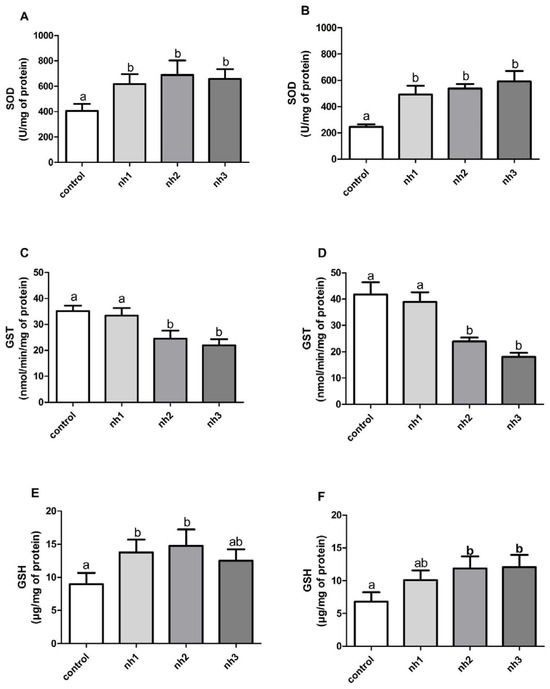
Figure 5.
Alterations in the antioxidant system of HaCaT cells exposed to natural herbicide (nh). SOD = superoxide dismutase (U/mg of protein) 24 h (A) and 72 h (B); GST = glutathione S-transferase (nmol/min/mg of protein) 24 h (C) and 72 h (D); and GSH = glutathione reduced (μg/mg of protein) 24 h (E) and 72 h (F). The results are expressed as mean ± standard error of the mean. The p-value considered significant was p < 0.05; groups with the same letters do not differ statistically, and groups with different letters present a statistically significant difference. ANOVA, Tukey’s post-hoc. n = 12.
4. Discussion
In this study, the effects of NH on human cells are reported for the first time, presenting possible risks to the health of individuals exposed to this compound. Only one other study evaluating the effects of NH was found in the literature, carried out by Lechinovski and collaborators. In this research, a comparative analysis was carried out between the conventional herbicides atrazine and glyphosate and Natural Herbicide in zebrafish (Danio rerio). Nuclear abnormalities were found in the blood of fish exposed to all herbicides analyzed, although they were more prominent for conventional herbicides [16]. Accordingly, it is possible to see that NH is capable of causing significant damage at a cellular level.
Assays with Presto Blue™ indicated cytotoxicity of NH, both in SH-SY5Y cells and in HaCaT cells, being observed in the latter to a greater extent, which indicates greater sensitivity of cells of dermal origin to NH when compared to those of neural origin. Another study evaluating the effect of the pesticide monocrotophos on the lung tumor line A549 and the dermal cell line HaCaT found similar toxicity effects, with no disparity in effect between the tumorigenic and non-tumorigenic cell lines [33]. The toxicity of the insecticides deltamethrin and chlorpyrifos was tested on the human keratinocyte line HUKE and the neuronal line SH-SY5Y, and both lineages showed the same level of sensitivity [34]. These studies demonstrate that the effect of substances on cell lines was more related to their origin and tissue characteristics than to whether they were tumorigenic or non-tumorigenic.
Other studies analyzed the cytotoxicity of synthetic herbicides on SH-SY5Y cells, with a great reduction in cell viability. In a study developed by Abarikwu and Farombi, the cytotoxic effect of the broad-spectrum herbicide atrazine, used on a large scale worldwide in various types of crops, was evaluated. The authors showed, through MTT assays, the occurrence of dose and time-dependent cytotoxicity arising from atrazine in neuroblastoma cells [35]. Similarly, Martínez and colleagues demonstrated that the SH-SY5Y strain could be used as an in vitro model for analyzing pesticides and also indicated that glyphosate, a post-emergent herbicide, induces cytotoxic effects on neuronal development, oxidative stress, apoptosis, and necrosis [19]. Regarding HaCaT, cytotoxicity tests after exposure to herbicides such as glyphosate and insecticides such as imidacloprid demonstrated IC50 values considered low, being 30 mM (24 h) and 2.35 mM (18 h), respectively, indicating the sensitivity of this cell line to herbicides [36]. The herbicide glyphosate was also capable of causing stiffening of the cell membrane on HaCaT cells after 18 h of exposure, a result that, like the present work, points to the cytotoxicity caused by herbicides on the HaCaT cell line [37].
Exposure to NH caused changes in AChE activity in SH-SY5Y cells, indicating neurotoxicity. This enzyme is responsible for the hydrolysis of the neurotransmitter acetylcholine into acetate and choline, allowing the continuous release of the neurotransmitter and propagation of the nerve impulse [38]. Some related studies analyzed the neurotoxic potential of pesticides in vivo and in vitro [39]. Fernandez and colleagues analyzed the effects of the herbicide alachlor, commonly used on grasses and weeds that affect corn crops, on RTG-2 and SH-SY5Y cell lines. It was observed that in neuroblastoma cells, there was a large increase in AChE activity, with EC50 of 20 µM in 24 h of exposure being considered a very sensitive marker for this cell line [40]. Another study, which evaluated the acute effects of glyphosate in fish of the species Prochilodus lineatus, found that there was inhibition of AChE activity in the muscle and brain tissue of the species [41]. In addition, AChE was established as an apoptosis marker because it shows increased activity during the apoptosis process and after apoptotic stimuli in numerous cell types and in vivo models. Although this aspect was not evaluated in the present study, it has been demonstrated that in cells in which the expression of this enzyme is increased, sensitivity to the occurrence of apoptosis also increases, in the same way that AChE deficiency leads to an inhibition of apoptosis [42]. Such studies show the potential of herbicides to cause changes in AChE levels in vivo and in vitro and consequently lead to an abnormal decrease or increase in the propagation of nerve impulses in living beings, in addition to being related to the occurrence of cell death.
The present study also demonstrates the induction of the antioxidant system through the observation of alterations in the levels of oxidative stress markers. Similar results were observed in both cell lines in relation to the SOD enzyme, since both caused an increase in its activity. However, different results were observed in relation to the GST enzyme, indicating differences in the response of the antioxidant systems of neural and dermal cells to NH. Antioxidant system markers work together in different pathways in order to defend the cell against free radicals and other oxidative agents [43]. Therefore, the SOD enzyme acts on oxygen free radicals (O2•−), transforming them into hydrogen peroxide (H2O2). GPx then converts H2O2 into H2O and O2 at the expense of oxidizing GSH into GSSG. However, to maintain GSH levels, glutathione reductase (GR) reduces GSSG into GSH, using NADPH as a cofactor [44]. Yang and Tiffany-Castiglioni exposed neuroblastoma cells to the herbicide paraquat, which showed pro-oxidant properties, leading to the inhibition of GPx activity and elevation of GST activity after 24 h of herbicide exposure [45]. However, and similarly to what was observed here in the HaCaT cell line exposed to NH, Diken and coworkers demonstrated the inhibitory potential of glyphosate on GST activity in human erythrocytes exposed to it [46]. The authors point out that glyphosate was able to reduce the enzyme’s activity in a non-competitive way, that is, without preventing it from binding to its substrate (GSH), but rather making its catalytic activity impossible.
Another study evaluated the chronic effects of low doses of rotenone, a pesticide considered natural due to its spontaneous occurrence in the roots of some plants, on SH-SY5Y cells. This component can cross the blood–brain barrier and can cause systemic toxicity. The study demonstrated an increase in SOD levels after 4 weeks of treatment, indicating the presence of a reaction against superoxide [47].
Furthermore, although NH has cytotoxic potential, it can be considered less harmful to the environment and more advantageous due to its artisanal production process compared to synthetic herbicides. When comparing thirty-two synthetics and thirty-four natural pesticides, using the Toxicity Estimation Software Tool (T.E.S.T.; version 4.2), studies found that the persistence in the environment and toxicity levels of synthetic pesticides are larger than those of the natural pesticides [48]. However, although it presents usage advantages when compared to conventional herbicides, some studies, such as the one presented here, demonstrate that NH can affect the antioxidant system and potentially cause neurotoxicity. Therefore, further studies should be conducted to assess the effects of this herbicide on other non-target organisms that may be exposed to it.
5. Conclusions
The antioxidant system, AChE activity, and viability of SH-SY5Y and HaCaT cells were altered after exposure to all concentrations of NH for both periods analyzed (24 and 72 h). Therefore, it is possible to conclude that NH caused changes in cellular homeostasis, since the elevation in the activity of antioxidant enzymes indicates an increase in reactive oxygen species, while the enhancement of AChE activity points to a reduction in nerve impulse propagation. When comparing the results obtained between the two cell lines, it is observed that HaCaT cells present more significant changes than cells of neural origin, which may indicate that exposure to NH via the dermal route leads to changes of greater impact than those observed in cells of the nervous system.
However, other analyses are necessary to confirm such findings, including assessments of apoptosis and inflammatory markers. Moreover, the need for other studies on the subject is notable, using different methods, concentrations, and comparative tests with conventional herbicides, since research on this herbicide is scarce. There is also a need to analyze alternative NH use concentrations, evaluating its level of effectiveness on weeds to carry out possible changes aimed at reducing damage to health and the environment. In this way, it will be possible to reduce the risks to local fauna and flora, as well as to the health of rural workers and the general population.
Author Contributions
L.N.-O.: Investigation, Data curation, Formal analysis, Writing—original draft, Visualization. J.F.d.S.: Investigation. S.d.S.M.: Investigation, Writing—review and editing. L.L.: Investigation. A.C.d.D.B.K.: Writing—review and editing, Investigation. I.C.G.: Conceptualization, Funding acquisition, Project administration, Supervision, Writing—review and editing. All authors have read and agreed to the published version of the manuscript.
Funding
The authors thank the Fundação Araucária for the scholarship granted to Letícia Nominato-Oliveira, the Instituto de Pesquisa Pelé Pequeno Príncipe for the scholarship granted to Juliana Ferreira da Silva, and the Conselho Nacional de Desenvolvimento Científico e Tecnológico (CNPq) for granting a junior post-doctoral fellowship (PROFIX-JD; process 421691/2022–0) to Shayane da Silva Milhorini. We would also like to thank the Instituto de Pesquisa Pelé Pequeno Príncipe for the equipment and materials used for the development of the experiment. The funders were not involved in the design of the study; data collection, analysis, and interpretation of the data; or the writing of the manuscript.
Institutional Review Board Statement
Not applicable.
Informed Consent Statement
Not applicable.
Data Availability Statement
The original contributions presented in this study are included in the article. Further inquiries can be directed to the corresponding author.
Conflicts of Interest
The authors declare no conflicts of interest.
References
- Sharma, A.; Kumar, V.; Shahzad, B.; Tanveer, M.; Sidhu, G.P.S.; Handa, N.; Kohli, S.K.; Yadav, P.; Bali, A.S.; Parihar, R.D.; et al. Worldwide pesticide usage and its impacts on ecosystem. SN Appl. Sci. 2019, 1, 1446. [Google Scholar] [CrossRef]
- Kumar, V.; Sharma, N.; Sharma, P.; Pasrija, R.; Kaur, K.; Umesh, M.; Thazeem, B. Toxicity analysis of endocrine disrupting pesticides on non-target organisms: A critical analysis on toxicity mechanisms. Toxicol. Appl. Pharmacol. 2023, 474, 116623. [Google Scholar] [CrossRef] [PubMed]
- Costas-Ferreira, C.; Durán, D.; Faro, L.R.F. Toxic effects of glyphosate on the nervous system: A systematic review. Int. J. Mol. Sci. 2022, 23, 4605. [Google Scholar] [CrossRef] [PubMed]
- Zakharov, S.; Csomor, J.; Urbanek, P.; Pelclova, D. Toxic epidermal necrolysis after exposure to dithiocarbamate fungicide mancozeb. BCPT 2016, 118, 89–91. [Google Scholar] [CrossRef]
- Damalas, C.A.; Koutroubas, S.D. Farmers’ exposure to pesticides: Toxicity types and ways of prevention. Toxics 2016, 4, 1. [Google Scholar] [CrossRef]
- Tudi, M.; Li, H.; Li, H.; Wang, L.; Lyu, J.; Yang, L.; Tong, S.; Yu, Q.J.; Ruan, H.D.; Atabila, A.; et al. Exposure routes and health risks associated with pesticide application. Toxics 2022, 10, 335. [Google Scholar] [CrossRef]
- Ghazi, R.M.; Yusoff, N.R.N.; Halim, N.S.A.; Rahayu, I.; Wahab, A.; Latif, N.A.; Hasmoni, S.H.; Zaini, M.A.A.; Zakari, Z.A. Health effects of herbicides and its current removal strategies. Bioengineered 2023, 14, 2259526. [Google Scholar] [CrossRef]
- Favaretto, A.; Cantrell, C.L.; Fronczek, F.R.; Duke, S.O.; Wedge, D.E.; Ali, A.; Scheffer-Basso, S.M. New phytotoxic cassane-like diterpenoids from Eragrostis plana. J. Agric. Food Chem. 2019, 67, 1973–1981. [Google Scholar] [CrossRef]
- Poonpaiboonpipat, T.; Pangnakorn, U.; Suvunnamek, U.; Teerarak, M.; Charoenying, P.; Laosinwattana, C. Phytotoxic effects of essential oil from Cymbopogon citratus and its physiological mechanisms on barnyardgrass (Echinochloa crus-Galli). Ind. Crops Prod. 2013, 41, 403–407. [Google Scholar] [CrossRef]
- Refifa, T.; Chahdoura, H.; Flamini, G.; Adouni, K.; Hammami, M.; Helal, A.N. Allelopathic potential of Pinus halepensis needles. Allelopath. J. 2016, 38, 193–214. [Google Scholar]
- Haas, P.; Kuhn, D.; Cordeiro, S.G.; Schweizer, Y.A.; Costa, B.; Hoehne, L. Phytotoxic effect of Pinus elliottii extracts on invasive plants from agricultural cultivation systems. Rev. Ibero-Am. Ciênc. Ambie. 2021, 12, 121–131. [Google Scholar] [CrossRef]
- Al-Naib, F.A.G.; Rice, E.L. Allelopathic effects of Platanus occidentalis. Bull. Torrey Bot. Club. 1971, 98, 75–82. [Google Scholar] [CrossRef]
- Xuan, T.D.; Tsuzuki, E.; Hiroyuki, T.; Mitsuhiro, M.; Khanh, T.D.; Chung, I.M. Evaluation on phytotoxicity of neem (Azadirachta indica. A. Juss) to crops and weeds. Crop Prot. 2004, 23, 335–345. [Google Scholar] [CrossRef]
- Lee, G.H.; Choi, K.C. Adverse effects of pesticides on the functions of immune system. Comp. Biochem. Physiol. Part C Toxicol. Pharmacol. 2020, 235, 108789. [Google Scholar] [CrossRef]
- Sirin, G.S.; Zhang, Y. How is acetylcholinesterase phosphonylated by soman? An ab initio QM/MM molecular dynamics study. J. Phys. Chem. A 2014, 118, 9132–9139. [Google Scholar] [CrossRef] [PubMed]
- Lechinovski, L.; Bados, M.; Rosa, J.; Moda, D.B.; Krawczyk, A.C.D.B. Ecotoxicological effects of conventional herbicides and a natural herbicide on freshwater fish (Danio rerio). J. Environ. Sci. Health Part B 2022, 57, 812–820. [Google Scholar] [CrossRef]
- Kandárová, H.; Letašiová, S. Alternative methods in toxicology: Pre-validated and validated methods. Interdiscip. Toxicol. 2011, 4, 107–113. [Google Scholar] [CrossRef] [PubMed]
- Agholme, L.; Lindström, T.; Kågedal, K.; Marcusson, J.; Hallbeck, M. An in vitro model for neuroscience: Differentiation of SH-SY5Y cells into cells with morphological and biochemical characteristics of mature neurons. J. Alzheimer’s Dis. 2010, 20, 1069–1082. [Google Scholar] [CrossRef]
- Martínez, M.A.; Rodríguez, J.L.; Lopez-Torres, B.; Martínez, M.; Martínez-Larrañaga, M.R.; Maximiliano, J.E.; Anadón, A.; Ares, I. Use of human neuroblastoma SH-SY5Y cells to evaluate glyphosate-induced effects on oxidative stress, neuronal development and cell death signaling pathways. Environ. Int. 2020, 231, 105414. [Google Scholar] [CrossRef]
- Boukamp, P.; Petrussevska, R.T.; Breitkreutz, D.; Hornung, J.; Markham, A.; Fusenig, N.E. Normal keratinization in a spontaneously immortalized aneuploid human keratinocyte cell line. JCB 1988, 106, 761–771. [Google Scholar] [CrossRef]
- Gehin, A.; Guyon, C.; Nicod, L. Glyphosate-induced antioxidant imbalance in HaCaT: The protective effect of vitamins C and E. Environ. Toxicol Pharmacol. 2006, 22, 27–34. [Google Scholar] [CrossRef]
- Jang, Y.; Lee, A.Y.; Jeong, S.H.; Park, K.H.; Paik, M.K.; Cho, N.J.; Kim, J.E.; Cho, M.H. Chlorpyrifos induces NLRP3 inflammasome and pyroptosis/apoptosis via mitochondrial oxidative stress in human keratinocyte HaCaT cells. Toxicology 2015, 338, 37–46. [Google Scholar] [CrossRef]
- Sawickia, K.; Czajkaa, M.; Matysiak-Kuchareka, M.; Kruszewskia, M.; Skawińskia, W.; Brzóskac, K.; Kapka-Skrzypczaka, L. Chlorpyrifos stimulates expression of vitamin D3 receptor in skin cells irradiated with UVB. Pestic. Biochem. Physiol. 2019, 154, 17–22. [Google Scholar] [CrossRef]
- Mentz, L.A.; Petrovick, P.R. Farmacognosia: Da Planta ao Medicamento; Editora da UFSC: Florianópolis, Brazil, 2003. [Google Scholar]
- Strober, W. Trypan Blue Exclusion Test of Cell Viability. Curr. Protoc. Immunol. 2001, 111, A3.B.1–A3.B.3. [Google Scholar] [CrossRef]
- Sun, W.; Chen, L.; Zheng, W.; Wei, X.; Wu, W.; Duysen, E.G.; Jiang, W. Study of acetylcholinesterase activity and apoptosis in SH-SY5Y cells and mice exposed to ethanol. Toxicology 2017, 384, 33–39. [Google Scholar] [CrossRef] [PubMed]
- Lee, J.; Huchthausen, J.; Schlichting, R.; Scholz, S.; Henneberger, L.; Escher, B.I. Validation of an SH-SY5Y Cell–Based Acetylcholinesterase Inhibition Assay for Water Quality Assessment. Environ. Toxicol. Chem. 2022, 41, 3046–3057. [Google Scholar] [CrossRef]
- Gao, R.; Yuan, Z.; Zhao, Z.; Gao, X. Mechanism of pyrogallol autoxidation and determination of superoxide dismutase enzyme activity. Bioelectroch. Bioenerg. 1998, 45, 41–45. [Google Scholar] [CrossRef]
- Keen, J.H.; Habig, W.H.; Jakoby, W.B. Mechanism for the several activities of the glutathione S-transferases. J. Biol. Chem. 1976, 251, 6183–6188. [Google Scholar] [CrossRef]
- Sedlak, J.; Lindsay, R.H. Estimation of total, protein-bound, and nonprotein sulfhydryl groups in tissue with Ellman’s reagent. Anal. Biochem. 1968, 25, 192–205. [Google Scholar] [CrossRef]
- Ellman, G.L.; Courtney, K.D.; Andres, V.; Featherstone, R.M. A new and rapid colorimetric determination of acetylcholinesterase activity. Biochem. Pharmacol. 1961, 7, 88–95. [Google Scholar] [CrossRef]
- Bradford, M.M. A rapid and sensitive method for the quantitation of microgram quantities of protein utilizing the principle of protein-dye binding. Anal. Biochem. 1976, 72, 248–254. [Google Scholar] [CrossRef] [PubMed]
- Khare, P.; Singh, V.K.; Pathak, A.K.; Bala, L. Serum deprivation enhanced monocrotophos mediated cellular damages in human lung carcinoma and skin keratinocyte. Gene Rep. 2022, 27, 101562. [Google Scholar] [CrossRef]
- Lasalvia, M.; Perna, G.; Capozzi, V. Raman spectroscopy of human neuronal and epidermal cells exposed to an insecticide mixture of chlorpyrifos and deltamethrin. Appl. Spectrosc. 2014, 68, 1123–1131. [Google Scholar] [CrossRef] [PubMed]
- Abarikwu, S.O.; Farombi, E.O. Atrazine induces apoptosis of SH-SY5Y human neuroblastoma cells via the regulation of Bax/Bcl-2 ratio and caspase-3-dependent pathway. Pestic. Biochem. Physiol. 2015, 118, 90–98. [Google Scholar] [CrossRef]
- Singh, A.; Kar, A.K.; Singh, D.; Verma, R.; Shraogi, N.; Zehra, A.; Gautam, K.; Anbumani, S.; Ghosh, D.; Patnaik, S. pH-responsive eco-friendly chitosan modified cenosphere/alginate composite hydrogel beads as carrier for controlled release of Imidacloprid towards sustainable pest control. Chem. Eng. J. 2022, 427, 131215. [Google Scholar] [CrossRef]
- Heu, C.; Berquand, A.; Elie-Caille, C.; Nicod, L. Glyphosate-induced stiffening of HaCaT keratinocytes, a Peak Force Tapping study on living cells. J. Struct. Biol. 2012, 178, 1–7. [Google Scholar] [CrossRef]
- Sun, H.; Zhu, J.; Lin, H.; Gu, K.; Feng, F. Recent progress in the development of small molecule Nrf2 modulators: A patent review (2012–2016). Expert Opin. Ther. Pat. 2017, 27, 763–785. [Google Scholar] [CrossRef]
- Mota, W.M.; Barros, M.L.; Cunha, P.E.L.; Santana, M.V.A.; Stevam, C.S.; Leopoldo, P.T.G.; Fernandes, R.P.M. Avaliação da inibição da acetilcolinesterase por extratos de plantas medicinais. Rev. Bras. Plantas Med. 2012, 14, 624–628. [Google Scholar] [CrossRef]
- Fernandez, M.; Ríos, J.C.; Jos, A.; Repetto, G. Comparative Cytotoxicity of Alachlor on RTG-2 Trout and SH-SY5Y Human Cells. Arch. Environ. Contam. Toxicol. 2006, 51, 515–520. [Google Scholar] [CrossRef]
- Modesto, K.A.; Martinez, C.B.R. Effects of Roundup Transorb on fish: Hematology, antioxidant defenses and acetylcholinesterase activity. Chemosphere 2010, 81, 781–787. [Google Scholar] [CrossRef]
- Zhang, X.J.; Greenberg, D.S. Acetylcholinesterase involvement in apoptosis. Front. Mol. Neurosci. 2012, 5, 40. [Google Scholar] [CrossRef] [PubMed]
- Yoshikawa, T.; Naito, Y. What is oxidative stress? JMAJ 2002, 47, 271–276. [Google Scholar]
- Fujii, J.; Homma, T.; Osaki, T. Superoxide radicals in the execution of cell death. Antioxidants 2022, 11, 501. [Google Scholar] [CrossRef] [PubMed]
- Yang, W.; Tiffany-Castiglioni, E. The bipyridyl herbicide paraquat produces oxidative stress-mediated toxicity in human neuroblastoma SH-SY5Y cells: Relevance to the dopaminergic pathogenesis. J. Toxicol. Environ. Health Part A 2005, 68, 1939–1961. [Google Scholar] [CrossRef]
- Diken, M.E.; Dorgan, S.; Dorgan, M.; Turhan, Y. In vitro effects of some pesticides on glutathione-s transferase activity. Fresenius Environ. Bull. 2017, 26, 8023–8029. [Google Scholar]
- Shaikh, S.B.; Nicholson, L.F. Effects of chronic low dose rotenone treatment on human microglial cells. Mol. Neurodegener. 2009, 4, 55. [Google Scholar] [CrossRef]
- Smith, C.J.; Perfetti, T.A. A comparison of the persistence, toxicity, and exposure to high-volume natural plant-derived and synthetic pesticides. Toxicol. Res. Appl. 2020, 4, 1–4. [Google Scholar] [CrossRef]
Disclaimer/Publisher’s Note: The statements, opinions and data contained in all publications are solely those of the individual author(s) and contributor(s) and not of MDPI and/or the editor(s). MDPI and/or the editor(s) disclaim responsibility for any injury to people or property resulting from any ideas, methods, instructions or products referred to in the content. |
© 2025 by the authors. Licensee MDPI, Basel, Switzerland. This article is an open access article distributed under the terms and conditions of the Creative Commons Attribution (CC BY) license (https://creativecommons.org/licenses/by/4.0/).