Article

Adjuvant Pluronic F68 Is Compatible with a Plant Root-Colonizing Probiotic, Pseudomonas chlororaphis O6

by
Amanda R. Streeter
1,2,
Anthony Cartwright
3,
Mohammad Zargaran
3,
Anagha Wankhade
3,
Anne J. Anderson
3,* and
David W. Britt
3,*
1
Department of Biological Sciences, State University of New York at Oswego, Oswego, NY 13126, USA
2
Department of Chemistry, State University of New York at Oswego, Oswego, NY 13126, USA
3
Department of Biological Engineering, Utah State University, Logan, UT 84322, USA
*
Authors to whom correspondence should be addressed.
Agrochemicals 2024, 3(1), 1-11; https://doi.org/10.3390/agrochemicals3010001
Submission received: 15 November 2023 / Revised: 15 December 2023 / Accepted: 19 December 2023 / Published: 22 December 2023

Abstract

:
Plant probiotic bacteria are being increasingly used to maximize both the productivity and quality of field crops. Pseudomonas chlororaphis O6 (PcO6) is a plant root colonizer with probiotic activities. This bacterium produces an array of metabolites, including a group of phenazines that are functional in plant protection. The paper reports responses of PcO6 to a nonionic triblock copolymer surfactant, Pluronic F68. This Pluronic exhibits membrane “healing” activity and improves cryopreservation recovery in eukaryotic cells. The product is FDA-approved and is applied as an adjuvant in formulations used in agriculture, medicine, and biotechnology. Growth of PcO6 on lysogeny broth at 25 °C was unhindered by 0.1 and 1.0 g/L F68, reduced at 10 g/L, and with significant inhibition at 100 g/L F68; micelle formation could account for inhibited growth at higher doses. Phenazine production was not changed by F68, whereas the surfactant activity of F68 induced the spread of bacterial colonization on 0.5% agar. Exposure of cells to fluorescein-labeled F68 resulted in intense fluorescence, stable to washing, showing a direct association of the Pluronic with the bacterium. However, neither protection nor harm was found for PcO6 cells suspended in either 0.1% or 1% F68 after three freeze (−20 °C)/thaw cycles. These findings suggest that F68 could be compatible for use in agricultural formulations with little effect on probiotics such as PcO6.

1. Introduction

Polymers are present in numerous agroformulations as wetting agents and adjuvants to improve the spreading and bioactivity of active ingredients [1]. These surface-active polymer adjuvants may have direct benefits for plant health, particularly when the crop is under stress. Since climate change is imposing a greater frequency of unpredictable drought, temperature extremes, and increased soil salinity on crops, novel methods to protect plants against abiotic stresses are needed [2,3].
The use of Pluronic F68 may provide adjuvant functionality while introducing membrane-protective activity. Pluronic F68 protects eukaryotic cell membranes from shear stress and bubble-facilitated ruptures in bioreactors [4,5,6]. Its insertion into damaged cell membranes is reported to “heal” any permeabilized bilayers, thus limiting electrolyte loss from the cells [7,8,9]. Pluronic F68 is an 8400 D triblock copolymer, which is 80% hydrophilic with a molecular structure of HO-[PEO]76-[PPO]29-[PEO]76-OH, where PEO is polyethylene oxide and PPO is polypropylene oxide (Figure 1). Of the numerous Pluronics that differ in MW and PEO/PPO ratios, F68 has FDA approval for human use [10]. Field studies with F68 demonstrate enhanced shoot regeneration in some plants and cryoprotection of frozen plant tissues in others [11,12,13,14,15,16]. Initial studies from our group confirm that F68 at concentrations up to 10 g/L has no phytotoxicity on wheat seedlings [17].
An agricultural formulation application should not harm the association of plants with beneficial microbiomes. The plant’s microbiome is essential for the plant’s welfare, including greater tolerance to both abiotic and biotic stress [18,19,20]. The Gram-negative strain, Pseudomonas chlororaphis O6 (PcO6), isolated from field-grown wheat roots, has probiotic effects, promoting protection from pathogenic challenge and stress due to drought and salinity [21,22]. Direct inhibition of pathogen growth and induced systemic resistance to pathogens are in part due to the production of specific metabolites by the bacterium. The group of compounds called phenazines are antimicrobial and are one of the triggers of induced plant resistance [23,24,25]. Previous work found that certain Pluronics enhanced and others decreased phenazine production in isolate PcO6, although they all promoted pseudomonad swarming motility, consistent with their surfactant activity [26]. These Pluronics exhibit a range of MWs and PEO/PPO ratios that differ from F68 (Supplemental Table S1). Phenazines and other surfactants interact positively, causing inhibition of pathogenic pythium species [27]. Indeed, a nonionic detergent with a different structure to the F68 co-block polymer causes lysis of plant pathogenic Pythium and Phytophthora species zoospores but lacks activity on their mycelia [28]. This background information stimulated our studies, which were directed at understanding the potential interactions between the probiotic pseudomonad, PcO6, and F68.
The studies reported in this paper examined whether Pluronic F68 stimulated or inhibited the growth and the production of phenazines by PcO6. Several phenazines are formed by PcO6, including the orange-colored 2-hydroxyphenazine-1-carboxylate [26,29]. The ability of the polymer to alter the swarming motility of the bacterium on the surface of 0.5% nutrient agar was studied because increased spreading through flagella-involved motility is a consequence of surfactant activity [26]. Modification of F68 by tagging with fluorescein (fF68) previously shows its interaction with plant cells [17]. In this paper, we used fF68 to determine how bacterial cells become stained and the stability of any stain to washing. Potential cryoprotectant effects of F68 for the prokaryote were determined by assessment of culturability after freeze/thaw cycles at −20 °C in cell suspensions amended with F68 with survival being compared to suspension in glycerol or water.

2. Materials and Methods

2.1. Effects of F68 on Growth of PcO6 in Liquid Culture

Stocks of PcO6 were maintained frozen at −80 °C in 15% sterile glycerol. The parent culture of PcO6 was isolated from the roots of field-grown winter wheat at harvest time in late August [21]. The bacterium was purified by single colony isolation. The stocks were thawed and inoculated onto lysogeny broth (LB) or minimal medium (MM) 2% agar plates with growth at 22 °C for 30 h. To determine whether F68 showed dose-dependent toxicity for PcO6, cultures were prepared with a starting inoculum of 105 colony-forming units per mL (CFU/mL) in LB liquid medium containing defined doses of F68 (0, 0, 0.1, 1, 10, and 100 g/L g/L) with shaking at 125 rpm at 25 °C. Aliquots of 1 mL were withdrawn from each culture, and the optical density was measured at 600 nm using a BioTek Synergy HTX Multimode Reader (Agilent Technologies; Santa Clara, CA, USA) as a measure of cell density. The incubation period was halted at 72 h. Images of the color of the cultured cells were taken at a late stationary phase to record the production of an orange-pigmented phenazine 2-hydroxyphenazine-1-carboxylate [30] typical of this strain when grown on an LB medium [26].
The Pluronic F68 was obtained from the BASF Chemical Company (USA). To show whether F68 was utilized as a carbon source by PcO6, inocula of 105 CFU/mL were added to 100 mL defined minimal medium (MM) containing phosphate salts as a pH 7.1 buffer (10.5 g/L dibasic; 4.5 g/L monobasic K phosphates), ammonium ions as a N source from ammonium sulfate (0.125 g/L), and magnesium sulfate heptahydrate (0.125 g/L) as both a S source and Mg ion source. This medium was used without C sources (the no-C control) or with the addition of F68 (1.72 g/L). Additional comparative treatments were included with amendments of an osmolyte, glycine betaine (GB) (1.88 g/L), or the mix of 0.86 g/L F68 with 0.94 g/L GB. The GB was obtained from Sigma Chemical Co., St. Louis, MO, USA. Each treatment had three separate replicates, and the cultures were shaken at 125 rpm for 120 h at 25 °C when cell suspensions were in the late stationary phase. To assess the effects of the treatments on cell culturability, serial dilutions of each culture were prepared in sterile distilled water, and aliquots, in triplicate, were spread onto LB agar containing no F68 or GB in plates. Colonies were counted after 48 h growth on the agar surfaces at 22 °C, and CFU/mL for the original cultures was calculated. The data are means and standard errors of CFU/mL with three replicates/treatments and three replicated studies. The letters above the bars indicate significant differences between treatments as determined by ANOVA with Tukey’s HSD adjustment at p < 0.05. Images of the cultures were taken to record whether the phenazine pigments were produced.

2.2. F68 Effects on PcO6 Swarming Mobility

The effect of F68 as a surfactant on the swarming motility of PcO6 cells was determined on MM containing 20 g sucrose/L as the C source and 0.5% agar [31]. The agar was amended with defined concentrations of F68 (0, 0.0001 g/L, 0.001 g/L, 0.01 g/L, 0.1 g/L, 1 g/L, and 10 g/L). Above 10 g/L F68, the agar would not set. The agar plate was inoculated in the center with 2 µL applications of PcO6 cells at 104 CFU/mL. The PcO6 cells had been previously cultured on unamended MM broth with shaking at 125 rpm at 25 °C for 48 h. The diameters of the colonies on the MM agar surfaces were measured after 48 h incubation at 22 °C, and their morphologies were noted in images. All chemicals were from Fischer Scientific, Waltham, MA, USA.

2.3. Labeling of PcO6 Cells with fF68

Fluorescein-labeled F68 (fF68) was prepared using the method described in Cartwright et al. (2022) [17]. The fF68 was added to the nontreated commercial F68 product at a 5% v/v ratio for use with cell suspensions. Late logarithmic phase, PcO6 cells, grown in MM broth for 20 h at 25 °C, were exposed in 1 mL aliquots (1–2 × 10⁸ CFU/mL) to 0.1 mM fF68 for 3 h. One control study involved incubating PcO6 cells with F68 that was not modified with fluorescein and a second control examined PcO6 cells that had no treatment. The cells from three replicates for each treatment were observed for fluorescence directly and after washing. For washing, cells were pelleted by centrifugation at 10,000× g for 10 min, and the supernatant was discarded. The pelleted cells were suspended in 1 mL MM. The cells were pelleted a second time by centrifugation at 10,000× g for 10 min and centrifuged a second time using the same conditions. The wash solution was decanted, and the twice-washed cell pellet was suspended in 1 mL MM to obtain the suspension used for microscopy. The fluorescence of the cell was observed using a Nikon TE-2000 microscope with a FITC filter set for excitation at 488 nm and emission at 516 nm. The integration time was 1 s, and five fields of view were used for each sample.

2.4. Cryoprotection by F68 for PcO6 Cell Suspensions

Cells of PcO6, grown to the late logarithmic phase in LB in a 25 °C incubator with shaking at 125 rpm, were pelleted by centrifugation at 10,000× g for 10 min in four sterile 50 mL tubes. The cell pellet was resuspended in sterile double distilled water before centrifugation and removal of the wash solutions. The cells in each tube were suspended in either sterile 15% glycerol, 0.1% w/v F68, 1% w/v F68, or double distilled water. The mixtures were shaken to generate homogenous suspensions before 1 mL aliquots were transferred to sterile Eppendorf tubes. The initial concentrations were 3.6 ± 0.5 × 10⁸ culturable cells per mL, based on culturability on LB agar medium after 48 h growth at 22 °C. All tubes were placed into a temperature-cycling freezer that cycled between −20 °C for 48 h with a temperature rise to 20 °C for 15 min before returning to −20 °C. One tube for each of the four suspensions was removed after one, two, and three freeze/thaw cycles. The samples were allowed to thaw fully, at 22 °C for 12 min, before being sampled for culturable cell densities. These samples were serially diluted in sterile water, and aliquots were transferred to LB solidified with 2% agar plates. Each sample was plated in triplicate. Colonies were counted on the agar plates after 48 h incubation at 22 °C to determine culturable cell densities. These data were analyzed using a one-way ANOVA followed by Tukey’s HSD in GraphPad Prism (v10).

3. Results

3.1. Growth of PcO6 in Liquid Cultures and Its Phenazine Production Are Not Altered by the Presence of F68

On rich LB medium, the growth of PcO6 was indistinguishable from the control cultures (0 mg/L F68) with Pluronic concentrations of 0.1 and 1 g/L (Figure 2A). The growth rate decreased by 10 g/L F68 and more extensively with 100 g/L (Figure 2A). Cell densities were statistically different (p = 0.050) between the cultures without F68 and those with 100 g/L F68 at all time points from 16 h of culture and up to 56 h of culture for the 10 g/L amendments. In the LB medium, the presence of F68 did not alter the potential of PcO6 to produce the orange-pigmented phenazine, 2-hydroxyphenazine-1-carboxylate (Figure 2B).
When F68 was added to a defined medium lacking a C source, no growth of PcO6 was observed (Figure 3A). The CFU/mL at 120 h after inoculation for the medium with only F68 as a C source was equal to that of medium inoculated with PcO6 but lacking any C source. This assessment shows that the F68 was not toxic to the pseudomonad cells and was not used as a C source. Cell density increases were observed when an osmoprotectant, glycine betaine (GB) [32,33], was added to the defined medium as the C source. The co-addition of F68 with GB cultures did not affect the final CFU/mL values, indicating that the use of the GB was not impaired by F68. None of the additions to this defined medium caused the PcO6 cells to generate any pigmented phenazines, as shown in Figure 3B.

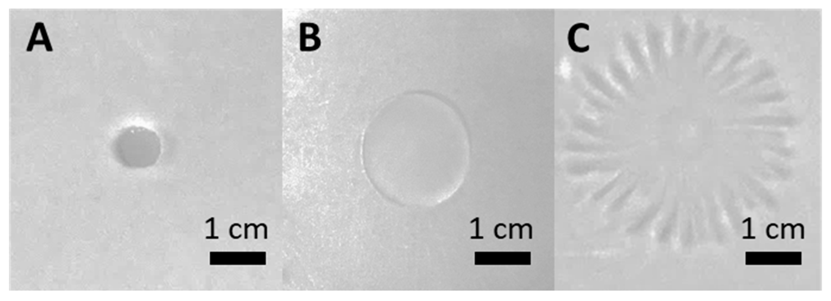
3.2. F68 Enhances the Swarming Behavior of PcO6

A positive effect of F68 was observed on swarming for PcO6. Inocula added to MM 0.5% agar plates with sucrose (20 g/L) as the C source grew to colonies of approximately 0.8 cm diameter in 48 h. The diameter of the colony and its tight-edged morphology were not affected by the addition of 0.0001 g/L F68 (Figure 4A). With 0.1 g/L F68, the colony diameter increased (Figure 4B), but a tight-edged morphology was retained so that the colony retained a circular shape. However, with the addition of 1 g/L F68, the colony diameter further increased, and a dendritic growth morphology was observed (Figure 4C). These two effects would increase the agar surface area supporting PcO6 growth. The change in morphology to the dendritic form occurs at a dose of F68 higher than the transition point in the relationship between surfactant activity and micelle formation for F68 (Supplemental Figure S1). Water surface tension measurements revealed that at F68 concentrations between 0.001 and 0.01 g/L, there was a sharp increase in surface activity (surface tension drop), which lessened for F68 concentrations between 0.01 and 0.1 g/L and dropped further above 2 g/L (Supplemental Figure S1A,B). These changes correlate with a shift from monomers (unimers) of F68 to the formation of micelles. Dynamic light scattering revealed particle sizes corresponding to micelles and larger aggregates (Supplemental Figure S1C).

3.3. PcO6 Cells Become Fluorescent When Exposed to fF68

Exposure to fF68 for 3 h caused PcO6 planktonic cells, grown to early stationary phase in rich LB in shake culture, to have bright fluorescence, as shown in Figure 5A,B. Some of these cells were in the process of cell division (Figure 5), and transfer of the cells treated with fF68 to LB agar medium resulted in normal, orange-pigmented colonies, indicating there was no impairment to culturability or phenazine formation. The fluorescence of PcO6 caused by treatment with fF68 was stable to extensive washing with the noninoculated minimal medium (Figure 5B). Two control treatments were examined; first, the PcO6 cells alone did not show fluorescence under these conditions (Supplemental Figure S2A), and second, incubation of PcO6 cells with nonlabelled F68 for 3 h (Supplemental Figure S2B) did not induce fluorescence.

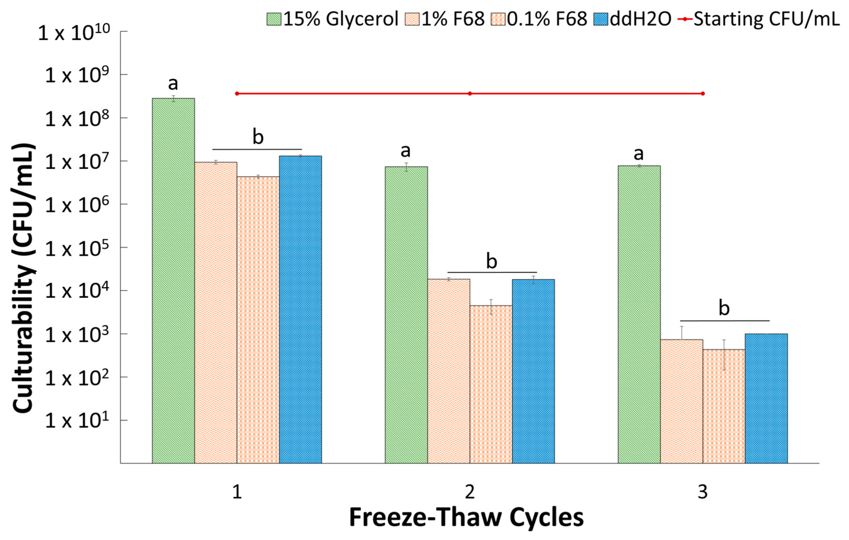
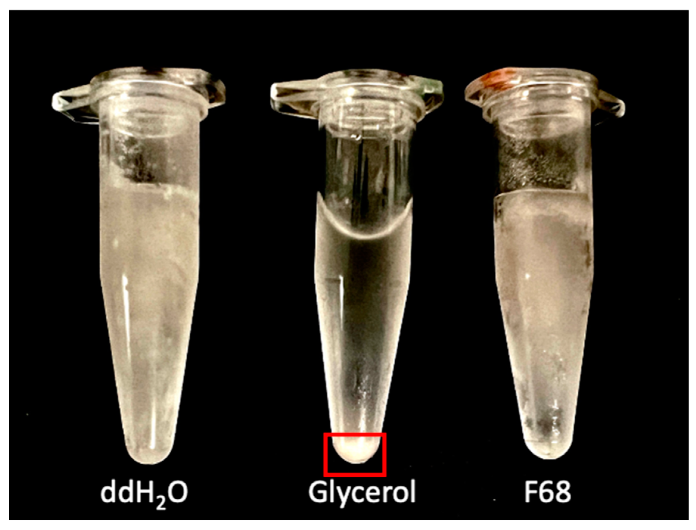
3.4. Suspension in F68 Does Not Provide Cryoprotection to PcO6

Suspensions of PcO6 cells, with a starting CFU/mL of 3.6 ± 0.5 × 10⁸, showed decreased culturability as the number of freeze/thaw cycles experienced during freezer storage increased. These cycles were imposed during the automatic defrost periods when warming for 15 min to 20 °C interrupted freezing at −20 °C. The data in Figure 6 show the culturability of fully thawed samples after they had undergone one, two, or three freeze/thaw events; loss in culturability was the least for the cell suspensions in glycerol. The glycerol suspensions showed no CFU loss after one freeze/thaw cycle, followed by a ~1.5 log CFU drop after the second cycle, with this value remaining constant after a third freeze/thaw event. The suspensions in sterile water and the F68 solutions showed a significantly greater (p < 0.05) loss in culturability than the cells suspended in glycerol. There was no significant difference in culturability for suspension in either 0.1% F68, 1% F68, or water for a given freeze/thaw cycle. Unlike glycerol, a continued loss in cell culturability occurred with each consecutive freeze/thaw cycle for suspensions in water or F68 (Figure 6).
During the thaw after the third cycle period, the glycerol suspensions were observed to become free of ice crystals faster than the samples in water or F68. The image in Figure 7 is for tubes after thawing at 22 °C for 7 min. In contrast, ice was still visible for the suspensions in F68 and water. Additionally, a pellet was formed with the glycerol suspension (Figure 7), whereas the thawing of the water—and F68—samples has an optical density typical of suspended cells. This pellet in the glycerol sample, when resuspended before the plating of the sample, produced the suspensions with the culturable cell number shown in Figure 6. In some of the published cryoprotection studies (Supplemental Table S2), F68 was active when added during the thawing process [15]. However, when 1% F68 was added to suspensions of cells frozen at −20 °C in either 15% glycerol or water and sampled without any freeze/thaw periods, no consistent effect, either protective or detrimental, was observed in studies repeated three times each with three replicates for the treatments.

4. Discussion

F68 was compatible with the growth of the plant probiotic PcO6 on both rich- and minimally defined media, supporting its classification as an FDA-approved adjuvant. These findings align with a previous publication [25] where cell densities of PcO6 grown in LB or MM liquid shake cultures were not affected by 0.5% (v/v) Pluronics P104, P108, P123 or the reverse Pluronic 25R2. Each of these Pluronics has different proportions of PEO and PPO, although all are surface-active, as shown in Supplemental Table S1.
F68 also did not alter the production of phenazines from PcO6, unlike the increases during growth on LB medium with 0.5% amendments seen with 25R2 and the decreases with P104 and P123; the null effect of F68 paralleled that of F108, which also has a PEO content of 80% [26]. These responses suggest that the presence of F68 in formulations would not alter the production of phenazines, which are important for the biocontrol activity of pseudomonads, including PcO6 [23,24]. Like all the Pluronics examined in previous studies by Housley et al. (2009) [26], the surface activity of F68 promoted the spreading of colonies of PcO6 on a soft agar surface. Such activity may enhance spread on the root surface during colonization, a factor essential for plant protection by biocontrol pseudomonads [34]. The finding that PcO6 does not utilize F68 as a C source for growth would add to the longevity of the Pluronic in the rhizosphere if it were present in applied agricultural formulations. PcO6’s ability to metabolize the plant osmolyte GB in the presence of F68 suggested that the catabolism of root exudate materials would not be impaired by F68.
PcO6 cells appeared to sorb a coating of F68, as determined by fluorescence imaging of cells exposed to fF68. Fluorescence of the bacterial cells from fF68 was stable to washing, but currently, it is not resolved whether any of the Pluronic is internalized by the bacterial cells. Labeling of organelles of fF68 within wheat root cells has been observed [17]. The internalization of F68 into bacterial cells may be restricted by the complexity of their surface layers. For the pseudomonad, an extracellular polymeric layer exists, followed by the lipopolysaccharides, which form the outer leaflet of the outer membrane. The periplasmic space is present between the inner leaflet of the outer membrane and the plasma membrane. Within the periplasm is the interconnected but thin layer of peptidoglycan. The bacterial plasma membrane is full of active components, including porins, transporters, as well as housing complexes and signal transduction sites for the insertion and function of flagella and other environmental sensors. The structures of the electron transport chain are also within the plasma membrane. This layered complexity in the bacterial membranes may limit the membrane healing effects observed for F68 action with eukaryotic membranes [7,8,9].
We were surprised by the finding that F68 did not act as a cryoprotectant for the PcO6 cells. As explained above, this result may be due to the complexity of the multiple layers that enclose the cytoplasm of the Gram-negative cell, preventing the healing of membranes damaged by ice crystal formation during freezing and thawing processes. It is unlikely that these results are due to the concentrations of F68 examined being too low; the values were within the range found in published literature (Supplemental Table S2). Indeed, 1% (w/v) F68 contains 7 × 10¹⁷ molecules F68/mL. Consequently, 2 × 10⁹ molecules of F68 would be available for each PcO6 cell in our studies using suspensions at 3.2 × 10⁸ CFU/mL. Further calculations, based on the size of PcO6 cells and an F68 molecule, propose that about 4 × 10³ molecules of F68 would be sufficient to fully cover a single cell of PcO6. The concentration of F68 in the 1% treatments thus exceeds the amount required for complete PcO6 cell coverage; these calculations support the observation of overall cell fluorescence after exposure to fF68. Supplemental Table S2 provides several examples where cryoprotection for eukaryotic cells is observed.
Agricultural formulations with a cryogenic-protective role would be valuable under the pressure of climate instability where field soil temperatures fluctuate to include freezing conditions. For instance, in Cache Valley, UT, USA, where field-grown winter wheat roots were the source of PcO6 [21], the winter conditions can result in the soil being frozen to depths of 20 cm multiple times before spring. Thus, the soil may experience several freeze/thaw cycles [35]. The depth to which the soils are frozen could subject PcO6 cells that are in the soil or attached to the roots of winter wheat to freeze/thaw events. Studies with another biocontrol-active pseudomonad, P. fluorescens Pf5, indicate that expression from the RpoS regulon protects against freeze damage; a comparative RpoS regulon active in stress response is present in PcO6 [36,37]. The consequences of expression from genes in the RpoS regulon could aid in the survival through the freeze/thaw cycles of the PcO6 cells under the conditions of our assays. The studies also support the cryoprotection offered by suspending the cells in the permeable osmolyte, 15% glycerol, where the loss in culturability was restricted to loss of about 1.5 log unit after two free/thaw cycles and no further loss after the third thaw event.

5. Conclusions

In summary, the findings support the adjuvant classification of F68. This Pluronic did not limit the growth of a plant probiotic PcO6 nor alter the production of the important biocontrol trait of phenazine biosynthesis, here monitored by synthesis of the orange-pigmented 2-hydroxyphenazine-1-carboxylate. The improved swarming in the presence of F68 might boost the colonization of the probiotic bacteria on plant surfaces. The biocompatible nature and surface activity of F68 could be valuable for its inclusion as an adjuvant in formulations for agricultural applications to improve crop production and quality, especially under environmental stress.

Supplementary Materials

The following supporting information can be downloaded at: https://www.mdpi.com/article/10.3390/agrochemicals3010001/s1. Table S1: Properties of Pluronics used in studies with PcO6; Table S2: Cell types to which Pluronic - F68 can serve as a cryoprotective agent under various research conditions [38,39,40,41,42]; Figure S1: Surfactant activity of F68 shown as a linear response (A) and a log scale (B); (C) Particle sizes detected in 0.1% F68 solution; Figure S2: A,B: Control studies for the responses of PcO6 cells to treatment with fF68.

Author Contributions

Conceptualization, D.W.B. and A.J.A.; methodology, A.R.S., A.C., M.Z., A.W., A.J.A. and D.W.B.; formal analysis, A.R.S., A.C., M.Z. and A.W.; investigation, A.R.S., A.C., M.Z. and A.W.; resources, A.J.A. and D.W.B.; data curation, A.R.S., A.C., M.Z., A.W., A.J.A. and D.W.B.; writing—original draft preparation, A.R.S., A.J.A. and D.W.B.; writing—review and editing, A.R.S., A.C., M.Z., A.W., A.J.A. and D.W.B.; visualization, A.R.S., A.W., M.Z., A.J.A. and D.W.B.; supervision, A.J.A. and D.W.B.; project administration, A.J.A. and D.W.B.; funding acquisition, A.J.A. and D.W.B. All authors have read and agreed to the published version of the manuscript.

Funding

This work was supported by grants from the Utah Agricultural Station Projects 1581 and 1746 and the National Science Foundation Research Experience for Undergraduate Program, 1950299.

Institutional Review Board Statement

Not Applicable.

Data Availability Statement

All the data from these studies are available upon request to the authors of the paper.

Conflicts of Interest

The authors declare no conflict of interest.

References

  1. Hazen, J.L. Adjuvants—Terminology, Classification, and Chemistry. Weed Technol. 2000, 14, 773–784. [Google Scholar] [CrossRef]
  2. Bertrand, A.; Castonguay, Y. Plant Adaptations to Overwintering Stresses and Implications of Climate Change. Can. J. Bot. 2003, 81, 1145–1152. [Google Scholar] [CrossRef]
  3. Willick, I.R.; Tanino, K.K.; Gusta, L.V. The Impact of Global Climate Change on the Freezing Tolerance of Winter Cereals in Western Canada. J. Agron. Crop Sci. 2021, 207, 88–99. [Google Scholar] [CrossRef]
  4. Murhammer, D.W.; Goochee, C.F. Sparged Animal Cell Bioreactors: Mechanism of Cell Damage and Pluronic F-68 Protection. Biotechnol. Prog. 1990, 6, 391–397. [Google Scholar] [CrossRef] [PubMed]
  5. Hua, J.; Erickson, L.E.; Yiin, T.-Y.; Glasgow, L.A. A Review of the Effects of Shear and Interfacial Phenomena on Cell Viability. Crit. Rev. Biotechnol. 1993, 13, 305–328. [Google Scholar] [CrossRef] [PubMed]
  6. Palomares, L.A.; González, M.; Ramírez, O.T. Evidence of Pluronic F-68 Direct Interaction with Insect Cells: Impact on Shear Protection, Recombinant Protein, and Baculovirus Production. Enzym. Microb. Technol. 2000, 26, 324–331. [Google Scholar] [CrossRef] [PubMed]
  7. Lee, R.C.; River, L.P.; Pan, F.S.; Ji, L.; Wollmann, R.L. Surfactant-Induced Sealing of Electropermeabilized Skeletal Muscle Membranes in Vivo. Proc. Natl. Acad. Sci. USA 1992, 89, 4524–4528. [Google Scholar] [CrossRef] [PubMed]
  8. Moloughney, J.G.; Weisleder, N. Poloxamer 188 (P188) as a Membrane Resealing Reagent in Biomedical Applications. Recent Pat. Biotechnol. 2012, 6, 200–211. [Google Scholar] [CrossRef]
  9. Houang, E.M.; Bates, F.S.; Sham, Y.Y.; Metzger, J.M. All-Atom Molecular Dynamics-Based Analysis of Membrane-Stabilizing Copolymer Interactions with Lipid Bilayers Probed under Constant Surface Tensions. J. Phys. Chem. B 2017, 121, 10657–10664. [Google Scholar] [CrossRef]
  10. Khaliq, N.U.; Lee, J.; Kim, S.; Sung, D.; Kim, H. Pluronic F-68 and F-127 Based Nanomedicines for Advancing Combination Cancer Therapy. Pharmaceutics 2023, 15, 2102. [Google Scholar] [CrossRef]
  11. Gill, M.I.S.; Cancino, G.O.; Anthony, P.; Davey, M.R.; Power, J.B.; Lowe, K.C. Pluronic F-68 Enhanced Shoot Regeneration in Micropropagated Citrus Rootstock and Passiflora Species. Acta Biotechnol. 2003, 23, 349–358. [Google Scholar] [CrossRef]
  12. Kumar, V.; Laouar, L.; Davey, M.R.; Mulligan, B.J.; Lowe, K.C. Pluronic F-68 Stimulates Growth of Solanum dulcamara in Culture. J. Exp. Bot. 1992, 43, 487–493. [Google Scholar] [CrossRef]
  13. Kok, A.D.-X.; Wan Abdullah, W.M.A.N.; Tan, N.-P.; Ong-Abdullah, J.; Sekeli, R.; Wee, C.-Y.; Lai, K.-S. Growth Promoting Effects of Pluronic F-68 on Callus Proliferation of Recalcitrant Rice Cultivar. 3 Biotechnol. 2020, 10, 116. [Google Scholar] [CrossRef] [PubMed]
  14. Kok, A.D.-X.; Mohd Yusoff, N.F.; Sekeli, R.; Wee, C.-Y.; Lamasudin, D.U.; Ong-Abdullah, J.; Lai, K.-S. Pluronic F-68 Improves Callus Proliferation of Recalcitrant Rice Cultivar via Enhanced Carbon and Nitrogen Metabolism and Nutrients Uptake. Front. Plant Sci. 2021, 12, 667434. [Google Scholar] [CrossRef] [PubMed]
  15. Anthony, P.; Jelodar, N.B.; Lowe, K.C.; Power, J.B.; Davey, M.R. Pluronic F-68 Increases the Post-Thaw Growth of Cryopreserved Plant Cells. Cryobiology 1996, 33, 508–514. [Google Scholar] [CrossRef]
  16. Lowe, K.C.; Anthony, P.; Davey, M.R.; Power, J.B. Beneficial Effects of Pluronic F-68 and Artificial Oxygen Carriers on the Post-Thaw Recovery of Cryopreserved Plant Cells. Artif. Cells Blood Substit. Biotechnol. 2001, 29, 297–316. [Google Scholar] [CrossRef] [PubMed]
  17. Cartwright, A.; Anderson, A.J.; Britt, D.W. Pluronic F68-Capped SiO2 Nanoparticles Are Compatible as Delivery Vehicles to Roots and Shoots. MRS Adv. 2022, 7, 327–332. [Google Scholar] [CrossRef]
  18. Pieterse, C.M.J.; Berendsen, R.L.; de Jonge, R.; Stringlis, I.A.; Van Dijken, A.J.H.; Van Pelt, J.A.; Van Wees, S.C.M.; Yu, K.; Zamioudis, C.; Bakker, P.A.H.M. Pseudomonas simiae WCS417: Star Track of a Model Beneficial Rhizobacterium. Plant Soil 2021, 461, 245–263. [Google Scholar] [CrossRef]
  19. Gamalero, E.; Glick, B.R. Recent Advances in Bacterial Amelioration of Plant Drought and Salt Stress. Biology 2022, 11, 437. [Google Scholar] [CrossRef]
  20. Dar, D.; Thomashow, L.S.; Weller, D.M.; Newman, D.K. Global Landscape of Phenazine Biosynthesis and Biodegradation Reveals Species-specific Colonization Patterns in Agricultural Soils and Crop Microbiomes. Elife 2020, 9, e59726. [Google Scholar] [CrossRef]
  21. Radtke, C.; Cook, W.S.; Anderson, A. Factors Affecting Antagonism of the Growth of Phanerochaete chrysosporium by Bacteria Isolated from Soils. Appl. Microbiol. Biotechnol. 1994, 41, 274–280. [Google Scholar] [CrossRef]
  22. Cho, S.M.; Kang, B.R.; Han, S.H.; Anderson, A.J.; Park, J.-Y.; Lee, Y.-H.; Cho, B.H.; Yang, K.-Y.; Ryu, C.-M.; Kim, Y.C. 2R,3R-Butanediol, a Bacterial Volatile Produced by Pseudomonas chlororaphis O6, Is Involved in Induction of Systemic Tolerance to Drought in Arabidopsis thaliana. Mol. Plant Microbe Interact. 2008, 21, 1067–1075. [Google Scholar] [CrossRef] [PubMed]
  23. Pierson, L.S., III; Pierson, E.A. Phenazine Antibiotic Production in Pseudomonas aureofaciens: Role in Rhizosphere Ecology and Pathogen Suppression. FEMS Microbiol. Lett. 1996, 136, 101–108. [Google Scholar] [CrossRef]
  24. Biessy, A.; Filion, M. Phenazines in Plant-Beneficial Pseudomonas Spp.: Biosynthesis, Regulation, Function and Genomics. Environ. Microbiol. 2018, 20, 3905–3917. [Google Scholar] [CrossRef] [PubMed]
  25. Kang, B.R.; Han, S.H.; Zdor, R.E.; Anderson, A.J.; Spencer, M.; Yang, K.Y.; Kim, Y.H.; Lee, M.C.; Cho, B.H.; Kim, Y.C. Inhibition of Seed Germination and Induction of Systemic Disease Resistance by Pseudomonas chlororaphis O6 Requires Phenazine Production Regulated by the Global Regulator, GacS. J. Microbiol. Biotechnol. 2007, 17, 586–593. [Google Scholar] [PubMed]
  26. Housley, L.; Anderson, T.; Sontag, N.; Han, S.-H.; Britt, D.W.; Anderson, A.J. Pluronics’ Influence on Pseudomonad Biofilm and Phenazine Production. FEMS Microbiol. Lett. 2009, 293, 148–153. [Google Scholar] [CrossRef]
  27. Perneel, M.; D’hondt, L.; De Maeyer, K.; Adiobo, A.; Rabaey, K.; Höfte, M. Phenazines and Biosurfactants Interact in the Biological Control of Soil-Borne Diseases Caused by Pythium Spp. Environ. Microbiol. 2008, 10, 778–788. [Google Scholar] [CrossRef]
  28. Stanghellini, M.E. Inhibitory and Lytic Effects of a Nonionic Surfactant on Various Asexual Stages in the Life Cycle of Pythium and Phytophthora Species. Phytopathology 1987, 77, 112. [Google Scholar] [CrossRef]
  29. Goodman, J.; Mclean, J.E.; Britt, D.W.; Anderson, A.J. Sublethal Doses of ZnO Nanoparticles Remodel Production of Cell Signaling Metabolites in the Root Colonizer Pseudomonas chlororaphis O6. Environ. Sci. Nanotechnol. 2016, 3, 1103–1113. [Google Scholar] [CrossRef]
  30. Pierson, L.S.; Pierson, E.A. Metabolism and Function of Phenazines in Bacteria: Impacts on the Behavior of Bacteria in the Environment and Biotechnological processes. Appl. Microbiol. Biotechnol. 2010, 86, 1659–1670. [Google Scholar] [CrossRef]
  31. Kearns, D.B. A Field Guide to Bacterial Swarming Motility. Nat. Rev. Microbiol. 2010, 8, 634–644. [Google Scholar] [CrossRef] [PubMed]
  32. Smith, L.T.; Pocard, J.A.; Bernard, T.; Le Rudulier, D. Osmotic Control of Glycine Betaine Biosynthesis and Degradation in Rhizobium meliloti. J. Bacteriol. 1988, 170, 3142–3149. [Google Scholar] [CrossRef] [PubMed]
  33. Annunziata, M.G.; Ciarmiello, L.F.; Woodrow, P.; Dell’Aversana, E.; Carillo, P. Spatial and Temporal Profile of Glycine Betaine Accumulation in Plants Under Abiotic Stresses. Front. Plant Sci. 2019, 10, 230. [Google Scholar] [CrossRef] [PubMed]
  34. de Weger, L.A.; van der Bij, A.J.; Dekkers, L.C.; Simons, M.; Wijffelman, C.A.; Lugtenberg, B.J.J. Colonization of the Rhizosphere of Crop Plants by Plant-Beneficial Pseudomonads. FEMS Microbiol. Ecol. 1995, 17, 221–227. [Google Scholar] [CrossRef]
  35. Cane, J. Global Warming, Advancing Bloom and Evidence for Pollinator Plasticity from Long-Term Bee Emergence Monitoring. Insects 2021, 12, 457. [Google Scholar] [CrossRef]
  36. Stockwell, V.O.; Loper, J.E. The Sigma Factor RpoS Is Required for Stress Tolerance and Environmental Fitness of Pseudomonas fluorescens Pf-5. Microbiology 2005, 151, 3001–3009. [Google Scholar] [CrossRef]
  37. Oh, S.A.; Kim, J.S.; Han, S.H.; Park, J.Y.; Dimkpa, C.; Edlund, C.; Anderson, A.J.; Kim, Y.C. The GacS-Regulated Sigma Factor RpoS Governs Production of Several Factors Involved in Biocontrol Activity of the Rhizobacterium Pseudomonas chlororaphis O6. Can. J. Microbiol. 2013, 59, 556–562. [Google Scholar] [CrossRef]
  38. Ashwood-Smith, M.J.; Voss, W.A.G.; Warby, C. Cryoprotection of Mammalian Cells in Tissue Culture with Pluronic polyols. Cryobiology 1973, 10, 502–504. [Google Scholar] [CrossRef]
  39. Doğan, A.; Yalvaç, M.E.; Yılmaz, A.; Rizvanov, A.; Şahin, F. Effect of F68 on Cryopreservation of Mesenchymal Stem cells derived from Human Tooth Germ. Appl. Biochem. Biotech. 2013, 171, 1819–1831. [Google Scholar] [CrossRef]
  40. Glauser, S.C.; Talbot, T.R. Some studies on freezing and thawing human erythrocytes. Am. J. Med. Sci. 1956, 231, 75–81. [Google Scholar] [CrossRef]
  41. González Hernández, Y.; Fischer, R.W. Serum-free Culturing of Mammalian Cells—Adaptation to and Cryopreservation in Fully Defined Media. ALTEX 2007, 24, 110–116. [Google Scholar] [CrossRef] [PubMed]
  42. Liu, C.; Lei, J.; Zhang, M.; Wu, F.; Ren, M.; Yang, J.; Wu, Q.; Shi, X. Optimization of Preservation Methods Provides Insights into Photosynthetic Picoeukaryotes in Lakes. Microbiol. Spectr. 2022, 10, e02557-21. [Google Scholar] [CrossRef] [PubMed]
Figure 1. The structure of F68.
Agrochemicals 03 00001 g001
Figure 2. Effect of F68 on growth and pigmentation of planktonic cells of PcO6 in shake culture on rich LB medium. The F68 was added to the culture medium at defined doses (0, 0.1, 1, 10 or 100 g/L). (A) The OD 600 nm for the cultures with and without F68 amendments at defined times of incubation up to 72 h. The means of the values and standard deviations for three replicates for each treatment are shown and are typical of three separate studies. (B) Orange coloration of the late stationary-phase cultures at 72 h of incubation.
Agrochemicals 03 00001 g002
Figure 3. Growth of planktonic PcO6 cells on defined minimal medium without added carbon source or with additions of F68 and/or glycine betaine. The defined minimal medium had no carbon sources (No C) or was amended with F68, glycine betaine (GB), or a mix of F68 and GB. The inoculum was added at the level of 104 CFU/mL. (A): Growth of PcO6 after 120 h of culture in CFU/mL. The data are means and standard errors of CFU/mL with three replicates/treatments and three replicated studies. The letters above the bars indicate significant differences between treatments as determined by ANOVA with Tukey’s HSD adjustment at p < 0.05. (B) Images of the 120 h cultures.
Agrochemicals 03 00001 g003
Figure 4. The effect of additions of F68 to minimal medium, solidified with 0.5% agar, on colony growth of PcO6. The minimal medium contained sucrose as the carbon source. The images shown are typical of six different replicates for each concentration of F68 added to the agar: (A) 0.0001 g/L, (B) 0.1 g/L, and (C) 1 g/L F68.
Agrochemicals 03 00001 g004
Figure 5. Fluorescence of PcO6 cells exposed to fF68. (A) Fluorescence of PcO6 cells exposed to fF68 for 3 h without washing; the medium contained free F68 and fF68 (A). (B) After washing cells two times with a noninoculated minimal medium when nonadsorbed fF68 was removed. The scale bars are 10 µm. The images were taken using a 100× NA 1.4 objective, a FITC filter with 488 nm and emission at 516 nm, and an integration time of 1 s. Images are typical of five fields of view examined from three independent studies.
Agrochemicals 03 00001 g005
Figure 6. Effect of freeze/thaw cycles in an automated freezer on culturability of PcO6. The data show the mean of culturable cells/mL obtained on LB agar after 1, 2, or 3 freeze/thaw cycles. Error bars are the standard deviations of three replicates. Letters denote significant (p < 0.01) differences among treatments for a given free/thaw cycle using one-way ANOVA followed by Tukey’s HSD.
Agrochemicals 03 00001 g006
Figure 7. Appearance of tubes of PcO6 cells suspended in water, glycerol, and F68 after three freeze (−20 °C)/thaw cycles. Thawing proceeded at room temperature (22 °C) for 7 min. This image showed the faster thaw for the glycerol suspensions versus the cells suspended in water or water amended with 1% F68 where ice is still visible. All samples were ice-free after 12 min thaw. The red box highlights the pellet in the glycerol sample. Ice crystals are apparent at the same time of thawing for PcO6 cells suspended in sterile water or 1% F68 when transferred into the freezer.
Agrochemicals 03 00001 g007
Disclaimer/Publisher’s Note: The statements, opinions and data contained in all publications are solely those of the individual author(s) and contributor(s) and not of MDPI and/or the editor(s). MDPI and/or the editor(s) disclaim responsibility for any injury to people or property resulting from any ideas, methods, instructions or products referred to in the content.

