Article

Contrast-Enhanced Mammography as a Functional Biomarker in Breast Cancer: Correlation of Enhancement Patterns with Ki-67 and Histological Grade

by
Marina Balbino
1,
Manuela Montatore
1,
Federica Masino
1,
Antonietta Ancona
2,
Francesca Anna Carpagnano
3,
Giulia Capuano
4,
Riccardo Guglielmi
5 and
Giuseppe Guglielmi
1,6,*
1
Department of Clinical and Experimental Medicine, Foggia University School of Medicine, Viale L. Pinto 1, 71121 Foggia, Italy
2
Section of Breast Imaging, Breast Care Unit, Santa Maria Hospital GVM-BA, Via Antonio De Ferrariis 22, 70124 Bari, Italy
3
Breast Unit, “Dimiccoli” Hospital, Viale Ippocrate 15, 70051 Barletta, Italy
4
Breast Unit, Istituto Tumori “Giovanni Paolo II” IRCCS, 70124 Bari, Italy
5
Department of Medical Imaging, Mount Sinai Hospital, Sinai Health, Toronto, ON M5G 1X5, Canada
6
Radiology Unit, “Dimiccoli” Hospital, Viale Ippocrate 15, 70051 Barletta, Italy
*
Author to whom correspondence should be addressed.
Targets 2025, 3(3), 29; https://doi.org/10.3390/targets3030029
Submission received: 6 August 2025 / Revised: 1 September 2025 / Accepted: 11 September 2025 / Published: 17 September 2025

Abstract

Background: Contrast-Enhanced Spectral Mammography (CESM) combines anatomical and functional imaging, showing promise in breast cancer diagnosis. Despite well-established lesion detection accuracy, few studies have investigated the link between CESM enhancement patterns and tumor aggressiveness biomarkers. Methods: We retrospectively evaluated 100 patients (mean age 59.5 years) undergoing CESM with complete histopathological data. Lesions were categorized by enhancement intensity (high, medium, low) and contrast homogeneity (homogeneous vs. heterogeneous), correlated with Ki-67 index and histological grade. Results: Lesion size measured by CESM closely matched histology (mean 2.16 cm vs. 2.25 cm). Mass-like lesions corresponded mainly to invasive ductal carcinoma, while non-mass patterns aligned with lobular or in situ carcinomas. Enhancement intensity correlated moderately with Ki-67 (Spearman ρ = 0.56, p < 0.001), and contrast heterogeneity showed a weaker but significant correlation with tumor grade (ρ = 0.22, p < 0.05). Conclusions: CESM accurately assesses tumor size and provides functional insight into tumor biology. Enhancement intensity may serve as a non-invasive proliferation marker, while contrast heterogeneity offers additional prognostic data, supporting CESM’s role in personalized breast cancer management.

1. Introduction

Contrast-Enhanced Spectral Mammography (CESM) has emerged as an innovative imaging modality that merges traditional mammography’s anatomical resolution with functional information derived from contrast enhancement. This combination enables detailed morphological lesion characterization alongside visualization of tumor vascularity, enhancing diagnostic precision for breast cancer [1,2,3].
Breast cancer remains the most commonly diagnosed cancer in women worldwide, with incidence rates rising in many regions. According to recent global data, it accounts for over 2.3 million new cases and 685,000 deaths annually [4]. In Europe alone, breast cancer incidence exceeds 500,000 cases yearly, underscoring the need for accurate diagnostic and prognostic tools [5].
While CESM’s sensitivity and specificity have been well studied, its correlation with biological tumor markers, such as the Ki-67 proliferation index and histological grade, remains underexplored [6].
Ki-67, a nuclear antigen expressed during active cell cycle phases, is a pivotal biomarker reflecting tumor proliferation and aggressiveness [7]. It is commonly used in clinical practice to guide treatment decisions, particularly in determining eligibility for adjuvant chemotherapy. However, despite its clinical value, imaging correlations with Ki-67 remain sparse, particularly in the context of CESM [8].
Histological grading assesses cellular differentiation, with higher grades indicating poor differentiation and worse prognosis [9]. Imaging features from CESM—specifically enhancement intensity and homogeneity—may reflect these biological properties by indicating vascularization and structural complexity, respectively [10].
Despite its increasing clinical use, CESM is predominantly applied for morphological evaluation, and its potential as a functional biomarker of tumor aggressiveness remains largely unexplored. This study aims to explore the relationship between CESM enhancement patterns and Ki-67 expression and tumor grade, potentially positioning CESM as a dual-purpose imaging tool for both detection and biological tumor profiling, aligned with precision oncology goals [11,12,13].

2. Materials and Methods

2.1. Study Population

This retrospective, single-center study included 100 female patients (mean age 59.5 years, range 34–81) selected from 450 individuals undergoing Contrast-Enhanced Spectral Mammography (CESM) from January 2020 to December 2022. Inclusion criteria were as follows: complete diagnostic and therapeutic work-up, histologically confirmed breast cancer, and preoperative CESM per protocol. Exclusion criteria included incomplete imaging data or unavailable histopathology [14].
The selection flowed from 450 initial cases to the final 100 patients, detailing exclusion due to missing histological data (n = 212), incomplete imaging protocol (n = 96), or loss to follow-up (n = 42).

2.2. Ethical Approval

Since the study was non-interventional and did not involve the collection of sensitive personal data, ethical approval from an Institutional Review Board (IRB) was not required under Italian national regulations (Ministerial Decree of 3 August 2007) and the internal guidelines of the University of Foggia. Participants were fully informed about the aims and procedures of the study.

2.3. Imaging Protocol

CESM was performed using a digital mammography system (Senographe Pristina, GE Healthcare) with dual-energy acquisition. A non-ionic iodinated contrast agent (1.5 mL/kg, iodine concentration 350 mg/mL; Iohexol, Singapore) was administered intravenously at 3 mL/s.
Imaging was initiated 2 min post-injection, with a craniocaudal (CC) and mediolateral oblique (MLO) view of each breast acquired in both low- and high-energy modes. Subtracted images were generated using the proprietary GE algorithm. Compression force and radiation dose were automatically adjusted by the device software. Additional imaging included tomosynthesis and ultrasound, followed by core needle biopsy [15,16].

2.4. Image Analysis

Three experienced breast radiologists independently reviewed CESM images. Lesions were classified by the following:
  • Enhancement intensity: high, moderate, low (visual signal relative to parenchyma),
  • Enhancement homogeneity: homogeneous vs. heterogeneous (uniformity of contrast distribution).
Interobserver agreement was high (κ = 0.84), demonstrating good reproducibility of qualitative assessment. However, we acknowledge that enhancement evaluation remains subjective; future studies should incorporate quantitative measures, such as radiomics or automated texture analysis, to improve objectivity and consistency [17].
Disagreements were resolved by consensus. Lesion size was measured using integrated digital calipers on the most enhanced view [18].

2.5. Histopathological Assessment

Histopathology was performed per the WHO 2019 criteria. Ki-67 was assessed by immunohistochemistry, with ≥20% defining high proliferation. Tumor grade was assigned using the Nottingham grading system (G1: well differentiated; G2: moderately; G3: poorly differentiated) [18,19].

2.6. Statistical Analysis

Analyses were conducted using SPSS 27. Continuous variables are means ± SD; categorical as counts and percentages. Correlations between CESM features and histopathological parameters were assessed with Spearman’s ρ. Associations between categorical variables were tested using Chi-square or Fisher’s exact tests. Standard errors were calculated and included in Figure 1 and Figure 2. Significance was set at p < 0.05 [20].
The sampling method used was consecutive sampling of all eligible patients meeting the inclusion criteria during the study period.

3. Results

3.1. Lesion Size Concordance

CESM measurements (mean 2.16 cm) closely matched histological size (mean 2.25 cm), differing by only 0.09 cm on average. Traditional mammography underestimated the size (mean 2.05 cm). Lesions ranged from 0.3 to 7.0 cm [21].

3.2. Lesion Morphology and Histology

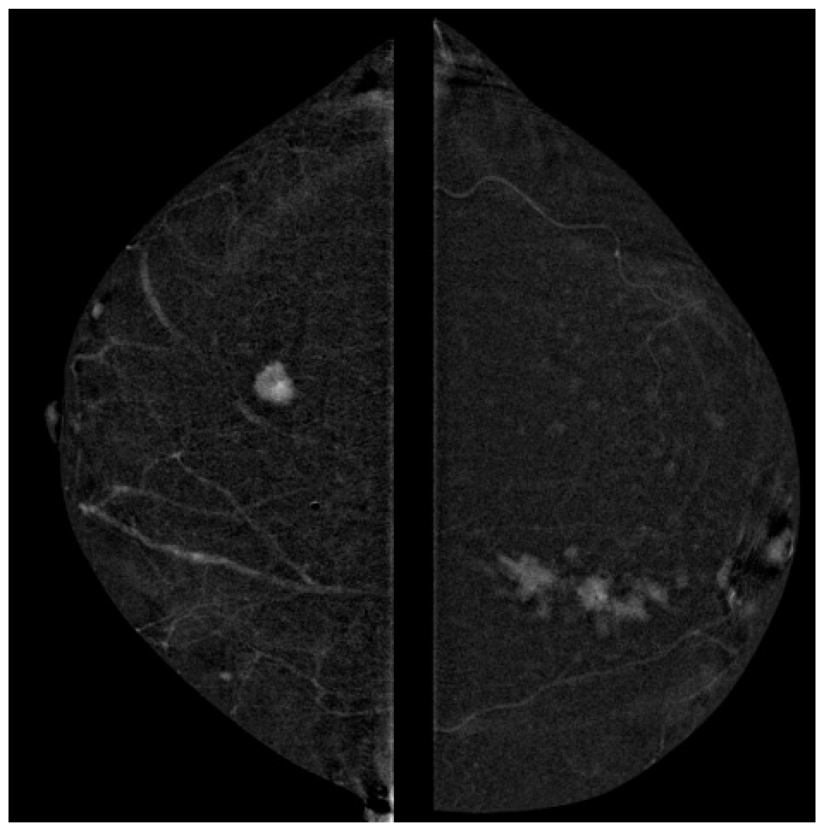
Among 100 lesions, 82 were mass-like and 18 were non-mass-like. Mass-like lesions were predominantly correlated with invasive ductal carcinoma (IDC), while non-mass-like lesions were associated with invasive lobular carcinoma (ILC) or ductal carcinoma in situ (DCIS), confirming prior reports [22,23].

3.3. Correlation of Ki-67 and Enhancement Intensity

A significant positive correlation was observed between Ki-67 and CESM enhancement intensity (ρ = 0.56, p < 0.001), indicating that higher proliferative tumors display stronger contrast uptake [24].

3.4. Correlation of Contrast Homogeneity and Tumor Grade

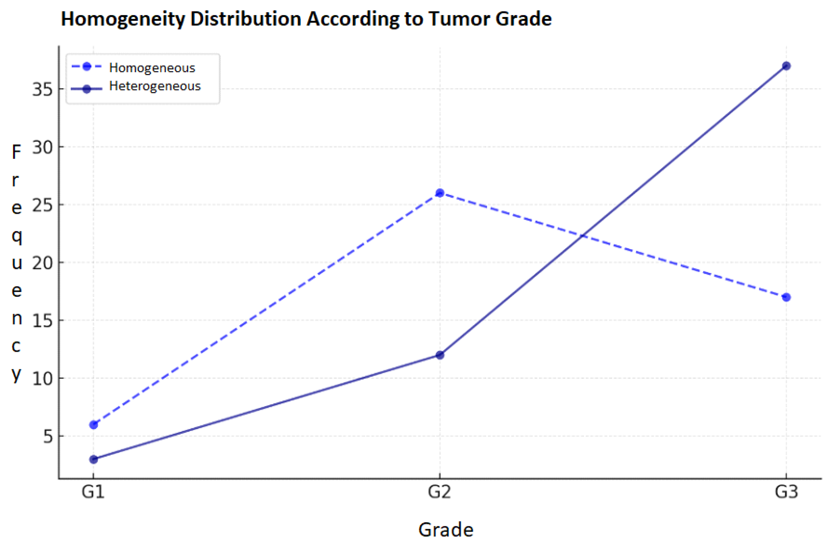
Contrast heterogeneity correlated weakly but significantly with tumor grade (ρ = 0.22, p < 0.05), with higher-grade tumors more often exhibiting heterogeneous enhancement. Chi-square test confirmed this association (χ2 = 8.14, p = 0.017) (Figure 3) [25].

4. Discussion

The strong concordance between CESM-derived tumor size and histological measurements observed in this study aligns with previous research highlighting CESM’s superiority over traditional mammography in tumor sizing accuracy [26,27]. The functional imaging capability of CESM, which highlights tumor vascularity, allows for more precise delineation of lesion margins, a critical factor in preoperative planning and surgical decision-making [28].
Morphological analysis confirmed established associations between CESM lesion types and histological subtypes: mass-like lesions predominantly corresponded to invasive ductal carcinoma, while non-mass-like lesions were more often linked to invasive lobular carcinoma or ductal carcinoma in situ. These imaging–pathology correlations enhance diagnostic confidence and may help guide therapeutic approaches, especially in cases where MRI is contraindicated or unavailable [29,30].
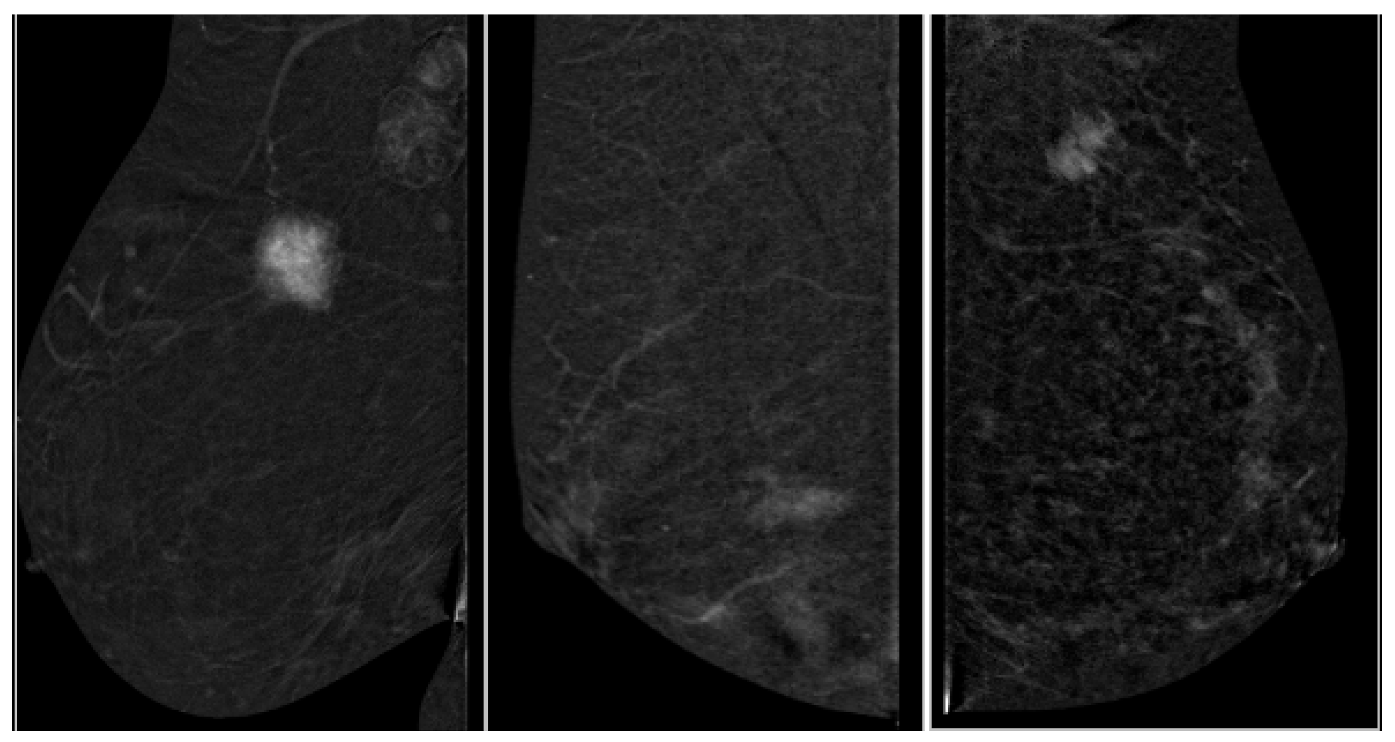
A key novel finding of our study is the moderate positive correlation between Ki-67 proliferation index and CESM enhancement intensity (ρ = 0.56, p < 0.001). Ki-67 is a validated marker of tumor aggressiveness, and its association with imaging features supports the hypothesis that enhancement intensity reflects tumor angiogenesis and metabolic activity [31]. This finding positions CESM as a potential non-invasive functional biomarker for proliferative activity, extending its role beyond anatomical imaging. Prior studies have reported similar correlations with dynamic contrast-enhanced MRI; however, data regarding CESM have been limited, making this contribution particularly relevant [32,33].
Clinically, the ability to estimate proliferative status non-invasively through CESM could inform risk stratification and treatment planning, particularly regarding decisions on neoadjuvant chemotherapy or targeted therapies. If validated in larger cohorts, this imaging biomarker could improve personalized treatment approaches in breast oncology [34].
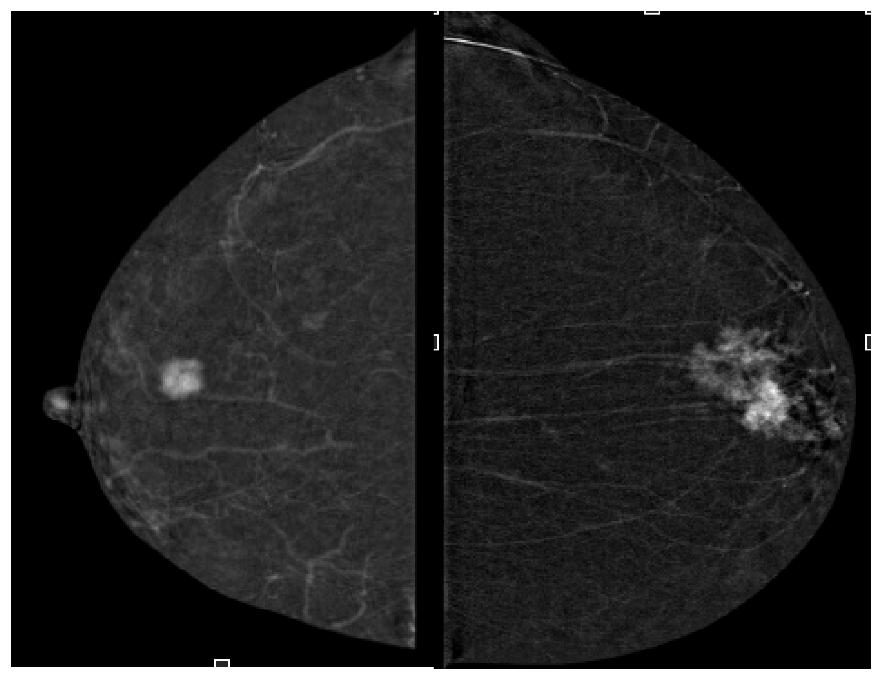
We also identified a statistically significant but weaker correlation between enhancement heterogeneity and histological tumor grade (ρ = 0.22, p < 0.05). Higher-grade tumors frequently exhibited heterogeneous contrast distribution, possibly reflecting vascular irregularity and complex tumor architecture typical of aggressive phenotypes [35]. While the modest strength of this correlation limits its standalone prognostic value, it may provide supplementary information when integrated with other clinical and imaging data. The observed association supports prior suggestions that enhancement patterns on CESM might serve as indicators of tumor biological complexity [36,37].
Our study confirms that CESM enhancement characteristics correlate with breast cancer molecular subtypes and proliferative activity, supporting CESM’s potential role as a non-invasive imaging biomarker (Figure 4 and Figure 5).
Limitations of our study include its retrospective and single-center design, which may limit the generalizability of findings. The qualitative nature of enhancement assessment could introduce observer bias, despite the high interobserver agreement. Additionally, the sample size, while adequate for exploratory correlations, requires expansion in future prospective multicenter studies to confirm the robustness of these results.
Future research should focus on quantitative evaluation of enhancement patterns using advanced imaging analytics and integration with molecular profiling and longitudinal clinical outcomes. Such approaches may enhance CESM’s utility as a non-invasive biomarker of tumor biology and aggressiveness, facilitating precision oncology strategies [38,39].

5. Conclusions

This study confirms the dual anatomical and functional utility of Contrast-Enhanced Spectral Mammography in breast cancer evaluation. CESM demonstrates excellent accuracy in tumor sizing, outperforming conventional mammography, and a strong correlation of lesion morphology with histological subtype. Importantly, enhancement intensity correlates moderately with Ki-67 proliferation index, supporting CESM’s role as a non-invasive biomarker of tumor aggressiveness. Contrast heterogeneity shows a weaker but significant association with histological grade, suggesting potential supplementary prognostic value.
Our findings highlight CESM’s evolving role beyond lesion detection towards biological tumor characterization, aligning with precision medicine principles. By potentially providing non-invasive insight into tumor proliferation and heterogeneity, CESM could inform personalized treatment planning, especially when MRI is contraindicated or unavailable. Validation in larger cohorts could position CESM as a pivotal tool for risk stratification and therapy tailoring in breast cancer management.

Author Contributions

Conceptualization, M.B. and A.A.; methodology, M.B., A.A. and M.M.; formal analysis, M.M.; investigation, M.B., F.A.C. and F.M.; data curation, M.M. and G.C.; writing—original draft preparation, M.B.; writing—review and editing, M.M. and F.M.; visualization, F.M. and R.G.; supervision, G.G. All authors have read and agreed to the published version of the manuscript.

Funding

This research received no external funding.

Institutional Review Board Statement

The study was conducted in accordance with the principles outlined in the Declaration of Helsinki (2013, revision). Ethical approval was not required for this type of study, as it did not involve any intervention or collection of sensitive personal data, in accordance with Italian national regulations (Ministerial Decree of 3 August 2007) and the university’s internal guidelines.

Informed Consent Statement

Verbal informed consent was obtained from all participants prior to their participation. Written consent was not required, in accordance with our institutional and national guidelines, due to the study’s non-invasive and fully anonymized nature.

Data Availability Statement

The original contributions presented in this study are included in the article. Further inquiries can be directed to the corresponding author.

Conflicts of Interest

The authors declare no conflict of interest.

References

  1. Jochelson, M.S.; Lobbes, M.B.I. Contrast-enhanced Mammography: State of the Art. Radiology 2021, 299, 36–48. [Google Scholar] [CrossRef]
  2. Tagliafico, A.S.; Bignotti, B.; Rossi, F.; Signori, A.; Sormani, M.P.; Valdora, F.; Calabrese, M.; Houssami, N. Diagnostic performance of contrast-enhanced spectral mammography: Systematic review and meta-analysis. Breast 2016, 28, 13–19. [Google Scholar] [CrossRef]
  3. Fallenberg, E.M.; Schmitzberger, F.F.; Amer, H.; Ingold-Heppner, B.; Balleyguier, C.; Diekmann, F.; Engelken, F.; Mann, R.M.; Renz, D.M.; Bick, U.; et al. Contrast-enhanced spectral mammography vs. mammography and MRI—clinical performance in a multi-reader evaluation. Eur. Radiol. 2017, 27, 2752–2764. [Google Scholar] [CrossRef]
  4. Wu, W.Y.-Y.; Tabar, L.; Tot, T.; Fann, C.-Y.; Yen, A.M.-F.; Chen, S.L.-S.; Chiu, S.Y.-H.; Ku, M.M.-S.; Hsu, C.-Y.; Beckmann, K.R.; et al. Imaging biomarkers as predictors for breast cancer death. J. Oncol. 2019, 2019, 2087983. [Google Scholar] [CrossRef]
  5. Davey, M.G.; Hynes, S.O.; Kerin, M.J.; Miller, N.; Lowery, A.J. Ki-67 as a Prognostic Biomarker in Invasive Breast Cancer. Cancers 2021, 13, 4455. [Google Scholar] [CrossRef] [PubMed]
  6. Elston, C.W.; Ellis, I.O. Pathological prognostic factors in breast cancer. I. The value of histological grade in breast cancer: Experience from a large study with long-term follow-up. Histopathology 1991, 19, 403–410. [Google Scholar] [CrossRef] [PubMed]
  7. Dowsett, M.; Nielsen, T.O.; A’Hern, R.; Bartlett, J.; Coombes, R.C.; Cuzick, J.; Ellis, M.; Henry, N.L.; Hugh, J.C.; Lively, T.; et al. Assessment of Ki67 in breast cancer: Recommendations from the International Ki67 in Breast Cancer working group. J. Natl. Cancer Inst. 2011, 103, 1656–1664. [Google Scholar] [CrossRef] [PubMed]
  8. O’Flynn, E.A.; DeSouza, N.M. Functional magnetic resonance: Biomarkers of response in breast cancer. Breast Cancer Res. 2011, 13, 204, Erratum in Breast Cancer Res. 2011, 13, 405. [Google Scholar] [CrossRef] [PubMed] [PubMed Central]
  9. Buadu, L.D.; Murakami, J.; Murayama, S.; Hashiguchi, N.; Sakai, S.; Masuda, K.; Toyoshima, S.; Kuroki, S.; Ohno, S. Breast lesions: Correlation of contrast medium enhancement patterns on MR images with histopathologic findings and tumor angiogenesis. Radiology 1996, 200, 639–649. [Google Scholar] [CrossRef]
  10. Li, L.; Roth, R.; Germaine, P.; Ren, S.; Lee, M.; Hunter, K.; Tinney, E.; Liao, L. Contrast-enhanced spectral mammography (CESM) versus breast magnetic resonance imaging (MRI): A retrospective comparison in 66 breast lesions. Diagn. Interv. Imaging 2017, 98, 113–123. [Google Scholar] [CrossRef]
  11. Kalia, M. Biomarkers for personalized oncology: Recent advances and future challenges. Metabolism 2015, 64, S16–S21. [Google Scholar] [CrossRef]
  12. Qi, Y.J.; Su, G.H.; You, C.; Zhang, X.; Xiao, Y.; Jiang, Y.Z.; Shao, Z.M. Radiomics in breast cancer: Current advances and future directions. Cell Rep. Med. 2024, 5, 101719. [Google Scholar] [CrossRef] [PubMed] [PubMed Central]
  13. Uematsu, T. Non-mass-like lesions on breast ultrasonography: A systematic review. Breast Cancer 2012, 19, 295–301. [Google Scholar] [CrossRef] [PubMed]
  14. Sakorafas, G.H.; Tsiotou, A.G. Selection criteria for breast conservation in breast cancer. Eur. J. Surg. 2000, 166, 835–846. [Google Scholar] [CrossRef] [PubMed]
  15. Zanardo, M.; Cozzi, A.; Trimboli, R.M.; Labaj, O.; Monti, C.B.; Schiaffino, S.; Carbonaro, L.A. Technique, protocols and adverse reactions for contrast-enhanced spectral mammography (CESM): A systematic review. Insights Imaging 2019, 10, 76. [Google Scholar] [CrossRef]
  16. James, J. Contrast-enhanced spectral mammography (CESM)-guided breast biopsy as an alternative to MRI-guided biopsy. Br. J. Radiol. 2022, 95, 20211287. [Google Scholar] [CrossRef]
  17. Schell, A.M.; Rosenkranz, K.; Lewis, P.J. Role of breast MRI in the preoperative evaluation of patients with newly diagnosed breast cancer. Am. J. Roentgenol. 2009, 192, 1438–1444. [Google Scholar] [CrossRef]
  18. Tan, P.H.; Ellis, I.; Allison, K.; Brogi, E.; Fox, S.B.; Lakhani, S.; Lazar, A.J.; Morris, E.A.; Sahin, A.; Salgado, R.; et al. The 2019 World Health Organization classification of tumours of the breast. Histopathology 2020, 77, 181–185. [Google Scholar] [CrossRef]
  19. Rakha, E.A.; El-Sayed, M.E.; Lee, A.H.S.; Elston, C.W.; Grainge, M.J.; Hodi, Z.; Blamey, R.W.; Ellis, I.O. Prognostic Significance of Nottingham Histologic Grade in Invasive Breast Carcinoma. J. Clin. Oncol. 2008, 26, 3153–3158. [Google Scholar] [CrossRef]
  20. Cox, T.F. Medical Statistics for Cancer Studies; Chapman & Hall/CRC: Boca Raton, FL, USA, 2022; ISBN 9780367486150. [Google Scholar]
  21. Barra, F.R.; de Souza, F.F.; Camelo, R.E.F.A.; Ribeiro, A.C.O.; Farage, L. Accuracy of contrast-enhanced spectral mammography for estimating residual tumor size after neoadjuvant chemotherapy in patients with breast cancer: A feasibility study. Radiol. Bras. 2017, 50, 224–230. [Google Scholar] [CrossRef]
  22. Montemurro, F.; Martincich, L.; Sarotto, I.; Bertotto, I.; Ponzone, R.; Cellini, L.; Redana, S.; Sismondi, P.; Aglietta, M. Relationship between DCE-MRI morphological and functional features and histopathological characteristics of breast cancer. Eur. Radiol. 2007, 17, 1490–1497. [Google Scholar] [CrossRef]
  23. Kim, S.H.; Cha, E.S.; Park, C.S.; Kang, B.J.; Whang, I.Y.; Lee, A.W.; Song, B.J.; Park, J. Imaging features of invasive lobular carcinoma: Comparison with invasive ductal carcinoma. Jpn. J. Radiol. 2011, 29, 475–482. [Google Scholar] [CrossRef]
  24. Kontzoglou, K.; Palla, V.; Karaolanis, G.; Karaiskos, I.; Alexiou, I.; Pateras, I.; Konstantoudakis, K.; Stamatakos, M. Correlation between Ki67 and breast cancer prognosis. Oncology 2013, 84, 219–225. [Google Scholar] [CrossRef] [PubMed]
  25. Rudnicki, W.; Heinze, S.; Niemiec, J.; Kojs, Z.; Sas-Korczynska, B.; Hendrick, E.; Luczynska, E. Correlation between quantitative assessment of contrast enhancement in contrast-enhanced spectral mammography (CESM) and histopathology—Preliminary results. Eur. Radiol. 2019, 29, 6220–6226. [Google Scholar] [CrossRef] [PubMed]
  26. Lobbes, M.B.; Lalji, U.C.; Nelemans, P.J.; Houben, I.; Smidt, M.L.; Heuts, E.; de Vries, B.; Wildberger, J.E.; Beets-Tan, R.G. The quality of tumor size assessment by contrast-enhanced spectral mammography and the benefit of additional breast MRI. J. Cancer 2015, 6, 144–150. [Google Scholar] [CrossRef] [PubMed]
  27. Boyd, N.F.; Jensen, H.M.; Cooke, G.; Han, H.L. Relationship between mammographic and histological risk factors for breast cancer. J. Natl. Cancer Inst. 1992, 84, 1170–1179. [Google Scholar] [CrossRef] [PubMed]
  28. Iotti, V.; Ravaioli, S.; Vacondio, R.; Coriani, C.; Caffarri, S.; Sghedoni, R.; Nitrosi, A.; Ragazzi, M.; Gasparini, E.; Masini, C.; et al. Contrast-enhanced spectral mammography in neoadjuvant chemotherapy monitoring: A comparison with breast magnetic resonance imaging. Breast Cancer Res. 2017, 19, 106. [Google Scholar] [CrossRef]
  29. Łuczyńska, E.; Heinze-Paluchowska, S.; Hendrick, E.; Dyczek, S.; Ryś, J.; Herman, K.; Blecharz, P.; Jakubowicz, J. Comparison between breast MRI and contrast-enhanced spectral mammography. Med. Sci. Monit. 2015, 21, 1358–1367. [Google Scholar] [CrossRef]
  30. Wong, C.Y.Y.; Lee, S.Y.S.; Mahmood, R.D. Contrast-enhanced spectral mammography. Singap. Med. J. 2024, 65, 195–201. [Google Scholar] [CrossRef]
  31. Nielsen, T.O.; Leung, S.C.Y.; Rimm, D.L.; Dodson, A.; Acs, B.; Badve, S.; Denkert, C.; Ellis, M.J.; Fineberg, S.; Flowers, M.; et al. Assessment of Ki67 in Breast Cancer: Updated Recommendations From the International Ki67 in Breast Cancer Working Group. J. Natl. Cancer Inst. 2021, 113, 808–819. [Google Scholar] [CrossRef]
  32. Juan, M.W.; Yu, J.; Peng, G.X.; Jun, L.J.; Feng, S.P.; Fang, L.P. Correlation between DCE-MRI radiomics features and Ki-67 expression in invasive breast cancer. Oncol. Lett. 2018, 16, 5084–5090. [Google Scholar] [CrossRef]
  33. Khan, Q.J.; Kimler, B.F.; O’Dea, A.P.; Zalles, C.M.; Sharma, P.; Fabian, C.J. Mammographic density does not correlate with Ki-67 expression or cytomorphology in benign breast cells obtained by random periareolar fine needle aspiration from women at high risk for breast cancer. Breast Cancer Res. 2007, 9, R35. [Google Scholar] [CrossRef] [PubMed]
  34. James, J.R.; Pavlicek, W.; Hanson, J.A.; Boltz, T.F.; Patel, B.K. Breast Radiation Dose With CESM Compared With 2D FFDM and 3D Tomosynthesis Mammography. AJR Am. J. Roentgenol. 2017, 208, 362–372. [Google Scholar] [CrossRef] [PubMed]
  35. Tang, S.; Xiang, C.; Yang, Q. The diagnostic performance of CESM and CE-MRI in evaluating the pathological response to neoadjuvant therapy in breast cancer: A systematic review and meta-analysis. Br. J. Radiol. 2020, 93, 20200301. [Google Scholar] [CrossRef] [PubMed]
  36. Macura, K.J.; Ouwerkerk, R.; Jacobs, M.A.; Bluemke, D.A. Patterns of enhancement on breast MR images: Interpretation and imaging pitfalls. Radiographics 2006, 26, 1719–1734, quiz 1719. [Google Scholar] [CrossRef]
  37. Zhou, J.; Zhang, Y.; Shi, S. Ultrasound elastography: Advances and challenges in early detection of breast cancer. Front. Oncol. 2025, 15, 1589142. [Google Scholar] [CrossRef]
  38. Marino, M.A.; Leithner, D.; Sung, J.; Avendano, D.; Morris, E.A.; Pinker, K.; Jochelson, M.S. Radiomics for Tumor Characterization in Breast Cancer Patients: A Feasibility Study Comparing Contrast-Enhanced Mammography and Magnetic Resonance Imaging. Diagnostics 2020, 10, 492. [Google Scholar] [CrossRef]
  39. Chen, Y.; Shao, X.; Shi, K.; Rominger, A.; Caobelli, F. AI in Breast Cancer Imaging: An Update and Future Trends. Semin. Nucl. Med. 2025, 55, 358–370. [Google Scholar] [CrossRef]
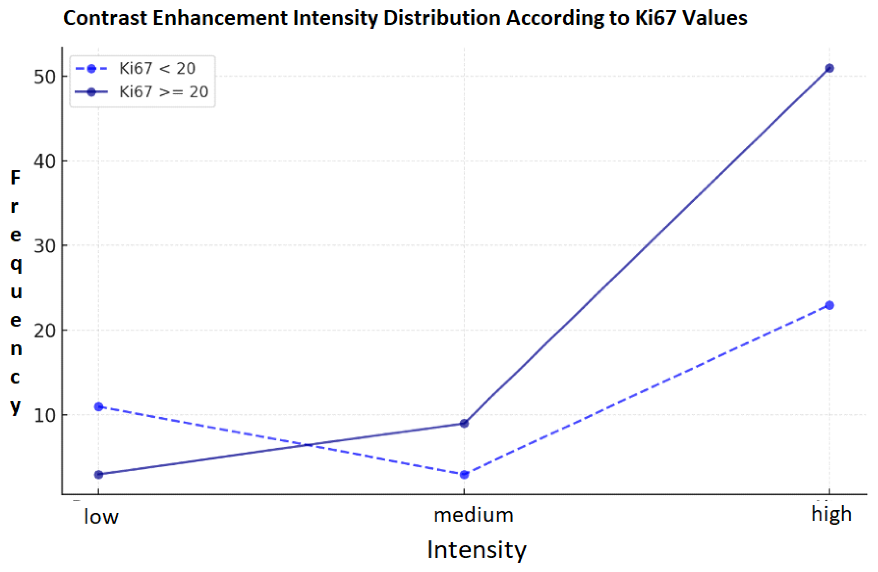
Figure 1. Frequency related to contrast intensity in patients with Ki-67 < or >20. Includes counts, percentages, standard error, and p-values.
Targets 03 00029 g001
Figure 2. Frequency related to grading in patients with homogeneous or non-homogeneous contrast enhancement. Includes counts, percentages, standard error, and p-values.
Targets 03 00029 g002
Figure 3. On the left: CESM CC image of the right breast—mass area; on the right: CESM CC image of the left breast—non-mass area.
Targets 03 00029 g003
Figure 4. Three CESM MLO images, from left to right: high, medium, and low intensity.
Targets 03 00029 g004
Figure 5. On the left: CESM CC image of the right breast—homogeneous contrast enhancement; on the right: CESM CC image of the left breast—non-homogeneous contrast enhancement.
Targets 03 00029 g005
Disclaimer/Publisher’s Note: The statements, opinions and data contained in all publications are solely those of the individual author(s) and contributor(s) and not of MDPI and/or the editor(s). MDPI and/or the editor(s) disclaim responsibility for any injury to people or property resulting from any ideas, methods, instructions or products referred to in the content.