Article

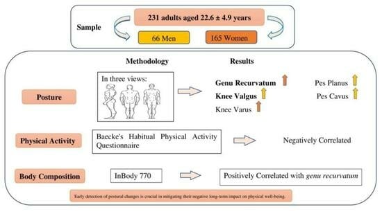
Association between Body Composition, Physical Activity Profile, and Occurrence of Knee and Foot Postural Alterations among Young Healthy Adults

1
Department of Physical Education and Sport, University of Madeira, 9020-105 Funchal, Portugal
2
Laboratory for Robotic and Engineering Systems, Interactive Technologies Institute, 9020-105 Funchal, Portugal
3
Research Center in Sports Sciences, Health Sciences and Human Development (CIDESD), 5000-801 Vila Real, Portugal
*
Author to whom correspondence should be addressed.
Future 2024, 2(1), 16-29; https://doi.org/10.3390/future2010002
Submission received: 10 November 2023 / Revised: 16 January 2024 / Accepted: 17 January 2024 / Published: 25 January 2024

Abstract

:
Knee and foot deformities refer to structural abnormalities in the knee and foot bones, joints, ligaments, or muscles. Various factors, including genetics, injury, disease, or excessive use, can cause these deformities. These musculoskeletal conditions can significantly impact individuals’ quality of life. This study examined foot and knee deformities in 231 young healthy adults (165 men, 66 women) aged 22.6 ± 4.9 years and their association with physical activity and body composition. The postural assessment was performed by two Physiotherapists, with the subject standing in three views: side, anterior, and posterior. Physical activity (Baecke’s Habitual Physical Activity Questionnaire) and body composition (InBody 770) were assessed. Results showed that the most common foot deformity was pes planus, while the genu recurvatum was the most common knee deformity among the individuals. Physical activity level was negatively associated with knee and foot deformities. Conversely, body composition differed with the presence of genu recurvatum. These findings present a starting point to understand the occurrence of knee and foot postural alterations according to the individuals’ body composition and physical activity profiles, which could support the deployment of tailored interventions among healthy adults. In addition, early detection of postural changes is crucial in mitigating their negative long-term impact on physical well-being.

Graphical Abstract

1. Introduction

Posture is an attitude or body position maintained at rest, adopted in an activity, or an exact way of supporting the body. It makes quick modifications over time to reduce the body’s energy, leading to a rise in muscle stress to maintain proper balance [1]. Posture is maintained by skeletal muscle contraction, coordinated by a series of external stimuli, and through continuous neuromuscular type modifications, representing the body’s reaction to the force of gravity [2]. Each individual’s distinctive posture may be influenced by several factors, such as ligament elasticity, breathing issues, or bone malformations [3]. Thus, postural changes have been related to unbalanced body components, leading to increased muscle stress to maintain proper balance [4], resulting in discomfort and functional impairment [5].
Studies emphasize that one of the most urgent issues facing contemporary society is the widespread nature of postural abnormalities [6,7,8,9], which might eventually lead to inevitable injuries and pain in the corresponding body organ. Foot and knee postures are known to determine how well the lower extremity operates and may contribute to the individual’s propensity for recurrent trauma [10,11]. For instance, a study conducted to assess the prevalence of foot pain and deformity among the Danish population reported high correlations between foot pain and foot alterations [12]. Additionally, the previous literature has emphasized the relationship between knee and foot misalignment, with changes in foot alignment before and after knee surgery [13]. Foot postures like pronated foot and flat foot (pes planus) are significantly associated with changes in dynamic function, alignment, and medial compartment knee osteoarthritis [14]. Several elements, including lifestyle choices, employment, and the environment, endanger the skeletal system [15]. According to previous studies, decreased physical activity (PA) in overweight and obese individuals contributes to altered lower-limb joint loading compared to normal-weight adults [16]. Differences in walking patterns between overweight and normal-weight individuals are associated with the attempt to increase stability due to impaired balance, minimize external work, and decrease load at the knee [17]. On the other hand, foot problems might be derived from the effects of increased weight on plantar loading, underlining a link between obesity and the development of foot pain [18].
Previous research has identified the most common knee and foot deformities, such as genu recurvatum, genu varum, genu varus, pes planus, and pes cavus. Genu Recurvatum is the position of a tibiofemoral joint in which the range of motion occurs beyond neutral or 0° of extension [19]. In genu valgus deformity, ankle joints are spaced apart when knees are in contact with each other in a weight-bearing position [20]. In contrast, in genu varus, the internal condyles of the femur become spaced apart if they are in weight-bearing contact with the media, malleolus of the ankle [20]. The medial longitudinal arch is clinically significant in diseases and the functioning of joints and muscles of the ankle and knee [21]. When the foot’s medial arch is disturbed, deformities occur, whereby considerable reduction in the arch causes pes planus, whereas the increase in the arch exceeding 18 mm causes pes cavus [22].
Meanwhile, the literature has reported that an increase in body mass index (BMI) leads to postural instability in young adults who are obese and non-obese [23,24,25]. A study on youngsters aged between 7 and 16 years revealed a substantial correlation between age, BMI, and genu varum [26]. Weakness in the lower extremities can lead to challenges in everyday activities and regular movements. Enhancing PA levels might be crucial to fighting overweight and obesity, diminishing the risk of injury, and improving the quality of life [27,28,29]. However, details regarding the relationship between PA, body composition variables, and the prevalence of foot and knee deformities are still lacking among healthy adults. Providing an early diagnosis of postural impairment allows designing targeted interventions that may reduce the detrimental effects of postural deformities [30]. It helps physical education teachers, coaches, and healthcare professionals design appropriate and corrective exercises to improve body alignment. Therefore, the present study aims to (1) determine the occurrence of postural alterations in the knee and foot among healthy adults and (2) compare the PA and body composition profiles between individuals with and without postural alterations in the knee and foot.

2. Methodology

2.1. Sample

This study included 231 adults who were students from the Physical Education and Sports course at the University of Madeira. The sample included 165 men and 66 women aged between 18 and 44 years old (22.5 ± 4.2 years). All participants were healthy and not injured at the time of data collection. The inclusion and exclusion criteria are presented in Figure 1. The optimal sample size was calculated using G*Power [31]. A priori independent samples t-test indicated a total sample of 128 participants (64 in each group) to attain 80% power for an effect size of 0.50 at the 0.05 level of significance. All the procedures implemented in the current study received ethical approval from the Scientific Committee of The Faculty of Physical Education and Sports at the University of Madeira (reference: ACTA N.77-12 April 2016). All participants were volunteers, and informed consent was signed before data collection.

2.2. General Procedure

2.2.1. Postural Evaluation

The postural evaluation was conducted using the Postural Assessment Table, employing the visual assessment method. The evaluation was carried out by two experienced physiotherapists who were trained in postural assessment techniques. A similar process of postural assessment was employed, as described by [32], in a study involving rhythmic gymnasts and non-trainees to observe the presence of postural alterations. The subjects under observation were assessed while standing in three distinct views: anterior, side, and posterior. Before the assessment, participants were instructed to present themselves barefoot and in light clothing. They assumed a neutral standing position, with specific guidance given regarding their gaze direction (looking forward and fixing their gaze on a point on a wall). Data collection spanned two weeks, during which assessments were conducted for each relevant class. These assessments took place in a dedicated physical performance laboratory between 10 a.m. and 2 p.m. on weekdays. Each evaluation session lasted approximately 40 min, allowing sufficient time for participants to complete consent forms and undergo testing. The decision to conduct assessments during regular working hours was deliberate, aiming to mitigate potential biases related to participants’ alertness and concentration levels. Moreover, this approach ensured consistent conditions for all participants, promoting the overall validity of the research findings. By allocating uniform time slots for assessment sessions, the study sought to enhance the reliability and credibility of the collected data.
(a)
In the Anterior View
Alignment and symmetry of knee and foot were observed, including alignment of the knee (internal, external rotation, varus, and valgus) and malleolus (symmetry and asymmetry).
(b)
In the Side View
Alignment and symmetry of knee and foot were observed, including Knees (genu recurvatum) and feet (medial longitudinal arch for pes planus and pes cavus).
(c)
In the Posterior View
Alignment and symmetry of knee and foot were observed, including popliteal lines (symmetry and asymmetry) and feet (valgus and varus).

2.2.2. Body Composition

Stature was measured to the nearest 0.01 cm using a stadiometer (SECA 213, Hamburg, Germany). Body composition variables were assessed using hand-to-foot bioelectrical impedance analysis (InBody 770, Cerritos, CA, USA). Body mass, body mass index (BMI), fat mass percentage (FM%), total body water (TBW), Intracellular Water, Extracellular Water, protein, minerals, and skeletal muscle mass (SMM), and waist–hip ratio WHR (cm).

2.2.3. Physical Activity

Baecke’s Habitual PA questionnaire [33] was used to calculate PA, which includes the following variables:
(a)
Formal PA
This variable assesses an individual’s engagement in formal PA and structured sports. Participants report the activity type, each associated with an intensity factor (0.76 for light, 1.26 for moderate, and 1.76 for vigorous). Time spent per week (0.5 to 4.5 h) and proportion of the year (0.04 to 0.92) are considered. Scores are calculated by multiplying intensity, time, and proportion. The final score, within predefined ranges, reflects overall activity.
(b)
Informal PA
This variable assesses informal activities in daily life unrelated to structured exercise. Responses to Likert scale questions gauge frequency and intensity. Higher scores indicate greater activity levels.
(c)
Total Practical History
Scores from Formal PA and Informal PA are combined to measure overall habitual PA. This encompasses both structured and informal activities.
(d)
Frequency of PA
Assesses planned exercise sessions beyond daily activities. The score typically reflects reported exercise sessions per week.

2.3. Statistical Analysis
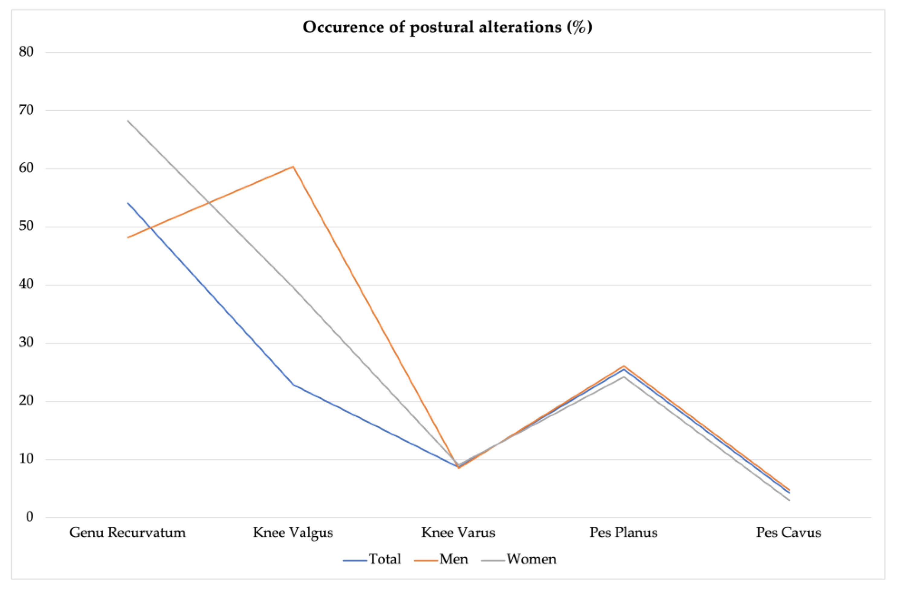
Data exploration was carried out to identify possible data entry errors and the presence of outliers. Descriptive statistics were presented as means ± standard deviation. All data were checked for normality using the Kolmogorov–Smirnov test. The independent samples t-test was conducted to examine the differences between participants with and without postural changes in quantitative variables with a normal distribution. The software used was SPSS version 27.0 (SPSS Inc., Chicago, IL, USA), and the significance level adopted was 5%.

3. Results

Figure 2 summarizes the occurrence of postural alterations of the knee and foot among the participants based on gender. Overall, genu recurvatum was the most frequent postural alteration, representing 54.1%. In contrast, pes cavus (4.3%) and genu varus (8.7%) were the least frequent postural alterations. A higher proportion of females presented genu recurvatum, while knee valgus was more prevalent among men. At the same time, the opposite occurs in knee valgus (60.4% vs. 39.6%), with the diagnosis occurring in a greater proportion in males than females.
Table 1 presents the descriptive statistics for body composition variables according to knee postural changes and the comparison results between groups (with and without knee postural changes). Although without statistically significant differences, body mass (M = 70.7 ± 11.7 kg) and BMI (M = 23.4 ± 2.6 kg/m2) in individuals without genu recurvatum alteration were higher than body mass (M = 68.6 ± 12.2 kg) and BMI (M = 23.3 ± 3.2 kg/m2) in individuals with genu recurvatum alteration. Furthermore, WHR (M = 0.86 ± 0.07 cm) and FM (M = 20.8 ± 10.1%) in individuals with genu recurvatum alteration were significantly higher than WHR (M = 0.82 ± 0.04 cm) and FM (M = 14.9 ± 7.0%) in individuals without genu recurvatum alteration. Stature, TBW, Intracellular Water, Extracellular Water, protein, minerals, and SMM in individuals without genu recurvatum alteration were significantly higher in individuals with an alteration (p < 0.05).
Body composition, including stature, body mass, TBW, intracellular and extracellular water, protein, FM%, and SMM in individuals without genu valgus and genu varus was higher than in individuals with genu valgus and genu varus. The difference was not statistically significant.
Overall, average body mass and BMI were not statistically significant, with a prevalence of genu recurvatum. At the same time, other body composition parameters were statistically significant with the occurrence of genu recurvatum (p < 0.05). Furthermore, Body composition showed no statistically significant relation with knee valgus and varus prevalence.
Table 2 resumes the descriptive statistics for body composition variables according to foot postural changes and the comparison results between groups (with and without postural changes). Overall, no statistically significant differences were observed between groups regarding body composition on the pes planus deformities. The same trend was seen concerning pes cavus, except for stature, which was significantly larger among individuals who showed pes cavus postural changes (p = 0.035).
Table 3 displays the descriptive statistics for PA variables according to knee postural changes and the comparison results between groups (with and without postural changes). The results indicate a significantly larger Score of Formal PA among individuals who presented knee valgus compared to those unaffected by this deformity (p = 0.048). In addition, a substantially higher Score of Formal PA (p = 0.003) and Total Score (p = 0.004) was identified in the group with knee varus compared to the individuals that did not present this postural change.
Finally, Table 4 resumes the descriptive statistics for PA variables according to the foot and the comparison results between groups (with and without postural changes). Among the PA variables, a statistically significant difference was only observed in the Frequency of PA for the pes planus condition, since the individuals without postural change reported a higher score than the ones affected by postural change (p = 0.035).

4. Discussion

This study aimed to evaluate the occurrence of knee and foot postural alterations and the differences in body composition and PA among young healthy adults. The frequency of knee alterations (genu recurvatum 54.1%, knee valgus 22.9%, and knee varus 8.7%) and foot alterations (pes planus 25.5% and pes cavus 4.3%) in the present study were substantial. Concerning postural alterations for gender, a distinction emerged. Genu recurvatum was more common among females (68.2%) compared to males (48.2%), while knee valgus exhibited a significantly higher occurrence in males (60.4%) than in females (39.6%).
Indeed, the literature has underlined a heightened occurrence of genu recurvatum among females compared to men [34,35,36]. In line with these findings, previous authors Penha, et al. [37] reported a comparable prevalence of genu recurvatum (54%) in school-aged children. Conversely, Gh, et al. [38] revealed that 22% of children exhibited genu recurvatum at birth, which showed slight variation with age or gender. The reason for the higher prevalence of genu recurvatum is not well known. However, repetitive and harmful habits that led to posterior capsule laxity could be associated [39], as well as a lack of strength/weakness of the gastrocnemius muscle [40] and the quadriceps muscle, which allows hyperextension of the knee [41]. In addition, gender differences may arise from factors such as greater knee laxity exhibited by females [42,43], knee geometry variations, and smaller anterior cruciate ligaments [44,45]. Further insights are drawn from another study highlighting the predominance of genu varus as a knee alteration, particularly pronounced among females [46]. In contrast, an alternative investigation by Odding, et al. [47] described varus deformities as more pronounced in males, while valgus deformities exhibited greater prevalence in females. A study conducted on the prevalence of dynamic knee valgus among children indicates 26.3% and 26.9% in the right and left lower limbs, with females exhibiting more knee valgus in the left limb [48]. Concerning foot alterations, prior research showcased the presence of pes planus in a staggering 90.8% of elementary school students [49]. However, among young, healthy individuals, pes planus was 29% in the South Indian population [50]. Notably, the occurrence of pes cavus (4.3%) was consistent with previous studies on the topic that reported a prevalence ranging between 0.2 to 3.7% [49,51,52,53,54].
Furthermore, in the current study, genu recurvatum condition was statistically significantly related to body composition parameters, particularly %FM and WHR. However, individuals without genu recurvatum showed statistically significant higher mean values of TBW, protein, minerals, and SMM. These findings align with the concept that diverse body types give rise to disparities in fat distribution, often manifesting as increased fat mass [55]. The potential connection between body composition and genu recurvatum can be attributed to muscle strength imbalances stemming from these varying fat distributions, thus contributing to muscular weaknesses and imbalances [56,57]. In the case of knee varus and valgus, no statistically significant connection was established with body composition, given the particular locations and age group in our sample with normal BMI, excluding obesity as a confounding factor. Nevertheless, a study involving individuals aged 11 and 13 years established a significant relationship between weight and knee varus and stature and knee varus [58]. In addition, the literature presents varying perspectives on the nexus between genu varus, genu valgum, and obesity. While, Soheilipour, et al. [59] proposed a weaker connection between genu varus and obesity, the authors noted a heightened prevalence of genu valgum in obese individuals. This observation gains reinforcement from multiple studies collectively indicating an increased occurrence of genu valgum [59,60,61,62]. Moreover, in the present study, no statistically significant relationship was observed between pes planus, pes cavus, and BMI, except between pes cavus and stature. This contrasts with previous research that asserts a robust and highly significant connection between pes planus and BMI [49,63,64].
Concerning PA and postural alterations, no statistical difference was observed between groups with and without genu recurvatum; however, formal PA was significantly linked to knee valgus and varus. The total PA (formal and informal) also differed notably among knee varus conditions. Strenuous knee-involved activities can lead to muscle tightness and joint strain, potentially contributing to knee varus. This aligns with Lee [65], who emphasized that stress during PA, especially among overweight individuals aiming to lose weight, might precipitate knee varus before weight reduction. The reason for knee valgus and varus relation with PA is still unknown.
Nonetheless, prevailing research emphasizes how high-intensity athletic performance can induce global and regional muscle fatigue, impairing postural stability [66,67,68]. Moreover, training parameters affect general fatigue, leading to a strong relationship between the type of exercise, fatigue, and postural deficits [68,69]. Notably, varying postural stability levels were observed in physical education students after short, intense, or prolonged moderate exercise [69]. Evidence suggests that training with poor posture can deteriorate the muscles’ proprioceptive feedback mechanism, limiting their ability to correct stance and maintain proper posture due to reduced sensory system input [67,70,71]. Muscle fatigue in stabilizing muscles (such as the gastrocnemius and soleus) misaligns joints and weakens neuromuscular control, contributing to decreased postural stability [66,72]. Furthermore, a statistically significant relationship is found between pes planus and practical history and physical exercise frequency. Possible causes include the laxity of soft tissues supporting the arch, including the tibialis posterior muscle, plantar fascia, intrinsic foot muscles, and calcaneonavicular ligaments [73]. A study conducted in Turkey reported weakness of foot plantar flexor group muscles, reduced flexibility of gastrocnemius and soleus muscles, and decreased balance with physical activity in individuals with pes planus [74]. In the current study, no statistical significance was found between groups with and without pes cavus and physical activity. The lower occurrence could be attributed to congenital causes or neuromuscular disorders like muscular dystrophy and Charcot–Marie–Tooth disease [75,76]. Traumatic injuries can also impact the tarsal bone position and lead to hypertonicity of the longitudinal arches. In pes cavus, the peroneus longus and posterior tibialis muscles tend to overpower the peroneus brevis, tibialis anterior, and intrinsic foot muscles, often resulting in plantar flexion due to peroneus longus contracture [77]. Studies have shown that foot postural alterations like pes planus and pes cavus are associated with an increased risk of various lower extremity injuries compared to individuals with a neutral arch [78,79,80].
Although these are important findings, this study presents some limitations that should be mentioned. Notably, the study’s sample size is relatively modest, suggesting the potential for enhanced robustness by broadening the scope to encompass a more diverse and extensive population. Future efforts could emphasize recruitment across different demographic groups and regions to fortify the generalizability of findings. Moreover, this study’s methodology relies on an observational analysis of postures, which, while informative, represents only a partial assessment of the complexities involved. Future investigations could incorporate a multifaceted approach to enhance the depth of research. A further limitation lies in the absence of interventions to address observed postural alterations among the student participants. A valuable avenue for future research lies in implementing interventions tailored to correct identified postural discrepancies and measuring the resultant changes. This longitudinal approach would not only elucidate the potential efficacy of intervention strategies but also contribute to developing evidence-based recommendations for managing and improving posture-related concerns. This study presents a starting point to understand the occurrence of knee and foot postural alterations according to the individuals’ body composition and PA profiles, which could support the deployment of tailored interventions among healthy adults.

5. Conclusions

Understanding the prevalence of knee and foot postural alterations holds substantial importance in preventing issues like shin splints, stress fractures, and plantar fasciitis during both physical exertion and routine activities. This study sought to pinpoint key determinants that play a pivotal role in discerning shifts in knee and foot posture while exploring their connections with body composition and physical activity. Ultimately, the investigation highlighted noteworthy factors such as BMI, WHR, and past PA experiences, among others, that could significantly contribute to devising effective strategies for addressing postural alterations in the knee and foot.

Author Contributions

Conceptualization, S.A., R.V., R.T.O. and A.R.; methodology, S.A., R.V., R.T.O. and A.R.; software, A.R.; validation, S.A., R.V., C.F., R.T.O. and A.R.; formal analysis, S.A., C.F. and A.R.; investigation, S.A. and R.V.; resources, S.A., R.V., C.F., R.T.O. and A.R.; writing—original draft preparation, S.A., C.F. and A.R.; writing—review and editing, S.A., R.V., C.F., R.T.O. and A.R.; visualization, C.F., R.T.O. and A.R.; supervision, R.T.O. and A.R.; project administration, R.T.O. and A.R.; funding acquisition, C.F., R.T.O. and A.R. All authors have read and agreed to the published version of the manuscript.

Funding

C.F. recognizes the support from LARSyS—the Portuguese national funding agency for science, research, and technology (FCT) pluriannual funding 2020–2023 (ITI-LX UIDP/50009/2020—IST-ID).

Institutional Review Board Statement

The study was conducted according to the guidelines of the Declaration of Helsinki and approved by the Scientific Committee of The Faculty of Physical Education and Sports at the University of Madeira (reference: ACTA N.77-12 April 2016).

Informed Consent Statement

Informed consent was obtained from all individuals involved in the study.

Data Availability Statement

The data presented in this study are available upon request from the corresponding author.

Conflicts of Interest

The authors declare no conflict of interest.

References

  1. Smith, L.K.; Weiss, L.; Lehmkuhl, L.D. Brunnstrom’s Clinical Kinesiology; FA Davis: Philadelphia, PA, USA, 1996. [Google Scholar]
  2. Barker, V. Postura, Posizione, Movimento per Potenziare le Prestazioni Sessuali, Rimodellare il Corpo, Ritardare L’invecchiamento, Ridurre L’obesità, Sviluppare le Capacità; Edizioni Mediterranee: Roma, Italy, 1998. [Google Scholar]
  3. Teixeira, L. Importância do Movimento Humano na Relação Homem/Trabalho: Aspectos Posturais; Cepeusp: São Paulo, Brazil, 1996. [Google Scholar]
  4. Bernard, P.; Geraci, M.; Hue, O.; Amato, M.; Seynnes, O.; Lantieri, D. Influence of obesity on postural capacities of teenagers. Preliminary study. Ann. Readapt. Med. Phys. 2003, 46, 184–190. [Google Scholar]
  5. Detsch, C.; Luz, A.M.H.; Candotti, C.T.; Oliveira, D.S.D.; Lazaron, F.; Guimarães, L.K.; Schimanoski, P. Prevalência de alterações posturais em escolares do ensino médio em uma cidade no Sul do Brasil. Rev. Panam. De Salud Publica 2007, 21, 231–238. [Google Scholar] [CrossRef] [PubMed]
  6. Andrews, D.M.; Fiedler, K.M.; Weir, P.L.; Callaghan, J.P. The effect of posture category salience on decision times and errors when using observation-based posture assessment methods. Ergonomics 2012, 55, 1548–1558. [Google Scholar] [CrossRef] [PubMed]
  7. Iedynak, G.; Galamandjuk, L.; Kyselytsia, O.; Nakonechnyi, I.; Hakman, A.; Chopik, O. Special aspects of changes in physical readiness indicators of young men with different somatotypes between 15 and 17 years of age. J. Phys. Educ. Sport 2017, 17, 2690–2696. [Google Scholar]
  8. Kashuba, V.; Dudko, M. Sovremennye podkhody, metodiki i tekhnologii k formirovaniiu zdorovogo obraza zhizni studentov v processe fizicheskogo vospitaniia [Modern approaches to, methodic and technologies of students’ healthy life style formation in process of physical education]. Fiz. Vikhovannia Sport I Kul’tura Zdorov’ia U Suchasnomu Suspil’stvi 2015, 1, 52–57. [Google Scholar]
  9. Torlakovic, A.; Muftic, M.; Radjo, I.; Talovic, M.; Mahmutovic, I. Evolution of sports-medical team management in the program of posture correction in children. Mater. Socio-Medica 2014, 26, 104. [Google Scholar] [CrossRef] [PubMed]
  10. Dahle, L.K.; Mueller, M.; Delitto, A.; Diamond, J.E. Visual assessment of foot type and relationship of foot type to lower extremity injury. J. Orthop. Sports Phys. Ther. 1991, 14, 70–74. [Google Scholar] [CrossRef] [PubMed]
  11. Nigg, B.M.; Cole, G.K.; Nachbauer, W. Effects of arch height of the foot on angular motion of the lower extremities in running. J. Biomech. 1993, 26, 909–916. [Google Scholar] [CrossRef]
  12. Mølgaard, C.; Lundbye-Christensen, S.; Simonsen, O. High prevalence of foot problems in the Danish population: A survey of causes and associations. Foot 2010, 20, 7–11. [Google Scholar] [CrossRef]
  13. Chandler, J.T.; Moskal, J.T. Evaluation of knee and hindfoot alignment before and after total knee arthroplasty: A prospective analysis. J. Arthroplast. 2004, 19, 211–216. [Google Scholar] [CrossRef]
  14. Abourazzak, F.; Kadi, N.; Azzouzi, H.; Lazrak, F.; Najdi, A.; Nejjari, C.; Harzy, T. A positive association between foot posture index and medial compartment knee osteoarthritis in moroccan people. Open Rheumatol. J. 2014, 8, 96. [Google Scholar]
  15. Sheng, B.; Li, X.; Nussler, A.K.; Zhu, S. The relationship between healthy lifestyles and bone health: A narrative review. Medicine 2021, 100, e24684. [Google Scholar] [CrossRef] [PubMed]
  16. Browning, R.C. Locomotion mechanics in obese adults and children. Curr. Obes. Rep. 2012, 1, 152–159. [Google Scholar] [CrossRef]
  17. Kim, D.; Lewis, C.L.; Gill, S.V. Effects of obesity and foot arch height on gait mechanics: A cross-sectional study. PLoS ONE 2021, 16, e0260398. [Google Scholar] [CrossRef] [PubMed]
  18. Butterworth, P.A.; Urquhart, D.M.; Landorf, K.B.; Wluka, A.E.; Cicuttini, F.M.; Menz, H.B. Foot posture, range of motion and plantar pressure characteristics in obese and non-obese individuals. Gait Posture 2015, 41, 465–469. [Google Scholar] [CrossRef]
  19. Brownstein, B.; Noyes, F.R.; Mangine, R.E.; Kryger, S. Physical Therapy of the Knee: Anatomy and Biomechanics; Mangine, R.E., Ed.; Churchill Livingstone: New York, NY, USA, 1988. [Google Scholar]
  20. Jalalvand, H.; Fatahi, A.; Entezari Khorasani, Z. Comparison of the Kinematic Pattern of Knee Joint Flexion in Healthy and Genu Valgum and Genu Varum Children During Jumping. J. Sport Biomech. 2021, 7, 270–279. [Google Scholar] [CrossRef]
  21. Franco, A.H. Pes cavus and pes planus: Analyses and treatment. Phys. Ther. 1987, 67, 688–694. [Google Scholar] [CrossRef]
  22. Oatis, C.A. Kinesiology the Mechanics and Pathomechanics of Human Movement; Wolters Kluwer: Alphen aan den Rijn, The Netherlands, 2009. [Google Scholar]
  23. Berrigan, F.; Simoneau, M.; Tremblay, A.; Hue, O.; Teasdale, N. Influence of obesity on accurate and rapid arm movement performed from a standing posture. Int. J. Obes. 2006, 30, 1750–1757. [Google Scholar] [CrossRef]
  24. Hue, O.; Simoneau, M.; Marcotte, J.; Berrigan, F.; Doré, J.; Marceau, P.; Marceau, S.; Tremblay, A.; Teasdale, N. Body weight is a strong predictor of postural stability. Gait Posture 2007, 26, 32–38. [Google Scholar] [CrossRef]
  25. Maffiuletti, N.; Agosti, F.; Proietti, M.; Riva, D.; Resnik, M.; Lafortuna, C.; Sartorio, A. Postural instability of extremely obese individuals improves after a body weight reduction program entailing specific balance training. J. Endocrinol. Investig. 2005, 28, 2–7. [Google Scholar] [CrossRef]
  26. Ghandi, A.R.; Hadi, H.A.; Behruzi, A.R.; Holakooie, A.R. The prevalence of genu-varum in students aged 7–16 in Arak city. J. Arak Univ. Med. Sci. 2012, 15, 63–68. [Google Scholar]
  27. Bray, G.A. Medical consequences of obesity. J. Clin. Endocrinol. Metab. 2004, 89, 2583–2589. [Google Scholar] [CrossRef] [PubMed]
  28. Janssen, I.; Katzmarzyk, P.T.; Ross, R. Body mass index, waist circumference, and health risk: Evidence in support of current National Institutes of Health guidelines. Arch. Intern. Med. 2002, 162, 2074–2079. [Google Scholar] [CrossRef] [PubMed]
  29. Kopelman, P.G. Obesity as a medical problem. Nature 2000, 404, 635–643. [Google Scholar] [CrossRef] [PubMed]
  30. Tomaszewska, A.; Pawlicka-Lisowska, A. Evaluation of an influence of systematic motor activity on the body posture of young people. Pol. Merkur. Lek. Organ Pol. Tow. Lek. 2014, 36, 336–340. [Google Scholar]
  31. Faul, F.; Erdfelder, E.; Buchner, A.; Lang, A.-G. Statistical power analyses using G* Power 3.1: Tests for correlation and regression analyses. Behav. Res. Methods 2009, 41, 1149–1160. [Google Scholar] [CrossRef] [PubMed]
  32. Radaš, J.; Bobić, T.T. Posture in top-level Croatian rhythmic gymnasts and non-trainees. Kinesiology 2011, 43, 64–73. [Google Scholar]
  33. Florindo, A.A.; Latorre, M.D.R.D.D.O.; Santos, E.C.M.D.; Negrão, C.E.; Azevedo, L.F.; Segurado, A.A.C. Validity and reliability of the Baecke questionnaire for the evaluation of habitual physical activity among people living with HIV/AIDS. Cad. De Saúde Pública 2006, 22, 535–541. [Google Scholar] [CrossRef]
  34. Loudon, J.K.; Jenkins, W.; Loudon, K.L. The relationship between static posture and ACL injury in female athletes. J. Orthop. Sports Phys. Ther. 1996, 24, 91–97. [Google Scholar] [CrossRef]
  35. Nguyen, A.-D.; Shultz, S.J. Sex differences in clinical measures of lower extremity alignment. J. Orthop. Sports Phys. Ther. 2007, 37, 389–398. [Google Scholar] [CrossRef]
  36. Stanley, H. Physical Examination of the Spine and Extremities; Appleton-Century-Crofts: New York, NY, USA, 1976. [Google Scholar]
  37. Penha, P.J.; João, S.M.A.; Casarotto, R.A.; Amino, C.J.; Penteado, D.C. Postural assessment of girls between 7 and 10 years of age. Clinics 2005, 60, 9–16. [Google Scholar] [CrossRef]
  38. Gh, M.E.; Alilou, A.; Ghafurinia, S.; Fereydounnia, S. Prevalence of faulty posture in children and youth from a rural region in Iran. Biomed. Hum. Kinet. 2012, 4, 121–126. [Google Scholar] [CrossRef]
  39. Hutchinson, M.R.; Ireland, M.L. Knee injuries in female athletes. Sports Med. 1995, 19, 288–302. [Google Scholar] [CrossRef] [PubMed]
  40. Kendall, F. Kendall-McCreary, and Provance PG. Muscles Testing and Function; Lippincott Williams & Wilkins: Philadelphia, PA, USA, 1993. [Google Scholar]
  41. Stauffer, R.N.; Chao, E.Y.; Györy, A.N. Biomechanical gait analysis of the diseased knee joint. Clin. Orthop. Relat. Res. 1977, 126, 246–255. [Google Scholar] [CrossRef]
  42. Scerpella, T.A.; Stayer, T.J.; Makhuli, B.Z. Ligamentous laxity and non-contact anterior cruciate ligament tears: A gender-based comparison. Orthopedics 2005, 28, 656–660. [Google Scholar] [CrossRef] [PubMed]
  43. Shultz, S.J.; Schmitz, R.J.; Nguyen, A.-D.; Chaudhari, A.M.; Padua, D.A.; McLean, S.G.; Sigward, S.M. ACL Research Retreat V: An update on ACL injury risk and prevention, March 25–27, 2010, Greensboro, NC. J. Athl. Train. 2010, 45, 499–508. [Google Scholar] [CrossRef]
  44. Anderson, A.F.; Dome, D.C.; Gautam, S.; Awh, M.H.; Rennirt, G.W. Correlation of anthropometric measurements, strength, anterior cruciate ligament size, and intercondylar notch characteristics to sex differences in anterior cruciate ligament tear rates. Am. J. Sports Med. 2001, 29, 58–66. [Google Scholar] [CrossRef]
  45. Chandrashekar, N.; Mansouri, H.; Slauterbeck, J.; Hashemi, J. Sex-based differences in the tensile properties of the human anterior cruciate ligament. J. Biomech. 2006, 39, 2943–2950. [Google Scholar] [CrossRef]
  46. Maciałczyk-Paprocka, K.; Stawińska-Witoszyńska, B.; Kotwicki, T.; Sowińska, A.; Krzyżaniak, A.; Walkowiak, J.; Krzywińska-Wiewiorowska, M. Prevalence of incorrect body posture in children and adolescents with overweight and obesity. Eur. J. Pediatr. 2017, 176, 563–572. [Google Scholar] [CrossRef]
  47. Odding, E.; Valkenburg, H.; Algra, D.; Vandenouweland, F.; Grobbee, D.; Hofman, A. The association of abnormalities on physical examination of the hip and knee with locomotor disability in the Rotterdam Study. Rheumatology 1996, 35, 884–890. [Google Scholar] [CrossRef]
  48. Sanchis, G.J.B.; Lopes, J.M.; de Assis, S.J.C.; Catão, R.L.A.; Santos, T.R.T.; Roncalli, A.G.D.C.O. Dynamic knee valgus prevalence in children and its association with pain intensity, foot mobility, and sex—A cross-sectional study. Heliyon 2022, 8, e10984. [Google Scholar] [CrossRef]
  49. Ganeb, S.S.; Egaila, S.E.-S.; Younis, A.A.; El-Aziz, A.M.A.; Hashaad, N.I. Prevalence of lower limb deformities among primary school students. Egypt. Rheumatol. Rehabil. 2021, 48, 1–7. [Google Scholar] [CrossRef]
  50. Azhagiri, R.; Malar, A.; Hemapriya, J.; Sumathi, G. The cause and frequency of PES Planus (Flat Foot) problems among young adults. Asian J. Med. Sci. 2021, 12, 107. [Google Scholar]
  51. Kharbuja, R.; Dhungel, S. Prevalence of Pes Cavus and Pes Planus Among School Going Children of Bhaktapur District, Nepal. Nepal. Med. Coll. J. 2017, 19, 9–12. [Google Scholar]
  52. Bafor, A.; Chibuzom, C. Foot and ankle abnormalities among a cohort of Nigerian school children: An epidemiological study. Int. Orthop. 2020, 44, 1169–1175. [Google Scholar] [CrossRef] [PubMed]
  53. Chou, L.-W.; Chen, F.-F.; Lo, S.-F.; Yang, P.-Y.; Meng, N.-H.; Lin, C.-L.; Liao, F.-Y.; Kao, M.-J. The prevalence of four common pathomechanical foot deformities in primary school students in Taichung county. Mid Taiwan J. Med. 2009, 14, 1–9. [Google Scholar]
  54. Kordi, Y.A. The evaluation and prevalence of foot problems among iranian students using “alfoot” company scanner. Health Sci. J. 2014, 8, 393. [Google Scholar]
  55. St-Onge, M.-P. Relationship between body composition changes and changes in physical function and metabolic risk factors in aging. Curr. Opin. Clin. Nutr. Metab. Care 2005, 8, 523–528. [Google Scholar] [CrossRef]
  56. Loudon, J.K.; Goist, H.L.; Loudon, K.L. Genu recurvatum syndrome. J. Orthop. Sports Phys. Ther. 1998, 27, 361–367. [Google Scholar] [CrossRef]
  57. Ahn, S.-H.; Kwon, O.-Y.; Hwang, U.-J.; Jung, S.-H.; Kim, H.-A.; Kim, J.-H. The association between genu recurvatum angle and the strength of the hip and knee muscles in standing workers. Work 2020, 66, 173–181. [Google Scholar] [CrossRef]
  58. Lasjouri, G.; Mirzaei, B. The prevalence of postural deformities in pupils aged 11–13 and their relationship with age, height and weight factors. Res. Sport Sci. 2005, 3, 123–133. [Google Scholar]
  59. Soheilipour, F.; Pazouki, A.; Mazaherinezhad, A.; Yagoubzadeh, K.; Dadgostar, H.; Rouhani, F. The prevalence of genu varum and genu valgum in overweight and obese patients: Assessing the relationship between body mass index and knee angular deformities. Acta Bio Medica Atenei Parm. 2020, 91, e2020121. [Google Scholar]
  60. Ciaccia, M.C.C.; Pinto, C.N.; Golfieri, F.d.C.; Machado, T.F.; Lozano, L.L.; Silva, J.M.S.; Rullo, V.E.V. Prevalence of genu valgum in public elementary schools in the city of Santos (SP), Brazil. Rev. Paul. De Pediatr. 2017, 35, 443–447. [Google Scholar] [CrossRef] [PubMed]
  61. Shapouri, J.; Aghaali, M.; Aghaei, M.; Iranikhah, A.; Ahmadi, R.; Hovsepian, S. Prevalence of lower extremities’ postural deformities in overweight and normal weight school children. Iran. J. Pediatr. 2019, 29, 6. [Google Scholar] [CrossRef]
  62. Taylor, E.D.; Theim, K.R.; Mirch, M.C.; Ghorbani, S.; Tanofsky-Kraff, M.; Adler-Wailes, D.C.; Brady, S.; Reynolds, J.C.; Calis, K.A.; Yanovski, J.A. Orthopedic complications of overweight in children and adolescents. Pediatrics 2006, 117, 2167–2174. [Google Scholar] [CrossRef] [PubMed]
  63. Chen, J.-P.; Chung, M.-J.; Wang, M.-J. Flatfoot prevalence and foot dimensions of 5- to 13-year-old children in Taiwan. Foot Ankle Int. 2009, 30, 326–332. [Google Scholar] [CrossRef] [PubMed]
  64. Suciati, T.; Adnindya, M.R.; Septadina, I.S.; Pratiwi, P.P. Correlation between flat feet and body mass index in primary school students. In Journal of Physics: Conference Series; IOP Publishing: Bristol, UK, 2019; p. 012063. [Google Scholar]
  65. Lee, J. Comparisons of incidence of spinal and lower extremity deformities according to the physical characteristics between sports major and nonmajor college students. J. Exerc. Rehabil. 2017, 13, 240. [Google Scholar] [CrossRef] [PubMed]
  66. Gribble, P.A.; Hertel, J. Effect of lower-extremity muscle fatigue on postural control. Arch. Phys. Med. Rehabil. 2004, 85, 589–592. [Google Scholar] [CrossRef] [PubMed]
  67. Cetin, N.; Bayramoglu, M.; Aytar, A.; Surenkok, O.; Yemisci, O.U. Effects of lower-extremity and trunk muscle fatigue on balance. Open Sports Med. J. 2008, 2, 16–22. [Google Scholar] [CrossRef]
  68. Susco, T.M.; McLeod, T.C.V.; Gansneder, B.M.; Shultz, S.J. Balance recovers within 20 minutes after exertion as measured by the balance error scoring system. J. Athl. Train. 2004, 39, 241. [Google Scholar]
  69. Zemková, E.; Hamar, D. Postural Sway Response to Exercise: The Effect of Intensity and Duration. Int. J. Appl. Sports Sci. 2005, 17, 1. [Google Scholar]
  70. Simoneau, M.; Bégin, F.; Teasdale, N. The effects of moderate fatigue on dynamic balance control and attentional demands. J. Neuroeng. Rehabil. 2006, 3, 1–9. [Google Scholar] [CrossRef] [PubMed]
  71. Vuillerme, N.; Anziani, B.; Rougier, P. Trunk extensor muscles fatigue affects undisturbed postural control in young healthy adults. Clin. Biomech. 2007, 22, 489–494. [Google Scholar] [CrossRef] [PubMed]
  72. Yaggie, J.; Armstrong, W.J. Effects of lower extremity fatigue on indices of balance. J. Sport Rehabil. 2004, 13, 312–322. [Google Scholar] [CrossRef]
  73. Deland, J.T. The adult acquired flatfoot and spring ligament complex: Pathology and implications for treatment. Foot Ankle Clin. 2001, 6, 129–135. [Google Scholar] [CrossRef]
  74. Kızılcı, H.; Erbahçeci, F. Pes planus olan ve olmayan erkeklerde fiziksel uygunluğun değerlendirilmesi. Fiz. Rehabil. 2016, 27, 25–33. [Google Scholar] [CrossRef]
  75. Aleander, I.J.; Johnson, K.A. Assessment and management of pes cavus in Charcot-Marie-tooth disease. Clin. Orthop. Relat. Res. 1989, 246, 273–281. [Google Scholar]
  76. Sabir, M.; Lyttle, D. Pathogenesis of pes cavus in Charcot-Marie-Tooth disease. Clin. Orthop. Relat. Res. 1983, 175, 173–178. [Google Scholar] [CrossRef]
  77. Babu, D.; Bordoni, B. Anatomy, bony pelvis and lower limb, medial longitudinal arch of the foot. In StatPearls [Internet]; StatPearls Publishing: Treasure Island, FL, USA, 2021. [Google Scholar]
  78. Şahin, F.N.; Ceylan, L.; Küçük, H.; Ceylan, T.; Arıkan, G.; Yiğit, S.; Sarşık, D.Ç.; Güler, Ö. Examining the Relationship between Pes Planus Degree, Balance and Jump Performances in Athletes. Int. J. Environ. Res. Public Health 2022, 19, 11602. [Google Scholar] [CrossRef]
  79. Zhao, X.; Tsujimoto, T.; Kim, B.; Tanaka, K. Association of arch height with ankle muscle strength and physical performance in adult men. Biol. Sport 2017, 34, 119–126. [Google Scholar] [CrossRef]
  80. Neal, B.S.; Griffiths, I.B.; Dowling, G.J.; Murley, G.S.; Munteanu, S.E.; Franettovich Smith, M.M.; Collins, N.J.; Barton, C.J. Foot posture as a risk factor for lower limb overuse injury: A systematic review and meta-analysis. J. Foot Ankle Res. 2014, 7, 1–13. [Google Scholar] [CrossRef]
Figure 1. Participants’ inclusion and exclusion criteria (n = 231).
Future 02 00002 g001
Figure 2. Occurrence of knee and foot postural alterations among the participants.
Future 02 00002 g002
Table 1. Descriptive statistics for body composition variables according to knee postural changes and the comparison results between groups (with and without knee postural changes).
Genu RecurvatumKnee ValgusKnee Varus
VariableWithWithoutComparisonWithWithoutComparisonWithWithoutComparison
Mean ± SDMean ± SDtpMean ± SDMean ± SDtpMean ± SDMean ± SDtp
Stature (cm)171.21 ± 9.79174.31 ± 8.442.4970.013 *171.35 ± 9.97172.93 ± 9.121.0520.294172.57± 9.50172.95 ± 7.56−0.1670.867
Body Mass (kg)68.64 ± 12.2370.68 ± 11.721.2220.21868.89 ± 11.5369.72 ± 12.210.6790.67067.43 ± 7.8469.64 ± 12.310.1680.279
BMI (kg/m2)23.27 ± 3.1523.35 ± 2.64−0.2930.76923.43 ± 3.4023.41 ± 2.79−0.0300.9762340 ± 2.9123.09 ± 2.160.4680.640
TBW (L)40.48 ± 8.3745.18 ± 7.672.8970.005 **40.21 ± 8.4343.76 ± 8.141.9070.06042.71 ± 8.3445.07 ± 9.47−0.4810.632
Intracellular Water (L)25.56 ± 5.4128.61 ± 4.962.9090.005 **25.39 ± 5.4327.69 ± 5.281.9110.05927.01 ± 5.4028.60 ± 6.16−0.4910.624
Extracellular Water (L)14.92 ± 2.9116.57 ± 2.722.8630.005 **14.82 ± 3.0116.07 ± 2.871.8920.06216.50 ± 3.3115.71 ± 2.96−0.4590.647
Protein (kg)11.05 ± 2.3712.37 ± 2.142.9110.004 **10.97 ± 2.3211.97 ± 2.291.9090.05911.67 ± 2.3312.30 ± 2.64−0.4570.649
Minerals (kg)3.92 ± 0.804.34 ± 0.762.7040.008 **4.22 ± 0.793.87 ± 0.791.9840.050 *4.48 ± 0.994.12 ± 0.80−0.7730.441
FM (%)20.78 ± 10.1214.92 ± 7.01−3.3330.001 **17.05 ± 8.4620.16 ± 10.75−1.5020.13617.77 ± 5.9517.91 ± 9.310.0270.979
SMM (kg)31.32 ± 7.0535.32 ± 6.472.9180.004 **31.09 ± 7.0734.11 ± 6.891.9210.05833.22 ± 7.0435.27 ± 8.01−0.4950.622
WHR (cm)0.86 ± 0.070.82 ± 0.04−3.0450.003 **0.85 ± 0.060.84 ± 0.06−0.6650.5070.85 ± 0.060.84 ± 0.06−0.2130.832
BMI (body mass index), TBW (total body water), FM (fat mass), SMM (skeletal muscle mass), WHR (waist–hip ratio), SD (standard deviation), * p ≤ 0.05, ** p ≤ 0.01.
Table 2. Descriptive statistics for body composition variables according to foot postural changes and the comparison results between groups (with and without postural changes).
Pes PlanusPes Cavus
VariableWithWithoutComparisonWithWithoutComparison
Mean ± SDMean ± SDtpMean ± SDMean ± SDtp
Stature (cm)171.47 ± 9.20172.94 ± 9.391.0190.309176.50 ± 5.10172.36 ± 9.46−2.3720.035 *
Body Mass (kg)69.56 ± 12.2769.51 ±11.98−0.0220.98272.37 ± 6.5569.38 ± 12.23−0.7650.445
BMI (kg/m2)23.96 ± 3.3123.22 ±2.760.0930.10823.35 ± 1.7723.42 ± 2.96−1.6130.945
TBW (L)42.99 ± 8.2742.99 ± 8.270.4790.63342.65 ± 8.3645.83 ± 8.080.4790.458
Intracellular Water (L)26.54 ± 5.5827.19 ± 5.370.4880.62726.96 ± 5.4129.05 ± 5.220.4880.453
Extracellular Water (L)15.56 ± 3.1315.80 ± 2.910.4600.64715.68 ± 2.9616.78 ± 2.870.4600.471
Protein (kg)11.76 ± 2.3211.46 ± 2.390.5110.61111.65 ± 2.3412.53 ± 2.250.5110.469
Minerals (kg)4.15 ± 0.8014.03 ± 0.820.5870.5584.11 ± 0.804.54 ± 0.840.5870.301
FM (%)17.53 ± 8.6119.29 ± 11.25−0.7760.43917.98 ± 9.3716.25 ± 3.49−0.7760.715
SMM (kg)33.46 ± 7.0132.61 ± 7.270.5900.62733.17 ± 7.0635.90 ± 6.810.4870.450
WHR (cm)0.85 ± 0.060.84 ± 0.06−0.4230.6730.84 ± 0.030.84 ± 0.06−0.4230.863
BMI (body mass index), FM (fat mass), TBW (total body water), WHR (waist–hip ratio), SMM (skeletal muscle mass), SD (standard deviation), * p ≤ 0.05.
Table 3. Descriptive statistics for PA variables according to knee postural changes and the comparison results between groups (with and without postural changes).
Genu RecurvatumKnee ValgusKnee Varus
VariableWithWithoutComparisonWithWithoutComparisonWithWithoutComparison
Mean ± SDMean ± SDtpMean ± SDMean ± SDtpMean ± SDMean ± SDtp
Score Formal PA11.93 ± 2.9112.06 ± 3.100.2920.77012.70 ± 3.1111.72 ± 2.91−1.9890.048 *13.62 ± 1.6611.85 ± 3.03−3.4270.003 **
Score Informal PA2.78 ± 0.582.81 ± 0.520.2830.7782.76 ± 0.572.81 ± 0.550.5820.5612.78 ± 0.672.79 ± 0.540.1320.895
Score Total14.89 ± 3.2814.73 ± 3.100.3390.73515.46 ± 3.2214.54 ± 3.13−1.7300.08516.57 ± 1.9314.64 ± 3.21−3.2790.004 **
Practical History9.90 ± 5.649.34 ± 5.44−0.6560.51310.39 ± 5.119.38 ± 5.69−1.0540.2939.13 ± 4.689.75 ± 5.660.4570.648
Frequency of PA8.42 ± 4.577.91 ± 3.61−0.6960.4888.89 ± 5.107.94 ± 3.79−1.1520.2518.77 ± 4.668.16 ± 4.18−0.4940.622
PA (physical activity); * p ≤ 0.05, ** p ≤ 0.01.
Table 4. Descriptive statistics for physical activity variables according to foot postural changes (n = 231).
Pes PlanusPes Cavus
VariablesWithWithoutComparisonWithWithoutComparison
Mean ± SDMean ± SDtpMean ± SDMean ± SDtp
Score Formal PA11.32 ± 2.5712.24 ± 3.101.8690.06311.44 ± 2.0112.08 ± 3.030.5600.576
Score Informal PA2.82 ± 0.552.79 ± 0.56−0.3160.7522.79 ± 0.562.79 ± 0.38−0.6850.494
Score Total14.13 ± 2.8615.05 ± 3.261.7380.06814.36 ± 2.2214.82 ± 3.210.4210.674
Practical History7.42 ± 4.4710.52 ± 5.693.3930.001 *12.17 ± 5.989.52 ± 5.51−1.4010.163
Frequency of PA6.78 ± 3.128.69 ± 4.412.1300.035 *8.64 ± 2.788.19 ± 4.29−0.2720.786
PA (physical activity); * p ≤ 0.05.
Disclaimer/Publisher’s Note: The statements, opinions and data contained in all publications are solely those of the individual author(s) and contributor(s) and not of MDPI and/or the editor(s). MDPI and/or the editor(s) disclaim responsibility for any injury to people or property resulting from any ideas, methods, instructions or products referred to in the content.

