Review

Therapeutic Potential of Mineralocorticoid Receptors in Skeletal Muscle Aging

by
Ricardo Aparecido Baptista Nucci
1,*,
Otávio de Toledo Nóbrega
2 and
Wilson Jacob-Filho
3
1
Laboratory of Neuroscience, Hospital Sírio-Libanês, São Paulo 01308-050, Brazil
2
Faculty of Health Sciences, University of Brasília, Brasília 70910-900, Brazil
3
Laboratory of Medical Research in Aging (LIM-66), Division of Geriatrics, Faculty of Medicine of the University of São Paulo, São Paulo 01246-903, Brazil
*
Author to whom correspondence should be addressed.
Receptors 2025, 4(3), 13; https://doi.org/10.3390/receptors4030013
Submission received: 29 December 2024 / Revised: 15 February 2025 / Accepted: 4 June 2025 / Published: 3 July 2025

Abstract

Skeletal muscle aging, or sarcopenia, involves progressive muscle mass and function loss, which limits mobility and independence in elderly populations. This decline is driven by chronic inflammation, oxidative stress, and insulin resistance, all of which impair muscle regeneration and accelerate protein breakdown. Mineralocorticoid receptors (MRs), known for their roles in electrolyte balance, have emerged as key regulators of these processes in skeletal muscle. MR activation promotes inflammatory signaling, increases oxidative stress, and worsens insulin resistance, accelerating sarcopenia progression. This review examines the impact of MRs on muscle health and highlights the therapeutic potential of targeting these receptors to counteract age-related muscle loss. MR antagonists, such as spironolactone, show promise in reducing inflammation and oxidative damage, potentially slowing sarcopenia. Physical exercise, an established intervention for muscle health, may enhance MR antagonism effects by improving insulin sensitivity and reducing inflammation. However, more research is needed to determine the efficacy and safety of combined MR antagonists and exercise protocols for preventing sarcopenia in older adults.

Graphical Abstract

1. Introduction

Maintaining muscle mass and functionality is essential during the aging process to avoid decline in mobility, independence, and in the overall quality of life among older adults [1]. Aging naturally leads to a progressive loss of muscle mass and strength that is highly influenced by lifestyle habits and leads to a condition known as sarcopenia [2], which, in turn, is associated with increased frailty, a higher risk of falls and injuries, and slower recovery following trauma or surgical procedures [2,3].
The current literature highlights that, in addition to muscle mass loss, aging is linked to metabolic alterations within muscle tissue [4]. In this sense, mitochondrial dysfunction is a key contributor to muscle mass decline during aging, as it impairs energy production, increases oxidative stress, and disrupts metabolic homeostasis [5,6]. These changes include elevated inflammation and insulin resistance along with impaired autophagy in skeletal muscle [4,7,8]. Such metabolic disruptions impair the muscle’s regenerative capacity and enhance the catabolism of muscle proteins, resulting in reduced strength and functionality [4,7,8].
Consequently, practices aiming to maintain adequate muscle mass tend to enhance functional independency so as to alleviate the burden on healthcare systems [9]. Older individuals with preserved musculature experience fewer complications related to mobility and metabolic health [1,9]. Furthermore, the preservation of muscle function directly influences the ability to perform physical activities and maintain an active lifestyle, which is crucial for overall cardiovascular and metabolic well-being [10].
In this context, mineralocorticoid receptors (MRs) play a pivotal role in muscle metabolism and function [11,12,13]. MRs influence critical processes necessary for maintaining electrolyte balance, modulating inflammatory responses, and regulating energy metabolism [14,15,16,17]. Traditionally recognized for their role in maintaining sodium and potassium homeostasis and controlling water retention in the kidneys, recent studies have revealed that MRs are also present in the skeletal muscle tissue and significantly contribute to muscle homeostasis [11,12].

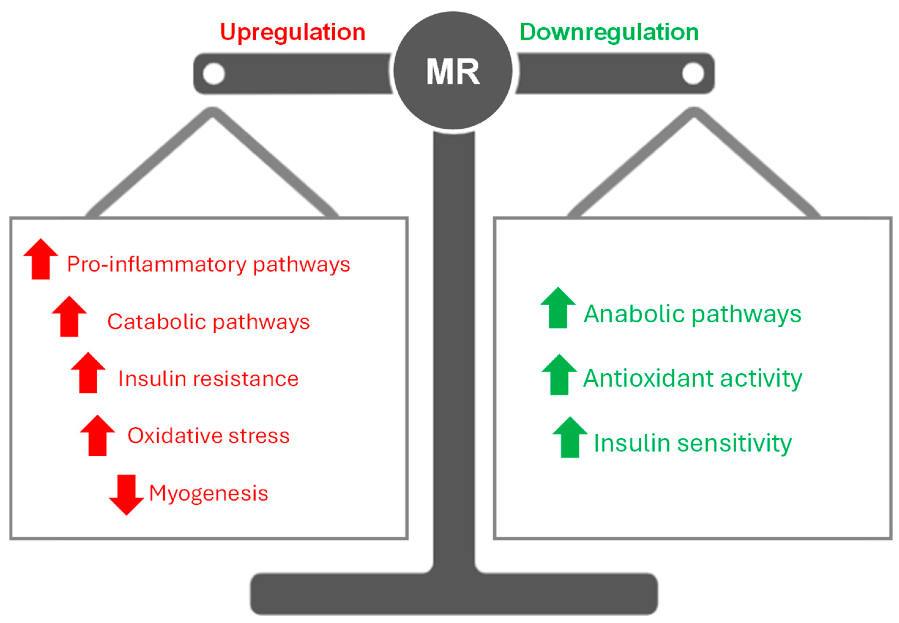
2. Role of Mineralocorticoid Receptors in Skeletal Muscle

MRs exist as two distinct isoforms, MR-1α and MR-1β, which are differentiated by their unique promoter sequences, P1 and P2, respectively [18,19]. These isoforms share core functional domains that exhibit distinct regulatory patterns and tissue-specific expression [18]. P1 is typically active in classic MR-responsive tissues, such as the kidneys, where the receptor plays a pivotal role in blood pressure regulation by electrolyte homeostasis [20]. In contrast, P2 drives expression in non-classical tissues, including skeletal muscle, where MRs influence processes such as inflammation, oxidative stress, and glucose metabolism [21,22]. While both isoforms share the same functional domains, their regulation by distinct transcriptional mechanisms may influence downstream gene expression and cellular responses. Although it remains unclear whether these isoforms have different intracellular targets or signaling pathways, their tissue-specific expression highlights the potential for selective therapeutic interventions aimed at modulating MR activity in aging skeletal muscle.
The excessive activation of MRs induces insulin resistance in skeletal muscle by impairing GLUT4 translocation to the cell membrane, which reduces glucose uptake by muscle fibers [23]. This dysfunction results in reduced glucose availability for anabolic processes, contributing to the catabolic state characteristic of sarcopenia [24]. Thus, the effects of MRs on glucose metabolism are an extension of their ability to amplify inflammatory and oxidative stress pathways, creating a deleterious cycle that exacerbates muscle atrophy. This duality in promoter activity allows a fine-tuned regulation of MR expression, aligning receptor function with the physiological demands of specific tissues [25]. By understanding the molecular mechanisms through which MRs influence muscle physiology, we can identify novel interventions to prevent or mitigate sarcopenia, thereby enhancing the quality of life and functional independence of the aging population.

2.1. Skeletal Muscle Aging: Insights into the Drivers of Sarcopenia

Sarcopenia is marked by a systemic and progressive loss of muscle mass, quality, and strength, which significantly impacts mobility and independence, especially among older people [2]. This degenerative process is driven by several key factors that collectively contribute to sarcopenia. Chronic low-grade inflammation, characterized by elevated levels of pro-inflammatory cytokines such as tumor necrosis factor-alpha (TNF-α) and interleukins (IL-1β, IL-6), accelerates muscle protein degradation and impairs muscle regeneration [26,27]. Aged skeletal muscle exhibits reduced mitochondrial biogenesis and efficiency, leading to decreased ATP availability and the excessive production of reactive oxygen species (ROS) [6]. This increase in ROS drives oxidative stress, causing damage to cellular proteins, lipids, and DNA, further compromising muscle integrity and accelerating muscle atrophy. This pro-inflammatory environment with increased oxidative stress leads to satellite cell dysfunction as both conditions impair these cells’ viability and self-renewal capacity via cellular injury [8]. In this sense, the dysfunction of satellite cells, which play a crucial role in muscle regeneration, hampers the tissue’s ability to recover and adapt, intensifying the loss of muscle mass and strength [28]. Finally, insulin resistance contributes to muscle atrophy by disrupting anabolic signaling pathways, particularly through the attenuation of the PI3K/Akt/mTOR axis, which is essential for protein synthesis and muscle maintenance [29]. The impaired insulin signaling also leads to an overactivation of catabolic pathways, including the ubiquitin–proteasome and autophagy–lysosome systems, further accelerating protein degradation and muscle fiber loss [30].
As these factors overlap in terms of signaling pathways throughout the aging process, skeletal muscle fibers (mainly of type II) undergo a marked reduction in size and quantity, essentially due to greater protein degradation rates [31,32]. These changes collectively lead to the gradual loss of muscle mass and strength that underlies the sarcopenic phenotype.
Emerging evidence highlights the intricate role of MRs in skeletal muscle homeostasis, particularly under conditions of aging [33,34]. The interplay between MR activation and key catabolic processes creates an environment conducive to muscle degradation. This cascade of events not only drives protein catabolism but also disrupts the regenerative capacity of muscle fibers, compounding the loss of muscle mass and function characteristic of sarcopenia [33,34]. Refining our understanding of these mechanisms can inform targeted interventions to disrupt this cycle, offering a more nuanced approach to maintaining muscle health in the elderly population.

2.2. MR-Driven Ion Transport in Skeletal Muscle

Beyond their well-established roles in renal regulation [14,15,35], MRs have emerged as key modulators of skeletal muscle physiology [11]. In muscle tissue, MRs influence ion distribution, particularly sodium and potassium, which are critical for maintaining excitability and contractility [25]. This ion balance is integral to muscle function and resilience, particularly under conditions of metabolic stress or aging. By regulating these processes, MRs contribute to the broader homeostatic mechanisms that sustain muscle performance and adaptability, as under conditions of excessive MR activation, as observed in certain pathological states and with aging, an overload of sodium accumulates within muscle cells, leading to intracellular ion imbalance and water retention [11,15,25]. This ionic disruption can cause edema and increased stress on muscle fibers, creating an unfavorable environment for muscle contraction and regeneration, which may exacerbate the loss of muscle mass and function due to atrophy [36,37]. Consequently, prolonged MR activation and subsequent ionic imbalances may contribute to muscle dysfunction by impairing cellular excitability and increasing metabolic strain.

2.3. The Intersection of MRs and Insulin Resistance

Experimental evidence strongly supports the role of excessive MR activation in the development of skeletal muscle insulin resistance [21,37]. An experimental study using a diet-induced model of metabolic dysfunction demonstrated that MR activation contributes to intramyocellular lipid accumulation, mitochondrial dysfunction, and impaired insulin signaling [23]. This study used a murine model in which MR blockade with spironolactone prevented Western diet-induced glucose intolerance and insulin resistance by enhancing GLUT4 expression and reducing free fatty acid content in muscle tissue. Mechanistically, MR activation downregulated miR-99a, leading to increased CD36 expression, which facilitated lipid accumulation and exacerbated insulin resistance in muscle cells [23]. Additionally, a study in zebrafish provided further evidence that MR activation is a critical determinant of glucose metabolism in skeletal muscle [38]. In this study, MR deletion abolished insulin-stimulated glucose uptake, suggesting that MRs play a key role in modulating anabolic glucose metabolism under basal conditions [38]. This evidence indicates that MR signaling regulates metabolic flexibility, and its dysregulation contributes to insulin resistance. Together, these findings highlight the direct involvement of MR activation in disrupting insulin signaling in skeletal muscle, leading to a catabolic state that drives protein degradation and muscle mass loss—common characteristics in aging individuals. Studies suggest that this insulin resistance, amplified by MR activation, also elevates the risk of related metabolic disorders, such as metabolic syndrome and type 2 diabetes [39,40,41,42].
These adverse effects of excessive MR activation may further be exacerbated by aging, a period during which skeletal muscle naturally experiences a decline in regenerative and functional capacity [43,44]. Prolonged MR activation thus contributes not only to muscle deterioration but also to metabolic dysfunction within skeletal muscle, positioning MRs as a promising therapeutic target for interventions aimed at preserving muscle metabolism in the elderly.

2.4. Linking MRs to Inflammation and Oxidative Stress

The activation of MRs leads to chronic inflammation and oxidative stress [34,45], processes that accelerate muscle aging and drive the progression of sarcopenia [6,26,27]. In response to MR activation, a cascade of cellular events is triggered, leading to the elevated production of pro-inflammatory mediators and ROS [46]. Briefly, MR activation induces an inflammatory response through the activation of the NF-κB transcription factor [47]. Thus, MR activation in immune cells, such as macrophages and lymphocytes, leads to the release of pro-inflammatory cytokines, including TNF-α, IL-6, and MCP-1. These cytokines increase the expression of adhesion molecules and promote immune cell recruitment to skeletal muscle, which may exacerbate muscle degradation. Furthermore, MR activation drives macrophage polarization toward the M1 pro-inflammatory phenotype, resulting in the increased secretion of TNF-α and IL-1β, thereby sustaining a chronic inflammatory environment. This process represents a potential mechanism by which MRs contribute to enhanced protein degradation and impaired muscle regeneration [5].
In this scenario, TNF-α plays a key role in muscle catabolism by activating proteolytic pathways, including the ubiquitin–proteasome and autophagy–lysosome systems, which impair satellite cell function and hinder muscle regeneration [48]. Elevated TNF-α levels, often observed in chronic inflammation and aging, induce the expression of E3 ubiquitin ligases like MuRF1 and Atrogin-1, which target myofibrillar proteins for degradation [49]. Additionally, TNF-α impairs myogenesis by inhibiting myogenic regulatory factors such as MyoD and myogenin by promoting mitochondrial dysfunction [50], which exacerbates oxidative stress and further contributes to muscle atrophy.
MRs have been implicated in amplifying TNF-α signaling [51]. Thus, MR activation enhances the expression of inflammation-associated genes, including those encoding key mediators such as pro-inflammatory cytokines and interleukins [52,53]. One key mechanism involves the activation of NF-κB, where MR signaling promotes the nuclear translocation of the p65 subunit, upregulating the transcription of pro-inflammatory cytokines such as TNF-α and IL-6 [51]. Additionally, MR activation engages the MAPK signaling cascade, specifically ERK1/2 and JNK pathways, leading to the activation of the AP-1 transcription complex (c-Fos/c-Jun), which further amplifies cytokine gene expression [47,51].
These pro-inflammatory mediators are known to recruit immune cells and exacerbate tissue damage [54], particularly in aged muscle, where cellular turnover is already limited. This creates a feedback loop where pro-inflammatory mediators, such as TNF-α, induce a catabolic signaling which is sustained, perpetuating muscle protein degradation and impairing regeneration. This chronic inflammatory state negatively impacts muscle function by promoting muscle fiber degradation and inhibiting protein synthesis, processes essential for maintaining muscle mass and strength [55]. Additionally, muscle-derived myokines play a pivotal role in modulating systemic inflammation and metabolic processes, acting as signaling molecules released during muscle contraction which are known to influence muscle regeneration [56,57].
In addition to driving inflammation, excessive MR activation increases ROS production [46], which may contribute to oxidative stress in muscle tissue. It has been demonstrated that MR activation induces the expression of the p47phox subunit of NADPH oxidase, leading to an increased production of superoxide anions (O2) [58]. This triggers NF-κB activation, which, in turn, upregulates the expression of additional pro-inflammatory mediators, creating a positive feedback loop between inflammation and oxidative stress. Moreover, MR activation can interact with angiotensin II (Ang II) signaling, further enhancing the activation of ERK1/2 and JNK kinases, which are crucial for amplifying oxidative stress and inflammation [47,58]. This suggests that the effects of MRs on ROS production do not occur in isolation but rather within a complex network of interconnected pathways that contribute to muscle degradation [5].
The buildup of ROS inflicts damage on cellular structures, including lipids, proteins, and DNA, compromising muscle cell integrity and diminishing the tissue’s regenerative capacity [59]. With aging, the natural antioxidant defenses become less effective, amplifying the detrimental effects of MRs-induced ROS. This sustained oxidative stress accelerates muscle degeneration, creating a cycle of incomplete repair that perpetuates sarcopenia [60]. This suggests a complex interaction where excessive MR activation may also alter the balance of myokine secretion, potentially amplifying muscle catabolism and impairing regenerative processes. Thus, this pathological mechanism highlights MRs as a potential therapeutic target for mitigating age-related muscle deterioration by reducing inflammation and oxidative stress, with the goal of preserving muscle mass and function in the elderly.

3. Counteracting Muscle Deterioration in Aging by Tackling MRs

3.1. Harnessing MR Antagonists for Therapy

The pathological activation of MRs contributes to molecular disturbances which suppress anabolic pathways and enhances catabolic signaling, driving excessive protein degradation in skeletal muscle (Figure 1). A key mechanism underlying this process is the suppression of myogenesis, coupled with the increased apoptosis of myocytes [61]. MR activation directly impairs myogenic differentiation by downregulating critical regulatory factors such as MyoD and Myogenin while increasing oxidative stress, which promotes mitochondrial dysfunction and further compromises satellite cell viability, ultimately disrupting muscle regeneration [62,63,64]. This dysfunction leads to the release of pro-apoptotic factors, including cytochrome c, which activates caspase-mediated apoptotic pathways [24,65]. These events result in myonuclear loss and a reduction in muscle cross-sectional area and strength, hallmark features of sarcopenia.
MR antagonists, such as spironolactone [52,66,67] and finerenone [68], show promise in mitigating these effects by reducing ROS production, modulating inflammatory cytokine expression, and limiting apoptosis. Additionally, these agents have been shown to promote anti-inflammatory macrophage phenotypes, reduce muscle fibrosis, and enhance the regenerative capacity of muscle tissue. These findings highlight the therapeutic utility of MR antagonists in targeting the molecular drivers of sarcopenia [25]. However, targeted clinical studies are still needed to confirm the efficacy and safety of these antagonists in healthy older adults and in the early stages of muscle wasting. Future research should focus on optimizing dosage and safety profiles to minimize adverse effects while promoting muscle preservation. This approach could position MR antagonists as a viable and effective non-invasive therapy to counteract age-related muscle decline, supporting muscular health and functionality in the elderly population.

3.2. Exercise as a Modulator of MRs

Physical exercise plays a critical role in regulating the activity of MRs [67,68,69] and serves as an effective adjunct in preventing sarcopenia associated with aging [70]. A study with three experimental groups of mdx mice, a Duchenne muscular dystrophy model, subjected to different regimens (exercised, sedentary, and isoproterenol-treated) treated the mice with spironolactone, an MR antagonist, in combination with lisinopril, an ACE inhibitor [67]. Functional and histological assessments revealed no significant benefit of MR blockade in the exercised or aged mice, with the authors concluding that MR antagonists are most effective in the early inflammatory stages of the disease, which were less prevalent in the exercise and aging protocols.
Exercise enhances energy metabolism and insulin sensitivity in skeletal muscle [71], which counteracts the adverse effects of excessive MR activation, avoiding muscle catabolism. By reducing the production of pro-inflammatory mediators and ROS [59,72], exercise can limit the harmful effects linked to MR activation, thereby preserving muscle fiber integrity and improving muscle functionality. In addition, studies suggest that regular exercise, particularly resistance training [73], helps modulate skeletal muscle metabolism, fostering an environment less prone to inflammation and oxidative stress, both of which are key factors in preserving muscle health during aging. Interestingly, a study focusing on ovariectomized mice, a model of post-menopausal metabolic disorders, treated with finerenone (1 mg/kg/day) for one month assessed cardiovascular function, glucose tolerance, and exercise capacity [68]. The results demonstrated that finerenone improved left ventricular filling pressure, insulin sensitivity, and exercise performance on a treadmill, with the authors concluding that MR blockade enhanced exercise capacity and cardiovascular function in a model of heart failure with preserved ejection fraction.
Additionally, the effects of MR blockade on the hypothalamic–pituitary–adrenal (HPA) axis in response to exercise were investigated in 12 healthy male participants using a randomized, double-blind, cross-over design [69]. Participants received either canrenoate (200 mg) or placebo 24 and 8 h prior to exercise, and blood samples were taken throughout the experiment to measure cortisol, ACTH, and growth hormone levels. The results revealed significantly elevated cortisol levels in the canrenoate condition both at rest and post-exercise, while ACTH and growth hormone remained unchanged. The authors concluded that MR blockade alters the set point for cortisol feedback inhibition, resulting in elevated cortisol secretion during exercise. Exercise acts on counteracting the dysregulation of the HPA axis [74,75], particularly under conditions of sleep disruption [76], which may be associated with MR activation [77]. Sleep disturbances impair anabolic signaling pathways, including those mediated by IGF-1, while enhancing catabolic processes such as the ubiquitin–proteasome and autophagy–lysosome systems [78]. Regular physical activity restores this balance, reducing HPA axis-driven catabolism and improving muscle health [79], which consequently may reduce MR expression.
Taken together, these studies suggest a complex interaction between exercise and MR activity, with MR antagonism potentially influencing exercise capacity and hormonal responses to physical stress. In this sense, combining MR antagonists with exercise interventions may provide a synergistic approach to addressing the multifactorial nature of sarcopenia. This integrated strategy offers a comprehensive framework for preserving muscle mass, strength, and functionality, ultimately improving the quality of life in aged populations due to functional independence.

3.3. Future Perspectives

Our understanding of the specific role of MRs in the interplay between oxidative stress and insulin resistance across different age groups remains incomplete. Although studies suggest that MR activation increases oxidative stress and disrupts insulin signaling [33,34], factors that contribute to insulin resistance in skeletal muscle, these interactions have largely been explored in pathological or animal models. How these mechanisms unfold across various aging stages and whether MR sensitivity to oxidative stress changes with age are still poorly understood. Addressing these gaps requires future research to investigate the potential of antioxidant interventions and MR antagonists as modulators of insulin response in aging populations. Studies examining whether antioxidants combined with MR blockers can effectively reduce oxidative stress and restore insulin signaling in healthy older adults and in early stages of insulin resistance could yield promising preventive and therapeutic strategies, ultimately offering new avenues to slow metabolic decline and improve quality of life in aging individuals. Additionally, the clinical applicability of MR antagonists in muscle health, particularly in patients with early-stage sarcopenia, insulin resistance, or high oxidative stress, is an area that warrants further investigation. In addition, future studies exploring the interplay between MRs and myokines could provide valuable insights into therapeutic strategies for mitigating muscle aging and enhancing overall health outcomes in aged populations.
While MR antagonists, such as spironolactone, have shown benefits in specific conditions like heart failure and muscular dystrophies [80,81], data on their impact in healthy older adults is limited. In particular, there is a pressing need for studies focusing on MR antagonists in the context of sarcopenia across age, which may be an essential factor in assessing the preventive and therapeutic potential of these agents under the aging process. Given the role of MRs in driving inflammation and oxidative stress [34,45], MR antagonists may be valuable in mitigating age-related muscle mass and strength loss. However, the absence of clinical trials targeting healthy aged individuals limits our ability to fully understand the efficacy and safety of these antagonists in preventing muscle decline. Furthermore, future clinical studies should also investigate aged populations without significant comorbidities to evaluate whether MR antagonists can serve as effective preventative strategies for delaying muscle wasting progression and enhancing muscle function throughout aging.
The combined effects of physical exercise and MR antagonism on muscle function in the elderly remain largely unexplored. Although the benefits of physical exercise in preserving muscle mass and strength are well-established [70,73,82], and MR antagonists have shown potential to reduce catabolic signaling in skeletal muscle, the synergistic potential of these approaches has yet to be fully investigated. Emerging evidence suggests that these interventions may complement each other, with exercise potentially mitigating the adverse effects of MR activation and fostering a more favorable muscle environment in aging populations. To maximize muscle preservation, clinical trials are needed to evaluate the efficacy of integrated protocols combining exercise and MR antagonists in aged populations. Such studies should aim to identify optimal dosages and frequencies for both exercise and MR antagonist use to enhance muscle strength, mass, and functionality, offering a comprehensive approach to preventing sarcopenia and physical decline in older adults.

4. Conclusions

MRs are central to skeletal muscle aging, driving inflammation, oxidative stress, and insul in resistance, which are factors that contribute to sarcopenia. This review underscores the importance of MRs as therapeutic targets, given their involvement in critical catabolic pathways. These mechanisms collectively contribute to muscle protein degradation and the progressive loss of muscle mass and function. Therapeutic interventions targeting MRs, such as spironolactone, have demonstrated potential in mitigating these detrimental effects. When combined with physical exercise, MR antagonism may offer an effective approach to managing sarcopenia. However, further research is needed to clarify MR function across aging stages and to establish the safety and efficacy of combining MR antagonists with exercise in both healthy and sarcopenic aged populations. These findings highlight the central role of MRs in muscle health and reinforce the need for innovative strategies to preserve muscle mass, strength, and overall quality of life in older adults.

Funding

This research received no external funding.

Institutional Review Board Statement

Not applicable.

Informed Consent Statement

Not applicable.

Data Availability Statement

No new data were created or analyzed in this study. Data sharing is not applicable to this article.

Conflicts of Interest

The authors declare no conflict of interest.

References

  1. Trombetti, A.; Reid, K.F.; Hars, M.; Herrmann, F.R.; Pasha, E.; Phillips, E.M.; Fielding, R.A. Age-associated declines in muscle mass, strength, power, and physical performance: Impact on fear of falling and quality of life. Osteoporos. Int. 2016, 27, 463–471. [Google Scholar] [CrossRef]
  2. Kamel, H.K. Sarcopenia and Aging. Nutr. Rev. 2003, 61, 157–167. [Google Scholar] [CrossRef] [PubMed]
  3. Landi, F.; Liperoti, R.; Russo, A.; Giovannini, S.; Tosato, M.; Capoluongo, E.; Bernabei, R.; Onder, G. Sarcopenia as a risk factor for falls in elderly individuals: Results from the ilSIRENTE study. Clin. Nutr. 2012, 31, 652–658. [Google Scholar] [CrossRef]
  4. Dirks, A.J.; Hofer, T.; Marzetti, E.; Pahor, M.; Leeuwenburgh, C. Mitochondrial DNA mutations, energy metabolism and apoptosis in aging muscle. Ageing Res. Rev. 2006, 5, 179–195. [Google Scholar] [CrossRef] [PubMed]
  5. Meng, S.-J.; Yu, L.-J. Oxidative Stress, Molecular Inflammation and Sarcopenia. Int. J. Mol. Sci. 2010, 11, 1509–1526. [Google Scholar] [CrossRef] [PubMed]
  6. Bellanti, F.; Buglio, A.L.; Vendemiale, G. Oxidative stress and sarcopenia. In Aging; Elsevier: Amsterdam, The Netherlands, 2020; pp. 95–103. [Google Scholar] [CrossRef]
  7. Shou, J.; Chen, P.-J.; Xiao, W.-H. Mechanism of increased risk of insulin resistance in aging skeletal muscle. Diabetol. Metab. Syndr. 2020, 12, 14. [Google Scholar] [CrossRef]
  8. Chen, M.; Wang, Y.; Deng, S.; Lian, Z.; Yu, K. Skeletal muscle oxidative stress and inflammation in aging: Focus on antioxidant and anti-inflammatory therapy. Front. Cell Dev. Biol. 2022, 10, 964130. [Google Scholar] [CrossRef]
  9. Álvarez-Bustos, A.; Rodríguez-Sánchez, B.; Carnicero-Carreño, J.A.; Sepúlveda-Loyola, W.; Garcia-Garcia, F.J.; Rodríguez-Mañas, L. Healthcare cost expenditures associated to frailty and sarcopenia. BMC Geriatr. 2022, 22, 747. [Google Scholar] [CrossRef]
  10. Tian, D.; Meng, J. Exercise for Prevention and Relief of Cardiovascular Disease: Prognoses, Mechanisms, and Approaches. Oxid. Med. Cell. Longev. 2019, 2019, 1–11. [Google Scholar] [CrossRef]
  11. Chadwick, J.A.; Hauck, J.S.; Lowe, J.; Shaw, J.J.; Guttridge, D.C.; Gomez-Sanchez, C.E.; Gomez-Sanchez, E.P.; Rafael-Fortney, J.A. Mineralocorticoid receptors are present in skeletal muscle and represent a potential therapeutic target. FASEB J. 2015, 29, 4544–4554. [Google Scholar] [CrossRef]
  12. Howard, Z.M.; Gomatam, C.K.; Rabolli, C.P.; Lowe, J.; Piepho, A.B.; Bansal, S.S.; Accornero, F.; Rafael-Fortney, J.A. Mineralocorticoid receptor antagonists and glucocorticoids differentially affect skeletal muscle inflammation and pathology in muscular dystrophy. JCI Insight 2022, 7, e159875. [Google Scholar] [CrossRef] [PubMed]
  13. Howard, Z.M.; Gomatam, C.K.; Piepho, A.B.; Rafael-Fortney, J.A. Mineralocorticoid Receptor Signaling in the Inflammatory Skeletal Muscle Microenvironments of Muscular Dystrophy and Acute Injury. Front. Pharmacol. 2022, 13, 942660. [Google Scholar] [CrossRef]
  14. Nguyen, E.T.; Berman, S.; Streicher, J.; Estrada, C.M.; Caldwell, J.L.; Ghisays, V.; Ulrich-Lai, Y.; Solomon, M.B. Effects of combined glucocorticoid/mineralocorticoid receptor modulation (CORT118335) on energy balance, adiposity, and lipid metabolism in male rats. Am. J. Physiol. Endocrinol. Metab. 2019, 317, E337–E349. [Google Scholar] [CrossRef] [PubMed]
  15. Gomez-Sanchez, E.; Gomez-Sanchez, C.E. The Multifaceted Mineralocorticoid Receptor. In Comprehensive Physiology, 1st ed.; Terjung, R., Ed.; Wiley: Hoboken, NJ, USA, 2014; pp. 965–994. [Google Scholar] [CrossRef]
  16. Zennaro, M.-C.; Caprio, M.; Fève, B. Mineralocorticoid receptors in the metabolic syndrome. Trends Endocrinol. Metab. 2009, 20, 444–451. [Google Scholar] [CrossRef]
  17. Young, M.J. Mechanisms of mineralocorticoid receptor-mediated cardiac fibrosis and vascular inflammation. Curr. Opin. Nephrol. Hypertens. 2008, 17, 174–180. [Google Scholar] [CrossRef]
  18. Pascual-Le Tallec, L.; Lombès, M. The Mineralocorticoid Receptor: A Journey Exploring Its Diversity and Specificity of Action. Mol. Endocrinol. 2005, 19, 2211–2221. [Google Scholar] [CrossRef] [PubMed]
  19. Gaudenzi, C.; Mifsud, K.R.; Reul, J.M.H.M. Insights into isoform-specific mineralocorticoid receptor action in the hippocampus. J. Endocrinol. 2023, 258, e220293. [Google Scholar] [CrossRef]
  20. Le Menuet, D.; Viengchareun, S.; Muffat-Joly, M.; Zennaro, M.-C.; Lombès, M. Expression and function of the human mineralocorticoid receptor: Lessons from transgenic mouse models. Mol. Cell. Endocrinol. 2004, 217, 127–136. [Google Scholar] [CrossRef]
  21. Ibarrola, J.; Lu, Q.; Zennaro, M.-C.; Jaffe, I.Z. Mechanism by Which Inflammation and Oxidative Stress Induce Mineralocorticoid Receptor Gene Expression in Aging Vascular Smooth Muscle Cells. Hypertension 2023, 80, 111–124. [Google Scholar] [CrossRef]
  22. Kuhn, E.; Lombès, M. The mineralocorticoid receptor: A new player controlling energy homeostasis. Horm. Mol. Biol. Clin. Investig. 2013, 15, 59–69. [Google Scholar] [CrossRef]
  23. Hulse, J.L.; Habibi, J.; Igbekele, A.E.; Zhang, B.; Li, J.; Whaley-Connell, A.; Sowers, J.R.; Jia, G. Mineralocorticoid Receptors Mediate Diet-Induced Lipid Infiltration of Skeletal Muscle and Insulin Resistance. Endocrinology 2022, 163, bqac145. [Google Scholar] [CrossRef] [PubMed]
  24. Marzetti, E.; Privitera, G.; Simili, V.; Wohlgemuth, S.E.; Aulisa, L.; Pahor, M.; Leeuwenburgh, C. Multiple Pathways to the Same End: Mechanisms of Myonuclear Apoptosis in Sarcopenia of Aging. Sci. World J. 2010, 10, 340–349. [Google Scholar] [CrossRef] [PubMed]
  25. Burton, L.A.; McMurdo, M.E.T.; Struthers, A.D. Mineralocorticoid antagonism: A novel way to treat sarcopenia and physical impairment in older people? Clin. Endocrinol. 2011, 75, 725–729. [Google Scholar] [CrossRef]
  26. Bano, G.; Trevisan, C.; Carraro, S.; Solmi, M.; Luchini, C.; Stubbs, B.; Manzato, E.; Sergi, G.; Veronese, N. Inflammation and sarcopenia: A systematic review and meta-analysis. Maturitas 2017, 96, 10–15. [Google Scholar] [CrossRef]
  27. Dalle, S.; Rossmeislova, L.; Koppo, K. The Role of Inflammation in Age-Related Sarcopenia. Front. Physiol. 2017, 8, 1045. [Google Scholar] [CrossRef]
  28. Alway, S.E.; Myers, M.J.; Mohamed, J.S. Regulation of Satellite Cell Function in Sarcopenia. Front. Aging Neurosci. 2014, 6, 246. [Google Scholar] [CrossRef]
  29. Ramasubbu, K.; Devi Rajeswari, V. Impairment of insulin signaling pathway PI3K/Akt/mTOR and insulin resistance induced AGEs on diabetes mellitus and neurodegenerative diseases: A perspective review. Mol. Cell. Biochem. 2023, 478, 1307–1324. [Google Scholar] [CrossRef] [PubMed]
  30. Cleasby, M.E.; Jamieson, P.M.; Atherton, P.J. Insulin resistance and sarcopenia: Mechanistic links between common co-morbidities. J. Endocrinol. 2016, 229, R67–R81. [Google Scholar] [CrossRef]
  31. McPhee, J.S.; Cameron, J.; Maden-Wilkinson, T.; Piasecki, M.; Yap, M.H.; Jones, D.A.; Degens, H. The Contributions of Fiber Atrophy, Fiber Loss, In Situ Specific Force, and Voluntary Activation to Weakness in Sarcopenia. J. Gerontol. Ser. A 2018, 73, 1287–1294. [Google Scholar] [CrossRef]
  32. Nishikawa, H.; Fukunishi, S.; Asai, A.; Yokohama, K.; Nishiguchi, S.; Higuchi, K. Pathophysiology and mechanisms of primary sarcopenia (Review). Int. J. Mol. Med. 2021, 48, 156. [Google Scholar] [CrossRef]
  33. Jia, G.; Lockette, W.; Sowers, J.R. Mineralocorticoid receptors in the pathogenesis of insulin resistance and related disorders: From basic studies to clinical disease. Am. J. Physiol.-Regul. Integr. Comp. Physiol. 2021, 320, R276–R286. [Google Scholar] [CrossRef]
  34. Kuster, G.M.; Kotlyar, E.; Rude, M.K.; Siwik, D.A.; Liao, R.; Colucci, W.S.; Sam, F. Mineralocorticoid Receptor Inhibition Ameliorates the Transition to Myocardial Failure and Decreases Oxidative Stress and Inflammation in Mice With Chronic Pressure Overload. Circulation 2005, 111, 420–427. [Google Scholar] [CrossRef] [PubMed]
  35. Krishna, G.G.; Kapoor, S.C. Potassium supplementation ameliorates mineralocorticoid-induced sodium retention. Kidney Int. 1993, 43, 1097–1103. [Google Scholar] [CrossRef] [PubMed]
  36. Jurkat-Rott, K.; Weber, M.-A.; Fauler, M.; Guo, X.-H.; Holzherr, B.D.; Paczulla, A.; Nordsborg, N.; Joechle, W.; Lehmann-Horn, F. K+ -dependent paradoxical membrane depolarization and Na + overload, major and reversible contributors to weakness by ion channel leaks. Proc. Natl. Acad. Sci. USA 2009, 106, 4036–4041. [Google Scholar] [CrossRef]
  37. Eisenhut, M.; Wallace, H. Ion channels in inflammation. Pflüg. Arch. Eur. J. Physiol. 2011, 461, 401–421. [Google Scholar] [CrossRef] [PubMed]
  38. Faught, E.; Vijayan, M.M. The Mineralocorticoid Receptor Functions as a Key Glucose Regulator in the Skeletal Muscle of Zebrafish. Endocrinology 2022, 163, bqac149. [Google Scholar] [CrossRef]
  39. Feraco, A.; Marzolla, V.; Scuteri, A.; Armani, A.; Caprio, M. Mineralocorticoid Receptors in Metabolic Syndrome: From Physiology to Disease. Trends Endocrinol. Metab. 2020, 31, 205–217. [Google Scholar] [CrossRef] [PubMed]
  40. Bender, S.B.; McGraw, A.P.; Jaffe, I.Z.; Sowers, J.R. Mineralocorticoid Receptor–Mediated Vascular Insulin Resistance. Diabetes 2013, 62, 313–319. [Google Scholar] [CrossRef]
  41. Korol, S.; Mottet, F.; Perreault, S.; Baker, W.L.; White, M.; De Denus, S. A systematic review and meta-analysis of the impact of mineralocorticoid receptor antagonists on glucose homeostasis. Medicine 2017, 96, e8719. [Google Scholar] [CrossRef]
  42. Barrera-Chimal, J.; Lima-Posada, I.; Bakris, G.L.; Jaisser, F. Mineralocorticoid receptor antagonists in diabetic kidney disease—mechanistic and therapeutic effects. Nat. Rev. Nephrol. 2022, 18, 56–70. [Google Scholar] [CrossRef]
  43. Michelucci, A.; Liang, C.; Protasi, F.; Dirksen, R.T. Altered Ca2+ Handling and Oxidative Stress Underlie Mitochondrial Damage and Skeletal Muscle Dysfunction in Aging and Disease. Metabolites 2021, 11, 424. [Google Scholar] [CrossRef] [PubMed]
  44. Carmeli, E.; Coleman, R.; Reznick, A.Z. The biochemistry of aging muscle. Exp. Gerontol. 2002, 37, 477–489. [Google Scholar] [CrossRef] [PubMed]
  45. Gorini, S.; Kim, S.K.; Infante, M.; Mammi, C.; La Vignera, S.; Fabbri, A.; Jaffe, I.Z.; Caprio, M. Role of Aldosterone and Mineralocorticoid Receptor in Cardiovascular Aging. Front. Endocrinol. 2019, 10, 584. [Google Scholar] [CrossRef]
  46. Fuller, P.J.; Young, M.J. Mechanisms of Mineralocorticoid Action. Hypertension 2005, 46, 1227–1235. [Google Scholar] [CrossRef]
  47. Muñoz-Durango, N.; Vecchiola, A.; Gonzalez-Gomez, L.M.; Simon, F.; Riedel, C.A.; Fardella, C.E.; Kalergis, A.M. Modulation of Immunity and Inflammation by the Mineralocorticoid Receptor and Aldosterone. BioMed Res. Int. 2015, 2015, 1–14. [Google Scholar] [CrossRef] [PubMed]
  48. Reid, M.B.; Li, Y.-P. Tumor necrosis factor-α and muscle wasting: A cellular perspective. Respir. Res. 2001, 2, 269. [Google Scholar] [CrossRef]
  49. Clavel, S.; Coldefy, A.-S.; Kurkdjian, E.; Salles, J.; Margaritis, I.; Derijard, B. Atrophy-related ubiquitin ligases, atrogin-1 and MuRF1 are up-regulated in aged rat Tibialis Anterior muscle. Mech. Ageing Dev. 2006, 127, 794–801. [Google Scholar] [CrossRef]
  50. Langen, R.C.J.; Velden, J.L.J.; Schols, A.M.W.J.; Kelders, M.C.J.M.; Wouters, E.F.M.; Janssen-Heininger, Y.M.W. Tumor necrosis factor-alpha inhibits myogenic differentiation through MyoD protein destabilization. FASEB J. 2004, 18, 227–237. [Google Scholar] [CrossRef]
  51. Chantong, B.; Kratschmar, D.V.; Nashev, L.G.; Balazs, Z.; Odermatt, A. Mineralocorticoid and glucocorticoid receptors differentially regulate NF-kappaB activity and pro-inflammatory cytokine production in murine BV-2 microglial cells. J. Neuroinflamm. 2012, 9, 260. [Google Scholar] [CrossRef]
  52. Thangaraj, S.S.; Oxlund, C.S.; Fonseca, M.P.D.; Svenningsen, P.; Stubbe, J.; Palarasah, Y.; Ketelhuth, D.F.J.; Jacobsen, I.A.; Jensen, B.L. The mineralocorticoid receptor blocker spironolactone lowers plasma interferon-γ and interleukin-6 in patients with type 2 diabetes and treatment-resistant hypertension. J. Hypertens. 2022, 40, 153–162. [Google Scholar] [CrossRef]
  53. Sønder, S.U.S.; Mikkelsen, M.; Rieneck, K.; Hedegaard, C.J.; Bendtzen, K. Effects of spironolactone on human blood mononuclear cells: Mineralocorticoid receptor independent effects on gene expression and late apoptosis induction. Br. J. Pharmacol. 2006, 148, 46–53. [Google Scholar] [CrossRef] [PubMed]
  54. Tu, H.; Li, Y.-L. Inflammation balance in skeletal muscle damage and repair. Front. Immunol. 2023, 14, 1133355. [Google Scholar] [CrossRef] [PubMed]
  55. Beenakker, K.G.M.; Ling, C.H.; Meskers, C.G.M.; De Craen, A.J.M.; Stijnen, T.; Westendorp, R.G.J.; Maier, A.B. Patterns of muscle strength loss with age in the general population and patients with a chronic inflammatory state. Ageing Res. Rev. 2010, 9, 431–436. [Google Scholar] [CrossRef]
  56. Gomarasca, M.; Banfi, G.; Lombardi, G. Myokines: The endocrine coupling of skeletal muscle and bone. In Advances in Clinical Chemistry; Elsevier: Amsterdam, The Netherlands, 2020; Volume 94, pp. 155–218. [Google Scholar]
  57. Lightfoot, A.P.; Cooper, R.G. The role of myokines in muscle health and disease. Curr. Opin. Rheumatol. 2016, 28, 661–666. [Google Scholar] [CrossRef] [PubMed]
  58. Fiebeler, A.; Luft, F.C. The Mineralocorticoid Receptor and Oxidative Stress. Heart Fail. Rev. 2005, 10, 47–52. [Google Scholar] [CrossRef]
  59. Powers, S.K.; Ji, L.L.; Kavazis, A.N.; Jackson, M.J. Reactive Oxygen Species: Impact on Skeletal Muscle. In Comprehensive Physiology, 1st ed.; Prakash, Y.S., Ed.; Wiley: Hoboken, NJ, USA, 2011; pp. 941–969. [Google Scholar] [CrossRef]
  60. Fulle, S.; Protasi, F.; Di Tano, G.; Pietrangelo, T.; Beltramin, A.; Boncompagni, S.; Vecchiet, L.; Fanò, G. The contribution of reactive oxygen species to sarcopenia and muscle ageing. Exp. Gerontol. 2004, 39, 17–24. [Google Scholar] [CrossRef]
  61. Burniston, J.; Saini, A.; Tan, L.; Goldspink, D. Aldosterone induces myocyte apoptosis in the heart and skeletal muscles of rats in vivo. J. Mol. Cell. Cardiol. 2005, 39, 395–399. [Google Scholar] [CrossRef]
  62. Lefranc, C.; Friederich-Persson, M.; Palacios-Ramirez, R.; Nguyen Dinh Cat, A. Mitochondrial oxidative stress in obesity: Role of the mineralocorticoid receptor. J. Endocrinol. 2018, 238, R143–R159. [Google Scholar] [CrossRef]
  63. Lee, J.Y.; Kim, D.A.; Choi, E.; Lee, Y.S.; Park, S.J.; Kim, B.-J. Aldosterone Inhibits In Vitro Myogenesis by Increasing Intracellular Oxidative Stress via Mineralocorticoid Receptor. Endocrinol. Metab. 2021, 36, 865–874. [Google Scholar] [CrossRef]
  64. Lefranc, C.; Friederich-Persson, M.; Braud, L.; Palacios-Ramirez, R.; Karlsson, S.; Boujardine, N.; Motterlini, R.; Jaisser, F.; Nguyen Dinh Cat, A. MR (Mineralocorticoid Receptor) Induces Adipose Tissue Senescence and Mitochondrial Dysfunction Leading to Vascular Dysfunction in Obesity. Hypertension 2019, 73, 458–468. [Google Scholar] [CrossRef]
  65. Marzetti, E.; Wohlgemuth, S.E.; Lees, H.A.; Chung, H.-Y.; Giovannini, S.; Leeuwenburgh, C. Age-related activation of mitochondrial caspase-independent apoptotic signaling in rat gastrocnemius muscle. Mech. Ageing Dev. 2008, 129, 542–549. [Google Scholar] [CrossRef] [PubMed]
  66. Lastra, G.; Whaley-Connell, A.; Manrique, C.; Habibi, J.; Gutweiler, A.A.; Appesh, L.; Hayden, M.R.; Wei, Y.; Ferrario, C.; Sowers, J.R. Low-dose spironolactone reduces reactive oxygen species generation and improves insulin-stimulated glucose transport in skeletal muscle in the TG(mRen2)27 rat. Am. J. Physiol.-Endocrinol. Metab. 2008, 295, E110–E116. [Google Scholar] [CrossRef]
  67. Lowe, J.; Kadakia, F.K.; Zins, J.G.; Haupt, M.; Peczkowski, K.K.; Rastogi, N.; Floyd, K.T.; Gomez-Sanchez, E.P.; Gomez-Sanchez, C.E.; Elnakish, M.T.; et al. Mineralocorticoid Receptor Antagonists in Muscular Dystrophy Mice During Aging and Exercise. J. Neuromuscul. Dis. 2018, 5, 295–306. [Google Scholar] [CrossRef] [PubMed]
  68. Pieronne-Deperrois, M.; Guéret, A.; Djerada, Z.; Crochemore, C.; Harouki, N.; Henry, J.; Dumesnil, A.; Larchevêque, M.; Do Rego, J.; Do Rego, J.; et al. Mineralocorticoid receptor blockade with finerenone improves heart function and exercise capacity in ovariectomized mice. ESC Heart Fail. 2021, 8, 1933–1943. [Google Scholar] [CrossRef] [PubMed]
  69. Wellhoener, P.; Born, J.; Fehm, H.L.; Dodt, C. Elevated Resting and Exercise-Induced Cortisol Levels after Mineralocorticoid Receptor Blockade with Canrenoate in Healthy Humans. J. Clin. Endocrinol. Metab. 2004, 89, 5048–5052. [Google Scholar] [CrossRef]
  70. Yoo, S.-Z.; No, M.-H.; Heo, J.-W.; Park, D.-H.; Kang, J.-H.; Kim, S.H.; Kwak, H.-B. Role of exercise in age-related sarcopenia. J. Exerc. Rehabil. 2018, 14, 551–558. [Google Scholar] [CrossRef]
  71. Borghouts, L.B.; Keizer, H.A. Exercise and Insulin Sensitivity: A Review. Int. J. Sports Med. 2000, 21, 1–12. [Google Scholar] [CrossRef]
  72. Peake, J.M.; Della Gatta, P.; Suzuki, K.; Nieman, D.C. Cytokine expression and secretion by skeletal muscle cells: Regulatory mechanisms and exercise effects. Exerc. Immunol. Rev. 2015, 21, 8–25. [Google Scholar]
  73. Hurst, C.; Robinson, S.M.; Witham, M.D.; Dodds, R.M.; Granic, A.; Buckland, C.; De Biase, S.; Finnegan, S.; Rochester, L.; Skelton, D.A.; et al. Resistance exercise as a treatment for sarcopenia: Prescription and delivery. Age Ageing 2022, 51, afac003. [Google Scholar] [CrossRef]
  74. Vgontzas, A.N.; Chrousos, G.P. Sleep, the hypothalamic–pituitary–adrenal axis, and cytokines: Multiple interactions and disturbances in sleep disorders. Endocrinol. Metab. Clin. N. Am. 2002, 31, 15–36. [Google Scholar] [CrossRef]
  75. Duclos, M.; Tabarin, A. Exercise and the Hypothalamo-Pituitary-Adrenal Axis. In Frontiers of Hormone Research; Lanfranco, F., Strasburger, C.J., Eds.; S. Karger AG: Basel, Switzerland, 2016; Volume 47, pp. 12–26. [Google Scholar]
  76. Piovezan, R.D.; Abucham, J.; Dos Santos, R.V.T.; Mello, M.T.; Tufik, S.; Poyares, D. The impact of sleep on age-related sarcopenia: Possible connections and clinical implications. Ageing Res. Rev. 2015, 23, 210–220. [Google Scholar] [CrossRef] [PubMed]
  77. Valaiyapathi, B.; Calhoun, D.A. Role of Mineralocorticoid Receptors in Obstructive Sleep Apnea and Metabolic Syndrome. Curr. Hypertens. Rep. 2018, 20, 23. [Google Scholar] [CrossRef]
  78. Dattilo, M.; Antunes, H.K.M.; Medeiros, A.; Mônico Neto, M.; Souza, H.S.; Tufik, S.; De Mello, M.T. Sleep and muscle recovery: Endocrinological and molecular basis for a new and promising hypothesis. Med. Hypotheses 2011, 77, 220–222. [Google Scholar] [CrossRef] [PubMed]
  79. Dolezal, B.A.; Neufeld, E.V.; Boland, D.M.; Martin, J.L.; Cooper, C.B. Interrelationship between Sleep and Exercise: A Systematic Review. Adv. Prev. Med. 2017, 2017, 1–14. [Google Scholar] [CrossRef]
  80. Rafael-Fortney, J.A.; Chimanji, N.S.; Schill, K.E.; Martin, C.D.; Murray, J.D.; Ganguly, R.; Stangland, J.E.; Tran, T.; Xu, Y.; Canan, B.D.; et al. Early Treatment with Lisinopril and Spironolactone Preserves Cardiac and Skeletal Muscle in Duchenne Muscular Dystrophy Mice. Circulation 2011, 124, 582–588. [Google Scholar] [CrossRef] [PubMed]
  81. Pitt, B.; Zannad, F.; Remme, W.J.; Cody, R.; Castaigne, A.; Perez, A.; Palensky, J.; Wittes, J. The Effect of Spironolactone on Morbidity and Mortality in Patients with Severe Heart Failure. N. Engl. J. Med. 1999, 341, 709–717. [Google Scholar] [CrossRef]
  82. Spirduso, W.W.; Cronin, D.L. Exercise dose-response effects on quality of life and independent living in older adults. Med. Sci. Sports Exerc. 2001, 33, S598–S608. [Google Scholar] [CrossRef]
Figure 1. The role of MRs in skeletal muscle. The upregulation of MRs leads to an increase in pro-inflammatory pathways (TNFα), catabolic pathways (MuRF1 and Atrogin1), insulin resistance, oxidative stress (ROS), and a decrease in myogenesis (MyoD and Myog). On the other hand, MR downregulation leads to an increase in anabolic pathways, antioxidant activity, and insulin sensitivity.
Receptors 04 00013 g001
Disclaimer/Publisher’s Note: The statements, opinions and data contained in all publications are solely those of the individual author(s) and contributor(s) and not of MDPI and/or the editor(s). MDPI and/or the editor(s) disclaim responsibility for any injury to people or property resulting from any ideas, methods, instructions or products referred to in the content.

