Article

Human Stem Cell-Derived Neural Organoids for the Discovery of Antiseizure Agents

by
Hamed Salmanzadeh
and
Robert F. Halliwell
*
Thomas J. Long School of Pharmacy, University of the Pacific, Stockton, CA 95211, USA
*
Author to whom correspondence should be addressed.
Receptors 2025, 4(3), 12; https://doi.org/10.3390/receptors4030012
Submission received: 30 March 2025 / Revised: 15 May 2025 / Accepted: 10 June 2025 / Published: 20 June 2025

Abstract

:
Background: The development of cerebral organoids created from human pluripotent stem cells in 3D culture may greatly improve the discovery of neuropsychiatric medicines. Methods: In the current study we differentiated neural organoids from a human pluripotent stem cell line in vitro, recorded the development of neurophysiological activity using multielectrode arrays (MEAs) and characterized the neuropharmacology of synaptic signaling over 8 months in vitro. In addition, we investigated the ability of these organoids to display epileptiform activity in response to a convulsant agent and the effects of antiseizure medicines to inhibit this abnormal activity. Results: Single and bursts of action potentials from individual neurons and network bursts were recorded on the MEA plates and significantly increased and became more complex from week 7 to week 30, consistent with neural network formation. Neural spiking was reduced by the Na channel blocker tetrodotoxin but increased by the inhibitor of KV7 potassium channels XE991, confirming the involvement of voltage-gated sodium and potassium channels in action potential activity. The GABA antagonists bicuculline and picrotoxin each increased the spike rate, consistent with inhibitory synaptic signaling. In contrast, the glutamate receptor antagonist kynurenic acid inhibited the spike rate, consistent with excitatory synaptic transmission in the organoids. The convulsant 4-aminopyridine increased spiking, bursts and synchronized firing, consistent with epileptiform activity in vitro. The anticonvulsants carbamazepine, ethosuximide and diazepam each inhibited this epileptiform neural activity. Conclusions: Together, our data demonstrate that neural organoids form inhibitory and excitatory synaptic circuits, generate epileptiform activity in response to a convulsant agent and detect the antiseizure properties of diverse antiepileptic drugs, supporting their value in drug discovery.

1. Introduction

Identifying novel agents for the treatment of complex neurological disorders, including seizures (epilepsy), is a major neuroscientific challenge. Conventional tools in drug discovery include animal and cellular models to evaluate candidate antiseizure medicines (ASMs) with clear limitations in their predictive value for human therapies [1]. Since the marketing of phenobarbitone in 1912, more than 32 additional antiseizure medicines have been developed, with over 20 of these released in just the last 25 years [2,3,4]. These drugs have diverse mechanisms of action, including sodium channel blocking, calcium channel blocking, the potentiation of GABAA receptors, the inhibition of glutamate-subtype receptors and even 5HT-2 receptor activation. Nevertheless, around one third of patients with epilepsy remain refractory to currently available pharmacotherapy and continue to have seizures despite trials with single and combined anticonvulsants [3,5]. In the United States, over 3 million adults and children are affected by epilepsy (CDC, 2024, https://www.cdc.gov/epilepsy/data-research/facts-stats/index.html (accessed on 20 March 2025)), and globally there are approximately 50 million people with epilepsy (WHO, 2024, https://www.who.int/news-room/fact-sheets/detail/epilepsy (accessed on 20 March 2025)), meaning that approximately 12–15 million individuals are impacted by uncontrolled seizures each year. There is therefore a significant need for new antiseizure medicines.
The discovery and availability of human stem cells with their potential to differentiate into diverse types of neurons and glial cells now holds significant promise for disease modeling and improved neuropsychiatric drug discovery and development [6]. Our lab, for example, has recently reported that human stem cells differentiate into mature neurons and glia in ultra-long-term culture (≥1 year) to form complex 2D excitatory (glutamate) and inhibitory (GABA) synaptic circuits that display epileptiform activity in response to a range of convulsants, including 4-aminopyridine [7]. Moreover, neural activity in these neuron–glia circuits is modulated by a variety of ion channel- and receptor-targeted drugs, and spontaneous and epileptiform activity is inhibited by first-, second- and third-generation antiseizure agents, supporting their value for drug discovery. More recently, we have also reported that such mature human neuron–glia circuits are able to detect novel potential antiseizure agents [8].
An innovative advance in the culture of human stem cells enabled the development of neural organoids [9], also referred to as cerebral organoids or colloquially ‘mini-brains’. These organoids are self-organized 3D structures comprising diverse neurons and glia and are derived from pluripotent stem cells in the presence of neural induction molecules to replicate some of the complex architecture and physiology of the brain within a hydrogel scaffold [9,10]. Neural organoids therefore offer a powerful approach to study early nervous system development to model human neuropsychiatric diseases in a dish and improve drug discovery [10,11,12,13]. Although recent studies have utilized human cerebral organoids to model neurodevelopment disorders associated with seizures, including Rett, Dravet and Angelman syndromes (for review see [11]), their value in evaluating antiseizure agents remains in its infancy.
In the current study, therefore, we investigated the ability of a human pluripotent stem cell line previously shown to differentiate into mature inhibitory and excitatory neural circuits in 2D [7] to form 3D neural organoids and recorded the development of neurophysiological activity over 8 months in vitro. Secondly, we investigated the ability of these human neural organoids to model seizures by testing their ability to display epileptiform-like activity in response to the convulsant 4-aminopyridine; and thirdly we determined the effects of three antiseizures medicines with distinct mechanisms of action on the epileptiform activity.

2. Methods

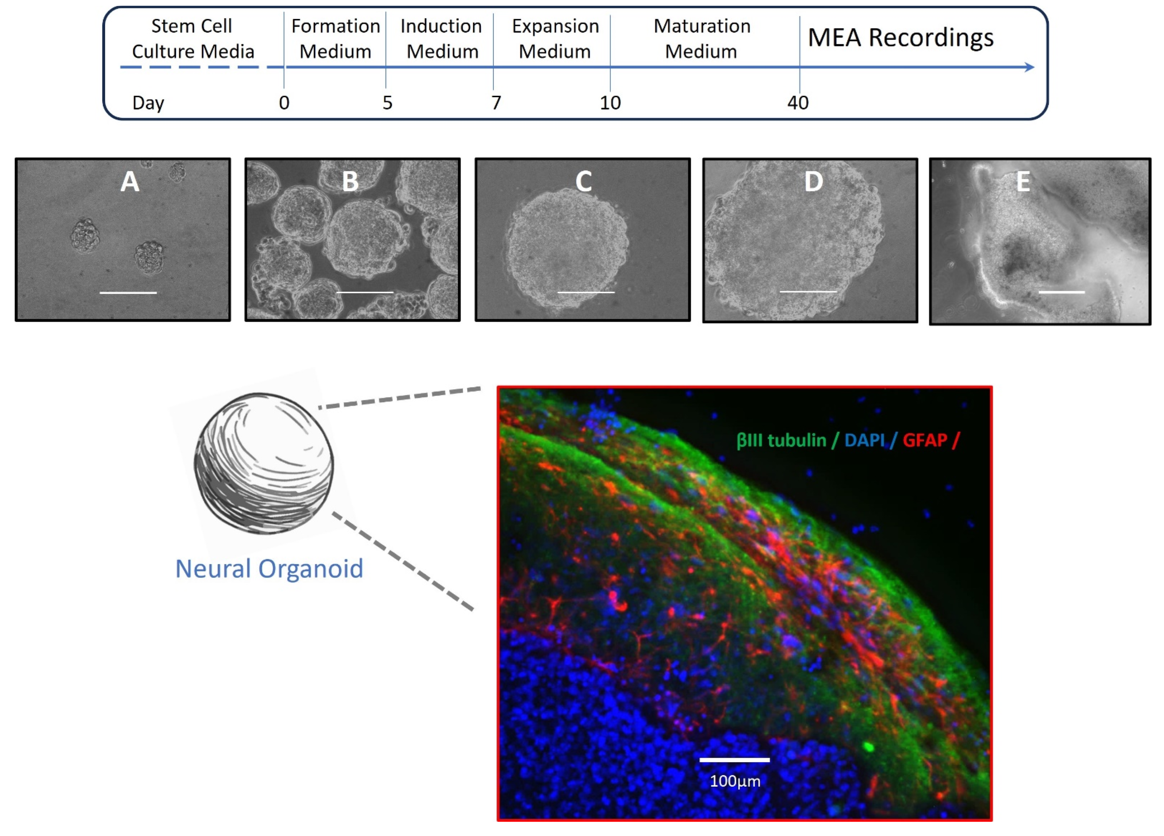
2.1. Culture and Neural Differentiation of Stem Cells

The human pluripotent embryonal stem cell line TERA2.cl.SP-12 (provided by SA Pryzborski, University of Durham, Durham, UK) was kept in 1.5 mL cryovials in liquid nitrogen until it was required for experiments. When thawed, cells were transferred to a sterile 75 mL culture flask and kept in an incubator (Caron Oasis, Marietta, OH, USA) at 37 °C, 5% CO2 and 100% relative humidity and maintained in Dulbecco’s Modified Eagle Medium (DMEM, Sigma-Aldrich, St Louis, MO, USA) with fetal bovine serum (FBS, 10% v/v; Invitrogen, Carlsbad, CA, USA), L-glutamine (2 mM; Sigma-Aldrich) and penicillin/streptomycin (100 U/mL, 100 μg/mL; Invitrogen). When the cells reached 80% confluence, they were passaged and re-seeded into new 75 mL tissue culture flasks. Cells between passages 3 and 35 were used for experiments.

2.2. Formation of Neural Organoids

Stem cells were cultured into three-dimensional (3D) structures to obtain organoids using the STEMdiff™ Cerebral Organoid Kit (Stemcell Technologies, Vancouver, BC, Canada) as follows: Cells were washed with PBS, dissociated with non-enzymatic Cell Dissociation Reagents (Stemcell Technologies), transferred to a sterile 50 mL conical tube and then rinsed with the Embryoid Body (EB) formation medium containing 10 μM of the Rho-associated protein kinase (ROCK) inhibitor, Y-27632 (Stemcell Technologies). Cells were then centrifuged at 500× g for 5 min, and the supernatant was replaced with fresh EB formation medium and resuspended. Approximately 9000 cells were added to every well in a 96-well round-bottom ultra-low attachment plate (faCellitate), placed back into the CO2 incubator and left undisturbed for ≥24 h.
After the first day of EB formation, small EBs (100–200 μm in diameter) could be observed with a layer of unincorporated cells around the central EBs. On day 2 and day 4, 100 μL of the EB formation medium (without the ROCK inhibitor) was added to each well. On day 5, EBs reached a diameter of 400–600 μm and exhibited round, smooth edges, and they were transferred to 24-well plates with the induction medium. Plates were treated with an Anti-Adherence Rinsing Solution (Stemcell Technologies), and the EBs were evenly distributed in the wells by gently shaking the plate and placing it back into the incubator for 48 h.
On day 7, EBs were embedded into Matrigel (Corning, Union City CA, USA) and transferred into the expansion medium as follows: Using a wide-bore pipette tip, 25–50 μL of the medium and an EB were drawn from a well of the 24-well plate and transferred onto Parafilm. Excess medium was removed, and 15 µL of Matrigel was added dropwise to the EB. The parafilm, retaining at least 12 EBs, was then placed into the incubator at 37 °C for 30 min to polymerize the Matrigel. The EBs were next transferred into a 6-well plate (pretreated with the Anti-Adherence Rinsing Solution) and kept in the expansion medium for 3 days. On day 10, the organoid media were replaced with 3 mL/well of the Maturation medium, and the plate was placed on an orbital shaker (Ohaus, Thermo Fisher Scientific, San Jose, CA, USA) in the incubator. After 40 days of growth, organoids generally showed dense cores with optically translucent edges.

2.3. Immunocytochemistry

Organoids were washed with PBbS (Gibco, Thermo Fisher Scientific) and fixed with 4% formaldehyde (Alfa Aesar, Thermo Fisher Scientific) for 2 h. The fixative was then removed, and cells were rinsed with PBS and then 0.3% Triton X-100 (Sigma-Aldrich) in PBS for 10 min to permeabilize them. Thereafter, cells were washed and placed in a blocking solution consisting of 5% normal donkey serum (Sigma-Aldrich) for 1 h. Cells were kept in dilutions of primary antibodies in blocking solutions that were directed against the protein of interest and incubated at 4 °C overnight. Thereafter, cells were washed with PBS and incubated for at least 2 h at room temperature with the appropriate secondary antibody, which was diluted 1:50 in the blocking solution, and washed with PBS and deionized water. Cells were counterstained with 4′, 6-diamidino-2-phenylindole (DAPI) (100 ng/mL; Sigma-Aldrich). Coverslips were mounted over slides by the anti-fade mounting solution (Prolong Gold Antifade Reagent; Invitrogen), and labeled cells were visualized using an EVOS FL auto microscope (Thermo Fisher). Antibodies used for detection included GFAP (sc-166481 AF594, Santa Cruz Biotechnology, Dallas, TX, USA), βIII-tubulin (sc-80005 FITC, Santa Cruz Biotechnology), MAP2 (sc-74421 AF594, Santa Cruz Biotechnology) and OCT3/4 (sc-5279 AF594, Santa Cruz Biotechnology).

2.4. MEA Electrophysiology

From day 40 onwards, human neural organoids were plated onto 6-well MEA plates (CytoviewMEA, Axion Biosystems, San Diego, CA, USA) pre-coated with polyethyleneimine (Sigma-Aldrich), maintained in the organoid medium in a final volume of 1 mL/well and kept in the cell culture incubator at 37 °C, 5% CO2 and 100% humidity for MEA recordings. Our electrophysiological recordings were conducted from neural organoids once each week from week 7 to week 30 using the MEA Maestro Edge system and the AxIS software (v3.12) (Axion Biosystems). MEA plates were placed into the Maestro Edge platform and allowed to stabilize for 10 min before starting a control baseline recording of at least 30 min. Electrical signals were recorded at a sampling rate of 12.5 kHz, with high and low band-pass filters of 0.2 kHz to 3 kHz and a spike threshold set at 6 times the estimated noise level. All recordings were conducted at 37 °C and 5% CO2. As previously described [7], the raw data were converted into Excel files using the AxIS software (v3.12), which included spike timing and network profile information. Only electrodes with at least 5 spikes per minute were analyzed. The AxIS Navigator software (v3.12) was used to obtain (i) the number of electrodes with an activity of ≥5 spikes/min, (ii) the weighted mean firing rate (WMFR) from all active electrodes, (iii) the number of single-electrode (single-cell) bursts (which are clusters of ≥5 spikes with a maximum inter-spike interval (ISI) of 100 ms), (iv) the number of network bursts (defined as ≥50 spikes with an ISI of ≤100 ms, involving at least 35% of the electrode array within a 20 ms window) and (v) the synchrony index (a measure of coordinated neural activity, with values between 0 and 1, where values closer to 1 indicate higher synchrony)

2.5. Drug Solutions and Drug Test Protocol

Stock solutions of drugs were freshly prepared and filtered using 0.22 μM solvent-resistant filters (Millipore, Burlington, MA, USA). Picrotoxin (Sigma-Aldrich), XE991 (Alamone) and kynurenic acid (Tocris, Bristol, UK) were dissolved in DMSO to obtain stock solutions of 10 mM. Bicuculline, 4-Aminopyridine (4-AP) (both Sigma-Aldrich) and tetrodotoxin (TTX) (Tocris) were dissolved in water to obtain stock solutions of 10 mM. Carbamazepine, ethosuximide and diazepam (all Sigma-Aldrich) were dissolved in ethanol to obtain stock solutions of 10 mM. The antiepileptic drugs tested in this study were at concentrations close to or within those measured in human serum following therapeutic dosing for seizure control [3].
Recordings of spontaneous activity were obtained for at least 30 min before drug treatment and again for a minimum of 30 min in the presence of test agents freshly added to the cell media. At the end of each drug experiment, the cell media was removed and replaced three times. Two days later, 90% of the culture media were again replenished with fresh media. To avoid possible residual effects of drug exposure, pharmacological testing was carried out at intervals of at least two days with the frequently at intervals of a week. To evaluate the validity of human stem cell-derived neural organoids as a 3D model for drug exploration, pilot experiments were conducted, and we determined that 4-AP (100 μM) reliably increased the spike activity, burst firing and synchronized neural network activity typical of epileptiform activity in vitro [7,8,14]. Control spontaneous firing was therefore recorded for a minimum of 15 min to establish the baseline before 4-AP was added; then activity was recorded for a further 60 min. This was followed by the addition of one of the following anticonvulsants: carbamazepine (10 μM), diazepam (10 μM) or ethosuximide (100 μM). The recordings continued for a further 40–50 min. All drugs were washed out once an asymptotic change in activity was established, and the cells were replenished with fresh culture media and placed back into the incubator.

2.6. Data Analysis

Analysis was conducted using the GraphPad Prism software (Version 10). Values reported are the means ± s.e.m, and n is the number of neural organoids. The one-sample t-test or Two-Way ANOVA and Holm-Šídák’s multiple comparisons post hoc tests were used to analyze neural spike amplitude, single-cell bursts, network bursts, and the synchrony index in the absence and presence of experimental drugs and antiseizure medicines. A p-value of 0.05 or less was considered statistically significant.

3. Results

3.1. Neural Organoids

In the current study, we used a commercial kit (STEMdiff™ Cerebral Organoid Kit) and a 4-step protocol based upon previous reports [9,15] to generate neural organoids from the pluripotent stem cell line TERA2.cl.SP-12 [16,17]. Previous studies of this human stem cell line have shown that it can differentiate into excitatory and inhibitory neurons; express diverse voltage-activated ion channels and ligand-gated receptors, along with glial cells; and form complex neuro–glial synaptic circuits when grown in 2D cell culture conditions [7,8,18,19]. Here, the stem cells went through EB formation, induction, expansion and organoid maturation in 3D structures over 30 weeks (see Figure 1). Our success rate in obtaining organoids with dense cores by day 40 was approximately 50%.
After 40 days of neural differentiation in low attachment plates, organoids were successfully transferred to MEA plates or dishes for morphological and functional analysis. Neural organoids were approximately 1 to 1.5 mm diameter spheroids at this time and subsequently expanded and developed lobe-like structures as they matured to around 2.5–3.0 mm (Figure 1). The presence of the neuroglial markers βIII-tubulin and GFAP was confirmed at 100 days of differentiation by immunohistochemistry and showed a complex network of neurons and glial cells present within the organoid structures (see Figure 1). We maintained the neural organoids in vitro for up to 30 weeks to focus on their electrophysiological development.

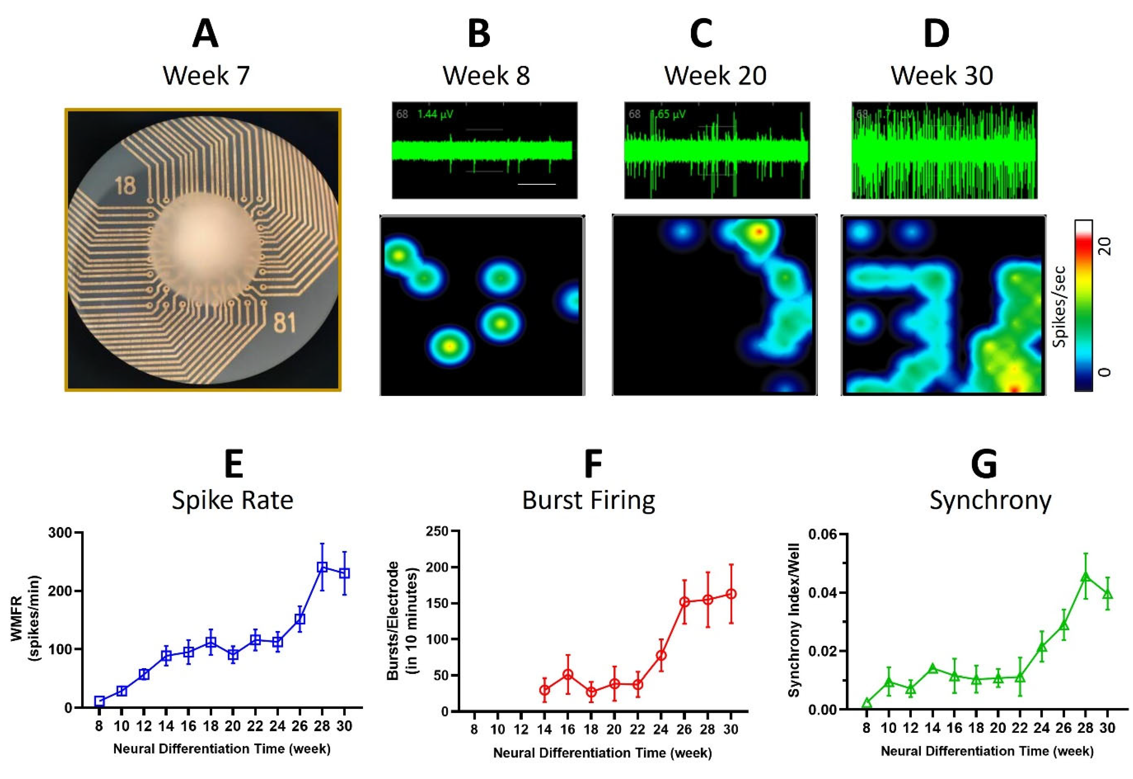
3.2. Multi-Electrode Array Recordings

Spontaneous neural activity was successfully recorded from organoids on the MEA plates from week 7 to week 30 of neural differentiation, with the neuropharmacology experiments conducted between 15 and 22 weeks in vitro. The weighted mean firing rate (WMFR) from all active electrodes at week 7 was 14 ± 3 spikes/min/electrode (n ≥ 3) and significantly increased over 30 weeks to a peak of 230 ± 33 spikes/min/electrode at weeks 28–30 (see Figure 2). In addition to increases in spontaneous neural activity, the number of active electrodes in each well increased from an average of 3 ± 1 electrodes (n ≥ 3) at week 7 to a peak of 17 ± 1 at week 15, where it remained stable. Single-cell burst firing was detected from week 13 with 18 ± 7 bursts over the 10 min recording (n ≥ 3), and these bursts significantly increased to 163 ± 40 by week 30 of neural maturation. Notably there was a 3-fold increase in single-cell burst firing between weeks 23 and 30 (see Figure 2). Additionally, the synchrony index, an indicator of neural network formation, was 0.002 per well at week 8 of differentiation (indicating low synaptic connectivity) but increased by over 20-fold to 0.046 at week 30 (Figure 2). Together, these data illustrate the development and functional maturation of stem cell-derived neurons within the organoids to form complex neural circuits.

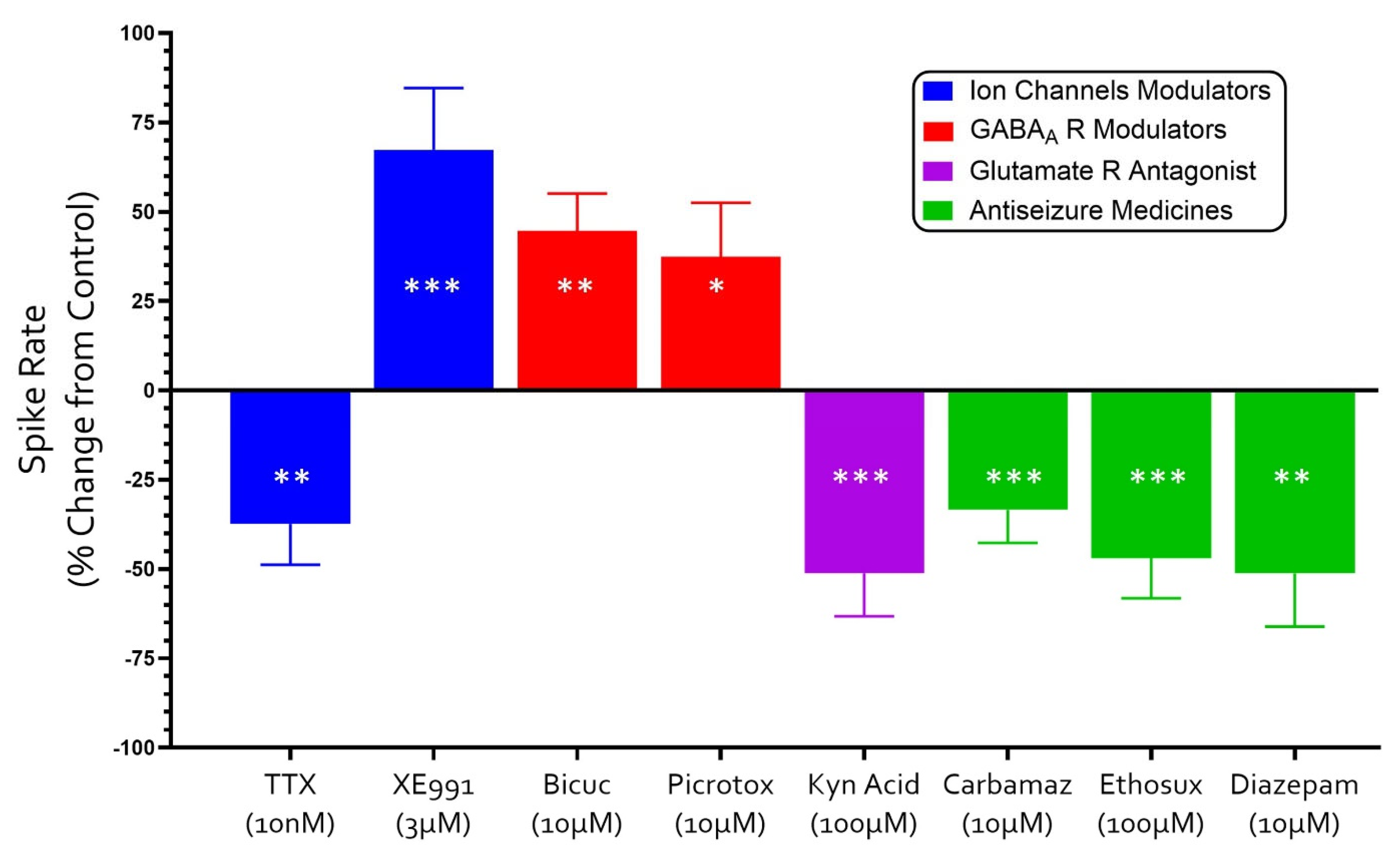
3.3. Neuropharmacology of Electrochemical Signaling in Neural Organoids

Neuronal spiking, burst firing and synaptic signaling across neural networks depend upon a complex and diverse array of ion channels and receptors in the brain [20]. To begin to determine the identity of some of the ion channels and receptors mediating signaling, we determined the impact of the sodium channel inhibitor TTX; the selective potassium (Kv7) ion channel blocker XE991, the GABAA antagonists picrotoxin and bicuculline and the broad-spectrum glutamate receptor antagonist kynurenic acid on spontaneous action potentials in our human stem cell-derived neural organoids, as described in the Methods Section.
The spontaneous spike rate was reduced by 37 ± 11% (n ≥ 3) in the presence of TTX (10 nM) but increased by 67 ± 17% (n ≥ 3) by XE991 (3 μM; see Figure 3). These two observations confirm that our MEA-recorded spiking was dependent on voltage-gated sodium (Na) and potassium (K) ion channels in the 3D neuroglia cultures and consistent with neuronal action potentials. Similarly, the competitive GABAA receptor antagonist bicuculline (10 µM) and the GABAA channel inhibitor picrotoxin (10 µM) each increased the spontaneous spike rate by 45 ± 10% (n ≥ 3) and 37 ± 15%, (n ≥ 3), respectively. In contrast, exposure of the neural organoids to the broad-spectrum glutamate receptor antagonist kynurenic acid (100 µM) reduced the spike rate by 51 ± 12% (n ≥ 3; see Figure 3). These data therefore indicate that synaptic communication in our neural organoids involves inhibitory and excitatory neurons that are mediated by GABAA and ionotropic glutamate receptors and are consistent with previous studies showing that these stem cell-derived neurons, which are grown in 2D cultures, express a broad range of neurotransmitter receptors and voltage-gated ion channels [7,8,18,19,21].
We then investigated the impact of the antiseizures agents carbamazepine, diazepam and ethosuximide on the rate of spontaneous action potentials generated in our neural organoids. These medicines were chosen because they have distinct and well-established mechanisms of action, with carbamazepine acting as a Na channel blocker, diazepam acting as a positive allosteric potentiator of GABAA receptors, and ethosuximide acting as a T-type Ca channel inhibitor [3,4]. Each of the three antiseizure agents reduced spontaneous firing as follows: carbamazepine (10 μM) by 33 ± 9% (n ≥ 3), ethosuximide (100 µM) by 47 ± 11% (n ≥ 3) and diazepam (10 µM) by 51 ± 14% (n ≥ 3; see Figure 3). Together, these observations show that the synaptic activity across the circuits of the neural organoids involves GABAA receptors, Na channels and T-type calcium channels.

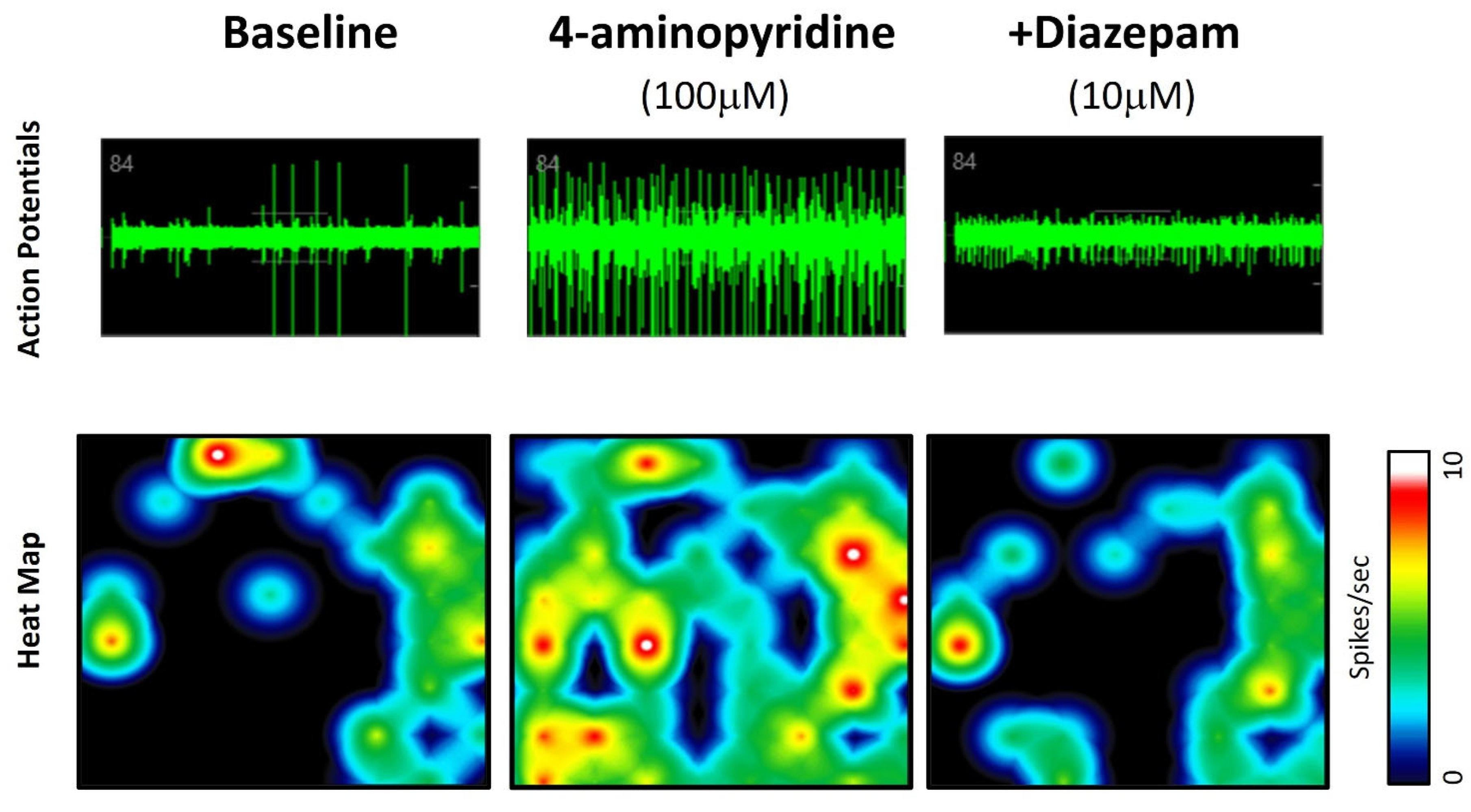
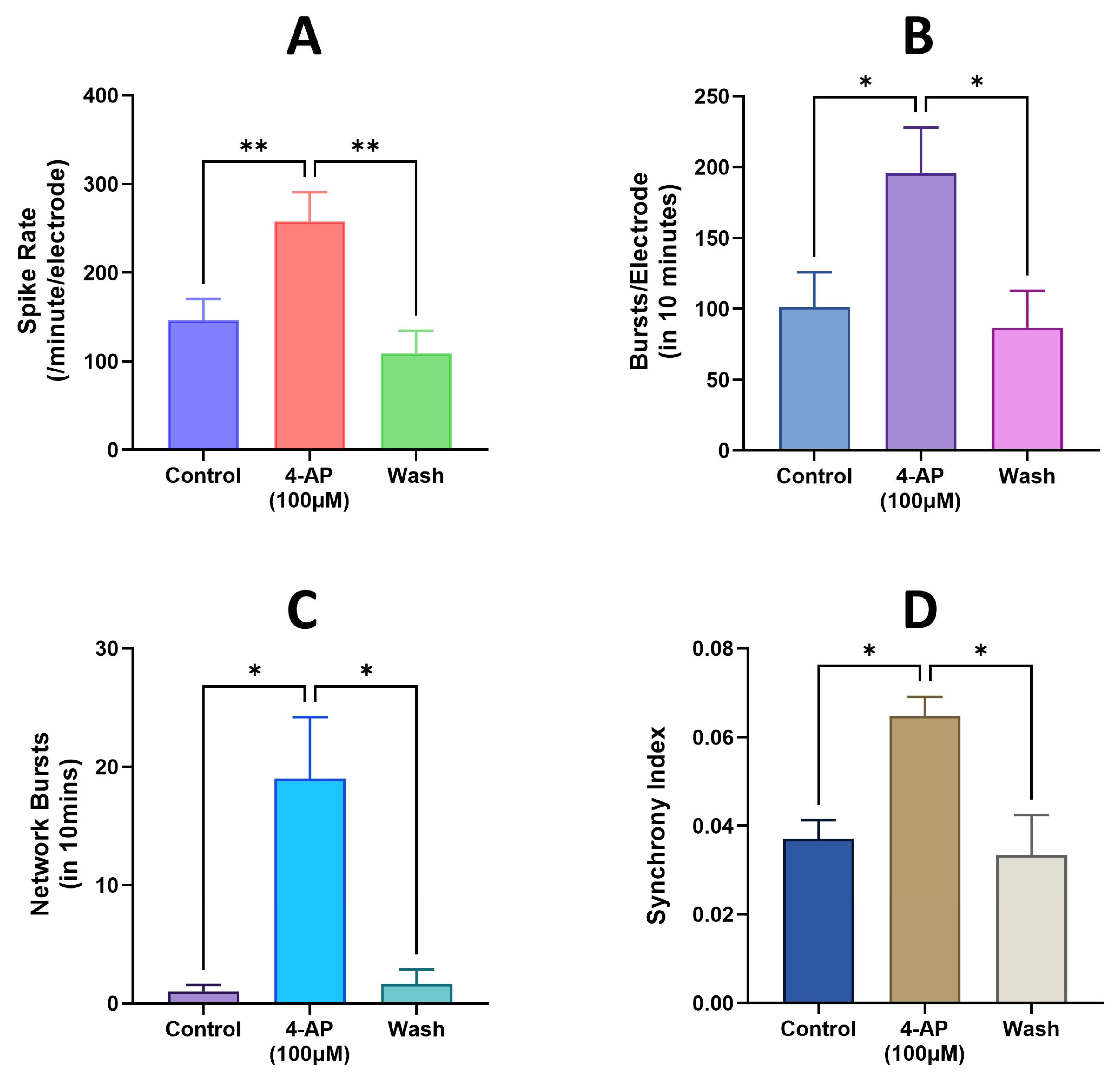
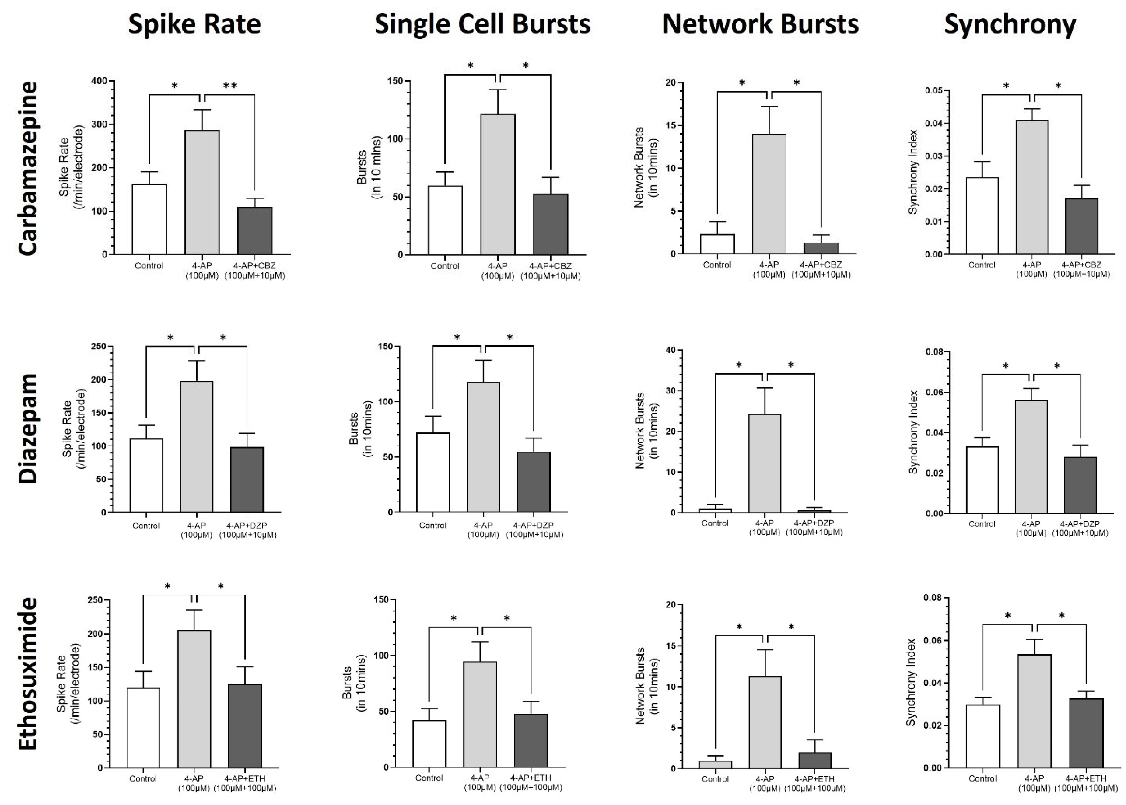
3.4. Modeling Epileptiform Activity in Neural Organoids and the Impact of Anticonvulsants

4-AP induces epileptiform activity both in vivo and in vitro [22,23]. To determine if we could model epileptiform activity in the neural organoids, we therefore tested the effects of 4-AP (100 µM) and found that it rapidly, consistently and, upon washout, reversibly increased the mean spike firing rate, single-cell bursting, network bursts and synchronized firing, consistent with seizure-like activity (Figure 4 and Figure 5). These data therefore demonstrate that the neural organoids can display abnormal epileptiform-like activity in response to a well-established convulsant.
Finally, we determined the impact of carbamazepine, diazepam and ethosuximide on 4-AP (100 µM)-evoked epileptiform activity in the neural organoids. Diazepam (10 μM) and carbamazepine (10 µM) each reduced the mean spike rate, single-cell bursts and synchrony index back to baseline levels and eliminated 4-AP (100 µM)-induced network bursts (see Figure 5). In addition, ethosuximide (100 µM) also fully reversed the 4-AP (100 µM)-induced epileptiform spiking, single-cell bursts, network bursts and synchrony back to baseline levels (Figure 6). Together, these data show that these human stem cell-derived neural organoids express the molecular targets for a range of antiseizure agents and are sensitive to their antiseizure actions.

4. Discussion

Self-organizing brain organoids derived from human pluripotent stem cells are generating great excitement because they provide a powerful 3D approach to study the developing nervous system, model neurological disorders in a dish and for personalized medicine [12,24,25,26]. Recent studies have exploited neural organoids to investigate structural and functional changes associated with epilepsy [11,13,27], and they could enhance the discovery of new medicines for epilepsy, although this approach is still in its infancy [28,29]. In this study, we therefore addressed the value of human stem cell-derived neural organoids to model seizure-like events in vitro and investigated their ability to detect, reliably, the antiseizure effects of three well-established anticonvulsants with distinct mechanisms of action.
Neural organoids derived from our human pluripotent embryonal stem cell line developed into 3D structures that are around 3 mm in size, with lobe-like features appearing bfrom week 14. The limited size of these organoids likely resulted from the lack of a circulatory system and limited availability for nutrient and oxygen exchange in the core, and these observations are consistent with those described by other groups [26,30,31]. Our immunocytochemistry results showed that the organoids were composed of βIII-tubulin+ and GFAP+ cells, consistent with the presence of neurons and glia in a complex matrix of cells and in line previous reports of the composition of cerebral organoids [32,33,34].
Over 8 months, we recorded the development of increased levels and complexity of electrophysiological activity in the organoids, represented by marked rises in spike rates, single-cell bursts, network bursts and synchronized firing patterns, which peaked around 7 months. In 2D culture, these neuron–glia circuits show peak electrophysiological activity around 9–10 months [7], suggesting that network formation may occur earlier in 3D. However, the planar multi-electrode array’s electrodes detect action potentials from neurons on the surface of the organoid and not in the core, suggesting that some neural activity and synchrony are missed. New mesh MEA wells might enable the recording of activity from within the organoids, though a recent study that utilized fine (Kirigami-inspired) flexible electrodes to record from within the core of human cortical organoids over 179 days did not see spontaneous activity until day 96, and the technology ‘required advanced expertise’ [35]. Nevertheless, our long-term recordings show remarkable maturation of electrophysiological activity in 3D culture around 6–7 months in vitro, a timeline consistent with cerebral organoids derived from human induced pluripotent stem cells (IPSCs), suggesting a genetically determined program of functional neural development in vitro similar to that described in vivo [34,36].
This study also conducted an extensive investigation of the functional ion channels and receptors mediating the spontaneous electrophysiological activity generated in the neural organoids. Our data shows that spiking was critically dependent on voltage-gated sodium channels and, for the first time, potassium channels, confirming that these signals are neuronal action potentials. We also show that the selective GABAA receptor antagonists bicuculline and picrotoxin rapidly and significantly increased the spike rate, burst firing and synchronized activity, and conversely, the broad-spectrum glutamate receptor antagonist kynurenic acid reduced this complex electrophysiological activity in the neural organoids. These data are therefore consistent with the formation of functional inhibitory and excitatory synaptic circuits. In line with our data, others have reported that TTX and the AMPA receptor antagonist CNQX, combined with the NMDA antagonist AP-5, reduced the activity in cerebral organoids derived from human IPSCs [33,36]. Our study also demonstrates that the anticonvulsant agents carbamazepine, the sodium channel blocker diazepam, an allosteric potentiator of GABAA receptors and ethosuximide, a T-type calcium channel blocker, modulate spontaneous synaptic activity in the organoids, showing that neurons express a diverse range of native ion channels and receptor sites and indicating that they may be a valuable resource in first-order screening of novel and new antiseizure agents.
The current study demonstrates that the convulsant 4-aminopyridine [22] induced significant increases in spike frequency, cell and network bursts and synchronized firing across synaptic networks in our neural organoids, paralleling epileptiform EEG patterns seen in human epilepsies [37]. Importantly, three antiseizure agents with distinctly different mechanisms and sites of action rapidly eliminated epileptiform activity in the organoids. To our knowledge, the sensitivity of a human 3D neural organoid to both model epileptiform activity and detect such a diversity of antiepileptics has not been reported previously. Consistent with our findings, Yokoi and colleagues [38] reported that human IPSC-derived cerebral organoids displayed ‘seizure-like waveforms’ in response to the convulsant pentylenetetrazol, and the antiepileptic agents phenytoin and perampanel reduced this abnormal electrophysiological activity. Together these studies support the power of 3D neural organoids to identify diverse anticonvulsant agents.

5. Conclusions

In conclusion, this study has demonstrated that human embryonal stem cells form 3D neural organoids comprising neurons and glial cells, mature into functionally complex inhibitory and excitatory circuits over four to six months and can be maintained in vitro for at least eight months. Neural organoids express a rich and diverse range of ion channels and receptors mediating spontaneous action potentials and inhibitory and excitatory synaptic signaling. Spontaneous synaptic activity can be modulated by antiseizure agents with diverse mechanisms of action, and epileptiform activity can be induced by proconvulsant drugs in these 3D neural networks and eliminated by diverse antiseizure agents. Additional work, in the future, will further characterize the neuronal subtypes in these functionally mature neural circuits. Overall, our data provide significant support for the value of 3D neural organoids in epilepsy disease modeling and for the investigation of novel antiepileptics in preclinical studies.

Author Contributions

Conceptualization, R.F.H.; data curation, H.S.; formal analysis, R.F.H. and H.S.; funding acquisition, R.F.H.; investigation, H.S. and R.F.H.; methodology, H.S. and R.F.H.; project administration, R.F.H.; writing—original draft, R.F.H.; writing—review and editing, R.F.H. and H.S. All authors have read and agreed to the published version of the manuscript.

Funding

This research received no external funding.

Institutional Review Board Statement

Not applicable.

Informed Consent Statement

Not applicable.

Data Availability Statement

The raw data supporting the conclusions of this article will be made available by the authors upon request.

Conflicts of Interest

The authors declare no conflicts of interest.

References

  1. Zhao, X.; Bhattacharyya, A. Human Models Are Needed for Studying Human Neurodevelopmental Disorders. Am. J. Hum. Genet. 2018, 103, 829–857. [Google Scholar] [CrossRef] [PubMed]
  2. Gunasekera, C.L.; Sirven, J.I.; Feyissa, A.M. The evolution of antiseizure medication therapy selection in adults: Is artificial intelligence-assisted antiseizure medication selection ready for prime time? J. Cent. Nerv. Syst. Dis. 2023, 15, 11795735231209209. [Google Scholar] [CrossRef] [PubMed]
  3. Hakami, T. Neuropharmacology of antiseizure drugs. Neuropsychopharmacol. Rep. 2021, 41, 336–351. [Google Scholar] [CrossRef] [PubMed]
  4. Sills, G.J.; Rogawski, M.A. Mechanisms of action of currently used antiseizure drugs. Neuropharmacology 2020, 168, 107966. [Google Scholar] [CrossRef]
  5. Kwan, P.; Brodie, M.J. Early identification of refractory epilepsy. N. Engl. J. Med. 2000, 342, 314–319. [Google Scholar] [CrossRef]
  6. Lybrand, Z.R.; Goswami, S.; Hsieh, J. Stem cells: A path towards improved epilepsy therapies. Neuropharmacology 2020, 168, 107781. [Google Scholar] [CrossRef]
  7. Salmanzadeh, H.; Poojari, A.; Rabiee, A.; Zeitlin, B.D.; Halliwell, R.F. Neuropharmacology of human TERA2.cl.SP12 stem cell-derived neurons in ultra-long-term culture for antiseizure drug discovery. Front. Neurosci. 2023, 17, 1182720. [Google Scholar] [CrossRef]
  8. Salmanzadeh, H.; Halliwell, R.F. Antiseizure properties of fenamate NSAIDs determined in mature human stem-cell derived neuroglial circuits. Front. Pharmacol. 2024, 15, 1385523. [Google Scholar] [CrossRef]
  9. Lancaster, M.A.; Renner, M.; Martin, C.A.; Wenzel, D.; Bicknell, L.S.; Hurles, M.E.; Homfray, T.; Penninger, J.M.; Jackson, A.P.; Knoblich, J.A. Cerebral organoids model human brain development and microcephaly. Nature 2013, 501, 373–379. [Google Scholar] [CrossRef]
  10. Birtele, M.; Lancaster, M.; Quadrato, G. Modelling human brain development and disease with organoids. Nat. Rev. Mol. Cell Biol. 2025, 26, 389–412. [Google Scholar] [CrossRef]
  11. Nieto-Estévez, V.; Hsieh, J. Human Brain Organoid Models of Developmental Epilepsies. Epilepsy Curr. 2020, 20, 282–290. [Google Scholar] [CrossRef] [PubMed]
  12. Smirnova, L.; Hartung, T. The Promise and Potential of Brain Organoids. Adv. Healthc. Mater. 2024, 13, e2302745. [Google Scholar] [CrossRef]
  13. Danačíková, Š.; Straka, B.; Daněk, J.; Kořínek, V.; Otáhal, J. In vitro human cell culture models in a bench-to-bedside approach to epilepsy. Epilepsia Open 2024, 9, 865–890. [Google Scholar] [CrossRef]
  14. Tukker, A.M.; Westerink, R.H.S. Novel test strategies for in vitro seizure liability assessment. Expert. Opin. Drug Metab. Toxicol. 2021, 17, 923–936. [Google Scholar] [CrossRef] [PubMed]
  15. Lancaster, M.; Knoblich, J. Generation of cerebral organoids from human pluripotent stem cells. Nat. Protoc. 2014, 9, 2329–2340. [Google Scholar] [CrossRef] [PubMed]
  16. Przyborski, S.A. Isolation of human embryonal carcinoma stem cells by immunomagnetic sorting. Stem Cells 2001, 19, 500–504. [Google Scholar] [CrossRef]
  17. Andrews, P.W. Human pluripotent stem cells: Tools for regenerative medicine. Biomater. Transl. 2021, 2, 294–300. [Google Scholar]
  18. Stewart, R.; Coyne, L.; Lako, M.; Halliwell, R.F.; Przyborski, S.A. Human embryonal carcinoma stem cells expressing green fluorescent protein form functioning neurons in vitro: A research tool for co-culture studies. Stem Cells Dev. 2004, 13, 646–657. [Google Scholar] [CrossRef]
  19. Coyne, L.; Shan, M.; Przyborski, S.A.; Hirakawa, R.; Halliwell, R.F. Neuropharmacological properties of neurons derived from human stem cells. Neurochem. Int. 2011, 59, 404–412. [Google Scholar] [CrossRef]
  20. Kandel, E.R.; Koester, J.D.; Mack, S.H.; Siegelbaum, S.A. (Eds.) Principles of Neural Science, 6th ed.; McGraw Hill: New York, NY, USA, 2021. [Google Scholar]
  21. Halliwell, R.F. Electrophysiological properties of neurons derived from human stem cells and iNeurons in vitro. Neurochem. Int. 2017, 106, 37–47. [Google Scholar] [CrossRef]
  22. Ventura-Mejía, C.; Nuñez-Ibarra, B.H.; Medina-Ceja, L. An update of 4-aminopyride as a useful model of generalized seizures for testing antiseizure drugs: In vitro and in vivo studies. Acta Neurobiol. Exp. 2023, 83, 63–70. [Google Scholar] [CrossRef]
  23. Heuzeroth, H.; Wawra, M.; Fidzinski, P.; Dag, R.; Holtkamp, M. The 4-Aminopyridine Model of Acute Seizures in vitro Elucidates Efficacy of New Antiepileptic Drugs. Front. Neurosci. 2019, 13, 677. [Google Scholar] [CrossRef] [PubMed]
  24. Qian, X.; Song, H.; Ming, G.L. Brain organoids: Advances, applications and challenges. Development 2019, 146, dev166074. [Google Scholar] [CrossRef] [PubMed]
  25. Hogberg, H.T.; Smirnova, L. The Future of 3D Brain Cultures in Developmental Neurotoxicity Testing. Front. Toxicol. 2022, 4, 808620. [Google Scholar] [CrossRef] [PubMed]
  26. Shafique, S. Stem cell-based region-specific brain organoids: Novel models to understand neurodevelopmental defects. Birth Defects Res. 2022, 114, 1003–1013. [Google Scholar] [CrossRef]
  27. Samarasinghe, R.A.; Miranda, O.A.; Buth, J.E.; Mitchell, S.; Ferando, I.; Watanabe, M.; Allison, T.F.; Kurdian, A.; Fotion, N.N.; Gandal, M.J.; et al. Identification of neural oscillations and epileptiform changes in human brain organoids. Nat. Neurosci. 2021, 24, 1488–1500. [Google Scholar] [CrossRef]
  28. Carvill, G.L.; Dulla, C.G.; Lowenstein, D.H.; Brooks-Kayal, A.R. The path from scientific discovery to cures for epilepsy. Neuropharmacology 2020, 167, 107702. [Google Scholar] [CrossRef] [PubMed]
  29. Brown, R.; Rabeling, A.; Goolam, M. Progress and potential of brain organoids in epilepsy research. Stem Cell Res. Ther. 2024, 15, 361. [Google Scholar] [CrossRef]
  30. Kelava, I.; Lancaster, M.A. Stem Cell Models of Human Brain Development. Cell Stem Cell. 2016, 18, 736–748. [Google Scholar] [CrossRef]
  31. Kim, S.H.; Chang, M.Y. Application of Human Brain Organoids-Opportunities and Challenges in Modeling Human Brain Development and Neurodevelopmental Diseases. Int. J. Mol. Sci. 2023, 24, 12528. [Google Scholar] [CrossRef]
  32. Yakoub, A.M.; Sadek, M. Development and Characterization of Human Cerebral Organoids: An Optimized Protocol. Cell Transplant. 2018, 27, 393–406. [Google Scholar] [CrossRef] [PubMed]
  33. Yakoub, A.M. Cerebral organoids exhibit mature neurons and astrocytes and recapitulate electrophysiological activity of the human brain. Neural Regen. Res. 2019, 14, 757–761. [Google Scholar] [CrossRef]
  34. Fair, S.R.; Julian, D.; Hartlaub, A.M.; Pusuluri, S.T.; Malik, G.; Summerfied, T.L.; Zhao, G.; Hester, A.B.; Ackerman, W.E., 4th; Hollingsworth, E.W.; et al. Electrophysiological Maturation of Cerebral Organoids Correlates with Dynamic Morphological and Cellular Development. Stem Cell Rep. 2020, 15, 855–868. [Google Scholar] [CrossRef] [PubMed]
  35. Yang, X.; Forró, C.; Li, T.L.; Miura, Y.; Zaluska, T.J.; Tsai, C.T.; Kanton, S.; McQueen, J.P.; Chen, X.; Mollo, V.; et al. Kirigami electronics for long-term electrophysiological recording of human neural organoids and assembloids. Nat. Biotechnol. 2024, 42, 1836–1843. [Google Scholar] [CrossRef]
  36. Trujillo, C.A.; Gao, R.; Negraes, P.D.; Gu, J.; Buchanan, J.; Preissl, S.; Wang, A.; Wu, W.; Haddad, G.G.; Chaim, I.A.; et al. Complex Oscillatory Waves Emerging from Cortical Organoids Model Early Human Brain Network Development. Cell Stem Cell 2019, 25, 558–569. [Google Scholar] [CrossRef] [PubMed]
  37. Lévesque, M.; Gnatkovsky, V.; Li, F.R.; Scalmani, P.; Uva, L.; Avoli, M.; de Curtis, M. Fast activity chirp patterns in focal seizures from patients and animal models. Epilepsia 2025, 66, 621–631. [Google Scholar] [CrossRef]
  38. Yokoi, R.; Shibata, M.; Odawara, A.; Ishibashi, Y.; Nagafuku, N.; Matsuda, N.; Suzuki, I. Analysis of signal components < 500 Hz in brain organoids coupled to microelectrode arrays: A reliable testbed for preclinical seizure liability assessment of drugs and screening of antiepileptic drugs. Biochem. Biophys. Rep. 2021, 28, 101148. [Google Scholar]
Figure 1. Differentiation and Growth of Neural Organoids. The top is a schematic of the timeline for the formation and maturation of neural organoids and the start of MEA recordings at day 40 in vitro. Middle panels (AE) are representative bright field images of embryoid bodies and organoid morphology at days 1, 5, 7,10 and 160, respectively. The calibration bar = 300 µm in all except (E), which = 50 μm. The lower panel is an immunofluorescence image of a fixed organoid stained with βIII-tubulin (green), GFAP (red) and DAPI (blue) at 130 days of neural maturation.
Receptors 04 00012 g001
Figure 2. Electrophysiological maturation of neural organoids in 3D: Panel (A) shows a neural organoid on an MEA at 7 weeks of differentiation; some of the electrode arrays can be seen at the edge of the organoid. Panels (BD) show extracellular recordings of action potentials (from electrode #68/384) at weeks 8, 20 and 30 with a snapshot beneath each trace of a heatmap of active electrodes in a single well. Note that more electrodes detect spiking, and cells also fire at higher frequencies as the organoid matures. Panels (EG) show plots of the mean (±s.e.m) of the spike rate (E), single-cell bursts (F) and synchronized firing (G), respectively, from weeks 7 to 30 of neural differentiation and maturation.
Receptors 04 00012 g002
Figure 3. Neuropharmacology of electrophysiological activity in organoids. The histogram summarizes the impact of drugs that act at specific synaptic receptors and voltage-gated ion channels on spike rates in the neural organoids. The sodium channel blocker TTX (10 nM) reduced the action potential rate, whereas the selective potassium (Kv7) channel blocker XE991 (3 μM) and the GABAA receptor antagonists bicuculline (10 μM) and picrotoxin (10 μM) increased the spike frequency. The glutamate receptor antagonist kynurenic acid (100 μM) and the three antiseizure agents carbamazepine (10 μM), ethosuximide (10 μM) and diazepam (10 μM) each reduced spontaneous spike activity. The effects of all drugs were reversible upon washout. All drugs significantly changed the spike rate; the level of significance (calculated from one sample t-tests) is indicated by the number of asterisks on each bar with the key as follows: * = p ≤ 0.05, ** = p ≤ 0.01 and *** = p ≤ 0.001.
Receptors 04 00012 g003
Figure 4. The convulsant 4-aminopyridine (4-AP) evokes epileptiform activity in neural organoids. The top half of the figure shows MEA recordings of action potentials (from electrode #84/384) at baseline (left), in the presence of 4-AP (100 μM, middle) and 4-AP (100 μM) + diazepam (10 μM, right); the lower half shows a snapshot of the heatmap of active electrodes in a single well in each of these conditions. As can be seen, 4-AP evokes increases in the number and rate of action potentials, as well as increases in burst firing and synchronized neural activity. The allosteric GABAA receptor potentiator diazepam (10 μM) eliminates the epileptiform activity in this neural organoid.
Receptors 04 00012 g004
Figure 5. 4-AP increases the number and rate of action potentials, high-frequency cell and network bursts and synchronized firings across neural networks in organoids. (AD) are histograms summarizing the WMFR from all active electrodes (spike rate), cell bursts, network bursts and synchronized firings, respectively, in the absence (control), presence and following washout of 4-aminopyridine (100 μM). All electrophysiological parameters were significantly increased by 4-AP, consistent with epileptiform behavior. The asterisks indicate the level of significance from the control and washout in the presence of 4-AP with * = p ≤ 0.05 and ** = p ≤ 0.01. These data are consistent with the epileptiform activity in the neural organoid.
Receptors 04 00012 g005
Figure 6. Epileptiform activity evoked by 4-AP is eliminated by 3 different antiseizure medicines. The histograms summarize the impact of carbamazepine (10 μM), diazepam (10 μM) and ethosuximide (100 μM) on changes in the WMFR from all active electrodes (spike rate), single-cell bursts, network bursts and synchronized firings evoked by 4-AP (100 μM). 4-AP significantly increased all 4 electrophysiological parameters, and these were reduced (significantly) by each of the antiseizure medicines in the neural organoids. Asterisks (*) indicate a significant difference, with * = p ≤ 0.05 and ** = p ≤ 0.01.
Receptors 04 00012 g006
Disclaimer/Publisher’s Note: The statements, opinions and data contained in all publications are solely those of the individual author(s) and contributor(s) and not of MDPI and/or the editor(s). MDPI and/or the editor(s) disclaim responsibility for any injury to people or property resulting from any ideas, methods, instructions or products referred to in the content.

