Review

Unraveling the Role of CHRNA6, the Neuronal α6 Nicotinic Acetylcholine Receptor Subunit

by
Yasamin Hajy Heydary
1,
Emily M. Castro
1,
Shahrdad Lotfipour
1,2,3 and
Frances M. Leslie
1,*
1
Department of Pharmaceutical Sciences, School of Pharmacy and Pharmaceutical Sciences, University of California, Irvine, CA 92697, USA
2
Department of Emergency Medicine, School of Medicine, University of California, Irvine, CA 92617, USA
3
Department of Pathology and Laboratory Medicine, School of Medicine, University of California, Irvine, CA 92617, USA
*
Author to whom correspondence should be addressed.
Receptors 2025, 4(1), 1; https://doi.org/10.3390/receptors4010001
Submission received: 1 October 2024 / Revised: 22 November 2024 / Accepted: 3 January 2025 / Published: 14 January 2025
(This article belongs to the Special Issue Nicotinic Receptors: From Molecule to Benchside)

Abstract

:
The increased prevalence of electronic cigarettes, particularly among adolescents, has escalated concerns about nicotine addiction. Nicotine, a potent psychostimulant found in tobacco products, exerts its effects by interacting with nicotinic acetylcholine receptors (nAChRs) in the brain. Recent findings in both pre-clinical and clinical studies have enhanced our understanding of nAChRs, overcoming the limitations of pharmacological tools that previously hindered their investigation. Of particular interest is the α6 subunit, whose expression peaks during adolescence, a critical period of brain development often marked by the initiation of substance use. Pre-clinical studies have linked α6-containing nAChRs (α6*nAChRs) to nicotine-induced locomotion, dopamine release, and self-administration behavior. Furthermore, clinical studies suggest an association between the α6 subunit and increased smoking behavior in humans. Specifically, a single nucleotide polymorphism in the 3′ untranslated region of the CHRNA6 gene that encodes for this subunit is linked to smoking behavior and other substance use. A comprehensive understanding of this subunit’s role in addiction is of high importance. This review aims to consolidate current knowledge regarding the α6 subunit’s functions and implications in addiction and other disorders, with the hope of paving the way for future research and the development of targeted therapies to address this pressing public health concern.

Graphical Abstract

1. Introduction

Nicotine use remains one of the most preventable threats to public health and leading causes of premature death in the United States [1]. Nicotine is a potent, addictive psychostimulant found in tobacco products. An estimated 46 million adults in the United States reported daily use of any tobacco product during 2021, including electronic cigarettes (e-cigarettes) [2]. Concerningly, an estimated 6.21 million middle and high school students reported trying any kind of tobacco product in the United States in 2022 [3]. Early use greatly increases the likelihood of developing dependence or escalation to more harmful substances [4,5,6]. Smoking and smoking-related diseases adversely impact individual healthcare and impose substantial economic burdens [7]. Such prevalence in tobacco use and cessation challenges make it necessary to gain a comprehensive understanding of nicotine’s impact on the brain and consequent behaviors.
Nicotinic acetylcholine receptors (nAChRs) serve as key regulators within the brain and their role in nicotine dependence has been well established over decades [8,9,10,11,12]. The expression patterns and binding properties of nAChR subunits vary across brain regions but follow distinct developmental events in the cholinergic system [9,13,14]. The subunit configuration of these receptors regulates pharmacological properties such as agonist affinity/efficacy, desensitization, and downstream signaling, and is heavily implicated in presynaptic neurotransmitter release [10,15]. The α6 nAChR subunit, highly expressed in dopamine neurons within the ventral tegmental area (VTA), has been found in preclinical studies to be crucial for nicotine-induced locomotion, dopamine release, and nicotine self-administration [16,17,18,19]. Genome-wide association studies have also associated the α6 subunit gene, CHRNA6, with nicotine and other substance use [20,21,22,23,24]. The goal of this review is to evaluate advances in the study of the α6 subunit, its role in substance use disorders and other diseases, evaluate genetic influences on behavior, and discuss potential implications for the development of future therapeutic treatments.

2. Structure and Distribution of α6-Containing Neuronal nAChRs

2.1. Structure of Neuronal nAChRs

Neuronal nAChRs are ligand-gated ion channels found in the central and peripheral nervous systems [25]. The endogenous neurotransmitter acetylcholine (ACh) and exogenous ligands such as nicotine bind to these pentameric receptors, which are composed of five subunits [26]. In vertebrates, nAChRs exhibit diverse subunit compositions, with nine α (α2–α10) and three β subunits (β2–β4) identified [25,27,28]. Each of these subunits is made up of a long extracellular N-terminus, which is involved in ligand binding; four transmembrane domains (M1–M4); an intracellular cytoplasmic loop; and a short extracellular c-terminus [26,29] (Figure 1A). The second transmembrane domain (M2) of each subunit lines the permeable pore of the receptor, and the M1 and M3 transmembrane domains shield the M2 domain from the cell membrane. The M4 transmembrane domain, on the other hand, has the most contact with the cell membrane [11].
nAChR subunits can assemble into either heteromeric receptors containing at least one α and one β subunit (Figure 1C) or homomeric receptors in which all subunits are identical (Figure 1B). Homomeric nAChRs have five ligand-binding sites, one between each pair of identical α subunits (Figure 1B). In contrast, most heteromeric nAChRs have two binding sites located between adjacent α and β subunits (Figure 1C), with the exception of α9α10 receptors, which have binding sites between two α subunits, similar to homomeric receptors [30,31]. The remaining subunit in heteromeric receptors is an accessory receptor that is not involved in ligand binding but is involved in activation and deactivation of the receptor [25,32,33]. The specific arrangement of subunits within each receptor is highly regulated and dictates its unique pharmacology and function [34]. The diverse homomeric and heteromeric nAChR subunit compositions contribute to a variety of functions in the central and peripheral nervous systems. In this review, however, we will focus on the understudied α6 subunit.

2.2. Distribution of the α6 Subunit

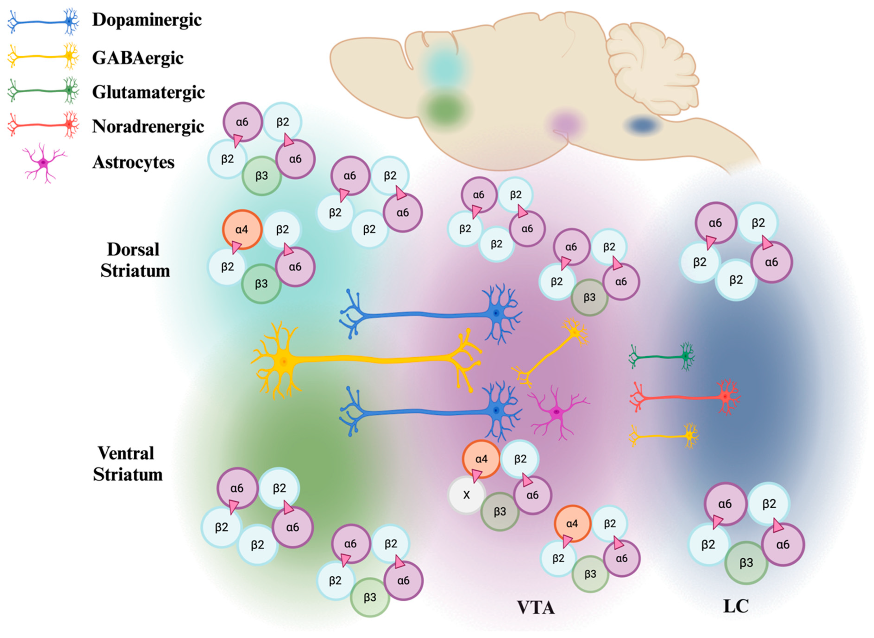
While nAChRs are widely distributed throughout the brain, exhibiting distinct receptor compositions in different regions, α6*nAChRs stand out due to their limited expression compared to some of the most abundant nAChRs. A variety of α6*nAChRs subunit combinations have been reported across many studies. These have shown the α6 subunit mRNA to be predominantly expressed in the locus coeruleus (LC), VTA, and substantia nigra (SN), with additional, lower expression in the reticular thalamic nucleus, supramammillary nucleus, inferior portion of ventral medial habenula (MHb), mesencephalic V nucleus, rostral subnucleus, and caudal subnucleus, as well as retinal ganglion cells, visual thalamus, superior colliculus, and the lateral geniculate nucleus of rodents [35,36,37,38,39,40,41,42]. nAChR composition studies in the dorsal nigrostriatal and ventral mesolimbic dopamine pathways have shown that about 90% of dorsal striatum α6*nAChRs co-express α4, suggesting the presence of both α6β2 and α4β2 binding interfaces in this region [18]. In the ventral striatum, about 60% of α6*nAChRs are composed of two α6β2 binding interfaces [18]. In addition to regional expression patterns, understanding the combinatorial patterns of the α6*nAChRs within reward-related regions is crucial for investigating the functional and pharmacological differences that influence substance use and other related conditions.
Recent work using fluorescent in situ hybridization has shed new light on the distribution of nAChR subunits in different cell types (dopaminergic, GABAergic, glutamatergic, and noradrenergic) of the VTA and LC of adult and adolescent C57BL/6J mice [43]. Previous findings demonstrating α6 transcript expression with α4, β2, and β3 transcripts in roughly 90% of VTA dopaminergic neurons have been confirmed [8,9,36]. It has been further proposed that VTA nAChRs are composed of two primary subtypes: α4α5α6β2β3 and α4α6β2β3, with each accounting for approximately half of the total nAChR population [43]. Notably, α6 subunit expression was previously believed to be restricted to VTA dopaminergic neurons [44]. However, recent work challenges that assumption by identifying α6 transcripts in roughly half of both GABAergic and glutamatergic neurons within the VTA [43,45,46], highlighting a broader distribution than previously recognized. While the α6 subunit had been previously identified in the LC of adolescent rats [35], the specific cell types expressing it were not determined. New findings now reveal α6 expression in GABAergic, glutamatergic, and noradrenergic neurons within the LC [43]. Furthermore, noradrenergic neurons may express α6(non-α4)β2β3 nAChRs, whereas GABAergic and glutamatergic neurons potentially express both α4α6β2β3 and α6β2 [43,47].
While neuronal nAChRs have been extensively researched, their expression in non-neuronal cells remains understudied and is valuable to gain a holistic overview of potential mechanisms underlying drug reward. Growing evidence supports the involvement of astrocytes in substance use disorder, specifically in drug seeking, extinction, and relapse [48,49,50,51,52]. Mouse astrocytes within the VTA express α4, α6, β2, and β3 transcripts, suggesting the potential for several subunit compositions of α6*nAChRs and a potential role for astrocytes in cholinergic system regulation [43]. Additionally, age- and sex-related differences in nAChR expression have been observed with higher relative α6 expression within VTA astrocytes of male adults compared to adolescents. Age and sex differences are critical to evaluate, since adolescence is a time of vulnerability to nicotine use and drug addiction and a significant period for the maturation of dopaminergic neurons [4,6,9]. This also emphasizes the importance of sex effects in studying α6*nAChRs and how the distribution patterns may differ for each sex. Sex differences have been observed in the neuronal excitability of the MHb, with adult males exhibiting heightened excitability compared to females when exposed to nicotine and menthol [53]. In MHb, α6 has been detected with β2 and β3, suggesting the receptor composition of α6β2β3 [40]. It has been suggested that combined exposure to high-dose nicotine and menthol may lead to a reduction in α6β2β3*nAChRs within this nucleus [53]. This shift in receptor population may explain the observed increase in neuronal excitability threshold in male subjects and highlight sex-specific variations in nAChR populations within the MHb, consistent with prior studies that have demonstrated sex differences in nAChR populations [12,54].
Taken together, recent work has advanced our understanding of α6*nAChR expression patterns (Figure 2; Table 1). We now recognize that these receptors are not limited to catecholaminergic neurons but are also expressed in a wider variety of neuronal and non-neuronal cell types, opening new avenues for therapeutic targeting. However, future studies expanding to other brain regions are essential for a comprehensive understanding of these receptor roles, along with more detailed analysis of subunit combinations.

3. Conventional and Novel Strategies in Studying α6

While homomeric α7 and heteromeric receptors containing α4 and β2 subunits have been extensively studied [30], other subtypes containing α6 remain under-researched. This is due to a combination of factors, including limited research tools, low expression levels of α6*nAChRs, and a lack of selective agonists for this receptor type [59]. The high sequence homology between the α6 and α3 subunits (61% amino acid similarity) further complicates research efforts [60]. Additionally, the absence of a specific antibody for the α6 subunit has hindered investigations into its expression patterns [61]. These challenges have resulted in limited understanding regarding the functional role of α6*nAChRs.
α-Conotoxins (α-Cntx), antagonistic peptides derived from cone snail venom, have been instrumental in investigating the role of the α6 subunit in nAChRs [56,62]. Various α-Cntxs can discriminate between different subtypes of nAChRs. For instance, a mutant of α-CntxPeIA, PeIA[S9H,V10A,E14N], offers high specificity, demonstrating a 290-fold preference for α6/α3β2β3 over α6/α3β4 nAChRs [63], highlighting the utility of these analogs in targeting specific nAChR subtypes. Notably, α-CntxMII and its analogs exhibit selectivity for α6-containing nAChRs over α3β2 nAChRs [64], leading to insights into the involvement of α6β2* and α4α6β2* receptors in striatal and NAc dopamine release [57,65,66]. Furthermore, α-contoxin PIA (α-CntxPIA) is the first conotoxin to distinguish between α6* and non-α6* nAChRs [67,68]. While α-Cntx BulA blocks multiple nAChR subtypes, its mutant form, α-Cntx BulA[T5A;P6O], exhibits a significantly higher affinity for the α6β4 subtype compared to the α6β2 subtype [69]. α-Cntxs have been used widely across both in vitro and in vivo studies exploring α6*nAChRs. While the use of α-Cntxs offers substantial mechanistic insight, the interpretation of results from in vivo studies must consider their regional administration, limited stability, and issues with bioavailability and crossing of the blood–brain barrier [70]. The following sections explore other methods of investigating α6*nAChRs and discuss developing novel strategies.

3.1. In Vitro Expression of α6*nAChRs

The potential of drugs targeting α6*nAChRs as therapeutic targets highlights the importance of pharmacological studies in this area. While in vitro systems offer the advantages of rapid, high-throughput experimentation and a smoother transition to in vivo studies, the challenge lies in achieving the functional receptor formation of α6*nAChRs. Nevertheless, the discovery of chaperone proteins has opened new avenues for in vitro investigations, providing valuable insights into this promising therapeutic target. Assembly of nAChRs is a highly regulated process and relies on the correct folding of the subunits. The assembled receptors need to be transported from their site of production, the endoplasmic reticulum, to the cell surface [71]. Some of the challenges in expressing α6*nAChRs in mammalian cells include aggregation of subunits, low receptor binding levels, poor cell surface transport, and, therefore, low cell surface expression [72,73]. As part of efforts to investigate the functional expression of these receptors, it has been established that novel acetylcholine receptor chaperone (NACHO), a transmembrane protein in the endoplasmic reticulum of neurons, is crucial for the assembly of α6β2β3 receptors, as evidenced by reduced binding sites in NACHO knockout mice [74]. However, these receptors remain intracellular even with NACHO, suggesting that additional factors are required for their functional expression [74,75]. Subsequently, four accessory proteins necessary for α6β2β3 nAChR expression were identified. In addition to NACHO, β-anchoring and regulatory protein (BARP), lysosomal-associated membrane protein 5 (LAMP5), and a cytosolic sulfotransferase (SULT2B1) are also involved. LAMP5 and SULT2B1 facilitate receptor expression on the cell surface, while BARP is responsible for channel gating [76]. Furthermore, there are differences in the accessory proteins necessary for the assembly of different α6*nAChRs compositions. Specifically, α6β4 does not necessitate NACHO but instead requires BARP, SULT2B1, and inositol-requiring enzyme-1α (IRE1α), which functions as an unfolded protein response (UPR) sensor and facilitates the assembly of α6β4 receptors through the UPR pathway [77]. The function of BARP in α6β4 is different from that of α6β2β3 receptors. BARP regulates the surface transport of these receptors suggesting distinct mechanisms for the regulatory role of BARP in different α6*nAChRs [77]. With the path now cleared for high-throughput screening, these discoveries will translate into a deeper understanding of α6*nAChRs in vitro, serving as a crucial step toward the development of targeted therapies.

3.2. Animal Models

Animal models have been pivotal in advancing our understanding of α6*nAChRs and helped establish the fundamental findings of these receptors’ function. Early work using knockout models has demonstrated the role of α6*nAChRs in nicotine-induced dopamine release and self-administration [8,16]. The α6-GFP mouse model, where there is a GFP fused in-frame into the M3–M4 intracellular loop of the subunit, has been a valuable tool that allows for high-resolution visualization of α6*nAChRs in the mouse visual system [39]. Given the broader expression of α6*nAChRs, including in astrocytes [43], this α6-GFP mouse model holds immense potential for mapping their expression patterns. Another useful animal model in the history of studying α6*nAChRs is the gain-of-function α6L9′S, in which leucine 9 of M2 transmembrane (leucine 280) is mutated to serine by homologous recombination [17]. This mutation has been shown to increase nicotine-induced locomotion activity even at a low dose (0.15 mg/kg, i.p.), illustrating a high-sensitivity function for α6L9′S nAChRs [17]. This model is particularly promising for investigating locomotor activity and dopamine release in response to novel α6*nAChR agonists and antagonists. Such innovative approaches have also been applied to other nAChR subunits [78,79,80,81].
Genomic studies have linked single-nucleotide polymorphisms (SNPs) within the CHRNA6 gene to conditions such as drug addiction. One such SNP, rs2304297, located in the 3′ untranslated (UTR) of the CHRNA6 gene, is associated with enhanced smoking behavior in humans (Table 2). To investigate the role of this SNP, a novel transgenic animal model was developed by replacing a rat CHRNA6 3′ UTR with the human version, creating two “humanized” rat lines: one homozygous GG (α6GG) and one homozygous CC (α6CC) [82]. The clinical and pre-clinical findings for this SNP will be further discussed in subsequent sections. Collectively, these animal models exemplify how invaluable they are for investigating the functional impact of SNPs. The models discussed here represent just a few examples of the powerful tools available for continued research or inspiration for the creation of novel models to further elucidate the role of α6*nAChRs.

3.3. Computational Methods

The utilization of lead compounds and computational modeling represents a well-established approach to drug discovery, and it has undergone significant refinement and advancement in recent years. This strategy leverages existing knowledge of compounds with known activity against a target, coupled with the computational power of protein modeling, virtual screening, and molecular dynamics simulations, to expedite the identification of promising drug candidates. A prime example of this approach lies in targeting the α6β2*nAChR receptor, implicated in nicotine addiction. Given the side effects of current treatments, such as depression, psychosis, and suicide [91], this receptor subtype represents a promising avenue for novel therapies. Researchers have employed computational methods using indolizidine (-)-237D, a known selective inhibitor, as a lead compound to screen over 2000 drugs. This has led to the identification of eight potential antagonists which need further investigation [92]. Although in vitro and in vivo validation remains essential, the integration of lead compounds and computational modeling offers a powerful strategy to significantly reduce the time and cost associated with initial drug discovery investigations.

3.4. Omics Approaches

The study of subunit distribution, particularly for those with low expression levels like α6, has been hindered at the protein level by technical limitations such as the lack of specific antibodies [59,61]. However, advancements in transcriptomics and proteomics offer new avenues for investigation in addiction neuroscience and nAChR biology. Methods like RNA sequencing, polysome profiling, and ribosome profiling allow researchers to probe transcripts directly, providing insights into gene expression, translation, and regulation that complement traditional protein-based approaches. These approaches have already helped reveal mechanisms underlying age and sex differences in substance use. Coupling a low dose of nicotine exposure paradigm that models smoking “initiation” with a NanoString multiplex gene panel, an mRNA-based approach showed that nicotine increased cocaine self-administration, decreased NAc inflammatory CX3CL1 transcripts, and increased NAc dopamine D2 receptor transcripts in adolescent rats [93]. Further evaluation revealed that NAc D2 receptors and CX3CL1 are a mechanistic interface for nicotine-induced microglial activation with resulting pruning of glutamate synapses and enhanced cocaine reinforcement in adolescents [93]. Large-scale patterns of protein expression using mass spectrometry in mice have also revealed baseline nicotine- and cocaine-induced sex differences in the NAc and VTA despite similar behavioral performance [94,95]. Further investigation into unique pathways identified in studies such as these and focusing efforts on specific nAChR subunits can identify novel therapeutic targets. Given potential difficulties in identifying nAChR subunits at the protein level, ribosome profiling may serve as a powerful tool, as it offers a global snapshot of translation [96], enabling the identification of actively translated subunits such as α6. By integrating these newer techniques, combined with established methods, we can gain a comprehensive understanding of potential receptor subunit compositions across diverse brain regions and cell types.

4. α6*nAChRs: Function and Impact

The diverse combinations of nAChR subunits are the primary determinants of both their function and pharmacological properties, including ion selectivity, signal intensity, desensitization, and recovery speed [26]. These receptors play crucial roles in both excitatory and inhibitory neurotransmission [29], impacting cognitive function, synaptic plasticity, learning and memory, and reward pathways [28,97,98,99,100]. α6*nAChRs, particularly within the dopaminergic neurons of the mesolimbic pathway, have a major role in modulating dopamine release in the striatum [17]. Dopamine activity within the mesolimbic pathway is highly implicated in drug response and the development of drug dependence [101]. Indeed, there is a wealth of literature illustrating that α6*nAChRs are crucial in mediating the addictive properties of stimulants such as nicotine and alcohol [17,55,102]. These receptors are associated with nicotine-induced dopamine release and locomotor activity in rats [17,35], as well as nicotine self-administration [16,18,103]. In vitro studies using mouse primary dopamine neuron cultures and iPSC-derived human dopamine neurons have shown that α6*nAChRs mediate nicotine-induced structural plasticity [104]. Neuroplasticity and synapse formation are other critical functions involved in substance use and dependence [93,105], with important implications for age- and sex-related differences in maturation of the dopamine system and drug response [9]. While these studies offer valuable insight into the complex functions of α6*nAChRs, many questions remain unanswered. This section will delve deeper into the drug-related functions of these receptors, paving the way for a more comprehensive understanding of their therapeutic potential.

4.1. Nicotine

4.1.1. A: α6*nAChRs and Dopamine Release

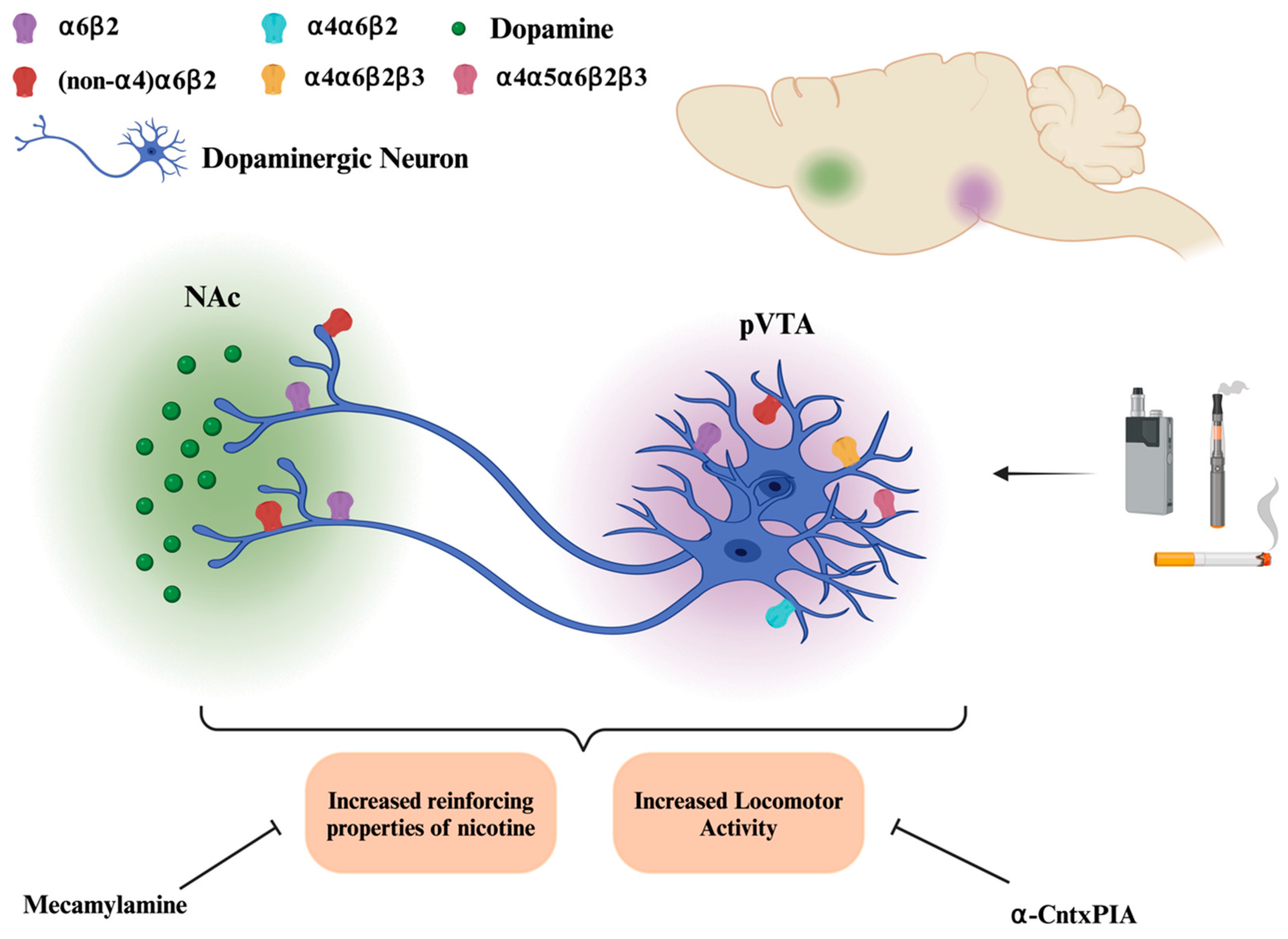
Nicotine exerts its rewarding effects by acting on nAChRs within the mesolimbic dopamine pathway [106]. Specifically, nAChRs on dopamine neurons in the VTA, which project to the nucleus accumbens (NAc), trigger dopamine release, a key factor in the addictive properties of psychostimulants like nicotine [107] (Figure 3). Given the diverse subunit composition of nAChRs in the mesolimbic dopamine pathway, identifying the specific subtypes involved in dopamine release has been a central research question. Notably, α6*nAChRs, predominantly localized to dopaminergic neurons [36,108], have been shown to modulate nicotine-induced dopamine release, particularly in the ventral striatal NAc compared to the dorsal striatal caudate putamen [66]. Additionally, intra-VTA perfusion of α-CntxMII, selective for α3/α6β2, and α-CntxPIA, selective for α6β2, suppresses nicotine-induced dopamine release in the NAc [18,109]. The VTA region can be divided into three subregions: anterior VTA (aVTA), posterior VTA (pVTA), and tail VTA (tVTA) [109]. Investigating the expression pattern of nAChR subunits in these regions has led to the finding that pVTA has significantly higher expression of α4, α6, and β3 subunits in comparison to aVTA dopaminergic neurons [109]. The crucial role of pVTA in mediating the rewarding properties of nicotine and its dopamine release was further illustrated when injection of a noncompetitive nAChR antagonist, mecamylamine, blocked these nicotine effects [109]. Further studies in knockout and wild-type mice demonstrated the importance of both α4 and α6 subunits in the modulation of dopaminergic neurons and suggest that these effects are mediated by α6α4β2*nAChRs in the pVTA [18,109,110].

4.1.2. B: α6*nAChRs and Locomotor Activity

One of the behavioral outputs of dopamine release in rodents is locomotor activity [111]. Oligonucleotides against the α6 subunit result in partial inhibition of nicotine-induced locomotion, suggesting the involvement of α6*nAChRs [35]. The highly specific α-CntxPIA infused into the pVTA inhibits nicotine-induced locomotion and dopamine release [18]. These effects suggest that α6*nAChRs are necessary and sufficient for nicotine-induced locomotor activity. Indeed, this is corroborated by studies revealing enhanced nicotine-induced locomotor activity via α6*nAChRs [12,17]. The hypersensitive α6L9′S mouse line was used to demonstrate increased baseline locomotion compared to wild-type mice [17]. Additionally, a dose-dependent increase in locomotor activity in response to nicotine (0.02–0.4 mg/kg, IP) was seen, which was blocked by the injection of α-CntxMII into the VTA [41]. Administration of the α6β2 antagonist, N, N-decane-1,10-diyl-bis-3-picolinium diiodide (bPiDI), to male and female mice induced a decrease in both locomotor activity in females between 10 to 30 min post-injection [112]. Using protein kinase C epsilon (PKCε) knockout female mice, enhanced nicotine-induced locomotor activity was associated with increased α6 and β3 expression [12]. PKCε is a critical signaling molecule that regulates the CHRNA6-CHRNB3 cluster [86] and may serve as a potential target for pharmacotherapies given its role in modulating nicotine sensitivity. However, it is important to note that the locomotor response to nicotine is not uniform across all mouse strains and experimental conditions. Of note, the previously described findings reflect the effects of low-dose nicotine. Acute exposure of C57BL/6J mice to a high dose of nicotine (1.5 mg/kg) did not alter locomotor activity [113]. These discrepancies highlight the complexity of nicotine’s effects and underscore the importance of considering factors such as mouse strain, nicotine dose, and administration route when interpreting experimental results. While the precise mechanisms underlying nicotine’s effects on locomotor activity and dopamine release remain to be fully elucidated, the available evidence points to a crucial role for α6*nAChRs in VTA dopaminergic neurons.

4.1.3. C: α6*nAChRs and Self-Administration

Nicotine self-administration is a critical behavioral measure to consider when investigating neurobiological mechanisms and developing potential treatments for addiction. In a self-administration study using wild-type and α6-knockout (α6-KO) mice, wild-type mice self-administered nicotine (26.3 µg/kg/Inj), whereas α6-KO mice did not, even at higher doses. Notably, re-expression of the α6 subunit in the VTA of KO mice restored nicotine self-administration [16]. Further investigation has shown that α6β2 receptors on dopamine terminals in the NAc shell are not only necessary for nicotine self-administration, but also for maintaining this behavior, as the blockade of these receptors reduced rats’ motivation for reinforcement [103]. Moreover, pretreatment with α-CntxMII in the VTA, blocking α6*nAChRs, decreased nicotine self-administration response in rats [18]. Systemic administration of the α6β2 antagonist, bPiDI, also impairs acquisition and maintenance of nicotine self-administration [114,115]. Collectively, these findings highlight the α6 subunit as a critical regulator of the reinforcing effects of nicotine and nicotine intake and may be a reliable target for smoking cessation therapies.

4.1.4. D: Clinical and Pre-Clinical Studies Investigating CHRNA6 Encoding for the α6 Subunit

Clinical research has established a strong genetic component to nicotine dependence, with heritability estimates ranging from 40–70% [116,117,118]. As nicotine is an agonist for nAChRs, genes encoding these receptors are prime candidates for investigation. In particular, identifying SNPs within nAChR genes and their association with smoking behavior is crucial for understanding the genetic underpinnings of nicotine dependence. While an SNP in the coding region of the CHRNA5 gene, encoding the α5 nAChR subunit, has been previously studied and linked to nicotine dependence [119,120], other nAChR subunits, such as α6, warrant further scrutiny. One study examined the SNPs in the CHRNB3-CHRNA6 gene cluster in a Chinese Han population. Various forms of analysis, such as haplotype analysis, gene–gene interactions, and meta-analysis, were employed to investigate the genetic factors underlying nicotine addiction. These analyses confirm that both genes within this cluster contribute significantly to nicotine dependence [121]. For CHRNA6, rs2304297, an SNP in the 3′ UTR of the gene has been found to be associated with nicotine dependence in Caucasians [21,89]. There is an abundance of evidence associating this SNP with nicotine-seeking behaviors and susceptibility to nicotine addiction in humans, particularly adolescents (Table 2) [22,87]. While most studies listed in Table 2 identify the “G” allele as the risk allele, some report it as protective. This SNP has also been associated with an increased positive perception of nicotine during adolescence and an increase in daily cigarette use (Table 2) [85,88].
Given the substantial clinical evidence linking this SNP (rs2304297) to nicotine dependence, our laboratory has created two rat lines facilitating pre-clinical investigations into the SNP’s functional role and elucidating its contribution to nicotine addiction. In the rat lines, the 3′ UTR of the rat CHRNA6 gene was replaced with that of humans. Using sub-chronic, low-dose nicotine exposure as a model of early adolescent nicotine use, sex and genotype differences were observed, with female α6CC and male α6GG exhibiting increased locomotor and anxiolytic behavior compared to saline controls [82]. Furthermore, adolescent male α6GG rats were more prone than male α6CC rats to relapse into nicotine-seeking behavior after drug plus cue exposure [122]. In α6CC females, sub-chronic nicotine treatment also resulted in the ability to discriminate between reinforced and non-reinforced responses in a methamphetamine self-administration paradigm [123] (Table 3).
In preliminary neurochemical studies, exposure to nicotine and methamphetamine increased dopamine overflow in α6GG males and α6CC females compared to their genotype counterparts [123] (Table 3). Tissue neurotransmitter levels were also measured in drug-naïve adults and adolescents carrying the CHRNA6 3′ UTR SNP, revealing baseline age and genotype differences in DA levels [123]. Adolescent male α6GG rats had significantly greater DA levels in the NAc core than their adult counterparts and adolescent male α6CC rats. No genotype or age differences were observed in females. The combined behavioral and neurochemical work using this model provides important insight into novel mechanisms in nicotine-induced behaviors driven by an SNP in a UTR of a nAChR subunit gene. While the precise mechanisms remain to be elucidated, these findings reveal new insights into how this SNP drives nicotine-induced behaviors.

4.2. Alcohol

The frequent co-use of alcohol and tobacco in humans is well established [124,125,126]. Indeed, individuals with alcohol use disorder (AUD) often have nicotine dependence [127,128]. Bidirectional associations between nicotine and alcohol have been described across several studies [129,130]. Although the field has put forth effort in evaluating these drugs, alone or combined, there is still limited understanding of underlying mechanisms or therapeutic treatments [131]. Human genetic variations in CHRNA6, encoding for the α6 subunit, have been linked to smoking and alcohol in some instances (Table 2) and support further investigation of the role of these receptor subunits in alcohol use and vulnerability.
Unlike nicotine, alcohol is not a direct agonist for nAChRs, thus suggesting an alternative modulatory role influencing how nAChRs respond to stimuli and regulate neurotransmitter release [132]. Injection of mecamylamine, a non-selective nAChR antagonist, into mouse VTA attenuates ethanol-induced increases in dopamine levels in the NAc [133,134,135]. nAChRs are also associated with alcohol-related behaviors such as sedation [136], locomotor activity [112,134], and intake [112,137]. Specifically, α6*nAChRs are necessary for ethanol-induced activation of VTA dopamine neurons and increased ACh release in mouse tissue slices [134,138]. Low-dose (0.1–5 mM) ethanol-induced dopamine release in the NAc can be blocked using α-CntxMII, suggesting ethanol acts as an allosteric modulator on α6*nAChRs [55]. Transgenic mice with heightened nicotine sensitivity, α6L9′S, also show enhanced ethanol sensitivity, alcohol intake and conditioned place preference [139], suggesting a role for the α6 subunit in mediating ethanol’s neurochemical and behavioral effects. Indeed, other animal models also indicate that α6*nAChRs regulate alcohol-induced dopamine release, locomotor activity, and voluntary intake in mice [55,139,140]. Collectively, these studies strongly suggest that α6*nAChRs modulate alcohol-induced dopamine function within the mesolimbic pathway with subsequent impacts on alcohol-related behaviors.
While there is a clear overlap in α6 subunit involvement for both nicotine- and alcohol-related dopamine function, a growing body of literature strongly implicates the regional and circuit-level influence of GABA [45,46,55]. α6*nAChRs are expressed on GABAergic terminals projecting onto both dopamine and GABAergic neurons within the VTA [43,45,141]. α6*nAChRs on the GABAergic terminals to VTA GABA neurons enhance low-dose ethanol-induced dopamine release in the NAc and subsequent ethanol-conditioned place preference [45,142] but inhibit these effects at high ethanol doses [55]. Taken together, the findings suggest that ethanol, at low doses, enhances GABA transmission onto VTA GABA neurons and increases overall activation of VTA dopamine neurons with parallel downstream increases in dopamine release within the NAc [45,46,55].
While α6*nAChRs are strongly implicated in mediating ethanol’s rewarding effects, the precise mechanisms by which high doses of ethanol inhibit evoked dopamine release while low doses enhance spontaneous dopamine release remains unclear. Discrepancies in species, ethanol doses, neuronal population, and experimental paradigms across studies complicate the interpretation and integration of findings. Moreover, the focus on dopaminergic neurons and the use of electrophysiological techniques in many studies limits our understanding of the complex interplay between different neurotransmitter systems in response to ethanol. Comprehensive investigations examining the interactions between dopaminergic and GABAergic neurons within different brain circuits are necessary to elucidate the full picture of ethanol’s effects on the brain.

4.3. α6 Subunit in Parkinson’s Disease

Parkinson’s disease (PD) is a progressive neurodegenerative disorder characterized by the loss of dopaminergic neurons in the nigrostriatal pathway, containing both α4β2* and α6β2*nAChR subtypes [25,57,143,144]. A major function of the nigrostriatal dopaminergic system is to facilitate motor activity. In PD, damage to nigrostriatal neurons results in reduced α4β2*- and α6β2*nAChR-mediated dopamine release, leading to the characteristic symptom of loss of motor function [145,146]. A prevalent therapeutic intervention employed for Parkinson’s disease patients is levo-dihydroxyphenylalanine (L-DOPA), which serves as a precursor for dopamine. The co-administration of L-DOPA along with carbidopa has demonstrated efficacy in alleviating motor symptoms [147] (Figure 4). L-DOPA crosses the blood–brain barrier and is converted to dopamine, whereas carbidopa inhibits DOPA decarboxylase and prevents its metabolism [147]. This treatment can lead to L-DOPA-induced dyskinesia (LID) within months of starting therapy [148,149]. The elucidation of the molecular mechanisms underpinning LID holds significant value and could potentially facilitate the optimization of L-DOPA therapy. Whereas nicotine inhibits LID via nAChR desensitization, this effect is eliminated in mice with the α6 subunit deleted [150,151]. A study with α6L9′S hypersensitive mice further emphasizes the critical role of α6*nAChRs in LID, suggesting a possible therapeutic role for α6β2*nAChR-selective drugs in treating LID [151,152].
Various studies have suggested that nicotine, despite its well-known risks, may offer unexpected neuroprotective benefits, particularly in the context of PD. Both in vitro studies on dopaminergic neurons and in vivo studies on PD animal models indicate that nicotine can protect against cell death and nigrostriatal damage, respectively [153,154,155,156]. In the 6-hydroxydopamine-induced nigrostriatal lesion model of PD, both α4β2*- and α6β2*-mediated dopamine release is progressively lost with increased nigrostriatal damage [146]. Although nicotine is neuroprotective, it is non-specific, interacting with all types of nAChRs. A novel α6β2*nAChR positive allosteric modulator, AN6001, has been shown to protect dopaminergic neurons from neurotoxic damage in vitro and has been suggested as a potential therapy for treatment of PD [157] (Figure 4). Positive allosteric modulators have reduced chances of side effects as they do not directly activate or overstimulate receptors. Additionally, they exhibit greater selectivity for specific receptor subtypes due to the less conserved nature of allosteric sites compared to the main binding site [140]. In this case, AN6001 exhibits significantly higher selectivity for α6β2* compared to α4β2* nAChRs, one of the most abundant nAChRs in the brain [157]. Although AN6001 has demonstrated potential in HEK293 cells and Xenopus oocytes, its efficacy must be evaluated in PD animal models to advance its therapeutic development.

4.4. α6 Subunit in Pain

Chronic pain patients often experience spontaneous pain and heightened sensitivity to normally non-painful stimuli, such as light touch (mechanical allodynia) [158,159,160]. P2X2/3 receptors are ionotropic receptors that are involved in the development of neuropathic pain [160]. Current treatments for this condition are ineffective, and we lack a thorough understanding of the underlying molecular mechanisms of symptoms such as mechanical allodynia. nAChRs have been identified and investigated as a potential drug target for chronic pain and attention has been on α4β2*, α7*, α9*, and α3* nAChRs [161,162,163]. Dorsal root ganglia (DRG), which contain neurons responsible for transmitting sensory information to the central nervous system, have been found to express α6β4 nAChRs in rats [164]. The immunohistochemistry of mouse DRG α6-containing neurons has also shown nociceptive neuron markers [165]. To better understand the molecular mechanisms, the genetic basis of mechanical allodynia has been examined using genome-wide gene expression levels in DRG of 25 inbred mouse strains [165]. The results suggest the involvement of the CHRNA6 gene and α6*nAChRs in influencing mechanical allodynia following nerve damage or inflammation. The proposed analgesic mechanism of action of α6* receptors is interaction with, and inhibition of, P2X2/3 receptors [165]. Additionally, the antiallodynic effect of nicotine has been investigated using α4 and α6 KO as well as wild-type mice [165]. Nicotine was administered via three types of administration, supraspinal, spinal, and peripheral, in both neuropathic and inflammatory pain models. The study illustrated that nicotine’s ability to alleviate mechanical allodynia is primarily mediated through α6*nAChRs, with the exception of supraspinal regions where both α6 and α4 receptors seem to play a role [165]. The α6 subunit, once overlooked, emerges from this study as a key player in pain processing, highlighting its potential as a therapeutic target.
Successful heterologous expression of α6β4*nAChRs has paved the way for further investigation and examination of the involvement of this receptor subtype in pain. The efficacy of two agonists, ABT-594 and ABT-894, has been tested on α6β4 and α4β2 receptors expressed in HEK293T cells. Whereas ABT-594 worked equally well on both, ABT-894 was less efficient on α6β4 [166]. In vivo testing of ABT-594 in BARP-KO mice which had reduced expression of α6β4 has shown blunted antiallodynic effects as compared to wild-type controls [166]. Although in vitro and in vivo studies have illuminated the role of α6β4*nAChRs in pain, bridging the gap to human applications remains crucial. Species-specific differences, such as those observed in α-conopeptide binding of human and rat α6β4*nAChRs [163], highlight the complexities of translating preclinical findings to clinical applications.
Genetic variations within the CHRNA6 gene have been studied in pain clinical studies [165]. More specifically, a rs7828365 SNP within the promoter region has been shown to be linked to altered pain sensitivity, such that homozygous individuals with the minor allele (TT) were more likely to experience persistent pain six months after surgery than subjects with TC and CC. While this suggests a role for this gene and α6*nAChRs in pain in both humans and mice, the authors acknowledge the infrequent occurrence of this genotype among humans and that other factors besides CHRNA6 likely contribute to the clinical variability of chronic pains. Additionally, the small number of homozygous TT individuals posed significant challenges and reduced the statistical power of the study. Therefore, a larger cohort of individuals would be beneficial in further understanding the role of CHRNA6 in chronic pain [165]. Collectively, the findings suggest that α6β4-containing receptors play a role in pain modulation. However, future research is necessary to fully elucidate the precise mechanisms involved and to identify additional compounds that could effectively target this receptor for pain relief.

5. Conclusions

Nicotine and other substance use disorders impose several threats to public health and exert a tremendous individual and socioeconomic strain. There is an urgent need to uncover neurobiological mechanisms mediating substance use and gather a comprehensive understanding of the role of nAChRs as therapeutic targets. In this review, we outline the key role of α6*nAChRs in nicotine and alcohol use and the genetic factors which may influence their function. We also review the implications for α6*nAChRs in alleviating dopamine-related side effects in PD treatment and potential considerations for α6*nAChRs in pain conditions. Historically, challenges like low expression levels and limited research tools have hindered the investigation of α6*nAChRs compared to other nAChR subtypes [59]. However, this review highlights recent advancements in research tools which have enabled significant progress. Improved methods for in vitro expression of functional and physiologically relevant α6*nAChRs will provide valuable insight into the basic mechanisms of action for substances and the development of effective treatments. High-throughput screening, using either in vitro or in vivo models, is a promising area of exploration for addiction research as it accelerates drug discovery and saves valuable time before pre-clinical and clinical trials. Furthermore, studies using innovative animal models underscore their indispensable role in understanding these receptors and potential cessation strategies.
Once believed to be restricted to dopaminergic neurons, α6*nAChRs are now known to be expressed in GABAergic, glutamatergic, and noradrenergic neurons [43,45,46]. This challenges previous research that focused solely on dopaminergic neurons and suggests a broader role for α6*nAChRs in cellular functions at the circuit level and beyond. Here, we review work focused on the impact of α6*nAChRs on VTA GABA neurons under various ethanol conditions. Future studies could evaluate the role of α6*nAChRs in the context of other substances and combined exposure such as nicotine and alcohol. The discovery of α6*nAChR expression in astrocytes, non-neuronal cells implicated in drug addiction, opens exciting avenues for research, particularly given the growing evidence linking these receptors to substance use disorders [106,107,131].
The adolescent brain is especially susceptible to the effects of nicotine [6], and it is essential to investigate how these receptors differ between sexes as well. Nicotine-induced dopamine levels in the NAc are greater in adolescent rats than in adult rats [166], and adolescent females show increased sensitivity to its rewarding effects [167]. α6 subunit mRNA expression peaks in VTA and substantia nigra dopamine neurons during adolescence in male rats [108]; thus, it is critical to evaluate how age impacts α6*nAChR function and substance use. Sex differences in α6 subunit mRNA expression have been observed in VTA astrocytes and MHb neurons [43,53]. As reviewed in Meon et al. (2021) [12], there are clear sex differences in nAChR expression patterns throughout the brain in adults with various implications for both nicotine- and alcohol-related behaviors. However, it is unclear exactly how α6*nAChR differs between sexes throughout development and the extent of their role in behavior. Thus, sex and age differences are critical considerations to be implemented into future studies to further explore α6*nAChR distribution and function.
Mounting evidence clearly suggests α6*nAChRs play a key role in reward-related behavior and substance use disorders, particularly nicotine and alcohol use. Polymorphisms in CHRNA6, the α6 subunit gene, are associated with both nicotine and alcohol susceptibility [87,168,169]. Common genetic factors underlying nicotine and alcohol codependency are important to evaluate since they may influence how a person responds to substances and any cessation treatments. In addition, identifying individuals who are more susceptible to the rewarding effects of abused drugs such as nicotine is critical for prevention strategies. Of note, the smoking cessation treatment with success in treating alcohol dependence, Varenicline, is a potent partial agonist of α4β2-containing and α6β2-containing nAChRs [151]. Genetic factors, including the SNPs highlighted in this review, may contribute to individual differences in response to treatments, offering insights into developing more effective treatments. Overall, the accumulating evidence highlights α6*nAChRs as promising therapeutic targets, underscoring the need for further investigation to fully realize their potential in treating various diseases.

Author Contributions

Conceptualization: Y.H.H., E.M.C. and F.M.L.; writing—original draft preparation, Y.H.H. and E.M.C.; visualization, Y.H.H., E.M.C. and F.M.L.; writing—review and editing, Y.H.H., F.M.L., E.M.C. and S.L.; supervision, F.M.L. and S.L. All authors have read and agreed to the published version of the manuscript.

Funding

This work was funded by the Tobacco and Related Disease Research Program (TRDRP) award (T33IP6417, T31IP1427, T34IR8049), University of California Irvine (UCI) NIDA T32 training grant (T32DA050558), and NIH DA048899.

Conflicts of Interest

The authors declare no conflicts of interest.

References

  1. Available online: https://www.hhs.gov/surgeongeneral/reports-and-publications/tobacco/index.html (accessed on 2 January 2025).
  2. Cornelius, M.E. Tobacco product use among adults–United States, 2021. MMWR Morb. Mortal. Wkly. Rep. 2023, 72, 475–483. [Google Scholar] [CrossRef] [PubMed]
  3. Birdsey, J. Tobacco product use among US middle and high school students—National Youth Tobacco Survey 2023. MMWR Morb. Mortal. Wkly. Rep. 2023, 72, 1173–1182. [Google Scholar] [CrossRef] [PubMed]
  4. Yuan, M.; Cross, S.J.; Loughlin, S.E.; Leslie, F.M. Nicotine and the adolescent brain. J. Physiol. 2015, 593, 3397–3412. [Google Scholar] [CrossRef] [PubMed]
  5. Ren, M.; Lotfipour, S. Nicotine gateway effects on adolescent substance use. West. J. Emerg. Med. 2019, 20, 696. [Google Scholar] [CrossRef] [PubMed]
  6. Leslie, F.M. Unique, long-term effects of nicotine on adolescent brain. Pharmacol. Biochem. Behav. 2020, 197, 173010. [Google Scholar] [CrossRef]
  7. Xu, X.; Shrestha, S.S.; Trivers, K.F.; Neff, L.; Armour, B.S.; King, B.A. US healthcare spending attributable to cigarette smoking in 2014. Prev. Med. 2021, 150, 106529. [Google Scholar] [CrossRef]
  8. Champtiaux, N.; Gotti, C.; Cordero-Erausquin, M.; David, D.J.; Przybylski, C.; Léna, C.; Clementi, F.; Moretti, M.; Rossi, F.M.; Le Novere, N.; et al. Subunit composition of functional nicotinic receptors in dopaminergic neurons investigated with knock-out mice. J. Neurosci. 2003, 23, 7820–7829. [Google Scholar] [CrossRef]
  9. Azam, L.; Chen, Y.; Leslie, F. Developmental regulation of nicotinic acetylcholine receptors within midbrain dopamine neurons. Neuroscience 2007, 144, 1347–1360. [Google Scholar] [CrossRef] [PubMed]
  10. Changeux, J.P. Nicotine addiction and nicotinic receptors: Lessons from genetically modified mice. Nat. Rev. Neurosci. 2010, 11, 389–401. [Google Scholar] [CrossRef]
  11. Zoli, M.; Pistillo, F.; Gotti, C. Diversity of native nicotinic receptor subtypes in mammalian brain. Neuropharmacology 2015, 96, 302–311. [Google Scholar] [CrossRef] [PubMed]
  12. Moen, J.K.; Lee, A.M. Sex differences in the nicotinic acetylcholine receptor system of rodents: Impacts on nicotine and alcohol reward behaviors. Front. Neurosci. 2021, 15, 745783. [Google Scholar] [CrossRef] [PubMed]
  13. Dwyer, J.B.; McQuown, S.C.; Leslie, F.M. The dynamic effects of nicotine on the developing brain. Pharmacol. Ther. 2009, 122, 125–139. [Google Scholar] [CrossRef]
  14. Thorpe, H.H.A.; Hamidullah, S.; Jenkins, B.W.; Khokhar, J.Y. Adolescent neurodevelopment and substance use: Receptor expression and behavioral consequences. Pharmacol. Ther. 2020, 206, 107431. [Google Scholar] [CrossRef]
  15. Engle, S.E.; Shih, P.Y.; McIntosh, J.M.; Drenan, R.M. α4α6β2* nicotinic acetylcholine receptor activation on ventral tegmental area dopamine neurons is sufficient to stimulate a depolarizing conductance and enhance surface AMPA receptor function. Mol. Pharmacol. 2013, 84, 393–406. [Google Scholar] [CrossRef] [PubMed]
  16. Pons, S.; Fattore, L.; Cossu, G.; Tolu, S.; Porcu, E.; McIntosh, J.M.; Changeux, J.P.; Maskos, U.; Fratta, W. Crucial role of α4 and α6 nicotinic acetylcholine receptor subunits from ventral tegmental area in systemic nicotine self-administration. J. Neurosci. 2008, 28, 12318–12327. [Google Scholar] [CrossRef] [PubMed]
  17. Drenan, R.M.; Grady, S.R.; Whiteaker, P.; McClure-Begley, T.; McKinney, S.; Miwa, J.M.; Bupp, S.; Heintz, N.; McIntosh, J.M.; Bencherif, M.; et al. In vivo activation of midbrain dopamine neurons via sensitized, high-affinity α6* nicotinic acetylcholine receptors. Neuron 2008, 60, 123–136. [Google Scholar] [CrossRef] [PubMed]
  18. Gotti, C.; Guiducci, S.; Tedesco, V.; Corbioli, S.; Zanetti, L.; Moretti, M.; Zanardi, A.; Rimondini, R.; Mugnaini, M.; Clementi, F.; et al. Nicotinic acetylcholine receptors in the mesolimbic pathway: Primary role of ventral tegmental area α6β2* receptors in mediating systemic nicotine effects on dopamine release, locomotion, and reinforcement. J. Neurosci. 2010, 30, 5311–5325. [Google Scholar] [CrossRef] [PubMed]
  19. Sanjakdar, S.S.; Maldoon, P.P.; Marks, M.J.; Brunzell, D.H.; Maskos, U.; McIntosh, J.M.; Bowers, M.S.; Damaj, M.I. Differential roles of α6β2* and α4β2* neuronal nicotinic receptors in nicotine-and cocaine-conditioned reward in mice. Neuropsychopharmacology 2015, 40, 350–360. [Google Scholar] [CrossRef]
  20. Dawes, C.T.; Loewen, P.J. The CHRNA6 gene, patience, and voter turnout. Patience Voter Turnout 2009. [Google Scholar] [CrossRef]
  21. Hoft, N.R.; Corley, R.P.; McQueen, M.B.; Schlaepfer, I.R.; Huizinga, D.; Ehringer, M.A. Genetic association of the CHRNA6 and CHRNB3 genes with tobacco dependence in a nationally representative sample. Neuropsychopharmacology 2009, 34, 698–706. [Google Scholar] [CrossRef] [PubMed]
  22. Lotfipour, S.; Leonard, G.; Perron, M.; Pike, B.; Richer, L.; Séguin, J.R.; Toro, R.; Veillette, S.; Pausova, Z.; Paus, T. Prenatal exposure to maternal cigarette smoking interacts with a polymorphism in the α6 nicotinic acetylcholine receptor gene to influence drug use and striatum volume in adolescence. Mol. Psychiatry 2010, 15, 6–8. [Google Scholar] [CrossRef] [PubMed]
  23. Cannon, D.S.; Mermelstein, R.J.; Hedeker, D.; Coon, H.; Cook, E.H.; McMahon, W.M.; Hamil, C.; Dunn, D.; Weiss, R.B. Effect of neuronal nicotinic acetylcholine receptor genes (CHRN) on longitudinal cigarettes per day in adolescents and young adults. Nicotine Tob. Res. 2014, 16, 137–144. [Google Scholar] [CrossRef]
  24. Pugach, O.; Cannon, D.S.; Weiss, R.B.; Hedeker, D.; Mermelstein, R.J. Classification tree analysis as a method for uncovering relations between CHRNA5A3B4 and CHRNB3A6 in predicting smoking progression in adolescent smokers. Nicotine Tob. Res. 2017, 19, 410–416. [Google Scholar] [PubMed]
  25. Gotti, C.; Clementi, F.; Fornari, A.; Gaimarri, A.; Guiducci, S.; Manfredi, I.; Moretti, M.; Pedrazzi, P.; Pucci, L.; Zoli, M. Structural and functional diversity of native brain neuronal nicotinic receptors. Biochem. Pharmacol. 2009, 78, 703–711. [Google Scholar] [CrossRef] [PubMed]
  26. Dani, J.A. Neuronal nicotinic acetylcholine receptor structure and function and response to nicotine. Int. Rev. Neurobiol. 2015, 124, 3–19. [Google Scholar]
  27. Corringer, P.J.; Sallette, J.; Changeux, J.P. Nicotine enhances intracellular nicotinic receptor maturation: A novel mechanism of neural plasticity? J. Physiol.-Paris 2006, 99, 162–171. [Google Scholar] [CrossRef]
  28. Hurst, R.; Rollema, H.; Bertrand, D. Nicotinic acetylcholine receptors: From basic science to therapeutics. Pharmacol. Ther. 2013, 137, 22–54. [Google Scholar] [CrossRef] [PubMed]
  29. Albuquerque, E.X.; Pereira, E.F.R.; Alkondon, M.; Rogers, S.W. Mammalian nicotinic acetylcholine receptors: From structure to function. Physiol. Rev. 2009, 89, 73–120. [Google Scholar] [CrossRef]
  30. Gotti, C.; Zoli, M.; Clementi, F. Brain nicotinic acetylcholine receptors: Native subtypes and their relevance. Trends Pharmacol. Sci. 2006, 27, 482–491. [Google Scholar] [CrossRef]
  31. Zoli, M.; Pucci, S.; Vilella, A.; Gotti, C. Neuronal and extraneuronal nicotinic acetylcholine receptors. Curr. Neuropharmacol. 2018, 16, 338–349. [Google Scholar] [CrossRef]
  32. Changeux, J.P. The acetylcholine receptor: An allosteric membrane protein. Biochem. Soc. Trans. 1981, 23, 195–205. [Google Scholar] [CrossRef]
  33. Changeux, J.P.; Devillers-Thiéry, A.; Chemouilli, P. Acetylcholine receptor: An allosteric protein. Science 1984, 225, 1335–1345. [Google Scholar] [CrossRef]
  34. Wills, L.; Ables, J.L.; Braunscheidel, K.M.; Caligiuri, S.P.; Elayouby, K.S.; Fillinger, C.; Ishikawa, M.; Moen, J.K.; Kenny, P.J. Neurobiological mechanisms of nicotine reward and aversion. Pharmacol. Rev. 2022, 74, 271–310. [Google Scholar] [CrossRef]
  35. Le Novere, N.; Zoli, M.; Changeux, J.P. Neuronal nicotinic receptor a6 subunit mRNA is selectively concentrated in catecholaminergic nuclei of the rat brain. Eur. J. Neurosci. 1996, 8, 2428–2439. [Google Scholar] [CrossRef]
  36. Azam, L.; Winzer-Serhan, U.H.; Chen, Y.; Leslie, F.M. Expression of neuronal nicotinic acetylcholine receptor subunit mRNAs within midbrain dopamine neurons. J. Comp. Neurol. 2002, 444, 260–274. [Google Scholar] [CrossRef]
  37. Champtiaux, N.; Han, Z.-Y.; Bessis, A.; Rossi, F.M.; Zoli, M.; Marubio, L.; McIntosh, J.M.; Changeux, J.-P. Distribution and pharmacology of α6-containing nicotinic acetylcholine receptors analyzed with mutant mice. J. Neurosci. 2002, 22, 1208–1217. [Google Scholar] [CrossRef] [PubMed]
  38. Gotti, C.; Moretti, M.; Zanardi, A.; Gaimarri, A.; Champtiaux, N.; Changeux, J.P.; Whiteaker, P.; Marks, M.J.; Clementi, F.; Zoli, M. Heterogeneity and selective targeting of neuronal nicotinic acetylcholine receptor (nAChR) subtypes expressed on retinal afferents of the superior colliculus and lateral geniculate nucleus: Identification of a new native nAChR subtype α3β2 (α5 or β3) enriched in retinocollicular afferents. Mol. Pharmacol. 2005, 68, 1162–1171. [Google Scholar]
  39. Mackey, E.D.; Engle, S.E.; Kim, M.R.; O’Neill, H.C.; Wageman, C.R.; Patzlaff, N.E.; Wang, Y.; Grady, S.R.; McIntosh, J.M.; Marks, M.J.; et al. α6* nicotinic acetylcholine receptor expression and function in a visual salience circuit. J. Neurosci. 2012, 32, 10226–10237. [Google Scholar] [CrossRef]
  40. Shih, P.Y.; Engle, S.E.; Oh, G.; Deshpande, P.; Puskar, N.L.; Lester, H.A.; Drenan, R.M. Differential expression and function of nicotinic acetylcholine receptors in subdivisions of medial habenula. J. Neurosci. 2014, 34, 9789–9802. [Google Scholar] [CrossRef] [PubMed]
  41. Berry, J.N.; Engle, S.E.; McIntosh, J.M.; Drenan, R.M. α6-Containing nicotinic acetylcholine receptors in midbrain dopamine neurons are poised to govern dopamine-mediated behaviors and synaptic plasticity. Neuroscience 2015, 304, 161–175. [Google Scholar] [CrossRef]
  42. Barloscio, D.; Cerri, E.; Domenici, L.; Longhi, R.; Dallanoce, C.; Moretti, M.; Vilella, A.; Zoli, M.; Gotti, C.; Origlia, N. In vivo study of the role of α6-containing nicotinic acetylcholine receptor in retinal function using subtype-specific RDP-MII (E11R) toxin. FASEB J. 2017, 31, 192–202. [Google Scholar] [CrossRef] [PubMed]
  43. Gao, R.; Schneider, A.M.; Mulloy, S.M.; Lee, A.M. Expression pattern of nicotinic acetylcholine receptor subunit transcripts in neurons and astrocytes in the ventral tegmental area and locus coeruleus. Eur. J. Neurosci. 2024, 59, 2225–2239. [Google Scholar] [CrossRef] [PubMed]
  44. Charpantier, E.; Barnéoud, P.; Moser, P.; Besnard, F.; Sgard, F. Nicotinic acetylcholine subunit mRNA expression in dopaminergic neurons of the rat substantia nigra and ventral tegmental area. Neuroreport 1998, 9, 3097–3101. [Google Scholar] [CrossRef] [PubMed]
  45. Steffensen, S.C.; Shin, S.I.; Nelson, A.C.; Pistorius, S.S.; Williams, S.B.; Woodward, T.J.; Park, H.J.; Friend, L.; Gao, M.; Gao, F.; et al. α6 subunit-containing nicotinic receptors mediate low-dose ethanol effects on ventral tegmental area neurons and ethanol reward. Addict. Biol. 2018, 23, 1079–1093. [Google Scholar] [CrossRef] [PubMed]
  46. Wadsworth, H.A.; Anderson, E.Q.; Williams, B.M.; Ronström, J.W.; Moen, J.K.; Lee, A.M.; McIntosh, J.M.; Wu, J.; Yorgason, J.T.; Steffensen, S.C. Role of α6-nicotinic receptors in alcohol-induced GABAergic synaptic transmission and plasticity to cholinergic interneurons in the nucleus accumbens. Mol. Neurobiol. 2023, 60, 3113–3129. [Google Scholar] [CrossRef]
  47. Yang, K.; Buhlman, L.; Khan, G.M.; Nichols, R.A.; Jin, G.; McIntosh, J.M.; Whiteaker, P.; Lukas, R.J.; Wu, J. Functional nicotinic acetylcholine receptors containing α6 subunits are on GABAergic neuronal boutons adherent to ventral tegmental area dopamine neurons. J. Neurosci. 2011, 31, 2537–2548. [Google Scholar] [CrossRef] [PubMed]
  48. Scofield, M.D. Exploring the role of astroglial glutamate release and association with synapses in neuronal function and behavior. Biol. Psychiatry 2018, 84, 778–786. [Google Scholar] [CrossRef] [PubMed]
  49. Linker, K.E.; Cross, S.J.; Leslie, F.M. Glial mechanisms underlying substance use disorders. Eur. J. Neurosci. 2019, 50, 2574–2589. [Google Scholar] [CrossRef] [PubMed]
  50. Giacometti, L.L.; Chandran, K.; Figueroa, L.A.; Barker, J.M. Astrocyte modulation of extinction impairments in ethanol-dependent female mice. Neuropharmacology 2020, 179, 108272. [Google Scholar] [CrossRef]
  51. Kruyer, A.; Kalivas, P.W. Astrocytes as cellular mediators of cue reactivity in addiction. Curr. Opin. Pharmacol. 2021, 56, 1–6. [Google Scholar] [CrossRef]
  52. Tizabi, Y.; Getachew, B.; Hauser, S.R.; Tsytsarev, V.; Manhães, A.C.; da Silva, V.D.A. Role of Glial Cells in Neuronal Function, Mood Disorders, and Drug Addiction. Brain Sci. 2024, 14, 558. [Google Scholar] [CrossRef] [PubMed]
  53. Olszewski, N.A.; Tetteh-Quarshie, S.; Henderson, B.J. Neuronal excitability in the medial habenula and ventral tegmental area is differentially modulated by nicotine dosage and menthol in a sex-specific manner. eNeuro 2024, 11. [Google Scholar] [CrossRef] [PubMed]
  54. Verplaetse, T.L.; Morris, E.D.; McKee, S.A.; Cosgrove, K.P. Sex differences in the nicotinic acetylcholine and dopamine receptor systems underlying tobacco smoking addiction. Curr. Opin. Behav. Sci. 2018, 23, 196–202. [Google Scholar] [CrossRef] [PubMed]
  55. Gao, F.; Chen, D.; Ma, X.; Sudweeks, S.; Yorgason, J.T.; Gao, M.; Turner, D.; Eaton, J.B.; McIntosh, J.M.; Lukas, R.J.; et al. Alpha6-containing nicotinic acetylcholine receptor is a highly sensitive target of alcohol. Neuropharmacology 2019, 149, 45–54. [Google Scholar] [CrossRef]
  56. Azam, L.; McIntosh, J.M. Alpha-conotoxins as pharmacological probes of nicotinic acetylcholine receptors. Acta Pharmacol. Sin. 2009, 30, 771–783. [Google Scholar] [CrossRef]
  57. Grady, S.R.; Salminen, O.; Laverty, D.C.; Whiteaker, P.; McIntosh, J.M.; Collins, A.C.; Marks, M.J. The subtypes of nicotinic acetylcholine receptors on dopaminergic terminals of mouse striatum. Biochem. Pharmacol. 2007, 74, 1235–1246. [Google Scholar] [CrossRef] [PubMed]
  58. Grady, S.R.; Moretti, M.; Zoli, M.; Marks, M.J.; Zanardi, A.; Pucci, L.; Clementi, F.; Gotti, C. Rodent habenulo–interpeduncular pathway expresses a large variety of uncommon nAChR subtypes 2009, but only the α3β4 and α3β3β4 subtypes mediate acetylcholine release. J. Neurosci. 2009, 29, 2272–2282. [Google Scholar] [CrossRef]
  59. Letchworth, S.R.; Whiteaker, P. Progress and challenges in the study of α6-containing nicotinic acetylcholine receptors. Biochem. Pharmacol. 2011, 82, 862–872. [Google Scholar] [CrossRef]
  60. Le Novere, N.; Changeux, J.P. Molecular evolution of the nicotinic acetylcholine receptor: An example of multigene family in excitable cells. J. Mol. Evol. 1995, 40, 155–172. [Google Scholar] [CrossRef]
  61. Cardenas, A.; Elabd, M.; Lotfipour, S. Specificity of a rodent alpha (α) 6 nicotinic acetylcholine receptor subunit antibody. Psychopharmacology 2020, 237, 283–285. [Google Scholar] [CrossRef]
  62. Lebbe, E.K.; Peigneur, S.; Wijesekara, I.; Tytgat, J. Conotoxins targeting nicotinic acetylcholine receptors: An overview. Mar. Drugs 2014, 12, 2970–3004. [Google Scholar] [CrossRef] [PubMed]
  63. Hone, A.J.; Gajewiak, J.; Christensen, S.; Lindstrom, J.; McIntosh, J.M. α-Conotoxin PeIA [S9H, V10A, E14N] potently and selectively blocks α6β2β3 versus α6β4 nicotinic acetylcholine receptors. Mol. Pharmacol. 2012, 82, 972–982. [Google Scholar] [CrossRef] [PubMed]
  64. McIntosh, J.M.; Azam, L.; Staheli, S.; Dowell, C.; Lindstrom, J.M.; Kuryatov, A.; Garrett, J.E.; Marks, M.J.; Whiteaker, P. Analogs of α-conotoxin MII are selective for α6-containing nicotinic acetylcholine receptors. Mol. Pharmacol. 2004, 65, 944–952. [Google Scholar] [CrossRef]
  65. Kulak, J.M.; Nguyen, T.A.; Olivera, B.M.; McIntosh, J.M. α-Conotoxin MII blocks nicotine-stimulated dopamine release in rat striatal synaptosomes. J. Neurosci. 1997, 17, 5263–5270. [Google Scholar] [CrossRef] [PubMed]
  66. Exley, R.; Clements, M.A.; Hartung, H.; McIntosh, J.M.; Cragg, S.J. α6-containing nicotinic acetylcholine receptors dominate the nicotine control of dopamine neurotransmission in nucleus accumbens. Neuropsychopharmacology 2008, 33, 2158–2166. [Google Scholar] [CrossRef] [PubMed]
  67. Dowell, C.; Olivera, B.M.; Garrett, J.E.; Staheli, S.T.; Watkins, M.; Kuryatov, A.; Yoshikami, D.; Lindstrom, J.M.; McIntosh, J.M. α-Conotoxin PIA is selective for α6 subunit-containing nicotinic acetylcholine receptors. J. Neurosci. 2003, 23, 8445–8452. [Google Scholar] [CrossRef]
  68. Pucci, L.; Grazioso, G.; Dallanoce, C.; Rizzi, L.; De Micheli, C.; Clementi, F.; Bertrand, S.; Bertrand, D.; Longhi, R.; De Amici, M.; et al. Engineering of α-conotoxin MII-derived peptides with increased selectivity for native α6β2* nicotinic acetylcholine receptors. FASEB J. 2011, 25, 3775–3789. [Google Scholar] [CrossRef] [PubMed]
  69. Azam, L.; Maskos, U.; Changeux, J.P.; Dowell, C.D.; Christensen, S.; De Biasi, M.; McIntosh, J.M. α-Conotoxin BuIA [T5A; P6O]: A novel ligand that discriminates between α6β4 and α6β2 nicotinic acetylcholine receptors and blocks nicotine-stimulated norepinephrine release. FASEB J. 2010, 24, 5113. [Google Scholar] [CrossRef] [PubMed]
  70. Armishaw, C.J. Synthetic α-conotoxin mutants as probes for studying nicotinic acetylcholine receptors and in the development of novel drug leads. Toxins 2010, 2, 1471–1499. [Google Scholar] [CrossRef]
  71. Millar, N.S.; Harkness, P.C. Assembly and trafficking of nicotinic acetylcholine receptors. Mol. Membr. Biol. 2008, 25, 279–292. [Google Scholar] [CrossRef]
  72. Kuryatov, A.; Olale, F.; Cooper, J.; Choi, C. Human α6 AChR subtypes: Subunit composition, assembly, and pharmacological responses. Neuropharmacology 2000, 39, 2570–2590. [Google Scholar] [CrossRef]
  73. Evans, N.M.; Bose, S.; Benedetti, G.; Zwart, R.; Pearson, K.H.; McPhie, G.I.; Craig, P.J.; Benton, J.P.; Volsen, S.G.; Sher, E.; et al. Expression and functional characterisation of a human chimeric nicotinic receptor with α6β4 properties. Eur. J. Pharmacol. 2003, 466, 31–39. [Google Scholar] [CrossRef]
  74. Gu, S.; Matta, J.A.; Lord, B.; Harrington, A.W.; Sutton, S.W.; Davini, W.B.; Bredt, D.S. Brain α7 nicotinic acetylcholine receptor assembly requires NACHO. Neuron 2016, 89, 948–955. [Google Scholar] [CrossRef] [PubMed]
  75. Matta, J.A.; Gu, S.; Davini, W.B.; Lord, B.; Siuda, E.R.; Harrington, A.W.; Bredt, D.S. NACHO mediates nicotinic acetylcholine receptor function throughout the brain. Cell Rep. 2017, 19, 688–696. [Google Scholar] [CrossRef] [PubMed]
  76. Gu, S.; Matta, J.A.; Davini, W.B.; Dawe, G.B.; Lord, B.; Bredt, D.S. α6-Containing nicotinic acetylcholine receptor reconstitution involves mechanistically distinct accessory components. Cell Rep. 2019, 26, 866–874. [Google Scholar] [CrossRef]
  77. Knowland, D.; Gu, S.; Eckert, W.A.; Dawe, G.B.; Matta, J.A.; Limberis, J.; Wickenden, A.D.; Bhattacharya, A.; Bredt, D.S. Functional α6β4 acetylcholine receptor expression enables pharmacological testing of nicotinic agonists with analgesic properties. J. Clin. Investig. 2023, 130, 6158–6170. [Google Scholar] [CrossRef]
  78. A Lester, H.; Fonck, C.; Tapper, A.R.; McKinney, S.; Damaj, M.I.; Balogh, S.; Owens, J.; Wehner, J.M.; Collins, A.C.; Labarca, C. Hypersensitive knockin mouse strains identify receptors and pathways for nicotine action. Curr. Opin. Drug Discov. Dev. 2003, 6, 633–639. [Google Scholar]
  79. Lotfipour, S.; Mojica, C.; Nakauchi, S.; Lipovsek, M.; Silverstein, S.; Cushman, J.; Tirtorahardjo, J.; Poulos, A.; Elgoyhen, A.B.; Sumikawa, K.; et al. α2* Nicotinic acetylcholine receptors influence hippocampus-dependent learning and memory in adolescent mice. Learn. Mem. 2017, 24, 231–244. [Google Scholar] [CrossRef] [PubMed]
  80. Walker, N.B.; Yan, Y.; Tapia, M.A.; Tucker, B.R.; Thomas, L.N.; George, B.E.; West, A.M.; Marotta, C.B.; Lester, H.A.; Dougherty, D.A.; et al. β2 nAChR activation on VTA DA neurons is sufficient for nicotine reinforcement in rats. eNeuro 2023, 10. [Google Scholar] [CrossRef] [PubMed]
  81. Wells, A.C.; Mojica, C.; Lotfipour, S. Hypersensitivity of the nicotinic acetylcholine receptor subunit (CHRNA2L9′ S/L9′ S) in female adolescent mice produces deficits in nicotine-induced facilitation of hippocampal-dependent learning and memory. Neurobiol. Learn. Mem. 2024, 213, 107959. [Google Scholar] [CrossRef]
  82. Cardenas, A.; Bai, Y.; Hajy Heydary, Y.; Li, J.; Leslie, F.M.; Lotfipour, S. Sex-and genotype-dependent nicotine-induced behaviors in adolescent rats with a human polymorphism (rs2304297) in the 3′-UTR of the CHRNA 6 gene. Int. J. Mol. Sci. 2022, 23, 3145. [Google Scholar] [CrossRef] [PubMed]
  83. Fontana, M.A. Mitigating Risk: Smartphone Notifications, Adaptive Surveying, and Genetics. Ph.D. Thesis, University of Michigan, Ann Arbor, MI, USA, 2015. [Google Scholar]
  84. Fletcher, J.M. Why have tobacco control policies stalled? Using genetic moderation to examine policy impacts. PLoS ONE 2012, 7, e50576. [Google Scholar] [CrossRef] [PubMed]
  85. Pedneault, M.; Labbe, A.; Roy-Gagnon, M.H.; Low, N.C.; Dugas, E.; Engert, J.C.; O’Loughlin, J. The association between CHRN genetic variants and dizziness at first inhalation of cigarette smoke. Addict. Behav. 2014, 39, 316–320. [Google Scholar] [CrossRef]
  86. Lee, W.; Bergen, A.W.; E Swan, G.; Li, D.; Liu, J.; Thomas, P.; Tyndale, R.F.; Benowitz, N.L.; Lerman, C.; Conti, D.V. Gender-stratified gene and gene–treatment interactions in smoking cessation. Pharmacogenomics J. 2012, 12, 521–532.M. [Google Scholar] [CrossRef] [PubMed]
  87. Thorgeirsson, T.E.; Gudbjartsson, D.F.; Surakka, I.; Vink, J.M.; Amin, N.; Geller, F.; Sulem, P.; Rafnar, T.; Esko, T.; Walter, S.; et al. Sequence variants at CHRNB3–CHRNA6 and CYP2A6 affect smoking behavior. Nat. Genet. 2010, 42, 448–453. [Google Scholar] [CrossRef] [PubMed]
  88. Zeiger, J.S.; Haberstick, B.C.; Schlaepfer, I.; Collins, A.C.; Corley, R.P.; Crowley, T.J.; Hewitt, J.K.; Hopfer, C.J.; Lessem, J.; McQueen, M.B.; et al. The neuronal nicotinic receptor subunit genes (CHRNA6 and CHRNB3) are associated with subjective responses to tobacco. Hum. Mol. Genet. 2008, 17, 724–734. [Google Scholar] [CrossRef]
  89. Saccone, S.F.; Hinrichs, A.L.; Saccone, N.L.; Chase, G.A.; Konvicka, K.; Madden, P.A.; Breslau, N.; Johnson, E.O.; Hatsukami, D.; Pomerleau, O.; et al. Cholinergic nicotinic receptor genes implicated in a nicotine dependence association study targeting 348 candidate genes with 3713 SNPs. Hum. Mol. Genet. 2007, 16, 36–49. [Google Scholar] [CrossRef]
  90. Rigbi, A.; Kanyas, K.; Yakir, A.; Greenbaum, L.; Pollak, Y.; Ben-Asher, E.; Lancet, D.; Kertzman, S.; Lerer, B. Why do young women smoke? V. Role of direct and interactive effects of nicotinic cholinergic receptor gene variation on neurocognitive function. Genes Brain Behav. 2008, 7, 164–172. [Google Scholar] [CrossRef]
  91. Prochaska, J.J.; Benowitz, N.L. Current advances in research in treatment and recovery: Nicotine addiction. Sci. Adv. 2019, 5, eaay9763. [Google Scholar] [CrossRef]
  92. Acquah, F.A.; Paramel, M.; Kuta, A.; Hussaini, S.R.; Wallace, D.R.; Mooers, B.H. Simulations of Promising Indolizidine—α6-β2 Nicotinic Acetylcholine Receptor Complexes. Int. J. Mol. Sci. 2021, 22, 7934. [Google Scholar] [CrossRef]
  93. Linker, K.E.; Gad, M.; Tawadrous, P.; Cano, M.; Green, K.N.; Wood, M.A.; Leslie, F.M. Microglial activation increases cocaine self-administration following adolescent nicotine exposure. Nat. Commun. 2020, 11, 306. [Google Scholar] [CrossRef]
  94. Lee, A.M.; Mansuri, M.S.; Wilson, R.S.; Lam, T.T.; Nairn, A.C.; Picciotto, M.R. Sex differences in the ventral tegmental area and nucleus accumbens proteome at baseline and following nicotine exposure. Front. Mol. Neurosci. 2021, 14, 657064. [Google Scholar] [CrossRef]
  95. López, A.J.; Johnson, A.R.; Euston, T.J.; Wilson, R.; Nolan, S.O.; Brady, L.J.; Thibeault, K.C.; Kelly, S.J.; Kondev, V.; Melugin, P.; et al. Cocaine self-administration induces sex-dependent protein expression in the nucleus accumbens. Commun. Biol. 2021, 4, 883. [Google Scholar] [CrossRef]
  96. Ingolia, N.T.; Hussmann, J.A.; Weissman, J.S. Ribosome profiling: Global views of translation. Cold Spring Harb. Perspect. Biol. 2019, 11, a032698. [Google Scholar] [CrossRef]
  97. Jones, S.; Sudweeks, S.; Yakel, J.L. Nicotinic receptors in the brain: Correlating physiology with function. Trends Neurosci. 1999, 22, 555–561. [Google Scholar] [CrossRef] [PubMed]
  98. Galvin, V.C.; Arnsten, A.F.; Wang, M. Involvement of nicotinic receptors in working memory function. Behav. Pharmacol. Cholinergic Syst. 2020, 45, 89–99. [Google Scholar]
  99. Koukouli, F.; Changeux, J.P. Do nicotinic receptors modulate high-order cognitive processing? Trends Neurosci. 2020, 43, 550–564. [Google Scholar] [CrossRef] [PubMed]
  100. Mineur, Y.S.; Soares, A.R.; Etherington, I.M.; Abdulla, Z.I.; Picciotto, M.R. Pathophysiology of nAChRs: Limbic circuits and related disorders. Pharmacol. Res. 2023, 191, 106745. [Google Scholar] [CrossRef] [PubMed]
  101. Volkow, N.D.; Morales, M. The brain on drugs: From reward to addiction. Cell 2015, 162, 712–725. [Google Scholar] [CrossRef] [PubMed]
  102. Kuzmin, A.; Jerlhag, E.; Liljequist, S.; Engel, J. Effects of subunit selective nACh receptors on operant ethanol self-administration and relapse-like ethanol-drinking behavior. Psychopharmacology 2009, 203, 99–108. [Google Scholar] [CrossRef]
  103. Brunzell, D.H.; Boschen, K.E.; Hendrick, E.S.; Beardsley, P.M.; McIntosh, J.M. α-Conotoxin MII-sensitive nicotinic acetylcholine receptors in the nucleus accumbens shell regulate progressive ratio responding maintained by nicotine. Neuropsychopharmacology 2010, 35, 665–673. [Google Scholar] [CrossRef]
  104. Collo, G.; Cavalleri, L.; Zoli, M.; Maskos, U.; Ratti, E.; Merlo Pich, E. Alpha6-containing nicotinic acetylcholine receptors mediate nicotine-induced structural plasticity in mouse and human iPSC-derived dopaminergic neurons. Front. Pharmacol. 2018, 9, 572. [Google Scholar] [CrossRef] [PubMed]
  105. Natividad, L.A.; Buczynski, M.W.; McClatchy, D.B.; Yates, J.R., III. From synapse to function: A perspective on the role of neuroproteomics in elucidating mechanisms of drug addiction. Proteomes 2018, 6, 50. [Google Scholar] [CrossRef] [PubMed]
  106. Ebrahimi, M.N.; Banazadeh, M.; Alitaneh, Z.; Suha, A.J.; Esmaeili, A.; Hasannejad-Asl, B.; Siahposht-Khachaki, A.; Hassanshahi, A.; Bagheri-Mohammadi, S. The distribution of neurotransmitters in the brain circuitry: Mesolimbic pathway and addiction. Physiol. Behav. 2024, 284, 114639. [Google Scholar] [CrossRef] [PubMed]
  107. De Biasi, M.; Dani, J.A. Reward, addiction, withdrawal to nicotine. Annu. Rev. Neurosci. 2011, 34, 105–130. [Google Scholar] [CrossRef]
  108. Azam, L.; McIntosh, J.M. Characterization of nicotinic acetylcholine receptors that modulate nicotine-evoked [3H] norepinephrine release from mouse hippocampal synaptosomes. Mol. Pharmacol. 2006, 70, 967–976. [Google Scholar] [CrossRef] [PubMed]
  109. Zhao-Shea, R.; Liu, L.; Soll, L.G.; Improgo, M.R.; Meyers, E.E.; McIntosh, J.M.; Grady, S.R.; Marks, M.J.; Gardner, P.D.; Tapper, A.R. Nicotine-mediated activation of dopaminergic neurons in distinct regions of the ventral tegmental area. Neuropsychopharmacol. Off. Publ. Am. Coll. Neuropsychopharmacol. 2011, 36, 1021–1032. [Google Scholar] [CrossRef] [PubMed]
  110. Liu, L.; Zhao-Shea, R.; McIntosh, J.M.; Gardner, P.D.; Tapper, A.R. Nicotine persistently activates ventral tegmental area dopaminergic neurons via nicotinic acetylcholine receptors containing α4 and α6 subunits. Mol. Pharmacol. 2012, 81, 541–548. [Google Scholar] [CrossRef] [PubMed]
  111. Picciotto, M.R.; Zoli, M.; Rimondini, R.; Léna, C.; Marubio, L.M.; Pich, E.M.; Fuxe, K.; Changeux, J.-P. Acetylcholine receptors containing the β2 subunit are involved in the reinforcing properties of nicotine. Nature 1998, 391, 173–177. [Google Scholar] [CrossRef]
  112. Kamens, H.M.; Peck, C.; Garrity, C.; Gechlik, A.; Jenkins, B.C.; Rajan, A. α6β2 nicotinic acetylcholine receptors influence locomotor activity and ethanol consumption. Alcohol 2017, 61, 43–49. [Google Scholar] [CrossRef]
  113. Jackson, K.J.; McIntosh, J.M.; Brunzell, D.H.; Sanjakdar, S.S.; Damaj, M. The role of α6-containing nicotinic acetylcholine receptors in nicotine reward and withdrawal. J. Pharmacol. Exp. Ther. 2009, 331, 547–554. [Google Scholar] [CrossRef]
  114. Beckmann, J.S.; Meyer, A.C.; Pivavarchyk, M.; Horton, D.B.; Zheng, G.; Smith, A.M.; Wooters, T.E.; McIntosh, J.M.; Crooks, P.A.; Bardo, M.T.; et al. r-bPiDI, an α6β2* nicotinic receptor antagonist, decreases nicotine-evoked dopamine release and nicotine reinforcement. Neurochem. Res. 2015, 40, 2121–2130. [Google Scholar] [CrossRef]
  115. Maggio, S.E.; Saunders, M.A.; Nixon, K.; Prendergast, M.A.; Zheng, G.; Crooks, P.A.; Dwoskin, L.P.; Bell, R.L.; Bardo, M.T. An improved model of ethanol and nicotine co-use in female P rats: Effects of naltrexone, varenicline, and the selective nicotinic α6β2* antagonist r-bPiDI. Drug Alcohol Depend. 2018, 193, 154–161. [Google Scholar] [CrossRef] [PubMed]
  116. Li, M.D.; Cheng, R.; Ma, J.Z.; Swan, G.E. A meta-analysis of estimated genetic and environmental effects on smoking behavior in male and female adult twins. Addiction 2003, 98, 23–31. [Google Scholar] [CrossRef]
  117. Maes, H.H.; Sullivan, P.F.; Bulik, C.M.; Neale, M.C.; Prescott, C.A.; Eaves, L.J.; Kendler, K.S. A twin study of genetic and environmental influences on tobacco initiation, regular tobacco use and nicotine dependence. Psychol. Med. 2004, 34, 1251–1261. [Google Scholar] [CrossRef] [PubMed]
  118. Sharp, B.M.; Chen, H. Neurogenetic determinants and mechanisms of addiction to nicotine and smoked tobacco. Eur. J. Neurosci. 2019, 50, 2164–2179. [Google Scholar] [CrossRef]
  119. Bierut, L.J.; Stitzel, J.A. Genetic Contributions of the α5 Nicotinic Receptor Subunit to Smoking Behavior. In Nicotinic Receptors; Humana Press: New York, NY, USA, 2014; pp. 327–339. [Google Scholar]
  120. Morel, C.; Fattore, L.; Pons, S.; A Hay, Y.; Marti, F.; Lambolez, B.; De Biasi, M.; Lathrop, M.; Fratta, W.; Maskos, U.; et al. Nicotine consumption is regulated by a human polymorphism in dopamine neurons. Mol. Psychiatry 2014, 19, 930–936. [Google Scholar] [CrossRef]
  121. Wen, L.; Han, H.; Liu, Q.; Su, K.; Yang, Z.; Cui, W.; Yuan, W.; Ma, Y.; Fan, R.; Chen, J.; et al. Significant association of the CHRNB3-CHRNA6 gene cluster with nicotine dependence in the Chinese Han population. Sci. Rep. 2017, 7, 9745. [Google Scholar] [CrossRef] [PubMed]
  122. Carreño, D.; Lotfipour, S. Sex-and genotype-dependent nicotine plus cue-primed reinstatement is enhanced in adolescent Sprague Dawley rats containing the human CHRNA 6 3′-UTR polymorphism (rs2304297). Front. Psychiatry 2023, 13, 1064211. [Google Scholar] [CrossRef] [PubMed]
  123. Carreño, D.; Facundo, A.; Cardenas, A.; Lotfipour, S. Sub-chronic nicotine exposure influences methamphetamine self-administration and dopamine overflow in a sex-and genotype-dependent manner in humanized CHRNA 6 3′-UTR SNP (rs2304297) adolescent rats. Front. Pharmacol. 2024, 15, 1445303. [Google Scholar] [CrossRef] [PubMed]
  124. Daeppen, J.B.; Smith, T.L.; Danko, G.P.; Gordon, L.; Landi, N.A.; Nurnberger, J.I., Jr.; Bucholz, K.K.; Raimo, E.; Schuckit, M.A. Clinical correlates of cigarette smoking and nicotine dependence in alcohol-dependent men and women. Alcohol Alcohol. 2000, 35, 171–175. [Google Scholar] [CrossRef] [PubMed]
  125. John, U.; Meyer, C.; Rumpf, H.J.; Schumann, A.; Thyrian, J.R.; Hapke, U. Strength of the relationship between tobacco smoking, nicotine dependence and the severity of alcohol dependence syndrome criteria in a population-based sample. Alcohol Alcohol. 2003, 38, 606–612. [Google Scholar] [CrossRef] [PubMed]
  126. Grant, B.F.; Hasin, D.S.; Chou, S.P.; Stinson, F.S.; Dawson, D.A. Nicotine dependence and psychiatric disorders in the united states: Results from the national epidemiologic survey on alcohol and relatedconditions. Arch. Gen. Psychiatry 2004, 61, 1107–1115. [Google Scholar] [CrossRef] [PubMed]
  127. Weinberger, A.H.; Zhu, J.; Levin, J.; Moeller, S.J.; McKee, S.A.; Goodwin, R.D. Changes in alcohol use by cannabis use status among adolescents and young adults in the United States: Emerging evidence for both substitution and complementarity. Alcohol. Clin. Exp. Res. 2021, 45, 2536–2545. [Google Scholar] [CrossRef] [PubMed]
  128. Frie, J.A.; Nolan, C.J.; Murray, J.E.; Khokhar, J.Y. Addiction-related outcomes of nicotine and alcohol co-use: New insights following the rise in vaping. Nicotine Tob. Res. 2022, 24, 1141–1149. [Google Scholar] [CrossRef]
  129. Cannizzaro, E.; Lavanco, G.; Castelli, V.; Cirrincione, L.; Di Majo, D.; Martines, F.; Argo, A.; Plescia, F. Alcohol and nicotine use among adolescents: An observational study in a Sicilian cohort of high school students. Int. J. Environ. Res. Public Health 2022, 19, 6152. [Google Scholar] [CrossRef]
  130. Patrick, M.E.; Parks, M.J.; Carroll, D.M.; Mitchell, C. Feasibility of mailed biomarker data collection among US young adults: Saliva-based cotinine and self-reported nicotine use. Drug Alcohol Depend. 2023, 244, 109791. [Google Scholar] [CrossRef] [PubMed]
  131. Morikawa, H.; Morrisett, R.A. Ethanol action on dopaminergic neurons in the ventral tegmental area: Interaction with intrinsic ion channels and neurotransmitter inputs. Int. Rev. Neurobiol. 2010, 91, 235–288. [Google Scholar]
  132. Rahman, S.; Engleman, E.A.; Bell, R.L. Nicotinic receptor modulation to treat alcohol and drug dependence. Front. Neurosci. 2015, 8, 426. [Google Scholar] [CrossRef] [PubMed]
  133. Blomqvist, O.; Ericson, M.; Engel, J.A.; Söderpalm, B. Accumbal dopamine overflow after ethanol: Localization of the antagonizing effect of mecamylamine. Eur. J. Pharmacol. 1997, 334, 149–156. [Google Scholar] [CrossRef] [PubMed]
  134. Larsson, A.; Svensson, L.; Söderpalm, B.; Engel, J.A. Role of different nicotinic acetylcholine receptors in mediating behavioral and neurochemical effects of ethanol in mice. Alcohol 2002, 28, 157–167. [Google Scholar] [CrossRef] [PubMed]
  135. Larsson, A.; Jerlhag, E.; Svensson, L.; Söderpalm, B.; Engel, J.A. Is an α-conotoxin MII–sensitive mechanism involved in the neurochemical, stimulatory, and rewarding effects of ethanol? Alcohol 2004, 34, 239–250. [Google Scholar] [CrossRef] [PubMed]
  136. Kamens, H.M.; Hoft, N.R.; Cox, R.J.; Miyamoto, J.H.; Ehringer, M.A. The α6 nicotinic acetylcholine receptor subunit influences ethanol-induced sedation. Alcohol 2012, 46, 463–471. [Google Scholar] [CrossRef]
  137. Feduccia, A.A.; Chatterjee, S.; Bartlett, S.E. Neuronal nicotinic acetylcholine receptors: Neuroplastic changes underlying alcohol and nicotine addictions. Front. Mol. Neurosci. 2012, 5, 83. [Google Scholar] [CrossRef]
  138. Liu, L.; Zhao-Shea, R.; McIntosh, J.M.; Tapper, A.R. Nicotinic acetylcholine receptors containing the α6 subunit contribute to ethanol activation of ventral tegmental area dopaminergic neurons. Biochem. Pharmacol. 2013, 86, 1194–1200. [Google Scholar] [CrossRef] [PubMed]
  139. Powers, M.S.; Broderick, H.J.; Drenan, R.M.; Chester, J.A. Nicotinic acetylcholine receptors containing α6 subunits contribute to alcohol reward-related behaviours. Genes Brain Behav. 2013, 12, 543–553. [Google Scholar] [CrossRef] [PubMed]
  140. Wang, J.; Lindstrom, J. Orthosteric and allosteric potentiation of heteromeric neuronal nicotinic acetylcholine receptors. Br. J. Pharmacol. 2018, 175, 1805–1821. [Google Scholar] [CrossRef]
  141. Yang, K.C.; Jin, G.Z.; Wu, J. Mysterious α6-containing nAChRs: Function, pharmacology, and pathophysiology. Acta Pharmacol. Sin. 2009, 30, 740–751. [Google Scholar] [CrossRef] [PubMed]
  142. Schilaty, N.D.; Hedges, D.M.; Jang, E.Y.; Folsom, R.J.; Yorgason, J.T.; McIntosh, J.M.; Steffensen, S.C. Acute ethanol inhibits dopamine release in the nucleus accumbens via α6 nicotinic acetylcholine receptors. J. Pharmacol. Exp. Ther. 2014, 349, 559–567. [Google Scholar] [CrossRef] [PubMed]
  143. Halliday, G.; Lees, A.; Stern, M. Milestones in Parkinson’s disease—Clinical and pathologic features. Mov. Disord. 2011, 26, 1015–1021. [Google Scholar] [CrossRef]
  144. Rascol, O.; Lozano, A.; Stern, M.; Poewe, W. Milestones in Parkinson’s disease therapeutics. Mov. Disord. 2011, 26, 1072–1082. [Google Scholar] [CrossRef] [PubMed]
  145. Pérez, X.A.; Bordia, T.; McIntosh, J.M.; Quik, M. α6β2* and α4β2* nicotinic receptors both regulate dopamine signaling with increased nigrostriatal damage: Relevance to Parkinson’s disease. Mol. Pharmacol. 2010, 78, 971–980. [Google Scholar] [CrossRef] [PubMed]
  146. Pérez, X.A.; Quik, M. Focus on α4β2* and α6β2* nAChRs for Parkinson’s Disease Therapeutics. Mol. Cell. Pharmacol. 2011, 3, 1. [Google Scholar]
  147. Hansen, C.A.; Miller, D.R.; Annarumma, S.; Rusch, C.T.; Ramirez-Zamora, A.; Khoshbouei, H. Levodopa-induced dyskinesia: A historical review of Parkinson’s disease, dopamine, and modern advancements in research and treatment. J. Neurol. 2022, 269, 2892–2909. [Google Scholar] [CrossRef] [PubMed]
  148. Manson, A.; Stirpe, P.; Schrag, A. Levodopa-induced-dyskinesias clinical features, incidence, risk factors, management and impact on quality of life. J. Park. Dis. 2012, 2, 189–198. [Google Scholar] [CrossRef] [PubMed]
  149. LeWitt, P.A. Levodopa therapy for Parkinson’s disease: Pharmacokinetics and pharmacodynamics. Mov. Disord. 2015, 30, 64–72. [Google Scholar] [CrossRef] [PubMed]
  150. Quik, M.; Perez, X.A.; Bordia, T. Nicotine as a potential neuroprotective agent for Parkinson’s disease. Mov. Disord. 2012, 27, 947–957. [Google Scholar] [CrossRef]
  151. Bordia, T.; Hrachova, M.; Chin, M.; McIntosh, J.M.; Quik, M. Varenicline is a potent partial agonist at α6β2* nicotinic acetylcholine receptors in rat and monkey striatum. J. Pharmacol. Exp. Ther. 2012, 342, 327–334. [Google Scholar] [CrossRef]
  152. Quik, M.; Perez, X.A.; Grady, S.R. Role of α6 nicotinic receptors in CNS dopaminergic function: Relevance to addiction and neurological disorders. Biochem. Pharmacol. 2011, 82, 873–882. [Google Scholar] [CrossRef] [PubMed]
  153. Quik, M.; O’Neill, M.; Perez, X.A. Nicotine neuroprotection against nigrostriatal damage: Importance of the animal model. Trends Pharmacol. Sci. 2007, 28, 229–235. [Google Scholar] [CrossRef]
  154. Bordia, T.; Campos, C.; Huang, L.; Quik, M. Continuous and intermittent nicotine treatment reduces L-3, 4-dihydroxyphenylalanine (L-DOPA)-induced dyskinesias in a rat model of Parkinson’s disease. J. Pharmacol. Exp. Ther. 2008, 327, 239–247. [Google Scholar] [CrossRef]
  155. Picciotto, M.R.; Zoli, M. Neuroprotection via nAChRs: The role of nAChRs in neurodegenerative disorders such as Alzheimer’s and Parkinson’s disease. Front Biosci. 2008, 13, 492–504. [Google Scholar] [CrossRef] [PubMed]
  156. Zhang, D.; Mallela, A.; Sohn, D.; Carroll, F.I.; Bencherif, M.; Letchworth, S.; Quik, M. Nicotinic Receptor Agonists Reduce l-DOPA–Induced Dyskinesias in a Monkey Model of Parkinson’s Disease. J. Pharmacol. Exp. Ther. 2013, 347, 225–234. [Google Scholar] [CrossRef] [PubMed]
  157. van Hout, M.; Klein, J.; Ahring, P.K.; Brown, D.T.; Thaneshwaran, S.; Dos Santos, A.B.; Jensen, A.A.; Kohlmeier, K.A.; Christophersen, P.; Dyhring, T. Characterization of AN6001, a positive allosteric modulator of α6β2-containing nicotinic acetylcholine receptors. Biochem. Pharmacol. 2020, 174, 113788. [Google Scholar] [CrossRef] [PubMed]
  158. Fillingim, R.B.; Loeser, J.D.; Baron, R.; Edwards, R.R. Assessment of chronic pain: Domains, methods, and mechanisms. J. Pain 2016, 17, T10–T20. [Google Scholar] [CrossRef]
  159. Inoue, K. Nociceptive signaling of P2X receptors in chronic pain states. Purinergic Signal. 2021, 17, 41–47. [Google Scholar] [CrossRef] [PubMed]
  160. Dong, C.R.; Zhang, W.J.; Luo, H.L. Association between P2X3 receptors and neuropathic pain: As a potential therapeutic target for therapy. Biomed. Pharmacother. 2022, 150, 113029. [Google Scholar] [CrossRef] [PubMed]
  161. Papke, R.L.; Horenstein, N.A. Therapeutic targeting of α7 nicotinic acetylcholine receptors. Pharmacol. Rev. 2021, 73, 1118–1149. [Google Scholar] [CrossRef] [PubMed]
  162. Zheng, N.; Christensen, S.B.; Dowell, C.; Purushottam, L.; Skalicky, J.J.; McIntosh, J.M.; Chou, D.H.C. Discovery of methylene thioacetal-incorporated α-RgIA analogues as potent and stable antagonists of the human α9α10 nicotinic acetylcholine receptor for the treatment of neuropathic pain. J. Med. Chem. 2021, 64, 9513–9524. [Google Scholar] [CrossRef] [PubMed]
  163. Hone, A.J.; McIntosh, J.M. Nicotinic acetylcholine receptors: Therapeutic targets for novel ligands to treat pain and inflammation. Pharmacol. Res. 2023, 190, 106715. [Google Scholar] [CrossRef]
  164. Hone, A.J.; Meyer, E.L.; McIntyre, M.; McIntosh, J.M. Nicotinic acetylcholine receptors in dorsal root ganglion neurons include the α6β4* subtype. FASEB J. 2012, 26, 917. [Google Scholar] [CrossRef] [PubMed]
  165. Wieskopf, J.S.; Mathur, J.; Limapichat, W.; Post, M.R.; Al-Qazzaz, M.; Sorge, R.E.; Martin, L.J.; Zaykin, D.V.; Smith, S.B.; Freitas, K.; et al. The nicotinic α6 subunit gene determines variability in chronic pain sensitivity via cross-inhibition of P2X2/3 receptors. Sci. Transl. Med. 2015, 7, ra72–ra287. [Google Scholar] [CrossRef] [PubMed]
  166. Corongiu, S.; Dessì, C.; Cadoni, C. Adolescence versus adulthood: Differences in basal mesolimbic and nigrostriatal dopamine transmission and response to drugs of abuse. Addict. Biol. 2020, 25, e12721. [Google Scholar] [CrossRef]
  167. Xue, S.; Behnood-Rod, A.; Wilson, R.; Wilks, I.; Tan, S.; Bruijnzeel, A.W. Rewarding effects of nicotine in adolescent and adult male and female rats as measured using intracranial self-stimulation. Nicotine Tob. Res. 2020, 22, 172–179. [Google Scholar] [CrossRef]
  168. Schlaepfer, I.R.; Hoft, N.R.; Ehringer, M.A. The genetic components of alcohol and nicotine co-addiction: From genes to behavior. Curr. Drug Abus. Rev. 2008, 1, 124–134. [Google Scholar] [CrossRef] [PubMed]
  169. Hoft, N.R.; Corley, R.P.; McQueen, M.B.; Huizinga, D.; Menard, S.; Ehringer, M.A. SNPs in CHRNA6 and CHRNB3 are associated with alcohol consumption in a nationally representative sample. Genes Brain Behav. 2009, 8, 631–637. [Google Scholar] [CrossRef] [PubMed]
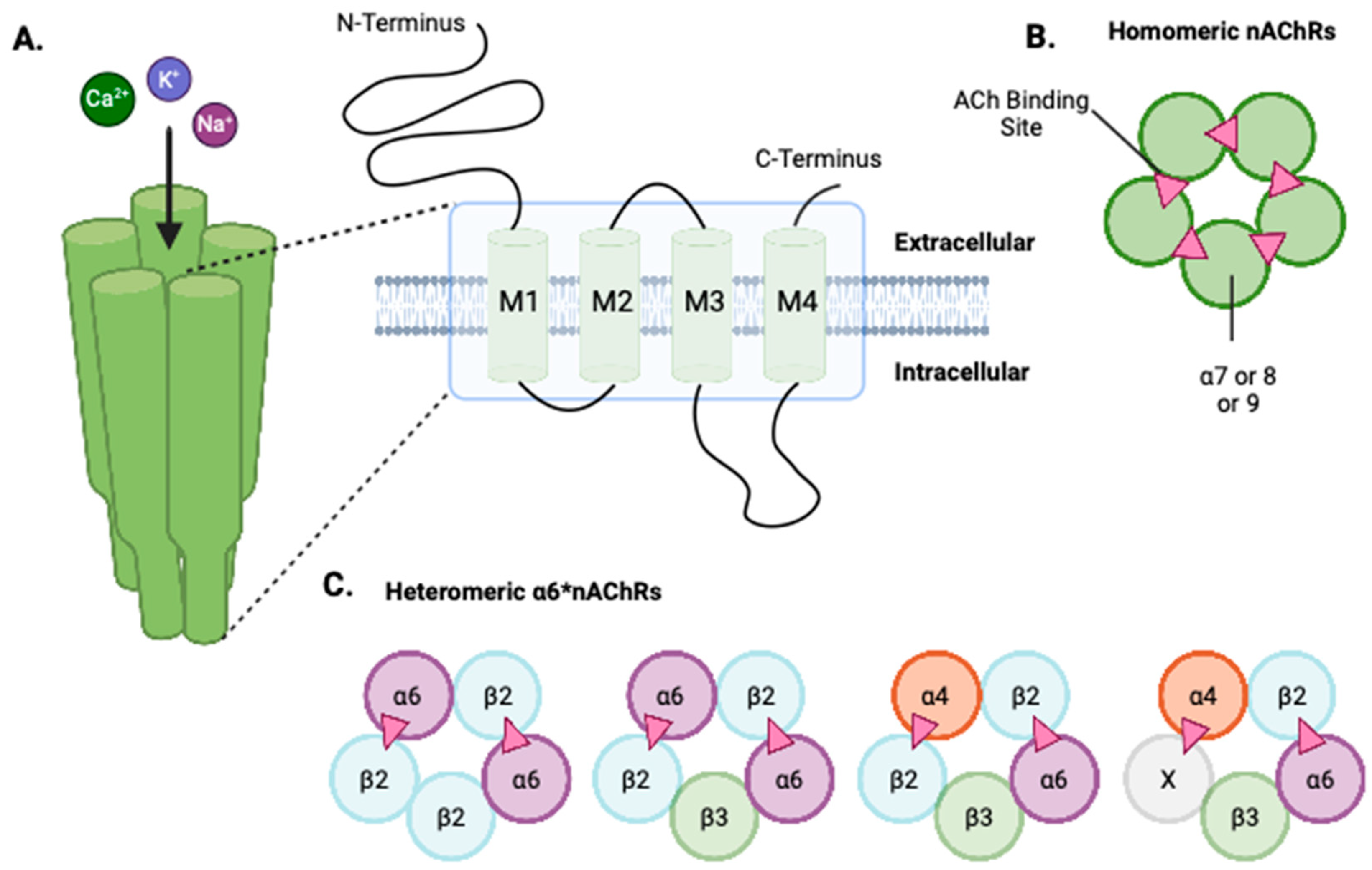
Figure 1. The structure of neuronal nAChRs. (A) nAChRs are made up of five subunits that come together to form a receptor. Each of these subunits has four transmembrane domains (M1–M4). M2 lines the aqueous pore while M1 and M3 protect M2 from the lipid bilayer. (B) Structure of a homomeric nAChR. These receptors have five agonist binding sites (depicted as pink triangles). (C) Structure of heteromeric α6*nAChRs and agonist binding sites. The X emphasizes that α6*nAChR can potentially combine with other subunits.
Receptors 04 00001 g001
Figure 2. Brain region and cell-type specific expression of α6*nAChRs. Schematic diagram illustrating the predominant α6*nAChR subunit arrangements found in the dorsal and ventral striatum (different shades of green), ventral tegmental area (purple), and locus coeruleus (blue). Different neuronal and non-neuronal cell populations are shown in different colors; dopaminergic (blue), GABAergic (yellow), glutamatergic (green), noradrenergic (red), astrocytes (purple). The X in the VTA region emphasizes that α6*nAChR can potentially combine with other subunits. Agonist binding sites are depicted as pink triangles.
Receptors 04 00001 g002
Figure 3. Nicotine’s effects on the mesolimbic dopamine system. Nicotine binds to the α6*nAChRs on dopaminergic neurons in the posterior ventral tegmental area (pVTA), leading to dopamine release in the nucleus accumbens (NAc). This dopamine release reinforces the rewarding properties of nicotine and increases locomotor activity, contributing to nicotine dependence. Injection of mecamylamine, an nAChR antagonist, and α-Ctx-PIA, an α6*nAChR antagonist, into the pVTA blocks dopamine release and therefore the reinforcing properties of nicotine and locomotor activity, respectively.
Receptors 04 00001 g003
Figure 4. Illustration of L-DOPA therapy and potential role of AN6001 in Parkinson’s patients. (A) The nigrostriatal pathway, crucial for motor control, with dopamine neurons projecting from the substantia nigra (SN) to the striatum. Degeneration of these neurons (shown in blue) in Parkinson’s causes dopamine deficiency and motor dysfunction. (B) L-DOPA, a precursor to dopamine, crosses the blood–brain barrier (BBB) and is converted into dopamine, offering symptomatic relief. However, L-DOPA therapy can lead to L-DOPA-induced dyskinesia (LID), which is linked to α6β2*nAChRs (shown in purple). (C) The positive allosteric modulator, AN6001 (red triangle), selectively enhances α6β2*nAChR activity, offering a potential avenue for neuroprotection and alleviating LID. AN6001′s targeted action on α6β2*nAChRs may provide a more refined therapeutic approach compared to non-specific agonists.
Receptors 04 00001 g004
Table 1. A summary of the distribution of various α6*nAChRs in the brain.
α6*nAChR SubtypeBrain RegionOrganism Supporting PublicationNeuronal Population
α6β2
-
Ventral and dorsal striatum
-
LC
Mouse
Rat
[34]
[55]
[47]
[18]
[56]
Dopamine
GABA
Glutamate
Norepinephrine
α6β4
-
Striatum
-
LC
-
Hippocampus
Mouse [56]Dopamine
Norepinephrine
α6β2β3
-
Striatum
-
Medial habenula
-
Superior colliculus
-
Lateral geniculate nucleus
Mouse[34]
[55]
[40]
[57]
[38]
Dopamine
Norepinephrine
α6β3β4
-
Interpeduncular nucleus
Mouse[58]-
α4α6β2
-
Striatum
-
VTA
Mouse [18]
[56]
Dopamine
GABA
α4α6β2β3
-
VTA
-
Retinal ganglion cells
-
Striatum
-
LC
-
Superior colliculus
-
Lateral geniculate nucleus
Mouse[43]
[34]
[55]
[39]
[38]
Dopamine
GABA
Glutamate
α4α5α6β2β3
-
VTA
Mouse[43]Dopamine
Table 2. Clinical studies of the rs2304297 SNP, located in the 3′ UTR of CHRNA6, associated with nicotine/tobacco use.
Supporting PublicationAdolescentSample Size % Male“G” as Risk Allele
[27]Yes48040Yes
[83]No12,50740.9No %
[23]Yes43941Yes
[84]No617848No %
[85]Yes129335No #
[86]No78948.9Yes *
[87]No76,681Not ReportedYes *
[22]Yes42345.4Yes
[21]Yes105149.2Yes
[21]Yes105149No +
[20]Yes267448Yes
[88]Yes105658.1Yes
[89]No192938Yes
[90]No2440Yes ^
* Does not survive multiple comparison correction; # carriers of minor allele (C) report decreased dizziness at first inhalation; % GG protective in the presence of higher taxes; + C not G listed as risk allele; ^ G is a risk genotype.
Table 3. Summary of pre-clinical findings of the rs2304297 SNP.
Behavioral Measure Male Rats
α6GG or α6CC
Female Rats
α6GG or α6CC
Locomotor Activity and Anxiolytic Behavior 1. Increased locomotor activity and anxiolytic behavior in nicotine-treated male α6GG rats compared to saline controls
2. Increased anxiolytic behavior in nicotine-treated α6GG males compared to α6CC males
Increased locomotor activity and anxiolytic behavior in nicotine-treated α6CC rats compared to saline controls as well as nicotine-treated α6GG rats
Dopamine ReleaseIncreased dopamine overflow in response to nicotine and methamphetamine in α6GG mice compared to α6CC ratsIncreased dopamine overflow in response to nicotine and methamphetamine in α6CC rats compared to α6GG rats
Methamphetamine Self-administrationNo genotype effect observed in methamphetamine self-administration after exposure to nicotineEnhanced discrimination between reinforced and non-reinforced response in nicotine-treated α6CC rats and saline-treated α6GG rats
Tissue Neurotransmitter Levels1. Nicotine + cue-treated adolescent α6GG rats had decreased dopamine levels in the NAc shell compared to other groups
2. Baseline differences: drug-naïve adolescent α6GG rats had greater dopamine levels in the NAc core compared to their adult counterparts and adolescent α6CC rats
No differences were observed
Disclaimer/Publisher’s Note: The statements, opinions and data contained in all publications are solely those of the individual author(s) and contributor(s) and not of MDPI and/or the editor(s). MDPI and/or the editor(s) disclaim responsibility for any injury to people or property resulting from any ideas, methods, instructions or products referred to in the content.