Review

Comparative Study between Mechanical Rotational Atherectomy Combined with Drug-Coated Balloon versus Drug-Coated Balloon Alone for Treatment of In-Stent Restenosis during Peripheral Endovascular Interventions: A Multicentric Trial

1
Department of General Surgery, Vascular Surgery Unit, Faculty of Medicine, Cairo University, Cairo 11562, Egypt
2
Department of Vascular Surgery, AL-Sheikh Zayed Specialized Hospital, Ministry of Health and Population, Giza 12622, Egypt
3
Department of General Surgery, Vascular Surgery Unit, Faculty of Medicine, Suez University, Suez 43511, Egypt
*
Author to whom correspondence should be addressed.
J. Vasc. Dis. 2024, 3(3), 290-305; https://doi.org/10.3390/jvd3030023
Submission received: 30 May 2024 / Revised: 6 July 2024 / Accepted: 22 July 2024 / Published: 12 August 2024
(This article belongs to the Section Peripheral Vascular Diseases)

Abstract

:
Purpose: To compare the efficacy and safety of percutaneous mechanical debulking (PMD) using mechanical rotational atherectomy combined with paclitaxel drug-coated balloon (DCB) versus using paclitaxel DCB alone in the treatment of in-stent restenosis. Material and Methods: This is a multicentric retrospective observational study conducted over a period of 2 years from 2020 to 2022. The study included 49 patients presented with chronic limb-threatening ischemia (CLTI) associated with in-stent restenosis, either acute (<14 days), subacute (<3 months) or chronic (>3 months). The enrolled patients underwent endovascular revascularization using either PMD combined with paclitaxel DCB or paclitaxel DCB only. They were followed up for 6 months after the intervention clinically and by duplex evaluation. Results: The lesion length was about 14.2 mm in the group treated by PMD+ DCB and 9.3 mm in the group treated by DCB alone. The technical success rate was the same between the two groups. However, the follow-up after 6 months showed that patencies for PMD + DCB and DCB alone were 15 (68.2%) patients and 15 (55.6%) patients, respectively (significant p value = 0.028). Procedural-related complications for PMD + DCB are distal embolization (9%) of cases and no vessel perforation. Regarding the candidates who were treated by DCB alone, there were minor groin hematomas (11.1%), distal arterial thrombosis (11.1%) and contrast-induced nephropathy (CIN) (11.1%) cases. Conclusion: The endovascular management of in-stent restenosis using percutaneous mechanical debulking (PMD) in conjunction with paclitaxel drug-coated balloon (DCB) showed that PMD combined with DCB is a safe and effective modality for achieving recanalization. It gives a satisfactory outcome in terms of technical success, freedom from clinically driven target lesion revascularization rate (CD-TLR) and mortality. Despite these promising results, further research with a large enrolled population may be required to determine the cost/benefit.

1. Introduction

In-stent restenosis (ISR) for the lower limb arteries is a significant problem that is shown to occur in up to 40% to 50% of cases following endovascular stenting, especially in the femoropopliteal segment. It is frequently observed after treating long, complex and heavily calcific lesions. According to the available published literature, in-stent thrombosis may occur in up to 4.3% of patients, and in half of these cases, it is an adverse consequence of late thrombosis within the stented segment of the artery [1,2].
Drug-coated balloon (DCB) angioplasty was considered one of the available treatment modalities that were proven to reduce the incidence of ISR, especially if applied in the femoropopliteal segment. However, some publications reported the need for the application of another stent in-stent as a bail-out solution. Currently, there are no solid data for a standard, widely accepted strategic treatment for ISR. A percutaneous mechanical debulking (PMD) strategy has been attempted to reduce the occlusive burden and consequently improve the patency rate and achieve freedom from clinically driven target lesion revascularization (CD-TLR). The debulking of the offending occlusive burden from within the previously deployed stent may potentially improve the effectiveness of paclitaxel DCB treatment by reducing the thickness of the thrombus and/or hyperplastic tissue and subsequently increasing drug delivery and transit to the intima [3].
Extensive arterial wall calcification has demonstrated a higher risk for restenosis even after the application of DCB. Thus, the debulking of atherosclerotic plaques by atherectomy was suggested as a vessel wall preparation to increase the potential benefit of DCB and improve clinical outcomes [4,5].

2. Materials and Methods

This is a multicentric retrospective observational study conducted over a period of 2 years between August 2020 and September 2022, including 49 patients presented with chronic limb-threatening ischemia (CLTI) and preprocedural investigations revealed restenosis of previously applied peripheral stent for which a special pre-determined protocol was applied in all patients aiming to perform endovascular procedures to treat in-stent restenosis (ISR) using percutaneous mechanical rotational atherectomy combined with drug-coated balloon versus drug-coated balloon alone, all interventions were performed at (name deleted to maintain the integrity of the review process) (30 patients) and (name deleted to maintain the integrity of the review process) (19 patients) by interventionalists share the same high learning curve and experience in both hospitals.
All patients were meticulously assessed by clinical examination, color-coded duplex ultrasonography (US) and computed tomography angiography (CTA). Study approval was obtained from the medical ethical committee, and informed consent was obtained from all patients.
Inclusion criteria included patients with CLTI, and investigations revealed in-stent restenosis/occlusion (thrombotic or intimal hyperplasia) with vessel diameter of more than 3 mm and the onset of ischemia either acute (less than 14 days), subacute (less than 3 months) or chronic (more than 3 months). Patients who refused intervention, patients with extensive gangrene (Rutherford 6) or in case of subintimal lesion crossing by the wire were excluded from the study.
Data regarding each patient at the time of the intervention and details about the procedure performed were entered retrospectively in the peripheral intervention report. The data were recorded using standardized forms and entered into a computerized database.
Full history was taken from all patients including name, gender, age, occupation, risk factors (smoking, diabetes, hypertension, +ve family history), affected lower limb, clinical presentation, duration of symptoms, past history (coronary disease, cardiomyopathy, congestive heart failure, stroke) and history of lower limb revascularization surgeries.
Clinical examinations with blood pressure, pulse, temperature, respiratory rate and local examination of the affected lower extremity (affected side, skin temperature, peripheral pulsations, trophic changes, ischemic ulcers, gangrene and previous amputation) were performed on all patients. Preoperative blood samples for serum creatinine level, fasting blood sugar, lipid profile (Cholesterol, LDL, HDL, TG) and CBC (Hemoglobin, WBC, PLT) were collected from all patients.
All patients were transferred to (Siemens Artis Zee and Philips FD20) machine catheter labs. After local anesthesia, we used US guidance (9-MHz linear probe, GE Logic E9; General Electric Medical Systems, Milwaukee, WI, USA) for access puncture to minimize the risk of bleeding.

2.1. Technique

All patients were loaded with Clopidogrel (300 mg), and an ipsilateral antegrade approach via femoral access was used as standard access. However, brachial and contralateral retrograde cross-over femoral accesses could be used in certain cases. A 6-F end-hole introducer sheath or a 7-F end-hole long cross-over sheath (Radifocus; Terumo, Tokyo, Japan) was advanced. After obtaining access, patients were anticoagulated with a 5000-unit bolus of intravenous unfractionated heparin.
Ultravist (Bayer Healthcare, Leverkusen, Germany) was used as a contrast medium for image acquisition during the endovascular intervention. Delayed imaging up to 30 s and large contrast volumes may be needed to opacify the tibial runoff vessels. In patients with impaired kidney functions, a CO2-guided endovascular procedure was performed with the aid of an automated injector system (Angiodroid ©, San Lazzaro di Savena, Italy).
After lesion localization, lesion crossing should be carefully performed trans-luminally, and subintimal passage should be avoided at all means. We used Command wire (Abbott Park, IL, USA) or V-18 standard wire (Watertown, MA, USA) with supporting catheter Trail-Blazer (Medtronic, Dublin, Ireland) or Renegade (Watertown, MA, USA) to achieve transluminal guidance of the wire through the lesion.
According to the lesion nature, morphology and length, the decision was tailored for the treatment plan, as we used a wire traversal test through the re-stenosed or occluded segment to raise the suspicion of thrombosis on top of hyperplasia, we tried to confirm the presence of thrombus by either obtaining different angiographic images in different projections or by applying suction to the thrombotic materials. If the diagnosis of thrombosis is confirmed, Rotarex® is preferred to clear the thrombus load and the underlying hyperplastic lesion. DCB is usually not preferred to avoid distal embolization and trashing with balloon inflation.
Regarding the hyperplastic lesions, the decision was variable; for single or multiple focal stenotic lesions, paclitaxel drug-coated balloon application was considered, while for long, diffuse and calcific lesions, percutaneous mechanical debulking using Rotarex in conjunction with paclitaxel drug-coated balloon was chosen. So, we can classify our treated patients into two groups:
Group 1: Patients were treated with an atherectomy and adjunctive drug-coated balloon.
Group 2: patients were treated with a drug-coated balloon only.
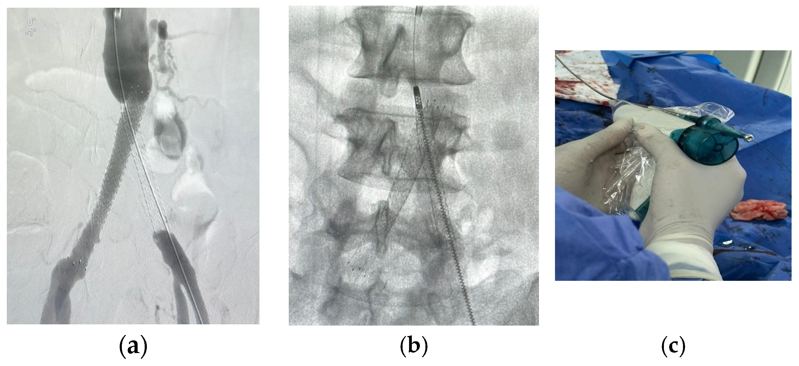
For patients in group 1, after securing the wire passage through the lesion, the mechanical rotational atherectomy device for percutaneous mechanical debulking (PMD) Rotarex® System (Straub Medical, Wangs, Switzerland), with 6-, 8- or 10-French (Fr) sheath compatible devices, depending on the vessel diameter to be treated, and variable length 85, 110 and 185 cm, was advanced over 0.018 wire.
The rotational thrombectomy device was slowly advanced and retracted in order to avoid embolic complications downstream of the occlusion. The rotations produce a continuous vacuum inside the catheter, which leads to aspiration of the thrombus into the catheter and transportation into the collection bag. The suction performance is about 0.66 mL/s with the 6-Fr system and 1.5 mL/s with the 8- and 10-Fr system. If necessary, the procedure was repeated until the blood flow was fully restored.
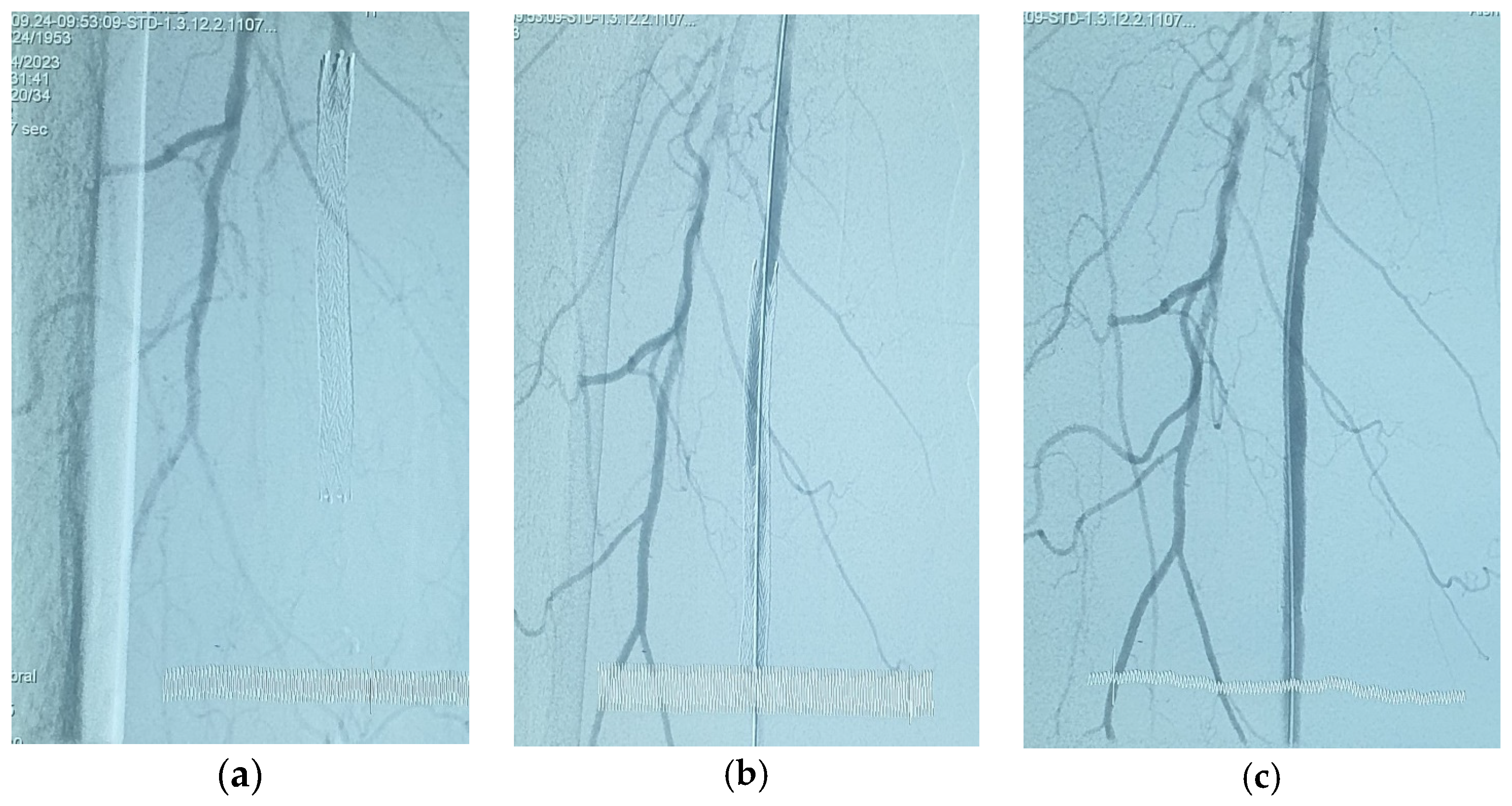
The Rotarex® catheter was then withdrawn while keeping the wire in place, a drug-coated balloon, IN.PACT Admiral paclitaxel-eluting balloon (Medtronic Cardiovascular, Santa Rosa, CA, USA) or Lutonix (Bard Peripheral Vascular, Tempe, AZ, USA), which were the FDA approved DCB, was advanced over the 0.018 wire with inflation time for 3 min at 8–12 atm pressure.
For patients in group 2, pre-dilatation with a plain balloon (1 mm smaller than the reference diameter) was performed first for optimal wall preparation, followed by using a paclitaxel drug-coated balloon, as previously mentioned.
All candidates were followed up clinically and by duplex ultrasonography after one, three and six months after the intervention.

2.2. Study Endpoints

Primary endpoints include safety and efficacy. The safety endpoint was procedure-related complications, while efficacy was based on procedure success (successful access and ≤30% residual stenosis by quantitative angiography with evidence of at least one patent tibial artery to the foot). Other efficacy outcomes included immediate hemodynamic success (restored pulse), defined as (ABI) improvement ≥ 0.15 compared to baseline, except for patients with falsely elevated ABI (defined as ABI ≥ 1.3).
Secondary endpoints include freedom from clinically driven target lesion revascularization CD-TLR, major limb adverse event (MALE) and thirty-day mortality.

2.3. Statistical Analysis

The collected data were coded, tabulated and statistically analyzed using the statistical package SPSS (Statistical Package for the Social Sciences) version 28. Continuous data were presented as mean ± SD (standard deviation) and minimum and maximum of the range, while categorical data were expressed as numbers and percentages. Comparisons between groups were performed using an unpaired t test. For comparing categorical data, a Chi-square (χ2) test was performed. The exact test was used instead when the expected frequency was less than 5. A level of significance of p value < 0.05 was considered significant; otherwise, it was non-significant.

3. Results

The current retrospective observational study conducted over a period of 2 years between August 2020 and September 2022, including 49 patients presented with chronic limb-threatening ischemia (CLTI) and preprocedural investigations revealed restenosis of previously applied peripheral stent, all interventions were performed at (name deleted to maintain the integrity of the review process) (30 patients) and (name deleted to maintain the integrity of the review process) (19 patients) by interventionalists share the same high learning curve and experience in both hospitals.

3.1. Patients’ Demographics, Risk Factors and Presentations

The baseline demographics of this study were age 61 ± 55, 28 male patients (57.1%) and 21 female (42.9%). A total of 33 (67.3%) were diabetic, 31 (63.3%) were hypertensive, smoking was found in 30 patients (61.2%), dyslipidemia was observed in 22 patients (44.9%), and ischemia heart disease was recorded in 14 patients (28.8%) (Table 1 and Table S1).
Patients’ presentations were variable, and claudication was recorded in nine (18.7%) patients. Rest pain was documented in 11 (22.4%) patients, cyanosis in 4 (8.1%), mottling in 1 patient and fixed color changes of the toes in 1 patient. Sudden onset of acute ischemic limb pain was recorded in three (6.1%) patients, and tissue loss in the form of toe gangrene, infected and/or non-healing ulcers, or development of foot infection was recorded in the rest of the patients (Table 2 and Table S2).

3.2. Lesion Distribution and Nature

The lesion distribution observed in the enrolled candidates revealed restenosis of previously deployed SFA stent in 35 patients (71.4%), restenosis in one iliac stent in 4 patients (8.2%) and in bilateral iliac stents in 2 patients (4.1%); combined restenosis in iliac + SFA stents was found in 6 patients (12.2%), and restenosis in popliteal “Supera” stent was found in 2 patients (4.1%) (Figure 1).
For restenosed SFA stent, the lesions were found to be focal single stenosis in 3 (6.1%) patients, multiple stenoses in 16 (32.7%) patients, total occlusion in 11 (22.4%) patients and segmental stenosis and occlusion in 5 (10.2%) patients. Regarding the unilateral iliac stents, restenosis was recorded in three (6.1%) patients and occlusion in one patient, while stenosis was only recorded for the restenosed bilateral iliac stents. For the patients with combined iliac + SFA stents, all of them showed stenosis of the iliac stent accompanied by occlusion of the SFA stent. Finally, of the patients with “Supera” stent, one of them presented with single focal stenosis while the other one presented with total occlusion of the stent (Table 3).
The lesion was documented as thrombotic in nature in 10 patients (20.4%) and intimal hyperplasia in 39 (79.6%). Lesions were single focal lesions in 20 patients (40.1%) and multiple levels in 29 (59.9%) patients. The lesion’s nature, morphology and characteristics are demonstrated in Table 3 and Table S3.

3.3. Procedural Data

In the current study, ipsilateral antegrade femoral access, “the most commonly used access”, was used in 25 (51.1%) patients, ipsilateral retrograde femoral access in 4 (8.1%) patients, contralateral cross-over femoral access in 14 (28.6%) patients and brachial access in 6 (12.2%) patients.
The technical success was achieved in all patients 100%, whether treated by Rotarex + DCB (Figure 2, Figure 3, Figure 4 and Figure 5) or DCB alone (Figure 6), and all procedures were completed successfully.
Procedural-related complications for Roatrex + DCB are two cases of distal embolization (managed conservatively by anticoagulation only and did not require further interventions) and one case of lost radial pulse after removal of brachial access sheath (compensated limb and managed conservatively); no vessel perforation occurred with Rotarex. Regarding the candidates who were treated by DCB alone, three cases developed minor groin hematomas (managed conservatively), three cases were complicated by distal arterial thrombosis (managed by anticoagulation only), three cases of contrast-induced nephropathy (CIN) occurred (managed conservatively with improvement of kidney function after one week). All procedural-related complications are demonstrated in Table 4 and Table S4.

3.4. Follow-Up Results

All patients were subjected to re-evaluation both clinically and by duplex ultrasonography. After one month, all patients (100%) treated by DCB are patent compared to 21 (95.5%) patients of patients treated by Rotarex + DCB.
After 3 months, follow-up for patency for Rotarex + DCB- and DCB-alone-treated patients were 19 (86.4%) and 21 (77.8%), respectively. In the Roatrex + DCB treated patient, only one patient (4.5%) developed in-stent occlusion, which was treated by surgical bypass between femoral and lower popliteal arteries, while in patients who were treated by DCB alone, two (7.4%) patients developed in-stent restenosis, and three (11.1%) patients developed in-stent occlusion (two of them treated by surgical bypass and one of them treated by re-do DCB + stent in-stent application) (Table 5 and Table S5).
In the follow-up after 6 months, patencies for Rotarex + DCB and DCB alone were 15 (68.2%) patients and 15 (55.6%) patients, respectively (significant p value = 0.028). Of the patients treated with Rotarex + DCB, three (13.6%) developed ISR treated by re-do angioplasty using DCB. While of patients treated by DCB alone, nine (33.3%) developed ISR (one of them was previously managed by the stent in-stent for ISR during previous follow-up), seven of them were treated by re-do angioplasty with DCB, and two of them were treated by stent in-stent application (Table 5 and Figure 7).
Among the patients treated with Rotarex + DCB, re-intervention was required in four patients; one of them required surgical bypass between femoral and lower popliteal arteries, and three of them required re-do angioplasty with DCB. While in DCB-alone-treated patients, 11 patients required re-intervention, 7 patients required re-do angioplasty with DCB, 2 patients required stent in-stent application (one of them required re-do angioplasty again with DCB in the next follow-up), and 2 patients required surgical bypass (Table 5 and Figure 7).
The clinically driven target lesion revascularization (CD-TLR) was variable between Rotarex + DCB- and DCB-alone-treated patients (18.2% vs. 40.7%, respectively). Regarding the major limb adverse event (MALE) and minor foot amputations, for Rotarex + DCB-treated patients, two cases resulted in above-knee amputation (AKA) and one case in below-knee amputation (BKA). For DCB-alone-treated patients, three cases resulted in AKA and one case in trans metatarsal amputation (Table 6 and Table S6).
For patients treated by Rotarex + DCB, three (13.5%) passed away; one patient died after 2 weeks from septicemia and MOSF, another one died after one month from myocardial infarction, and the last one died after 5 months from massive stroke. On the other hand, for DCB-alone-treated patients, there was only one case (3.7%) of death after 2 months from cerebrovascular stroke (Table 6).

4. Discussion

In the last few years, ISR has become a frequently faced endovascular challenge, which raises the requirement for developing a modality of treatment [1,6]. Reduction in the bulk of atheromatous plaque with subsequent modification of its nature through the endovascular atherectomy devices can allow better outcomes and lower rate of procedural complications [4,7]. Particularly, debulking of heavily calcified atheromatous plaques using mechanical rotational atherectomy devices showed improved lesion response to endovascular balloon dilation and subsequently provided better delivery of antiproliferative agents to the wall [5,8].
Several devices and techniques can be used to treat femoropopliteal segments in stent stenotic and/or occlusive lesions. Bare metal stent implantation is one of the most popular and commonly used bail-out solutions in these lesions, as stent application can resolve intra-procedural problems induced by elastic recoil, residual stenosis or dissection. However, ISR stays high, with an incidence of 15% to 32% in 12 months [9,10].
Recorded risk factors for ISR in many published literature include male gender, patients on regular hemodialysis, severe lesion calcifications, lesion length and hypercholesterolemia [11]. Surprisingly, diabetes was not found to be associated with an increased risk of ISR. On the other hand, insulin-dependent diabetes was reported as a predictor of CD-TLR [12].
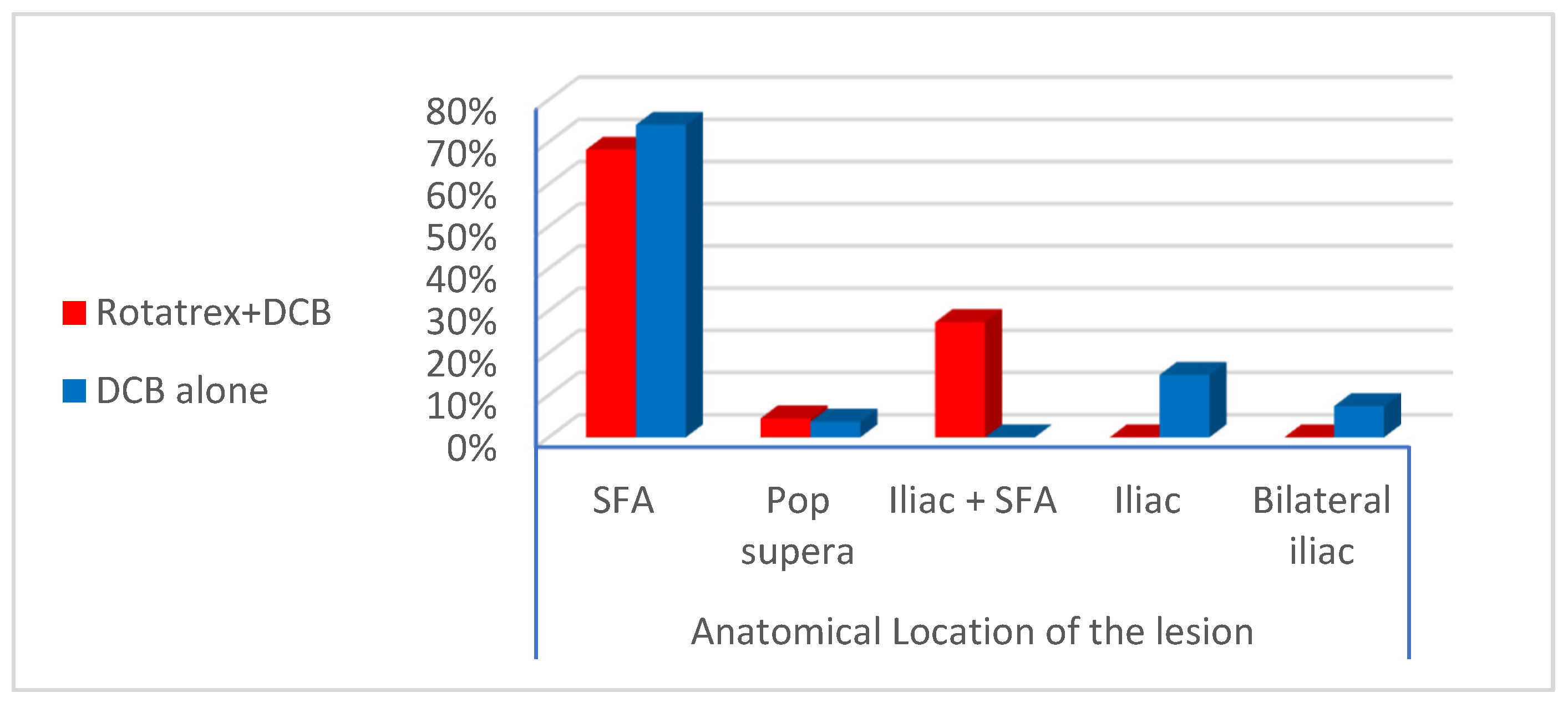
For most of the published studies and trials, the femoropopliteal segment is the arterial territory most frequently involved by ISR following previous endovascular stenting. The lesion distribution observed in the enrolled candidates revealed restenosis of previously deployed SFA stents in (71.4%) of patients while combined restenosis in iliac + SFA stents in (12.2%) of patients [1].
In our study, ISR lesions within the femoropopliteal segment were found to be focal single stenosis in 6.1% of patients, multiple stenoses in 32.7% of patients, total occlusion in 22.4% of patients, and combined segmental stenosis and occlusion in 10.2% patients. For the patients with combined iliac + SFA stents, all of them showed stenosis of the iliac stent accompanied by occlusion of the SFA stent. The mean length of the lesion was 141.8 mm for Rotarex + DCB patients and 92.9 mm for DCB alone patients. Most of these criteria were similar to published studies and trials by Tepe et al. and Kokkinidis et al. [10,13].
Zeller T et al. documented 78.8% in-stent stenosis and 30% in-stent occlusion at 12 months follow-up for lesions with a mean length of 133 ± 91.7 mm [14].
The outcome of several published studies and trials for the treatment modalities and techniques used for the treatment of femoropopliteal segment ISR is shown in Table 7 and Table S7. Also, multiple studies have documented more fruitful outcomes of hybrid treatment using the combination of atherectomy (laser, directional) with DCB angioplasty [15].
The EXCITE ISR trial is the first randomized prospective study that highlighted the superiority of laser atherectomy plus PTA versus PTA alone, giving procedural success (93.5% vs. 82.7%, p = 0.01) and few complications. Also, 6 months follow-up revealed freedom from CD-TLR (73.5% versus 51.8%, p < 0.005) [16].
The Rotarex® system is a percutaneous mechanical rotational endovascular atherectomy in combination with a thrombectomy device, which provides safety and effectiveness in eliminating large thrombus load [17,18].
A comparative study between PMD and local thrombolysis for the treatment of acute and subacute in-stent thrombotic occlusion was conducted by Kronlage et al. The results showed technical success, reaching up to 98% for both modalities. However, one year of primary and secondary patency after PMD documented significantly better results compared to local thrombolysis (63% and 85%, p < 0.05) [19].
Also, successful recanalization was reported in (95%) of performed interventions by Stanek et al. for patients presenting with acute and subacute thrombotic and/or atherosclerotic in-stent occlusion in the peripheral limb arteries using Rotarex® catheter system for PMD [20].
According to multiple published studies, the Rotarex® catheter system can be used for short or long occlusions with almost the same success rate, especially if combined with other adjunctive measures, particularly DCB, preferably in heavily calcified vessels and keeping in consideration guidewire lesion traversal should pass transluminal at all possible means [20,21,22].
This study included 49 patients with ISR in a previously deployed peripheral stent, conducted on a heterogeneous patch of the population with different target vessels (iliac, SFA, popliteal or combined lesions) and with different types of in-stent lesions (stenosis, occlusion or combined), (thrombotic or hyperplastic) and the use of different techniques (PMD + DCB or DCB alone).
Much recent literature has documented that pre-treatment with atherectomy devices prior to balloon dilatation and drug-coated balloon showed a significant reduction in dissection within the offending lesion with subsequent need for endovascular stenting. Despite these superior early and mid-term results, these studies did not support more favorable outcomes of combined use of atherectomy and drug-coated balloon when compared to the use of drug-coated balloon alone regarding CD-TLR. Also, a meta-analysis of five clinical studies revealed no statistically significant privilege of combined use of atherectomy and DCB in terms of the primary patency or TLR compared to DCB alone [23].
One of the major limitations of these published trials was the small study sample, giving rise to insufficient statistical power, making clear data on whether atherectomy with adjunctive DCB has clinical benefit in the treatment of ISR of the femoropopliteal segment inconclusive. Other explanations were concluded for these limitations as multiple types of atherectomy devices were used for the treatment of ISR or occlusion. It is still also unclear how much volume of the offending plaque should be debulked in order to obtain optimal early and late outcomes. In addition, too much lesion debulking can induce medial and adventitial injuries, which might promote subsequent restenosis [24].
A study by Fanelli et al. concluded that severe lesion calcification was considered a significant risk factor for losing patency after DCB alone [5]. Severe lesion calcification can easily limit the lesion response to balloon dilation and also impede the delivery of antiproliferative drugs. A trial published by Cioppa et al. has reported a 12-month primary patency rate of 90% after combined atherectomy and DCB in severely calcified ISR lesions [8] while in the long-lesion subgroups of the Lutonix and IN.PACT global registries, dissection of the lesion happened in 34.3% and 62.1%, and bail-out stenting was needed in 35.7% and 39.4%, respectively [25,26].
Drawbacks related to the use of atherectomy devices have been encountered in many studies, especially intra-procedural distal embolization (advocating the use of embolic protection devices) [27,28] and vessel wall perforation in 2.6% of cases, as presented by Zhen Y et al. in a meta-analysis study [23]. Meanwhile, a study by Boitet et al. suggested an association between the different types of DCB profiles and the occurrence of distal embolic events [29].
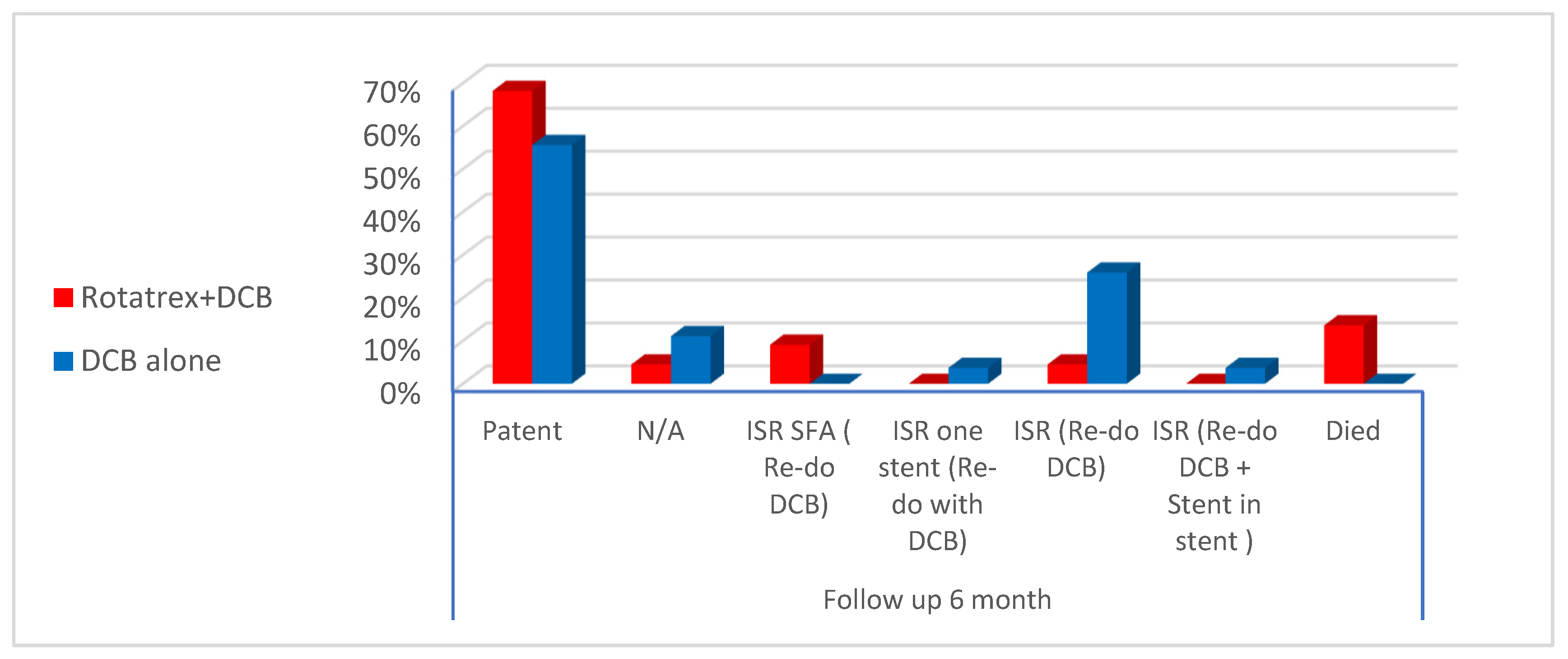
The current study showed intra-procedural distal embolization in (9%) of cases who were treated with Rotarex. Also, there was no statistical difference in distal embolization regarding the profile of DCB in both the Rotarex + DCB and DCB alone. To be noted, in the current study, we did not encounter vessel wall perforation while using Rotarex [21].
There is a paucity of studies comparing lower extremity bypass and different types of peripheral atherectomy devices. However, in 2024, Van Leeuwen G. et al. published a single-center retrospective study and discussed the technical success and only short-term outcomes of the BYCROSS® atherectomy device used for the management of long-segment femoropopliteal lesions (including both atherosclerotic disease and hyperplastic lesions of previously deployed stents) compared to standard lower extremity surgical bypass regarding both safety, feasibility and possible complications. They showed a technical success rate of 100%, a 30-day mortality of 5%, a 30-day MACE rate of 11%, and a 30-day MALE rate of 0% in a cohort of patients with significant comorbidities (i.e., 75% with American Society of Anesthesiologists Physical Status Classification (ASA) III and IV). The patency rate at 30 days was acceptable at 83% [30].

5. Limitations

This study showed two obvious limitations. First, this is a retrospective, non-randomized study with inherent limitations, such as variations in the baseline characteristics and possible potential confounding factors like the decision of the treating physician regarding the use of treatment modality. Second, in this study, the follow-up was performed for 6 months, so late clinical outcomes between the two treatment groups may not be properly evaluated. Finally, some data on the previous therapy received by the patients were lacking at the time of inclusion.

6. Conclusions

This study included 49 patients with ISR in a previously deployed peripheral stent, conducted on a heterogeneous patch of the population with different target vessels (iliac, SFA, popliteal or combined lesions) and with different types of in-stent lesions (stenosis, occlusion or combined) (thrombotic or hyperplastic) and using different techniques (PMD + DCB or DCB alone). This study showed that Rotarex® combined with DCB angioplasty is a safe and effective modality for achieving recanalization for patients with in-stent occlusion or restenosis, especially in the femoropopliteal arterial segment. It gives a satisfactory outcome in terms of technical success, freeform from CD-TLR, MALE and mortality. Despite these promising results, further research with a large enrolled population may be required to determine cost/benefit.

Supplementary Materials

The following supporting information can be downloaded at: https://www.mdpi.com/article/10.3390/jvd3030023/s1, Table S1: Recorded patients demographics and risk factors; Table S2: anatomical distribution, morphology and characteristics of re-stenotic of the lesion; Table S3: Patient presentation at the time of intervention; Table S4: Technical success and complication related to the procedure; Table S5: Follow up at 1, 3 and 6 months; Table S6: CD-TLR, MALE and mortality of the treated patients; Table S7: Comparison between current study and other studies regarding CR-TLR and follow up.

Author Contributions

The conceptualization and ideation of this work were attributed to M.A. and M.N.; the literature search was conducted by A.E.; the data were analyzed by A.T.; the work was drafted and revised by M.A. All authors have read and agreed to the published version of the manuscript.

Funding

This research received no external funding.

Institutional Review Board Statement

The study was conducted in accordance with the Declaration of Helsinki and approved by the research ethics committee of the faculty of medicine, Cairo University (protocol code N-9-2024 and date of approval 24 February 2024).

Informed Consent Statement

Informed consent was obtained from all subjects involved in the study, and written informed consent has been obtained from the patient(s) to publish this paper.

Data Availability Statement

The original contributions presented in the study are included in the article; further inquiries can be directed to the corresponding author.

Conflicts of Interest

The authors declare no conflicts of interest.

References

  1. Loffroy, R.; Edriss, N.; Goyault, G.; Chabanier, A.; Pernes, J.-M.; Sauguet, A.; Touil, M.; Woerly, B.; Pongas, D.; Chevallier, O.; et al. Percutaneous mechanical atherothrombectomy using the Rotarex®S device in peripheral artery in-stent restenosis or occlusion: A French retrospective multicenter study on 128 patients. Quant. Imaging Med. Surg. 2020, 10, 283–293. [Google Scholar] [CrossRef]
  2. Banerjee, S.; Sarode, K.; Mohammad, A.; Gigliotti, O.; Baig, M.S.; Tsai, S.; Shammas, N.W.; Prasad, A.; Abu-Fadel, M.; Klein, A.; et al. Femoropopliteal artery stent thrombosis: Report from the excellence in peripheral artery disease registry. Circ. Cardiovasc. Interv. 2016, 9, e002730. [Google Scholar] [CrossRef]
  3. Scheinert, D.; Duda, S.; Zeller, T.; Krankenberg, H.; Ricke, J.; Bosiers, M.; Tepe, G.; Naisbitt, S.; Rosenfield, K. The LEVANT I (Lutonix paclitaxel-coated balloon for the prevention of femoropopliteal restenosis) trial for femoropopliteal revascularization: First-in-human randomized trial of low-dose drug-coated balloon versus uncoated balloon angioplasty. JACC Cardiovasc. Interv. 2014, 7, 10–19. [Google Scholar] [CrossRef]
  4. Cha, J.-J.; Lee, J.-H.; Ko, Y.-G.; Roh, J.-H.; Yoon, Y.-H.; Lee, Y.-J.; Lee, S.-J.; Hong, S.-J.; Ahn, C.-M.; Kim, J.-S.; et al. Clinical Outcomes of Atherectomy Plus Drug-coated Balloon Versus Drug-coated Balloon Alone in the Treatment of Femoropopliteal Artery Disease. Korean Circ. J. 2022, 52, 123–133. [Google Scholar] [CrossRef]
  5. Fanelli, F.; Cannavale, A.; Gazzetti, M.; Lucatelli, P.; Wlderk, A.; Cirelli, C.; D’adamo, A.; Salvatori, F.M. Calcium burden assessment and impact on drug-eluting balloons in peripheral arterial disease. Cardiovasc. Interv. Radiol. 2014, 37, 898–907. [Google Scholar] [CrossRef]
  6. Lichtenberg, M.; Stahlhoff, F.W.; Boese, D. Endovascular treatment of acute limb ischemia and proximal deep vein thrombosis using rotational thrombectomy: A review of published literature. Cardiovasc. Revasc. Med. 2013, 14, 343–348. [Google Scholar] [CrossRef]
  7. Katsanos, K.; Spiliopoulos, S.; Reppas, L.; Karnabatidis, D. Debulking atherectomy in the peripheral arteries: Is there a role and what is the evidence? Cardiovasc. Interv. Radiol. 2017, 40, 964–977. [Google Scholar] [CrossRef]
  8. Cioppa, A.; Stabile, E.; Popusoi, G.; Salemme, L.; Cota, L.; Pucciarelli, A.; Ambrosini, V.; Sorropago, G.; Tesorio, T.; Agresta, A.; et al. Combined treatment of heavy calcified femoro-popliteal lesions using directional atherectomy and a paclitaxel coated balloon: One-year single centre clinical results. Cardiovasc. Revasc. Med. 2012, 13, 219–223. [Google Scholar] [CrossRef]
  9. Kim, W.; Choi, D. Treatment of femoropopliteal artery in-stent restenosis. Korean Circ. J. 2018, 48, 191–197. [Google Scholar] [CrossRef]
  10. Tepe, G.; Brodmann, M.; Micari, A.; Scheinert, D.; Choi, D.; Menk, J.; Zeller, T.; on behalf of the IN. PACT GlobalStudy Investigators. 5-Year Outcomes of Drug-CoatedBalloons for Peripheral Artery In-Stent Restenosis, Long Lesions, and CTOs. JACC Cardiovasc. Interv. 2023, 16, 1065–1078. [Google Scholar] [CrossRef]
  11. Scheinert, D.; Schmidt, A.; Zeller, T.; Müller-Hülsbeck, S.; Sixt, S.; Schröder, H.; Weiss, N.; Ketelsen, D.; Ricke, J.; Steiner, S.; et al. German center subanalysis of the LEVANT 2 global randomized study of the Lutonix drug-coated balloon in the treatment of femoropopliteal occlusive disease. J. Endovasc. Ther. 2016, 23, 409–416. [Google Scholar] [CrossRef]
  12. Kohi, M.P.; Brodmann, M.; Zeller, T.; Micari, A.; Baumgartner, I.; Wang, H.; Wall, B.; Razavi, M.K. Sex-related differences in the long-term outcomes of patients with femoropopliteal arterial disease treated with the IN.PACT drug-coated balloon in the IN.PACT SFA randomized controlled trial: A post hoc analysis. J. Vasc. Interv. Radiol. 2020, 31, 1410–1418.e10. [Google Scholar] [CrossRef]
  13. Kokkinidis, D.G.; Behan, S.; Jawaid, O.; Hossain, P.; Giannopoulos, S.; Singh, G.D.; Laird, J.R.; Valle, J.A.; Waldo, S.W.; Armstrong, E.J. Laser atherectomy and drug-coated balloons for the treatment of femoropopliteal in-stent restenosis: 2-Year outcomes. Catheter. Cardiovasc. Interv. 2020, 95, 439–446. [Google Scholar] [CrossRef]
  14. Zeller, T.; Dake, M.D.; Tepe, G.; Brechtel, K.; Noory, E.; Beschorner, U.; Kultgen, P.L.; Rastan, A. Treatment of femoropopliteal in-stent restenosis with paclitaxel-eluting stents. JACC Cardiovasc. Interv. 2013, 6, 274–281. [Google Scholar] [CrossRef]
  15. Milnerowicz, A.; Milnerowicz, A.; Kuliczkowski, W.; Protasiewicz, M. Rotational atherectomy plus drug-coated balloon angioplasty for the treatment of total in-stent occlusions in iliac and infrainguinal arteries. J. Endovasc. Ther. 2019, 26, 316–321. [Google Scholar] [CrossRef]
  16. Dippel, E.J.; Makam, P.; Kovach, R.; George, J.C.; Patlola, R.; Metzger, D.C.; Mena-Hurtado, C.; Beasley, R.; Soukas, P.; Colon-Hernandez, P.J.; et al. Randomized controlled study of excimer laser atherectomy for treatment of femoropopliteal in-stent restenosis: Initial results from the EXCITE ISR trial (EXCImer Laser Randomized Controlled Study for Treatment of FemoropopliTEal In-Stent Restenosis). JACC Cardiovasc. Interv. 2015, 8, 92–101. [Google Scholar] [CrossRef]
  17. Giusca, S.; Raupp, D.; Dreyer, D.; Eisenbach, C.; Korosoglou, G. Successful endovascular treatment in patients with acute thromboembolic ischemia of the lower limb including the crural arteries. World J. Cardiol. 2018, 10, 145–152. [Google Scholar] [CrossRef]
  18. Liu, J.; Li, T.; Huang, W.; Zhao, N.; Liu, H.; Zhao, H.; Wang, H. Percutaneous mechanical thrombectomy using Rotarex catheter in peripheral artery occlusion diseases—Experience from a single center. Vascular 2019, 27, 199–203. [Google Scholar] [CrossRef]
  19. Kronlage, M.; Printz, I.; Vogel, B.B.; Blessing, E.; Müller, O.J.; Katus, H.A.; Erbel, C. A comparative study on endovascular treatment of (sub)acute critical limb ischemia: Mechanical thrombectomy vs thrombolysis. Drug Des. Devel. Ther. 2017, 11, 1233–1241. [Google Scholar] [CrossRef]
  20. Stanek, F.; Ouhrabkova, R.; Prochazka, D. Mechanical thrombectomy using the Rotarex catheter in the treatment of acute and subacute occlusions of peripheral arteries: Immediate results, long-term follow-up. Int. Angiol. 2013, 32, 52–60. [Google Scholar]
  21. Silingardi, R.; Cataldi, V.; Moratto, R.; Azzoni, I.; Veronesi, J.; Coppi, G. Mechanical thrombectomy in in-stent restenosis: Preliminary experience at the iliac and femoropoliteal arteries with the Rotarex system. J. Cardiovasc. Surg. 2010, 51, 543–550. [Google Scholar]
  22. Liao, C.J.; Song, S.H.; Li, T.; Zhang, Y.; Zhang, W. Combination of rotarex thrombectomy and drug-coated balloon for the treatment of femoropopliteal artery in-stent restenosis. Ann. Vasc. Surg. 2019, 60, 301–307. [Google Scholar] [CrossRef] [PubMed]
  23. Zhen, Y.; Chang, Z.; Wang, C.; Liu, Z.; Zheng, J. Directional atherectomy with antirestenotic therapy for femoropopliteal artery disease: A systematic review and meta-analysis. J. Vasc. Interv. Radiol. 2019, 30, 1586–1592. [Google Scholar] [CrossRef] [PubMed]
  24. Tarricone, A.; Ali, Z.; Rajamanickam, A.; Gujja, K.; Kapur, V.; Purushothaman, K.-R.; Purushothaman, M.; Vasquez, M.; Zalewski, A.; Parides, M.; et al. Histopathological evidence of adventitial or medial injury is a strong predictor of restenosis during directional atherectomy for peripheral artery disease. J. Endovasc. Ther. 2015, 22, 712–715. [Google Scholar] [CrossRef] [PubMed]
  25. Thieme, M.; Von Bilderling, P.; Paetzel, C.; Karnabatidis, D.; Perez Delgado, J.; Lichtenberg, M.; Lutonix Global SFA Registry Investigators. The 24-month results of the Lutonix Global SFA Registry: Worldwide experience with Lutonix drug-coated balloon. JACC Cardiovasc. Interv. 2017, 10, 1682–1690. [Google Scholar] [CrossRef] [PubMed]
  26. Scheinert, D.; Micari, A.; Brodmann, M.; Tepe, G.; Peeters, P.; Jaff, M.; Wang, H.; Schmahl, R.; Zeller, T.; IN. PACT Global Study Investigators. Drug-coated balloon treatment for femoropopliteal artery disease. Circ. Cardiovasc. Interv. 2018, 11, e005654. [Google Scholar] [CrossRef] [PubMed]
  27. Krishnan, P.; Tarricone, A.; Purushothaman, K.R.; Purushothaman, M.; Vasquez, M.; Kovacic, J.; Baber, U.; Kapur, V.; Gujja, K.; Kini, A.; et al. An algorithm for the use of embolic protection during atherectomy for femoral popliteal lesions. JACC Cardiovasc. Interv. 2017, 10, 403–410. [Google Scholar] [CrossRef] [PubMed]
  28. Khakwani, M.Z.; Kotev, S.; Boiangiu, C.; Hasan, O.; Anna, M.; Tayal, R.; Kaid, K.; Baker, G.; Cohen, M.; Wasty, N. Ubiquitous nature of distal athero/thromboembolic events during lower extremity atherectomy procedures involving the superficial femoral artery. Int. J. Angiol. 2016, 25, 252–257. [Google Scholar] [CrossRef] [PubMed]
  29. Boitet, A.; Grassin-Delyle, S.; Louedec, L.; Dupont, S.; Lamy, E.; Coggia, M.; Michel, J.-B.; Coscas, R. An experimental study of paclitaxel embolisation during drug coated balloon angioplasty. Eur. J. Vasc. Endovasc. Surg. 2019, 57, 578–586. [Google Scholar] [CrossRef]
  30. van Leeuwen, G.L.; Bokkers, R.P.H.; Oldenziel, J.; Schuurmann, R.C.L.; Vos, C.G.; de Vries, J.-P.P.M. Safety and Feasibility of the BYCROSS® Atherectomy Device for the Treatment of Femoropopliteal Arterial Obstructions: Single-Center Short-Term Outcomes. J. Clin. Med. 2024, 13, 1809. [Google Scholar] [CrossRef]
Figure 1. Re-stenotic lesion distribution.
Jvd 03 00023 g001
Figure 2. Diagnostic angiography showing Rt SFA ISR (a) through cross-over antegrade access (b); PMD using Rotarex® system (c); DCB angioplasty (d); completion angiography revealed successful revascularization (e).
Jvd 03 00023 g002
Figure 3. CTA (a) and diagnostic angiography (b) showing LT SFA ISR through cross-over antegrade access, atheroma sample after PMD using Rotarex® system (c); completion angiography revealed successful revascularization before DCB angioplasty (d) (computed tomography angiography (CTA), in-stent restenosis (IRS), percutaneous mechanical debulking (PMD) and drug-coated balloon (DCB)).
Jvd 03 00023 g003
Figure 4. Diagnostic angiography showing LT SFA metal jacket ISR through cross-over antegrade access (ac); PMD using Rotarex® system (d,e); DCB angioplasty (f); completion angiography revealed successful revascularization (g) (in-stent restenosis (IRS), percutaneous mechanical debulking (PMD) and drug-coated balloon (DCB)).
Jvd 03 00023 g004aJvd 03 00023 g004b
Figure 5. Diagnostic angiography showing LT common iliac artery ISR (a) through ipsilateral retrograde access; PMD using Rotarex® system (bd); DCB angioplasty (e); completion angiography revealed successful revascularization (f) (in-stent restenosis (IRS), percutaneous mechanical debulking (PMD) and drug-coated balloon (DCB)).
Jvd 03 00023 g005aJvd 03 00023 g005b
Figure 6. Diagnostic angiography showing Rt SFA ISR (a) through cross-over antegrade access; angiography after plain balloon angioplasty with spiral dissection (b); DCB angioplasty; completion angiography revealed successful revascularization (c) (in-stent restenosis (IRS) and drug-coated balloon (DCB)).
Jvd 03 00023 g006
Figure 7. Documenting the follow-up after 6 months for patients with previously treated ISR.
Jvd 03 00023 g007
Table 1. Recorded patients’ demographics and risk factors.
TotalRotarex + DCBDCB Alonep Value
Count%Count%Count%
Male2857.1%1254.5%1659.3%0.740
Female2142.9%1045.5%1140.7%
DM3367.3%1463.6%1970.4%0.617
HTN3163.3%1254.5%1970.4%0.253
Smoking3061.2%1359.1%1763.0%0.782
Dyslipidemia2244.9%1045.5%1244.4%0.944
IHD1428.8%522.7%933.3%0.414
Table 2. Patient presentation at the time of intervention.
Rotarex + DCBDCB Alonep Value
Count%Count%
PresentationClaudication14.5%829.6%0.018
Rest pain418.2%725.9%
Cyanosis of the toes418.2%00.0%
Mottling of the toes14.5%00.0%
Fixed color changes14.5%00.0%
Tissue loss836.4%1244.4%
Sudden onset of acute ischemia313.5%00.0%
Table 3. Anatomical distribution, nature and characteristics of re-stenotic of the lesion.
Rotarex + DCBDCB Alonep Value
Count%Count%
Types of ISRINH1254.5%27100.0%-
Thrombosis1045.5%00.0%
single or multipleSingle1359.1%725.9%0.019
Multiple940.9%2074.1%
stenosis or occlusionStenosis and occlusion940.9%13.7%<0.001
Stenosis (iliac) and occlusion (SFA)14.5%00.0%
Stenosis00.0%2488.9%
focal stenosis00.0%13.7%
Occlusion1254.5%13.7%
Anatomical location of the lesionSFA1568.2%2074.1%0.003
Pop supera14.5%13.7%
Iliac + SFA627.3%00.0%
Iliac00.0%414.8%
Bilateral iliac00.0%27.4%
Table 4. Technical success and complications related to the procedure.
Rotarex + DCBDCB Alonep Value
Count%Count%
Technical successYes22100.0%27100.0%-
Procedure related complicationsArterial thrombosis in tibials (Anticoagulation)00.0%311.1%0.021
CIN (conservative)00.0%311.1%
Distal embolization (Cyanosis of big toe) Conservative14.5%00.0%
Distal embolization (cyanosis of medial 3 toes) conservative14.5%00.0%
Lost radial pulse but compensated limb14.5%00.0%
Minor groin hematoma00.0%311.1%
No1986.4%1866.7%
Table 5. Follow-up at 1, 3 and 6 months.
Rotarex + DCBDCB Alonep Value
Count%Count%
Follow-up 1 monthPatent2195.5%27100.0%0.449
Death14.5%00.0%
Follow-up 3 monthsPatent1986.4%2177.8%0.160
N/A (bypass, death or amputation)00.0%13.7%
ISR (re-do DCB + stent in-stent)00.0%27.4%
IS Occlusion (re-do DCB + stent in-stent)00.0%13.7%
IS Occlusion (fem-pop bypass)00.0%27.4%
IS Occlusion (bypass)14.5%00.0%
Died29.1%00.0%
Follow-up 6 monthsPatent1568.2%1555.6%0.028
N/A (Bypass, death or amputation)14.5%311.1%
ISR SFA (re-do DCB)29.1%00.0%
ISR one stent (re-do with DCB)00.0%13.7%
ISR (re-do DCB)14.5%725.9%
ISR (re-do DCB + stent in-stent)00.0%13.7%
Death313.6%00.0%
Table 6. Clinically driven target lesion revascularization (CD-TLR), major limb adverse event (MALE) and mortality of the treated patients.
Rotarex + DCBDCB Alonep Value
Count%Count%
CD-TLRYes418.2%1140.7%0.227
No1568.2%1348.1%
N/A (bypass, death or amputation)313.6%311.1%
MALETMA14.5%13.7%0.709
No1568.2%2281.5%
N/A (bypass/death)29.1%00.0%
BKA14.5%00.0%
Big toe amputation14.5%13.7%
AKA29.1%311.1%
MortalityYes313.6%13.7%0.314
No1986.4%2696.3%
Mortality detailsAfter 2 weeks (septicemia +MOSF)14.5%00.0%0.231
After 1 month (MI)14.5%00.0%
Died 2 months (massive stroke)00.0%13.7%
After 5 months (stroke)14.5%00.0%
No1986.4%2696.3%
Table 7. Comparison between current study and other studies regarding CR-TLR and follow-up.
Study DeviceFollow-UpLesions Free CD-TLRPrimary Patency
FAIRDCB126290.8%70.5%
PACUBADCB127449%40.7%
PLAISIRDCB125590.2%83.7%
EXCITE ISRLaser atherectomy + PTA616973.5%/
PATENTExcimer Laser + Turbo Booster129064.4%73.8%
Current studyRotational atherectomy + DCB614268.2%/
Disclaimer/Publisher’s Note: The statements, opinions and data contained in all publications are solely those of the individual author(s) and contributor(s) and not of MDPI and/or the editor(s). MDPI and/or the editor(s) disclaim responsibility for any injury to people or property resulting from any ideas, methods, instructions or products referred to in the content.

