Case Report

Covered Stent Herniation into Coronary Aneurysm Sac—A Case-Inspired Review of Neurointerventional Realignment Techniques

1
Department of Invasive Cardiology and Interventional Radiology, St. Adalbert’s Hospital, Copernicus PL, 80-462 Gdansk, Poland
2
Department of Cardiology, St. Vincent de Paul Hospital Pomeranian Hospitals, 81-348 Gdynia, Poland
3
Department of Cardiology and Interventional Angiology, Kashubian Center for Heart and Vascular Diseases, Pomeranian Hospitals, 84-200 Wejherowo, Poland
*
Author to whom correspondence should be addressed.
J. Vasc. Dis. 2023, 2(4), 402-412; https://doi.org/10.3390/jvd2040031
Submission received: 31 May 2023 / Revised: 30 August 2023 / Accepted: 4 September 2023 / Published: 1 November 2023
(This article belongs to the Section Cardiovascular Diseases)

Abstract

:
Background: Coronary aneurysms are an infrequent finding in diagnostic angiography, with a reported incidence of 0.35–0.7% in the largest contemporary registries. At least half of them have an atherosclerotic etiology and as such they are often diagnosed in the setting of acute coronary syndrome. The wiring of a thrombosed aneurysm is a difficult task and after successful recanalization, the operator has to decide on the optimal method of aneurysm exclusion. Covered stents are commonly deployed, but their use involves the risk of delivery failure as well as device dislodgement and loss due to their stiffness and size. Moreover, proper stent sizing and apposition is difficult in the case of thrombosed aneurysms. Case presentation: We present a case of coronary aneurysm recanalization and exclusion with a covered stent, with a postdilation-induced stent foreshortening. Due to the subsequent stent migration into the aneurysm sac, its repositioning was attempted. The pitfalls of coronary aneurysm stenting and neurointerventional techniques of prolapsed device realignment are discussed. Conclusions: An adequate landing zone is of the utmost importance in aneurysm exclusion with covered stents. In the case of a short stent anchoring in the normal vessel, another covered or conventional stent should be deployed to mitigate the risk of the device migration.

1. Introduction

Coronary artery aneurysms (focal dilatations of an artery segment > 1.5-fold the normal size of an adjacent reference vessel) are infrequently encountered in angiography, with a reported prevalence of 0.35–0.7% in the largest contemporary registries [1,2]. The main cause of coronary aneurysms is in most cases atherosclerosis, and less often Takayasu arteritis, connective tissue disorders, congenital anomalies, infection, specific drug reactions, trauma, and Kawasaki disease. In addition to ‘spontaneous’ aneurysms, cases of iatrogenic coronary artery dilation related to drug-eluting stent implantation have started accumulating [3]. The natural history of coronary aneurysm is unclear; however, potential complications include perforation, thrombosis, and distal embolism. Recommendations for optimal management are lacking, and treatment options include anticoagulation and antiplatelet therapy, percutaneous coronary intervention, and surgical procedures (aneurysm ligation, re-section, marsupialization with interposition graft, or most commonly bypass grafting with the vessel suturing at the aneurysm level).
Primary angioplasty of thrombosed coronary aneurysms is a difficult task in itself, involving technical challenges such as aneurysm crossing and distal vessel wiring; increased risk of distal embolization and the no-reflow phenomenon (up to 70%) due to large thrombus burden; higher incidence of procedure failure; and stent malapposition and dislodgement [1,4,5,6]. Several studies have also reported higher odds of mortality, stent thrombosis, and other adverse events both in-hospital and in the long-term follow-up [1,4,5,6]. Successful aneurysm recanalization still leaves open the question of optimal management to exclude the aneurysm sac. Self-expanding stents have been used in ectatic coronary segments, while treatment of large aneurysms has often involved the insertion of covered stents, notorious for their susceptibility to restenosis and thrombosis, due to, as has been suggested, delayed endothelialization and the increased susceptibility to thrombus formation of these stents [1,7,8,9,10]. Covered stent registries and retrospective studies reported high thrombosis rates in the short- and long-term observation, ranging from 4.4–8.9% in the 1-year period to 17.8% in the 10-year follow-up [8,11]. Some of them suggested that the risk may be higher for old polytetrafluoroethylene-covered stents than for newer polyurethane-covered prostheses (9.8% vs. 1.8%, respectively); however, these results may have also been affected by the usually prolonged dual antiplatelet therapy recommended with contemporary covered stents and more potent agents prescribed nowadays [12].
While coronary perforation remains the main indication for covered stent deployment, some studies suggest that stent thrombosis risk may be particularly high when they are implanted to treat coronary aneurysms. One large study of 127 Papyrus stents (Biotronik AG, Bülach, Switzerland) implanted in 108 patients reported a higher thrombosis rate for coronary aneurysm management compared to coronary artery perforation treatment (7.1% vs. 0%, respectively), with similar rates of major adverse cardiovascular events [9]. In contrast, another large retrospective analysis of 190 patients, in whom a covered stent had been implanted (mainly a polytetrafluoroethylene-covered Jostent Graftmaster, Abbott Vascular, Santa Clara, CA, USA), reported at the 10-year follow-up that target vessel occlusion, target vessel myocardial infarction, and stent thrombosis were not identified in the aneurysm group, in comparison to 38.4%, 20.6% and 23.9% of such events, respectively, in the coronary perforation group [8].
To solve the problem of restenosis, burying a covered stent under a drug-eluting stent has been proposed [13,14,15]. Another strategy of inhibiting edge restenosis is the implantation of a longer drug-eluting stent prior to covered stent deployment or an overlapping implantation of covered stents with drug-eluting stents at the edges [15,16,17]. Yet another solution is a double or triple stent technique, wherein a ‘coronary flow diverter’ is created by the deployment of two/three completely overlapping drug-eluting coronary stents [18,19]. Micro-mesh self-expanding nitinol carotid and coronary stents have also been used for coronary aneurysm exclusion [20,21]. In both strategies, a slow-velocity zone in the aneurysm sac is sufficient to induce its mural thrombosis and sealing-off, while the reduction of hydrodynamic pressure acting on the aneurysm wall may also induce its shrinkage [22]. Both drug-eluting and bare metal stents have routinely been used in the treatment of coronary aneurysms in the US due to the lack of FDA approval of covered stents [2].
We report a case of primary percutaneous recanalization of a thrombosed coronary aneurysm and its exclusion with a covered stent, with subsequent downstream migration and herniation of the stent into the aneurysm sac resulting in an impeded blood flow into the distal segment of the right coronary artery. An attempt at stent realignment was performed. The pitfalls of coronary aneurysm stenting and neurointerventional techniques for the herniated stent rearrangement are discussed further.

2. Case Presentation

A 91-year old woman with a history of arterial hypertension, chronic renal insufficiency, normocytic anaemia, chronic obstructive pulmonary disease, and prior tuberculosis was admitted due to chest pain of 1 h duration. In ECG, ST segment elevation in the inferior wall leads with a third-degree atrioventricular block and an escape rhythm of 50 bpm were present. Upon arrival, the patient was conscious with blood pressure of 85/60 mmHg. In an urgent coronary angiography, proximal occlusion of the dominant right coronary artery (RCA) was found, as well as a small ectopic left circumflex artery arising from the RCA and left coronary system without significant lesions (Figure 1A, Video S1). Balloon recanalization revealed a 15 × 20 mm thrombosed aneurysm of the mid-RCA (Figure 1B, Video S2). Due to the fact that balloon angioplasty alone was not enough to restore TIMI 3 flow in the RCA and due to the risk of reocclusion of the thrombus-laden aneurysm, the longest locally available covered stent, a 5.0 × 24 mm Begraft (Bentley Innomed GmbH, Hechingen, Germany) was deployed to exclude the aneurysm with the support of a 6 French guide extension catheter (Figure 1C). A minor residual leak into the thrombus-filled aneurysm was observed at its proximal edge after a postdilation-induced stent foreshortening (Figure 1D, Video S3). Soon after RCA reperfusion, the atrioventricular block subsided and blood pressure rose. The decision to deploy another covered stent was postponed due to the higher risk of restenosis and thrombosis associated with the implantation of overlapping stents. The operator assessed that a small leak would not interfere with the clotting of the aneurysm, which in turn should stabilize the stent. Control angiography was scheduled to decide the need for an additional covered stent placement if the aneurysm sac did not thrombose under the stent and the leak persisted. During hospitalization, the chest pain did not recur and the patient was mobilized without signs and symptoms of heart failure. The patient was discharged with a left ventricle ejection fraction of 55% and akinesia of the basal segments of the inferior and inferoseptal wall.
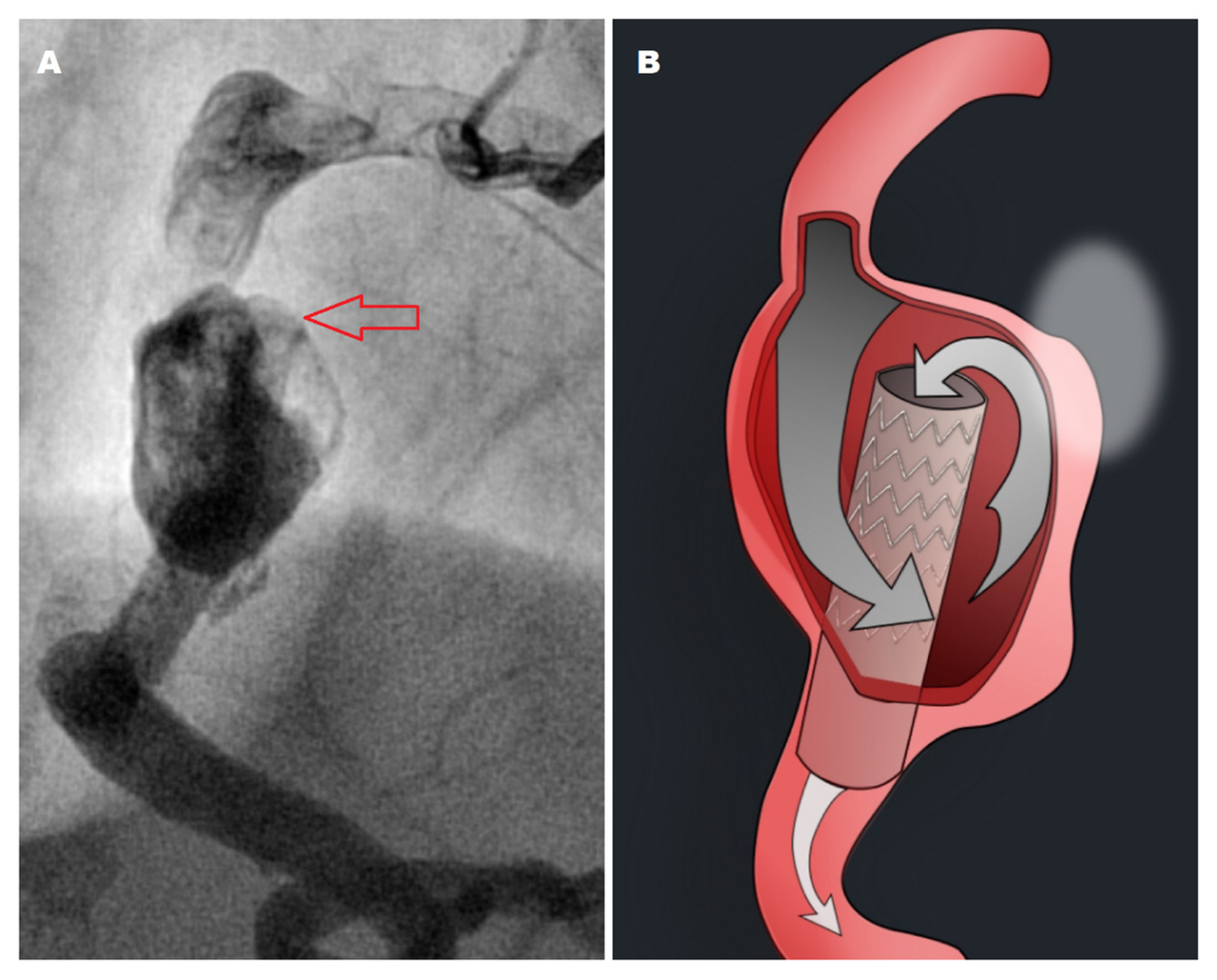
In a repeat angiography three weeks later, stent distal migration with prolapse of its proximal edge into the aneurysm sac was found (Figure 1E, Videos S4 and S5). The perfusion of the distal RCA was impeded, with contrast agent filling the aneurysm sac first, next passing through the prolapsed stent edge into the stent lumen, and then flowing downstream into the distal RCA (Figure 2A,B). The patient was informed about the complication and the risk of stent thrombosis and vessel perforation as well as the risks associated with the realignment attempt. Stent repositioning was attempted to deploy a second, overlapping covered stent. After wiring and microcatheter insertion (and wire exchange for an extra support wire), the system was pulled in a failed attempt to unloop the wire and realign the stent (Figure 1F, Videos S6 and S7). We were unable to introduce a balloon for distal anchoring, or to partially advance it into the stent and inflate it for stent edge realignment. A guide extension catheter was used to increase backup. Strong resistance was felt during attempts to realign the stent and the balloon passage into the stent was blocked as the stent was clinging closely to the aneurysm wall. As a bailout, we decided to bypass the stent at its distal edge with a chronic total occlusion wire and crush it with covered stents. However, the intervention had to be terminated due to poor tolerance of the prolonged procedure by the frail patient. The patient declined another attempt. As a result, the distal RCA was left to be supplied by the sequential flow (Video S8). Prolonged dual antiplatelet therapy was recommended to prevent stent thrombosis and so far the follow-up has been uneventful.

3. Discussion

Large thrombosed coronary aneurysms are infrequently encountered during primary angioplasty and coronary stent herniation is an extremely rare complication. In contrast to neurointerventionalists, cardiologists are seldom faced with device migration. In the present case, several factors led to the subsequent stent prolapse into the aneurysm sac, including too short a landing zone of the stent, postdilation-induced stent foreshortening, use of a stent with uncovered edges, and withholding the decision to insert another stent for fear of further increasing restenosis and thrombosis risk.

3.1. Pitfalls of Coronary Aneurysm Sizing

Our case shows the importance of adequate aneurysm sizing and ensuring a sufficient landing zone for a covered stent in a normal-sized vessel. Thrombosed aneurysms can lead to the underestimation of their true size and length, while aneurysm expansion may lead to late malapposition. For appropriate aneurysm sizing, stent apposition, and assessment of proper anchoring, intravascular ultrasound or optical coherence tomography imaging should be considered [23,24]. In covered stent sizing for aneurysm exclusion, it should be kept in mind that in some covered stents, neither end is covered with the impermeable expanded polytetrafluoroethylene sleeve (e.g., in Begraft by Bentley Innomed and Direct-Stent by InSitu Technologies) and thus enables blood penetration into the aneurysm sac. Moreover, postdilation-induced stent foreshortening should be considered. Other mechanisms such as the watermelon seed effect or stent end positioning at a vessel hinge point may also induce stent herniation. Devices implanted in the coronary arteries are not only under mechanical stress induced by blood flow and pressure, but also under that caused by heart motion and by the systolic-diastolic deformation of the vessel wall. In addition, in the case of covered stents, the forces acting on them are much greater than those acting on conventional stents, due to the much larger surface area of the former.
In the case of acute stent dislodgement or incomplete aneurysm exclusion, another stent graft should be deployed, or a drug-eluting stent should be used to provide a scaffolding for another covered device if in repeat angiography the aneurysm is still incompletely sealed off. The implantation of a long conventional coronary stent or stents to support the subsequent deployment and stabilization of covered stents has also been proposed [25,26,27,28].

3.2. Complications of Covered Stent Deployment

The complication and failure rate of covered stent implantation is high. It is related to the poor deliverability of these bulky devices with low flexibility and their susceptibility to dislodgement, especially in tortuous and calcified vessels [8,29]. In the case of conventional stent loss or dislocation, its retrieval may be considered. However, a covered stent is a rather bulky device so its retrograde traction may harm the coronary artery (especially the thin-walled aneurysm), and a large-bore catheter is needed to accommodate a crushed covered stent. Such a procedure has never been reported for coronary prostheses, only for peripheral covered stents used in much larger vessels that provide more space for retrieval manoeuvres [30]. So far only undeployed coronary covered stents were successfully removed from coronary arteries [30,31].
In the event of coronary covered stent migration, its realignment should be considered to reduce the likelihood of thrombosis and vessel occlusion. There is also a risk of mechanical erosion and perforation induced by the motion of a herniated stent edge against the aneurysm wall during the cardiac cycle [32]. However, before stent rearrangement is attempted, the risk of vessel perforation during the procedure should be taken into account; all the more so given that in all coronary aneurysms, medial thinning or complete destruction of the tunica media have been found, which is also one of the mechanisms of aneurysm formation [33]. In younger patients, coronary bypass grafting should be considered when myocardial perfusion is compromised by the herniated stent.

3.3. Coronary and Neurointerventional Techniques for Management of Stent Migration

Stent migration within a coronary aneurysm sac has rarely been described and we have found only one report of a successful repositioning of a prolapsed covered stent [32,34,35]. However, several cases of the herniation of Willis covered stents (WCS, MicroPort, Shanghai, China), Pipeline Flex embolic devices (PED, Medtronic-Covidien, Irvine, CA, USA), and other intracranial stents and flow diverters into intracranial aneurysms have been published [36,37,38,39,40,41,42]. Several techniques have been proposed for the realignment of these stent-like devices. They are presented in Table 1 [43,44,45,46,47,48,49,50,51,52,53].
Most of the techniques presented in Table 1 have primarily been used for proximal stent herniation, although some of them (e.g., balloon anchoring) have also been utilized in the case of distal stent herniation, which, at least theoretically, is easier to be wired. The technique of bypassing chronically occluded stents and their subintimal crush has been described in chronic total occlusion interventions (Table 1) [54]. This was the method we planned to use as a bailout in our patient, realizing however the risk of covered stent crushing (Table 1). A forceps-like Alligator retrieval device (ARD) (ev3, Irvine, CA, USA) has been used to grasp the proximal edge of a prolapsed intracranial stent and realign it [55]. Retrograde techniques were used in a patient with a chronically occluded coronary aneurysm and open collaterals from the contralateral coronary artery [56]. However, they may not be applicable to covered stent herniation. The use of some of the aforementioned techniques may also be considered for coronary stent retrieval in the event of its loss, embolization, or entrapment.
After successful stent realignment, an additional conventional or covered stent should be deployed to ensure its proper fixation. In the case of very large aneurysms, coil embolization may be considered for stent stabilization before another stent is deployed in an overlapping manner.

4. Conclusions

Incomplete coronary aneurysm coverage with a short landing zone in the normal vessel and residual leak should be considered a suboptimal result and as such resolved immediately due to the risk of covered stent dislodgement and herniation. Procedural complications should be thoroughly discussed and their risk factors and causes analyzed, with both preventive and rescue measures explored. However, it should be emphasized that the neurointerventional bailout techniques presented in the article may not be fully applicable to coronary aneurysms and their use is strictly off-label.

Supplementary Materials

The following supporting information can be downloaded at https://www.mdpi.com/article/10.3390/jvd2040031/s1: Video S1: Acute occlusion of the right coronary artery; Video S2: Right coronary artery balloon recanalization revealing a large coronary aneurysm; Video S3: Covered stent deployed to exclude the aneurysm with residual leak visible at its proximal edge after stent postdilation; Video S4: Proximal stent edge herniation in the aneurysm sac found at control angiography (LAO projection); Video S5: Proximal stent edge herniation in the aneurysm sac found at control angiography (RAO projection); Video S6: Prolapsed stent wiring; Video S7: Microcatheter passed through the prolapsed stent and extra support wire inserted with the whole system pulled back in an attempt to unloop the wire and realign the stent; Video S8: Final result after the unsuccessful attempt to realign the stent.

Author Contributions

Conceptualization, J.K., P.R., W.T. and M.G.; validation, K.P. and D.S.; formal analysis, J.K., K.P. and W.K.; writing—original draft preparation, J.K., P.R., W.T. and M.G.; writing—review and editing, J.K., P.R., D.S. and M.S.; visualization, P.R. and M.G.; supervision, J.K., W.K. and M.S. All authors have read and agreed to the published version of the manuscript.

Funding

This research received no external funding.

Institutional Review Board Statement

The study was conducted in accordance with the Declaration of Helsinki. The Institutional Review Board of Copernicus PL Sp. z o.o. gave ethical approval for this study and deemed it exempt from full review as it is an unsystematic clinical observation for educational purposes. All authors’ use of protected health information complied with the GDPR (General Data Protection Regulation) and all of the patient’s data were fully anonymized.

Informed Consent Statement

Written informed consent has been obtained from the patient to publish this paper.

Data Availability Statement

Additional data are available upon reasonable request. Inquiries can be directed to the corresponding author.

Conflicts of Interest

The authors declare no conflict of interest.

References

  1. Núñez-Gil, I.J.; Cerrato, E.; Bollati, M.; Nombela-Franco, L.; Terol, B.; Alfonso-Rodríguez, E.; Camacho Freire, C.J.; Villablanca, P.A.; Amat Santos, I.J.; de la Torre Hernández, J.M.; et al. Coronary artery aneurysms, insights from the international coronary artery aneurysm registry (CAAR). Int. J. Cardiol. 2020, 299, 49–55. [Google Scholar] [CrossRef] [PubMed]
  2. Khubber, S.; Chana, R.; Meenakshisundaram, C.; Dhaliwal, K.; Gad, M.; Kaur, M.; Banerjee, K.; Verma, B.R.; Shekhar, S.; Khan, M.Z.; et al. Coronary artery aneurysms: Outcomes following medical, percutaneous interventional and surgical management. Open Heart 2021, 8, e001440. [Google Scholar] [CrossRef] [PubMed]
  3. Aoki, J.; Kirtane, A.; Leon, M.B.; Dangas, G. Coronary artery aneurysms after drug-eluting stent implantation. JACC Cardiovasc. Interv. 2008, 1, 14–21. [Google Scholar] [CrossRef] [PubMed]
  4. Yip, H.K.; Chen, M.C.; Wu, C.J.; Hang, C.L.; Hsieh, K.Y.; Fang, C.Y.; Yeh, K.H.; Fu, M. Clinical features and outcome of coronary artery aneurysm in patients with acute myocardial infarction undergoing a primary percutaneous coronary intervention. Cardiology 2002, 98, 132–140. [Google Scholar] [CrossRef] [PubMed]
  5. Campanile, A.; Sozzi, F.B.; Consonni, D.; Piscione, F.; Sganzerla, P.; Indolfi, C.; Stabile, A.; Migliorini, A.; Antoniucci, D.; Ferraresi, R.; et al. Primary PCI for the treatment of ectatic infarct-related coronary artery. Minerva Cardioangiol. 2014, 62, 327–333. [Google Scholar]
  6. Ipek, G.; Gungor, B.; Karatas, M.B.; Onuk, T.; Keskin, M.; Tanik, O.; Hayiroglu, M.I.; Oz, A.; Borklu, E.B.; Bolca, O.; et al. Risk factors and outcomes in patients with ectatic infarct-related artery who underwent primary percutaneous coronary intervention after ST elevated myocardial infarction. Catheter. Cardiovasc. Interv. 2016, 88, 748–753. [Google Scholar] [CrossRef]
  7. Nagaraja, V.; Schwarz, K.; Moss, S.; Kwok, C.S.; Gunning, M. Outcomes of patients who undergo percutaneous coronary intervention with covered stents for coronary perforation: A systematic review and pooled analysis of data. Catheter. Cardiovasc. Interv. 2020, 96, 1360–1366. [Google Scholar] [CrossRef]
  8. Hachinohe, D.; Latib, A.; Laricchia, A.; Iannopollo, G.; Demir, O.M.; Ancona, M.B.; Mangieri, A.; Regazzoli, D.; Giannini, F.; Azzalini, L.; et al. Long-term follow-up of covered stent implantation for various coronary artery diseases. Catheter. Cardiovasc. Interv. 2019, 94, 571–577. [Google Scholar] [CrossRef]
  9. Jurado-Román, A.; Rodríguez, O.; Amat, I.; Romani, S.A.; García-Touchard, A.; Cruz-González, I.; Benito-González, T.; Fernández-Cisnal, A.; Córdoba-Soriano, J.G.; Subinas, A.; et al. Clinical Outcomes After Implantation of Polyurethane-Covered Cobalt-Chromium Stents: Insights from the Papyrus-Spain Registry. Cardiovasc. Revasc. Med. 2021, 29, 22–28. [Google Scholar] [CrossRef]
  10. Takano, M.; Yamamoto, M.; Inami, S.; Xie, Y.; Murakami, D.; Okamatsu, K.; Ohba, T.; Seino, Y.; Mizuno, K. Delayed endothelialization after polytetrafluoroethylene-covered stent implantation for coronary aneurysm. Circ. J. 2009, 73, 190–193. [Google Scholar] [CrossRef]
  11. Harnek, J.; James, S.K.; Lagerqvist, B. Very long-term outcome of coronary covered stents: A report from the SCAAR registry. EuroIntervention 2019, 14, 1660–1667. [Google Scholar] [CrossRef] [PubMed]
  12. Bartuś, J.; Januszek, R.; Hudziak, D.; Kołodziejczak, M.; Kuźma, L.; Tajstra, M.; Figatowski, T.; Pawłowski, T.; Gruz-Kwapisz, M.; Smolarek-Nicpoń, M.; et al. Clinical Outcomes following Large Vessel Coronary Artery Perforation Treated with Covered Stent Implantation: Comparison between Polytetrafluoroethylene- and Polyurethane-Covered Stents (CRACK-II Registry). J. Clin. Med. 2021, 10, 5441. [Google Scholar] [CrossRef] [PubMed]
  13. Bossard, M.; Cioffi, G.M.; Yildirim, M.; Moccetti, F.; Wolfrum, M.; Attinger, A.; Toggweiler, S.; Kobza, R.; Cuculi, F. “Burying” covered coronary stents under drug-eluting stents: A novel approach to ensure long-term stent patency. Cardiol. J. 2023, 14, 196–203. [Google Scholar] [CrossRef] [PubMed]
  14. Boi, A.; Sanna, F.; Rossi, A.; Loi, B. Exclusion of a giant saphenous vein graft pseudo-aneurysm with a “double-layer bridging” technique. Cardiovasc. Revasc. Med. 2018, 19, 8–12. [Google Scholar] [CrossRef] [PubMed]
  15. Will, M.; Kwok, C.S.; Nagaraja, V.; Potluri, R.; Weiss, T.W.; Mascherbauer, J.; Schwarz, K. Outcomes of Patients Who Undergo Elective Covered Stent Treatment for Coronary Artery Aneurysms. Cardiovasc. Revasc. Med. 2022, 36, 91–96. [Google Scholar] [CrossRef]
  16. Papafaklis, M.I.; Sianos, G.; Cost, B.; Vaina, S.; Manginas, A.; Dardas, P.S.; Tsikaderis, D.; van Mieghem, C.A.; Michalis, L.K.; Serruys, P.W. Clinical and angiographic follow-up after overlapping implantation of polytetrafluoroethylene covered stents with drug eluting stents. EuroIntervention 2006, 2, 218–223. [Google Scholar] [PubMed]
  17. Hou, J.; Jia, H.; Huang, X.; Yu, H.; Ren, X.; Fang, Y.; Han, Z.; Yang, S.; Meng, L.; Zhang, S.; et al. Optical coherence tomographic observations of polytetrafluoroethylene-covered sirolimus-eluting coronary arterial stent. Am. J. Cardiol. 2013, 111, 1117–1122. [Google Scholar] [CrossRef]
  18. Visrodia, K.; Matthews, R.; Shriki, J. Double-stent method: An alternative technique to treat coronary artery aneurysms. Catheter. Cardiovasc. Interv. 2011, 77, 1036–1041. [Google Scholar] [CrossRef]
  19. Dai, H.L.; Guang, X.F.; Jiang, L.H.; Xue, Q.; Zhang, W.H. Overlapping-Stent Intervention Treatment of a Giant Right Coronary Artery Pseudoaneurysm. JACC Cardiovasc. Interv. 2015, 8, e255–e256. [Google Scholar] [CrossRef]
  20. Barioli, A.; Pellizzari, N.; Favero, L.; Cernetti, C. Unconventional treatment of a giant coronary aneurysm presenting as ST-elevation myocardial infarction: A case report. Eur. Heart J. Case Rep. 2021, 5, ytab385. [Google Scholar] [CrossRef]
  21. Danzi, G.B.; Pomidossi, G.A.; Casolo, F.; Centola, M.; Ferraresi, R.; Lotan, C. A new device to seal large coronary aneurysms: A case report. J. Med. Case Rep. 2010, 4, 238. [Google Scholar] [CrossRef] [PubMed]
  22. Ohtsuka, M.; Uchida, E.; Yamaguchi, H.; Nakajima, T.; Akazawa, H.; Funabashi, N.; Kobayashi, Y.; Shiojima, I.; Komuro, I. Coronary aneurysm reduced after coronary stenting. Int. J. Cardiol. 2007, 121, 76–77. [Google Scholar] [CrossRef] [PubMed]
  23. Singh, G.; Khemani, H.; Singla, R.; Shaikh, S.; Patil, V.; Bansal, N.O. Intravascular Ultrasound-Guided Management of Proximal Left Anterior Descending Artery Aneurysm With Covered Stent—A Case Report. Cardiol. Res. 2018, 9, 318–323. [Google Scholar] [CrossRef] [PubMed]
  24. La Manna, A.; Geraci, S.; Tamburino, C. A self-expandable coronary stent system to treat complex coronary stenosis complicated by poststenotic aneurysm: An optical coherence tomographic evidence-based case report. J. Invasive Cardiol. 2011, 23, E277–E280. [Google Scholar]
  25. López-Meneses, M.; Alfonso, F.; Fernández-Ortíz, A.; Escaned, J.; Flores, A.; García, P.; Arias, J.; Sabaté, M.; Hernández, R.; Bañuelos, C.; et al. Treatment of a giant coronary aneurysm with a novel technique: Scaffolding (tunnel) stenting to support PTFE-covered stents: Insights from intravascular ultrasound. J. Invasive Cardiol. 2002, 14, 273–276. [Google Scholar]
  26. Dutary, J.; Zakhem, B.; DE Lucas, C.B.; Paulo, M.; Gonzalo, N.; Alfonso, F. Treatment of a giant coronary artery aneurysm: Intravascular ultrasound and optical coherence tomography findings. J. Interv. Cardiol. 2012, 25, 82–85. [Google Scholar] [CrossRef]
  27. Shah, S.C.; Patel, T.M.; Patel, G.A.; Pancholy, S.B. Percutaneous Treatment of Long Coronary Aneurysms: Novel Technique for Increased Efficacy and Safety. JACC Case Rep. 2019, 1, 628–632. [Google Scholar] [CrossRef]
  28. Orlic, D.; Vitrella, G.; Corvaja, N.; Colombo, A. New technique to seal a long giant coronary aneurysm with PTFE-covered stents: A case report. Catheter. Cardiovasc. Interv. 2006, 67, 41–45. [Google Scholar] [CrossRef]
  29. Megaly, M.; Zordok, M.; Mentias, A.; Chugh, Y.; Buttar, R.S.; Basir, M.B.; Burke, M.N.; Karmpaliotis, D.; Azzalini, L.; Alaswad, K.; et al. Complications and Failure Modes of Covered Coronary Stents: Insights From the MAUDE Database. Cardiovasc. Revasc. Med. 2022, 35, 157–160. [Google Scholar] [CrossRef]
  30. Zvavanjanja, R.C. Percutaneous retrieval of migrated Viabahn stent from a segmental pulmonary artery. CVIR Endovasc. 2020, 6, 1. [Google Scholar] [CrossRef]
  31. Al-Obaidi, F.R.; Al-Ali, M.H.; Al-Kinani, T.; Al-Myahi, M. Covered Stent Embolization: A case of acute coronary intervention with a series of multiple complications. Interv. Akut. Kardiol. 2020, 19, 232–235. [Google Scholar] [CrossRef]
  32. Panigrahi, N.K.; Raju, J.; Baruah, D.K.; Srinivas, M.; Varma, R.K. Extraluminal Migration of a Drug-Eluting Stent Into a Thrombosed Coronary Aneurysm With Preserved Antegrade Flow in the Right Coronary Artery. JACC Cardiovasc. Interv. 2017, 10, 834–836. [Google Scholar] [CrossRef]
  33. Virmani, R.; Robinowitz, M.; Atkinson, J.B.; Forman, M.B.; Silver, M.D.; McAllister, H.A. Acquired coronary arterial aneurysms: An autopsy study of 52 patients. Hum. Pathol. 1986, 17, 575–583. [Google Scholar] [CrossRef]
  34. Kaneko, U.; Kashima, Y.; Hashimoto, M.; Fujita, T. Very Late Stent Migration Within a Giant Coronary Aneurysm in a Patient With Kawasaki Disease: Assessment With Multidetector Computed Tomography. JACC Cardiovasc. Interv. 2017, 10, 1799–1800. [Google Scholar] [CrossRef] [PubMed]
  35. Chou, H.H.; Huang, H.L.; Ko, Y.L. Acute Myocardial Infarction Due to Prolapse of Covered Stent Into Coronary Artery Aneurysm. JACC Cardiovasc. Interv. 2018, 11, e147–e148. [Google Scholar] [CrossRef]
  36. Wu, Z.B.; Wang, S.; Song, L.G.; Yang, X.J.; Mu, S.Q. Pipeline Embolization Device for Salvage Treatment of a Willis Covered Stent Prolapse Into the Aneurysmal Sac. Front. Neurol. 2019, 10, 1099. [Google Scholar] [CrossRef] [PubMed]
  37. Srinivasan, V.M.; Carlson, A.P.; Mokin, M.; Cherian, J.; Chen, S.R.; Puri, A.; Kan, P. Prolapse of the Pipeline embolization device in aneurysms: Incidence, management, and outcomes. Neurosurg. Focus 2017, 42, E16. [Google Scholar] [CrossRef]
  38. Pan, L.; Hum, B.; David, C.; Lee, S.K. Management of intraprocedural spontaneous stent migration into target aneurysm during stent-assisted coiling procedure. J. Neurointerv. Surg. 2010, 2, 352–355. [Google Scholar] [CrossRef] [PubMed]
  39. El Ouadih, Y.; Coll, G.; Jean, B.; Coste, A.; Chaix, R.; Sakka, L.; Lemaire, J.J. A rare complication of flow diverter: Delayed migration causing aneurysm expansion and brainstem compression. Br. J. Neurosurg. 2019, 37, 741–744. [Google Scholar] [CrossRef]
  40. Lubicz, B.; Collignon, L.; Raphaeli, G.; Pruvo, J.P.; Bruneau, M.; De Witte, O.; Leclerc, X. Flow-diverter stent for the endovascular treatment of intracranial aneurysms: A prospective study in 29 patients with 34 aneurysms. Stroke 2010, 41, 2247–2253. [Google Scholar] [CrossRef] [PubMed]
  41. Cohen, J.E.; Gomori, J.M.; Moscovici, S.; Leker, R.R.; Itshayek, E. Delayed complications after flow-diverter stenting: Reactive in-stent stenosis and creeping stents. J. Clin. Neurosci. 2014, 21, 1116–1122. [Google Scholar] [CrossRef] [PubMed]
  42. Chalouhi, N.; Tjoumakaris, S.I.; Gonzalez, L.F.; Hasan, D.; Pema, P.J.; Gould, G.; Rosenwasser, R.H.; Jabbour, P.M. Spontaneous delayed migration/shortening of the pipeline embolization device: Report of 5 cases. AJNR Am. J. Neuroradiol. 2013, 34, 2326–2330. [Google Scholar] [CrossRef] [PubMed]
  43. Martínez-Galdámez, M.; Ortega-Quintanilla, J.; Hermosín, A.; Crespo-Vallejo, E.; Ailagas, J.J.; Pérez, S. Novel balloon application for rescue and realignment of a proximal end migrated pipeline flex embolization device into the aneurysmal sac: Complication management. BMJ Case Rep. 2016, 2016, bcr2016012263. [Google Scholar] [CrossRef] [PubMed]
  44. Crowley, R.W.; Abla, A.A.; Ducruet, A.F.; McDougall, C.G.; Albuquerque, F.C. Novel application of a balloon-anchoring technique for the realignment of a prolapsed pipeline embolization device: A technical report. J. Neurointerv. Surg. 2014, 6, 439–444. [Google Scholar] [CrossRef]
  45. Jagadeesan, B.D.; Sandhu, D.; Hong, K.J.; Roshan Khanipour, S.; Grande, A.W.; Tummala, R.P. Salvage of Herniated Flow Diverters Using Stent and Balloon Anchoring Techniques: A Technical Note. Interv. Neurol. 2017, 6, 31–35. [Google Scholar] [CrossRef]
  46. Ding, D.; Starke, R.M.; Evans, A.J.; Jensen, M.E.; Liu, K.C. Balloon anchor technique for pipeline embolization device deployment across the neck of a giant intracranial aneurysm. J. Cerebrovasc. Endovasc. Neurosurg. 2014, 16, 125–130. [Google Scholar] [CrossRef]
  47. Bhoopalan, K.; Rajendran, R.; Alagarsamy, S.; Kesavamoorthy, N. Successful extraction of refractory thrombus from an ectatic coronary artery using stent retriever during primary angioplasty for acute myocardial infarction: A case report. Eur. Heart J. Case Rep. 2019, 3, yty161. [Google Scholar] [CrossRef]
  48. Spirito, A.; Quagliana, A.; Coiro, M.; Melaku, G.D.; Vandenberghe, S.; Leibundgut, G.; Häner, J.; Moccetti, M.; Araco, M.; Garcia-Garcia, H.M.; et al. A prospective, first-in-human use of the NeVa mechanical thrombectomy device for patients with acute coronary syndromes. EuroIntervention 2022, 18, 242–252. [Google Scholar] [CrossRef]
  49. Hanna, E.B.; Rossen, J.; Eustes, A.S.; Dayal, S. Heavy lone coronary artery thrombosis treated by stent retriever, in the setting of COVID-19 infection. Catheter. Cardiovasc. Interv. 2022, 99, 457–461. [Google Scholar] [CrossRef]
  50. Chapot, R.; Stracke, C.P.; Wallocha, M.; Rikhtegar, R.; Yamac, E.; Mosimann, P.J. Bailout stentectomy of 47 self-expandable intracranial stents. J. Neurointerv. Surg. 2022, 14, 160–163. [Google Scholar] [CrossRef]
  51. Mitchell, B.; Jou, L.D.; Mawad, M. Retrieval of distorted pipeline embolic device using snare-loop. J. Vasc. Interv. Neurol. 2014, 7, 1–4. [Google Scholar] [PubMed]
  52. Hauck, E.F.; Natarajan, S.K.; Langer, D.J.; Hopkins, L.N.; Siddiqui, A.H.; Levy, E.I. Retrograde trans-posterior communicating artery snare-assisted rescue of lost access to a foreshortened pipeline embolization device: Complication management. Neurosurgery 2010, 67 (Suppl. S2), 495–502. [Google Scholar] [CrossRef] [PubMed]
  53. Kirby, J.M.; Guo, X.F.; Midia, M. Repositioning of covered stents: The grip technique. Cardiovasc. Interv. Radiol. 2011, 34, 615–619. [Google Scholar] [CrossRef] [PubMed]
  54. Capretti, G.; Mitomo, S.; Giglio, M.; Carlino, M.; Colombo, A.; Azzalini, L. Subintimal Crush of an Occluded Stent to Recanalize a Chronic Total Occlusion Due to In-Stent Restenosis: Insights From a Multimodality Imaging Approach. JACC Cardiovasc. Interv. 2017, 10, e81–e83. [Google Scholar] [CrossRef]
  55. Chen, M.; Goldstein-Smith, L. Endovascular rescue of a misshapen intracranial stent: Report of two cases. J. Neurointerv. Surg. 2011, 3, 25–26. [Google Scholar] [CrossRef]
  56. Samaan, A.; Amin, A.; Wagdy Shaker, K.; Osman, A.; El Mozy, W.; ElGuindy, A. Percutaneous Management of a Giant Left Anterior Descending Artery Aneurysm via the Retrograde Approach. Circ. Cardiovasc. Interv. 2018, 11, e006967. [Google Scholar] [CrossRef]
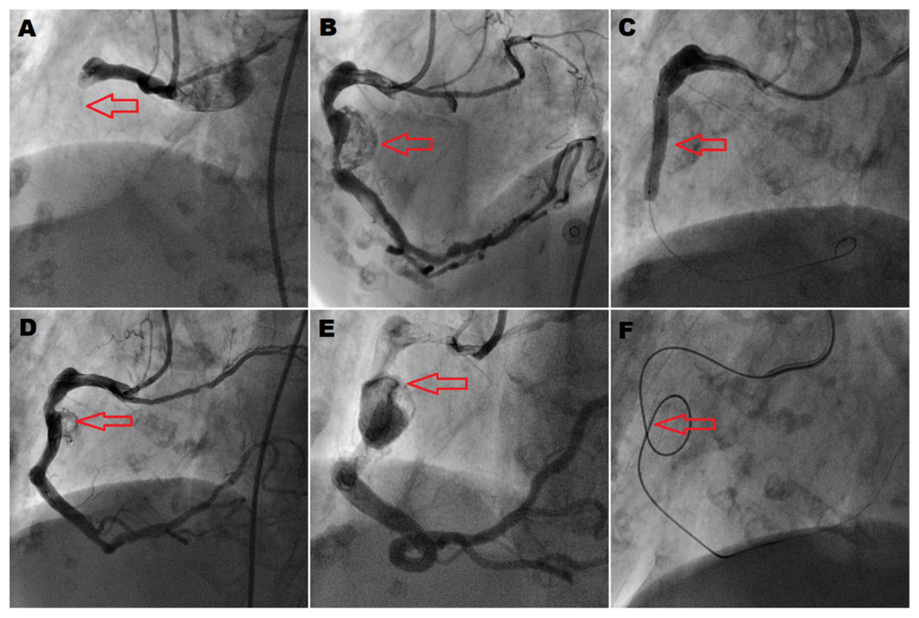
Figure 1. Primary recanalization of right coronary artery aneurysm and the attempts to exclude it. (A) Acute right coronary artery occlusion (RCA) (arrow); (B) aneurysm found after vessel recanalization (arrow) (ectopic circumflex artery arising from the proximal RCA); (C) covered stent deployment (arrow); (D) residual leak into the aneurysm sac (arrow) after postdilation-induced stent foreshortening; (E) stent prolapse into the aneurysm sac (arrow) found in control angiography; (F) microcatheter passed through the stent (arrow) during the realignment attempt.
Jvd 02 00031 g001
Figure 2. Stent herniation into the coronary aneurysm sac. (A) Right coronary artery (RCA) aneurysm with a prolapsed proximal edge of a covered stent (arrow); (B) schematic presentation of the blood flow sequence, from the proximal RCA into the aneurysm sac and after its filling, into the prolapsed stent edge, then through the covered stent into the distal RCA.
Jvd 02 00031 g002
Table 1. Techniques of prolapsed stent realignment.
Realignment TechniqueElements of the TechniqueTechnique Description
Parallel balloon inflationJvd 02 00031 i001A parallel balloon is inflated to push the stent out of the aneurysm sac and enable a rewiring of the distal vessel [43].
Balloon inflation at prolapsed stent edgeJvd 02 00031 i002If enough space is left at the herniated stent edge to wire it and pass another device, several balloon techniques may be employed, including balloon inflation at the prolapsed stent edge to coaxially realign the prosthesis [44,45,46].
Distal balloon anchoringJvd 02 00031 i003In this technique, a balloon is deployed distal to the stent with gentle traction applied to use the balloon shaft for stent repositioning.
Stent retriever-assisted repositioningJvd 02 00031 i004Stent retrievers may also be considered for stent rearrangement with a technique similar to the one used for bailout stentectomy of intracranial stents, whereby a stent retriever is deployed inside a prolapsed stent and pulled back to reposition it coaxially [50].
Snare-assisted stent realignmentJvd 02 00031 i005For proximal stent edge realignment, snares may be used, preferably with a deflated balloon inserted distally through the stent to expand the snare-crimped stent after its rearrangement [51,52,53].
Snare-and-balloon techniqueJvd 02 00031 i006A balloon is deployed at the prolapsed proximal edge of stent to be caught by a snare and repositioned coaxially.
Stent crushJvd 02 00031 i007The prolapsed stent is crushed with a balloon, with subsequent deployment of additional covered stents to exclude the aneurysm.
Disclaimer/Publisher’s Note: The statements, opinions and data contained in all publications are solely those of the individual author(s) and contributor(s) and not of MDPI and/or the editor(s). MDPI and/or the editor(s) disclaim responsibility for any injury to people or property resulting from any ideas, methods, instructions or products referred to in the content.