Abstract
Smartphone-based fundus cameras and telemedicine are an opportunity for accessing ocular health inequalities in under-resourced areas. The objective of this study is to evaluate the prevalence of retinal findings in a community in the Amazon and propose strategies to enhance ocular health. A retrospective study was conducted in a riverside community. Retinal photos from the posterior pole and optic disc were captured using a portable fundus camera. All photos and data were analyzed remotely by a retina specialist. The final sample was 107 participants, aged 52 ± 17. Retinal findings were detected in 37.4% (95%CI 28.7–46.8) of the sample; the three main retinal findings were epithelial changes (10.3%, 95%CI 5.6–17.1), chorioretinal scars (8.4%, 95%CI 4.2–14.8), and dry age-related macular degeneration (7.5%, 95%CI 3.6–13.6). This study detected retinal alterations in a similar prevalence to that of other under-resourced areas. Telemedicine is an opportunity to address health inequities, especially in ophthalmology, through relatively low-cost portable devices, supporting clinical decisions in areas with low health access; however, maintaining assistance after implementation is a challenge. Enhancing medical education and training local non-specialized health professionals in risk assessment, device handling, and data base use is reasonable to ensure follow-up.
1. Introduction
Currently, according to the World Health Organization (WHO), 2.2 billion people in the world suffer from some visual impairment, and of these, 1 billion are the result of preventable or treatable causes [1]. Glaucoma, age-related macular degeneration (AMD), and diabetic retinopathy are among the major causes of blindness. All of these can have their morbidity reduced via early detection through retinography and/or retinal mapping, thus permitting appropriate intervention [2].
The estimate of blindness in Brazil is 1.559 million people, and the main causes are in accordance with WHO’s declaration: cataracts, glaucoma, AMD, and diabetic retinopathy [3]. Brazil has as unbalanced geographical distribution of ophthalmologists, the predominance of the specialty in concentrated in southeast and there is a huge deficit in the northern region, particularly in communities outside the state capital [3].
Conventional instruments for fundus evaluation (e.g., indirect ophthalmoscope and slit lamp) are difficult to handle, require extensive prior training, and are examiner dependent, not allowing, in most cases, photographic recording of the retina. With traditional fundus cameras, it is possible to take wide-field high-resolution pictures of the retina—imaging with an angular field of view over 50 degrees [4]—allowing for different examiners’ evaluation and fundus record for follow up; however, they have elevated acquisition cost and logistical problems for transportation [5]. Capturing retinal images is important in ophthalmic practice, since it permits doctors to evaluate more details of the retina, to monitor diseases progression, and to apply analysis software and artificial intelligence, and it also plays a key role in the training of new doctors [5,6].
In the present era, a variety of portable fundus imaging techniques facilitate the acquisition of high-quality images of the retina. One of the techniques described is indirect ophthalmoscopy with a mobile phone camera. In this method, a 20 D condensing lens is positioned in close proximity to the patient’s eye in mydriasis, and the phone is set to video mode with the flash activated. However, the quality of the image depends on the examiner, demanding a highly trained professional [7,8].
This technique has been refined and is now employed in a number of handheld portable fundus cameras that are currently available on the market. These devices are equipped with a system of converging lenses attached to a smartphone, which enables the capture of high-resolution digital images of the retina [9]. These devices are light, small, less examiner depend, and, although still expensive, are much more affordable than the traditional fundus cameras [6]. Moreover, most do not require mydriasis, shortening the examination time and avoiding adverse effects, such as visual discomfort, photophobia, keratitis, and increased intraocular pressure [10]. These devices have already been implemented in remote communities for the screening of diabetic retinopathy, glaucoma, and other conditions. This has been achieved in locations with no access to specialists in screening, as evidenced by events held within eye camps. This indicates that it may be feasible to reduce barriers to eye care through the use of mobile devices [11,12].
With the advent of telemedicine and cloud-based data records, images are automatically sent to an online network, a secure data base with access that is restricted to those involved in the exam and the responsible doctor, enabling a specialized ophthalmologist to analyze them from anywhere in the world, bringing the patient a more reliable diagnosis and supporting local physicians to choose the most appropriate and up-to-date management [12].
Regarding the reality of telemedicine in Brazil, it is known that its introduction in the country is recent, and it was only regulated during the COVID-19 pandemic in 2020 [13]. Its dissemination throughout the country faces several obstacles, such as structural barriers—lack of access to internet and digital technologies—and techniques—few people have the necessary knowledge to effectively insert telemedicine into daily care [14]. This difference is even more important when we consider teleophthalmology, since this is one of the areas of telemedicine that most needs high technology density [15].
In wealthy regions, these interfaces are not major obstacles since there is enough investment to purchase modern devices and enough access to specialized professionals [5]. However, even in high-income countries, millions of people live in low-income areas without access to ophthalmologists and technology, and, hence, are left unattended and develop various blinding conditions. Added to the lack of access, these patients are also unaware of the need for early detection and treatment to improve their life quality [16]. New smartphone-based fundus camera and telemedicine, combined, show a promising opportunity for accessing ocular health inequalities in vulnerable populations. Thus, this study had the objective to investigate the prevalence of retinal findings through a smartphone-based fundus camera in a semi-isolated riverside community in the Amazon forest, to explore its applications, and to propose strategies to enhance ocular health through telemedicine in unaided populations. As a secondary objective, we intended to demonstrate the potential of low-cost devices and telemedicine in improving access to ophthalmological care, thus encouraging other communities to adopt similar approaches and raise the demand for other portable devices that could be applied with telemedicine in remote and undeveloped places.
2. Materials and Methods
2.1. Sample Selection
A retrospective observational study was conducted in the community of Calama, Rondônia, Brazil, using secondary data without primary reporting. Calama is a 2000-people riverside community in the Amazon forest, located around 15 h by boat away from the closest ophthalmology service. We conducted a both retrospective cross-sectional and case-control analysis in order to gain insight into the local epidemiology, investigate potential factors associated with the retinal alterations, and reduce biases pertaining to more rigorous associations and cause-and-effect relationships [17]. Participants included adults randomly selected during home visits or at the local general medicine care clinic (primary health care) during January–February 2022; to avoid bias, excluded from the sample were patients under 18 years old, or with anterior segment opacities, or those who looked for the clinics with known eye diseases.
2.2. Data Acquisition
Photos were captured by four previously trained fifth-year medical students, using a portable non-mydriatic smartphone-based fundus camera (Eyer, Phelcom Thecnologies®, São Carlos, Brazil). Interns were trained in a four-hour lection that included theoretical aspects (e.g., physics of the device, ophthalmological basic anamneses and exam, and retinal pa-thology) and practical activities in a simulation-based scenario. In the sitting position, two retinal photos were captured from each eye: (i) posterior pole and (ii) optic disc, using the mentioned portable fundus camera (Eyer, Phelcom Thecnologies®, São Carlos, Brazil).
After the photo acquisition, participants were questioned about their age, sex, and self-reported comorbidities, but no ethnic information was collected during the study. The portable fundus camera is equipped with artificial intelligence software that provides a probability rating for retinal alterations in percentages (EyerMap, Phelcom Thecnolo-gies®, São Carlos, Brazil). In instances where the system was unable to read the images, they were also excluded from the analysis. The students evaluated the quality of the photos, considering criteria such as the ability to read the images using artificial intelligence, the visualization of the macula, arteries, and vessels, as well as of the optic nerve head. All of the photos and data were automatically sent to an online, encrypted data base (EyerCloud, Phelcom Thecnologies®, São Carlos, Brazil).
A single retina specialist, located more than 3000 km away, received all data re-motely (demographic data, comorbidities, and retinal photos), checked all exams for retinal alterations, and, through the data base, sent back the signed results in less than two days to the local health agents, which individually contacted each screened patient. All participants were properly referred or oriented according to the final diagnosis given through telemedicine. The process was as follows: (i) fundus photo acquisition was achieved through the portable fundus camera; (ii) the images were sent to an online encrypted data base; (iii) a diagnosis was made by a remote retinal specialist; (iv) the remote fundus photo diagnosis and medical guidance were transmitted to the local population with the help of local health agents. The transportation of all the referred patients was guaranteed by the local health secretary.
2.3. Data Analysis
Epithelial changes were considered as areas of altered hyper- or hypopigmentation in the epithelium without a visual alteration or known cause. Chorioretinal scars were considered as areas of pigmentary change or fibrosis resulting from an uncomplete healing of the retina, showing a possible participation of the retinal pigmented epithelium (RPE) in a process that affected the retina and choroid. Dry AMD was considered as the presence of drusen in the macula without neovascularization. Enlarged optic discs were considered as disc relation >0.50. Hypertensive retinopathy was considered as the widening of the arteriole reflex, arteriovenous crossing signs, and copper or silver wire arterioles (copper- or silver-coloured arteriole light reflex), or as constricted and tortuous arterioles, retinal hemorrhage, or hard exudates in patients with a known history of arterial hypertension. Non-proliferative diabetic retinopathy was considered as the presence of microaneurysms, dot-blot hemorrhages, cotton wool spots, and hard exudates without neovascularization in patients with known diabetes.
In the development of this paper, the STROBE reporting guideline was followed [18]. Statistical analyses were performed using IBM SPSS® 28.0.1 (IBM, Inc., Chicago, IL, USA). Continuous variables were compared between groups using the Mann–Whitney U test for non-parametric independent samples, and nominal variables were compared using the chi-squared test. Proportion 95% confidence intervals (CI) were calculated considering a = 0.05 and binomial distributions.
2.4. Ethical Aspects
The study was performed in accordance with the ethical standards of The Declaration of Helsinki and approved by the Local Research Ethics Committee (number 5.325.976, approval date: 1 April 2022); informed consent was verbal.
3. Results
During the study, 122 individuals were screened for retinal alterations, 15 were excluded for not meeting the inclusion criteria (Figure 1A), 10 had dense cataracts, 1 had significant cornea opacity due to previous trauma, 1 had significant media opacity due to probable uveitis, the photos of 1 could not be obtained by students, and 2 were less than 18 years old. The final sample was composed of 107 participants, with a mean age of 52 ± 17.7 years (range 18 to 88), and 57.9% female. Comorbidities were found in 46.7% of the sample, and these were systemic arterial hypertension (SAH), type 2 diabetes mellitus (DM2), and dyslipidemia, with 22.4% having more than one associated comorbidity. Patients’ demographics are shown in Table 1. Altered retina were considered patients with a retinal alteration in the fundus photography of at least one eye.
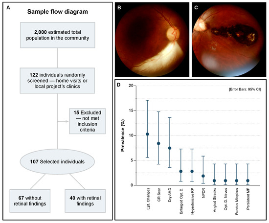
Figure 1.
Large chorioretinal scars were among the major retinal findings. (A) Sample flow diagram. (B) Persistent myeline fibres in the right eye, 45-year-old male. (C) Large chorioretinal scar involving the macula, 29-year-old male. (D) Prevalence and 95% confidence interval of identified retinal alterations in the population. AMD: age-related macular degeneration; CI: confidence intervals; D.: disc; Ept.: Epithelial; MF: myeline fibres; Opt.: optic; and RP: retinopathy.

Table 1.
Patients’ demographics.
Most of the population did not have any identifiable retinal alterations, at 62.6%, CI [53.2, 71.3]; however, findings were detected in 37.4%, CI [28.7, 46.8], of the sample. A large set of alterations were identified, ranging from non-visual threatening to severe blindness (e.g., inferior temporal persistent myeline fibres vs. large macular chorioretinal scar; Figure 1B,C). Interestingly, the three main retinal findings were epithelial changes (10.3%, CI [5.6, 17.1]), chorioretinal scars (8.4%, CI [4.2, 14.8]), and dry age-related macular degeneration (dAMD; 7.5%, CI [3.6, 13.6]). In lower rates, enlarged optic disc (2.8%, CI [0.8, 7.3]), hypertensive retinopathy (2.8%, CI [0.8, 7.3]), and non-proliferative diabetes retinopathy (NPDR; 1.9%, CI [0.4, 5.9]) were also identified. Other alterations appeared only once, 0.9%, CI [0.1, 4.3]—these included angioid streaks, fundus miopicus, optic disc nevus, and persistent myeline fibres (Figure 1D). All patients that showed NPDR had confirmed type 2 diabetes mellitus (DM2), and the prevalence of NPDR among DM2 participants was 8.0%, CI [1.7, 23.3]. Other ophthalmological findings that were not in the retina (e.g., refractive errors, pterygium, cataract, etc.) were not considered in the study.
Among the sample, participants with retinal alterations were significantly older (p < 0.001); however, as expected, when adjusted by age and in pairwise comparison with retinal findings, only dAMD had statistical significance (p < 0.001) and other findings had no correlation with age (p > 0.05). SAH was the only comorbidity that showed higher prevalence among participants with retinal findings (52.5%; CI 37.3, 67.3, p = 0.008). Sex, DM2, dyslipidemia, and smoking showed no impact on the findings (p > 0.05).
4. Discussion
Through telemedicine and a smartphone-based device, our study detected retinal alterations in 37.4% of the population in a remote riverside community in the Amazon forest, a prevalence which is similar to the average of other under-resourced areas [19]. The most prevalent retinal finding was epithelial changes (10.3%), which is a non-specific sign that could be related to a variety of diseases, e.g., light toxicity, macular dystrophies, and early-stage pachychorid [20,21,22], with etiologies probably related to the community’s social conditions, such as intense sun exposure and high rates of consanguinity. However, complementary exams are necessary to clearly state the management of the findings—OCT, blood testing, genetic panels, etc.—as in this study, the only available tool was a portable fundus camera.
Interestingly, the prevalence of chorioretinal scars was 8.4%, which is similar to other studies which showed a prevalence of 1.0% to 17.7%, depending on the design and characteristics of the population [23,24]. It is known that ocular toxoplasmosis is one of the most frequent causes of posterior uveitis worldwide, and that it is the leading cause of childhood blindness in Brazil since, when not treated, it may leave typical chorioretinal scars [25]. Toxoplasma gondii, the parasite associated with zoonosis, is endemic in the region, and its main source of dissemination is contaminated water, undercooked meat, and vertical transmission during pregnancy—risk factors to which the population of this study is largely exposed [26]. This finding highlights the necessity of intensive measures to detect contaminated pregnant women early, to increase access to treated water, and to intensify public policies aiming prophylaxis.
Prior studies have demonstrated the efficacy and quality of portable fundus cameras. A study conducted in India demonstrated a sensitivity of 95.8% and a specificity of 80.2% for detecting diabetic retinopathy using the Remidio ‘Fundus on phone’ device, thereby illustrating its potential for use in retinal screening [27]. In addition to DR, other studies have reported the efficacy of this portable method for detecting other pathologies, including retinopathy of prematurity, with a sensitivity of 100% and a specificity of 91% using Pictor™ (Volk Optical Inc., Mentor, OH), and optic disc cupping with a sensitivity of 92% and a specificity of 83% using a novel iPhone fundus camera [11,28]. These methods demonstrate efficacy in detecting a range of ocular pathologies, with a high degree of concordance with the established gold standard. Moreover, advancements in artificial intelligence (AI) and neural networks have influenced the field of imaging analysis, potentially rendering the diagnostic process more feasible, faster, and more accurate [29,30]. The combined results of the previously mentioned studies in conjunction with the findings presented in our own research demonstrate that portable fundus cameras have proven to be a valuable diagnostic tool in the evaluation of a wide range of common ophthalmological conditions, even when a specialist is geographically distant.
The potential applications of telemedicine and portable devices in ophthalmology are diverse. This study concentrated on the evaluation of the retina; however, other subspecialties within ophthalmology are also being investigated. The evaluation of structural and functional tests through telemedicine yielded comparable levels of agreement with those of in-person examinations for glaucoma [31,32]. Similarly, studies have demonstrated the efficacy of imaging devices in the diagnosis of corneal and external diseases. A recent review identified 51 such devices with potential applications in telemedicine and the remote diagnosis of prevalent diseases [33]. The integration of devices for comprehensive ophthalmological examinations represents a promising approach to the delivery of holistic eye care through the implementation of telemedicine and AI.
The riverside community of Calama is in a remote region of the Brazilian Amazon forest, and the only way to reach it is by boat, through the Madeira River. As in other isolated communities, it is marked by a difficult displacement caused by territorial dimensions, lack of transport systems, and a low concentration of health professionals [34,35]. This study shows that telemedicine, involving local support with distant specialists, is a huge opportunity to address health inequities, especially in ophthalmology, which is a medical field that demands extensively trained professionals and high-cost equipment.
The recognition of risk factors for non-preventable diseases is conducted by general doctors in the primary health care scenario, which is, in most of the places, represented by recently graduated physicians. However, studies showed that these doctors receive a huge deficit in ophthalmological knowledge during medical school and are uncertain when initially assisting and/or referring ophthalmological cases, compromising the proper approach of the patients [36,37,38]. This problem is possibly correlated to the exclusion of ocular health as a mandatory subject in the Brazilian medical curriculum, and less than half the pupils have the option of undertaking an elective clerkship in ophthalmology, increasingly distancing the specialty from the reality of the general practitioner [39,40]. Easy handling devices, artificial intelligence, and telemedicine can support medical education, and bring back clinicians’ security when initially managing and referring patients with risk factors for blinding ocular conditions. Extracurricular activities are also an important opportunity to support medical education and encourage knowledge developed outside the university walls, a fact highlighted by the exponential growth of The Brazilian Association of Ophthalmology Academic Leagues (ABLAO), an association that develops multicenter extension, research, and teaching activities, bringing together students interested in ophthalmology and providing opportunities for social impact and the improvement of medical education [39].
5. Conclusions
In this study, we showed that, through relatively low-cost and portable devices, distant highly specialized professionals can guarantee and support effective clinical decisions in remote areas with low health access. Furthermore, our findings demonstrated a concordant prevalence of retinal findings with other vulnerable populations, notably chorioretinal scars. However, this study also showed that maintaining assistance after initial implementation is still a challenge, since the visit of trained health professionals is periodic, and some conditions demand frequent follow-up. We suggest enhancing medical education with regard to the recognition of ocular pathologies, and training local non-specialized health professionals in risk assessment, device handling, and data base use to ensure early diagnosis and follow-up after the project implementation. Apart from that, we also suggest the use of combined portable devices with telemedicine and AI for complete ophthalmological exams, the development of new research aimed at unexplored portable devices (e.g., OCT and visual field), and larger investigations with a broader spectrum of exams to support public policies in blindness prevention.
Author Contributions
Conceptualization, J.S., L.E.S., J.V.M.L., L.R.d.S., I.U.N., C.P.C., A.C.F.d.A., A.M., Ê.L.D. and L.F.M.L.; methodology, J.S. and L.E.S.; software, L.E.S.; validation, A.M., Ê.L.D. and L.F.M.L.; formal analysis, J.S. and L.F.M.L.; investigation, J.S., L.E.S., J.V.M.L., L.R.d.S., I.U.N., C.P.C. and A.C.F.d.A.; resources, J.S., L.E.S., A.M., Ê.L.D. and L.F.M.L.; data curation, J.S. and L.E.S.; writing—original draft preparation, J.S., L.E.S., J.V.M.L., L.R.d.S., I.U.N., C.P.C. and A.C.F.d.A.; writing—review and editing, J.S., A.M., Ê.L.D. and L.F.M.L.; visualization, J.S., L.E.S., Ê.L.D. and L.F.M.L.; supervision, A.M., Ê.L.D. and L.F.M.L.; project administration, Ê.L.D. and L.F.M.L.; funding acquisition, J.S., L.E.S., A.M., Ê.L.D. and L.F.M.L. All authors have read and agreed to the published version of the manuscript.
Funding
This study was funded by the Cultural and Extension Dean’s Office of the University of São Paulo. LES and LRS are supported by São Paulo Research Foundation (respectively, grants numbers 2020/0365-8 and 2021/09023-0).
Institutional Review Board Statement
The study was conducted in accordance with the Declaration of Helsinki and approved by the Ethics Committee of Hospital de Reabilitação de Anomalias Craniofaciaias da Universidade de São Paulo—HRAC USP protocol code 5.325.976 from 1 April 2022.
Informed Consent Statement
Informed consent was obtained from all subjects involved in the study.
Data Availability Statement
The data presented in this study are available on request from the corresponding author due to (ethical restriction).
Acknowledgments
We would like to acknowledge Letícia Leite, Magali Caldana, and José R. M. Basto, coordinators of the project “Bauru School of Dentistry—Rondônia”, all members of the expedition, and Luiz F. F. Silva for the material, logistic and administrative support; Marisa Romangnolli for the technical and design support; the Cultural and Extension Dean’s Office of the University of São Paulo for the financial and administrative support; and Phelcom Technologies for the material and technical support.
Conflicts of Interest
The authors declare no conflicts of interest.
References
- Flaxman, S.R.; Bourne, R.R.A.; Resnikoff, S.; Ackland, P.; Braithwaite, T.; Cicinelli, M.V.; Das, A.; Jonas, J.B.; Keeffe, J.; Kempen, J.H.; et al. Global Causes of Blindness and Distance Vision Impairment 1990-2020: A Systematic Review and Meta-Analysis. Lancet Glob Health 2017, 5, e1221–e1234. [Google Scholar] [CrossRef]
- GBD 2019 Blindness and Vision Impairment Collaborators; Vision Loss Expert Group of the Global Burden of Disease Study. Causes of Blindness and Vision Impairment in 2020 and Trends over 30 Years, and Prevalence of Avoidable Blindness in Relation to VISION 2020: The Right to Sight: An Analysis for the Global Burden of Disease Study. Lancet Glob. Health 2021, 9, e144–e160. [Google Scholar] [CrossRef] [PubMed]
- Umbelino, C.C.; Avlia, M. As Condições de Saúde Ocular No Brasil, 1st ed.; Umbelino, C.C., Avlia, M., Eds.; Conselho Brasileiro de Oftalmologia: São Paulo, Brazil, 2023. [Google Scholar]
- Lucente, A.; Taloni, A.; Scorcia, V.; Giannaccare, G. Widefield and Ultra-Widefield Retinal Imaging: A Geometrical Analysis. Life 2023, 13, 202. [Google Scholar] [CrossRef] [PubMed]
- Garg, S.J. Applicability of Smartphone-Based Screening Programs. JAMA Ophthalmol. 2016, 134, 158–159. [Google Scholar] [CrossRef]
- Wintergerst, M.W.M.; Jansen, L.G.; Holz, F.G.; Finger, R.P. A Novel Device for Smartphone-Based Fundus Imaging and Documentation in Clinical Practice: Comparative Image Analysis Study. JMIR Mhealth Uhealth 2020, 8, e17480. [Google Scholar] [CrossRef]
- Shanmugam, M.; Mishra, D.; Madhukumar, R.; Ramanjulu, R.; Reddy, S.; Rodrigues, G. Fundus Imaging with a Mobile Phone: A Review of Techniques. Indian. J. Ophthalmol. 2014, 62, 960. [Google Scholar] [CrossRef]
- Toy, B.C.; Myung, D.J.; He, L.; Pan, C.K.; Chang, R.T.; Polkinhorne, A.; Merrell, D.; Foster, D.; Blumenkranz, M.S. Smartphone-based dilated fundus photography and near visual acuity testing as inexpensive screening tools to detect referral warranted diabetic eye disease. Retina 2016, 36, 1000–1008. [Google Scholar] [CrossRef] [PubMed]
- Chalam, K.V.; Chamchikh, J.; Gasparian, S. Optics and Utility of Low-Cost Smartphone-Based Portable Digital Fundus Camera System for Screening of Retinal Diseases. Diagnostics 2022, 12, 1499. [Google Scholar] [CrossRef]
- Hong, D.; Tripathy, K. Tropicamide. In StatPearls; StatPearls Publishing: Treasure Island, FL, USA, 2024. [Google Scholar] [PubMed]
- Cheng, D.; Babij, R.; Cabrera, D.; Yuan, M.; Port, A.; Mckenney, A.S.; Zhu, J.; Van Tassel, S.; Imperato-McGinley, J.; Sun, G. Effective Low-Cost Ophthalmological Screening With a Novel IPhone Fundus Camera at Community Centers. Cureus 2022, 14, e28121. [Google Scholar] [CrossRef]
- Jin, K.; Lu, H.; Su, Z.; Cheng, C.; Ye, J.; Qian, D. Telemedicine Screening of Retinal Diseases with a Handheld Portable Non-Mydriatic Fundus Camera. BMC Ophthalmol. 2017, 17, 89. [Google Scholar] [CrossRef]
- Lisboa, K.O.; Hajjar, A.C.; Sarmento, I.P.; Sarmento, R.P.; Gonçalves, S.H.R. A História Da Telemedicina No Brasil: Desafios e Vantagens. Saúde Soc. 2023, 32, e210170pt. [Google Scholar] [CrossRef]
- Nakayama, L.F.; Binotti, W.W.; Link Woite, N.; Fernandes, C.O.; Alfonso, P.G.; Celi, L.A.; Regatieri, C.V. The Digital Divide in Brazil and Barriers to Telehealth and Equal Digital Health Care: Analysis of Internet Access Using Publicly Available Data. J. Med. Internet Res. 2023, 25, e42483. [Google Scholar] [CrossRef]
- Grisolia, A.B.D.; Abalem, M.F.; Lu, Y.; Aoki, L.; Matayoshi, S. Teleophthalmology: Where Are We Now? Arq. Bras. Oftalmol. 2017, 80, 401–406. [Google Scholar] [CrossRef] [PubMed]
- Lodhia, V.; Karanja, S.; Lees, S.; Bastawrous, A. Acceptability, Usability, and Views on Deployment of Peek, a Mobile Phone MHealth Intervention for Eye Care in Kenya: Qualitative Study. JMIR Mhealth Uhealth 2016, 4, e30. [Google Scholar] [CrossRef]
- Mann, C.J. Observational Research Methods. Research Design II: Cohort, Cross Sectional, and Case-Control Studies. Emerg. Med. J. 2003, 20, 54–60. [Google Scholar] [CrossRef] [PubMed]
- von Elm, E.; Altman, D.G.; Egger, M.; Pocock, S.J.; Gøtzsche, P.C.; Vandenbroucke, J.P. The Strengthening the Reporting of Observational Studies in Epidemiology (STROBE) Statement: Guidelines for Reporting Observational Studies. Lancet 2007, 370, 1453–1457. [Google Scholar] [CrossRef]
- Jacobsen, B.H.; Shah, A.A.; Aggarwal, S.; Mwanansao, C.; McFadden, M.; Zouache, M.A.; Shakoor, A. Prevalence of Retinal Diseases and Associated Risk Factors in an African Population From Mwanza, Tanzania. Ophthalmic Surg. Lasers Imaging Retin. 2020, 51, S17–S25. [Google Scholar] [CrossRef]
- Youssef, P.N.; Sheibani, N.; Albert, D.M. Retinal Light Toxicity. Eye 2011, 25, 1–14. [Google Scholar] [CrossRef] [PubMed]
- Warrow, D.J.; Hoang, Q.V.; Freund, K.B. Pachychoroid Pigment Epitheliopathy. Retina 2013, 33, 1659–1672. [Google Scholar] [CrossRef]
- Halford, S.; Liew, G.; Mackay, D.S.; Sergouniotis, P.I.; Holt, R.; Broadgate, S.; Volpi, E.V.; Ocaka, L.; Robson, A.G.; Holder, G.E.; et al. Detailed Phenotypic and Genotypic Characterization of Bietti Crystalline Dystrophy. Ophthalmology 2014, 121, 1174–1184. [Google Scholar] [CrossRef] [PubMed]
- de Amorim Garcia, C.A.; Oréfice, F.; de Oliveira Lyra, C.; Gomes, A.B.; França, M.; de Amorim Garcia Filho, C.A. Socioeconomic Conditions as Determining Factors in the Prevalence of Systemic and Ocular Toxoplasmosis in Northeastern Brazil. Ophthalmic Epidemiol. 2004, 11, 301–317. [Google Scholar] [CrossRef] [PubMed]
- Glasner, P.D.; Silveira, C.; Kruszon-Moran, D.; Martins, M.C.; Burnier, M.; Silveira, S.; Camargo, M.E.; Nussenblatt, R.B.; Kaslow, R.A.; Belfort, R. An Unusually High Prevalence of Ocular Toxoplasmosis in Southern Brazil. Am. J. Ophthalmol. 1992, 114, 136–144. [Google Scholar] [CrossRef] [PubMed]
- de Angelis, R.E.; Veronese Rodrigues, M.L.; Passos, A.D.C.; Bollela, V.R.; Freitas E Silva, M.S.; Vieira, B.R.; de Lucena, M.M.; Moralles, T.D.; de Morais Vicente, L.; de Melo Rocha, G.; et al. Frequency and Visual Outcomes of Ocular Toxoplasmosis in an Adult Brazilian Population. Sci. Rep. 2021, 11, 3420. [Google Scholar] [CrossRef]
- Strang, A.G.G.F.; Ferrari, R.G.; do Rosário, D.K.; Nishi, L.; Evangelista, F.F.; Santana, P.L.; de Souza, A.H.; Mantelo, F.M.; Guilherme, A.L.F. The Congenital Toxoplasmosis Burden in Brazil: Systematic Review and Meta-Analysis. Acta Trop. 2020, 211, 105608. [Google Scholar] [CrossRef] [PubMed]
- Rajalakshmi, R.; Subashini, R.; Anjana, R.M.; Mohan, V. Automated Diabetic Retinopathy Detection in Smartphone-Based Fundus Photography Using Artificial Intelligence. Eye 2018, 32, 1138–1144. [Google Scholar] [CrossRef]
- Prakalapakorn, S.G.; Freedman, S.F.; Hutchinson, A.K.; Wallace, D.K.; Stinnett, S.S.; Riggins, J.W.; Gallaher, K.J. Evaluating a Portable, Noncontact Fundus Camera for Retinopathy of Prematurity Screening by Nonophthalmologist Health Care Workers. Ophthalmol. Retin. 2018, 2, 864–871. [Google Scholar] [CrossRef]
- Wu, C.-T.; Lin, T.-Y.; Lin, C.-J.; Hwang, D.-K. The Future Application of Artificial Intelligence and Telemedicine in the Retina: A Perspective. Taiwan. J. Ophthalmol. 2023, 13, 133–141. [Google Scholar] [CrossRef]
- Shi, C.; Lee, J.; Wang, G.; Dou, X.; Yuan, F.; Zee, B. Assessment of Image Quality on Color Fundus Retinal Images Using the Automatic Retinal Image Analysis. Sci. Rep. 2022, 12, 10455. [Google Scholar] [CrossRef]
- Balakrishnan, P.; Swain, T.A.; McGwin, G.; Owsley, C.; Girkin, C.A.; Rhodes, L.A. Comparison of Glaucoma Diagnosis by Telemedicine, In-Person Ophthalmologist, and Optometrist. J. Glaucoma 2024, 33, 619–623. [Google Scholar] [CrossRef]
- Christopher, M.; Hallaj, S.; Jiravarnsirikul, A.; Baxter, S.L.; Zangwill, L.M. Novel Technologies in Artificial Intelligence and Telemedicine for Glaucoma Screening. J. Glaucoma 2024, 33, S26–S32. [Google Scholar] [CrossRef]
- Cao, B.; Vu, C.H.V.; Keenan, J.D. Telemedicine for Cornea and External Disease: A Scoping Review of Imaging Devices. Ophthalmol. Ther. 2023, 12, 2281–2293. [Google Scholar] [CrossRef] [PubMed]
- Franco, E.C.; Santo, C.d.E.; Arakawa, A.M.; Xavier, A.; França, M.d.L.; de Oliveira, A.N.; Machado, M.A.M.d.P.; Bastos, R.d.S.; Bastos, J.R.d.M.; Caldana, M.d.L. Promoção Da Saúde Da População Ribeirinha Da Região Amazônica: Relato de Experiência. Rev. CEFAC 2015, 17, 1521–1530. [Google Scholar] [CrossRef]
- Leite, L.d.A. Levantamento Dos Fatores de Risco Para o Acidente Vascular Cerebral Em Sujeitos Adultos de Diferentes Regiões Do Estado de Rondônia; Assistidos Pelo Projeto \”FOB-USP Em Rondônia\”; Universidade de São Paulo: Bauru, Brazil, 2022. [Google Scholar]
- de Espíndola, R.F.; Teixeira, F.C.; Yamakami, I.M.; da Silva, H.R.F.; Freitas, J.A.H. de Análise Dos Conhecimentos Básicos Sobre Urgências Oftalmológicas Em Plantonistas Não-Oftalmologistas. Arq. Bras. Oftalmol. 2006, 69, 11–15. [Google Scholar] [CrossRef]
- Rached, C.R.; de Oliveira, T.C.; Sousa, C.L.M.d.M.; Escudeiro, I.M.; Mori, L.P.; Ferreira, F.P.; Xavier, J.C.B.; Milioni, B.H.d.M.; de Figueiredo, R.R.; Paula, M.A.d. Avaliação Do Conhecimento Sobre Urgências Oftalmológicas Dos Acadêmicos Da Faculdade de Medicina Da Pontifícia Universidade Católica de Campinas. Rev. Bras. Oftalmol. 2012, 71, 100–105. [Google Scholar] [CrossRef]
- Cobbs, L.; Tsui, E.; Haberman, I.; Kim, E.; Sperber, L.; Wu, M.; Schuman, J. Student Perceptions of the Ophthalmology Curriculum in Medical School. J. Acad. Ophthalmol. 2018, 10, e79–e82. [Google Scholar] [CrossRef]
- Gameiro, G.R.; Gameiro, G.R. Ligas Acadêmicas de Oftalmologia: Gerando Impacto Na Educação Médica. Arq. Bras. Oftalmol. 2020, 83, V–VI. [Google Scholar] [CrossRef]
- Ferreira, M.d.A.; Gameiro, G.R.; Cordeiro, F.d.M.; Santos, T.V.; Hilarião, A.A.V.B.P.; Souza, G.M.; Nassaralla Neto, J.J.; Carricondo, P.C.; Portes, A.J.F.; Portes, A.L.F. Perfil Multicêntrico Do Acadêmico de Medicina e Suas Perspectivas Sobre o Ensino Da Oftalmologia. Rev. Bras. Oftalmol. 2019, 78, 315–320. [Google Scholar]

Disclaimer/Publisher’s Note: The statements, opinions and data contained in all publications are solely those of the individual author(s) and contributor(s) and not of MDPI and/or the editor(s). MDPI and/or the editor(s) disclaim responsibility for any injury to people or property resulting from any ideas, methods, instructions or products referred to in the content. |
© 2025 by the authors. Licensee MDPI, Basel, Switzerland. This article is an open access article distributed under the terms and conditions of the Creative Commons Attribution (CC BY) license (https://creativecommons.org/licenses/by/4.0/).