Proceeding Paper

Enhanced Lung Disease Detection Using Double Denoising and 1D Convolutional Neural Networks on Respiratory Sound Analysis †

by
Reshma Sreejith
,
R. Kanesaraj Ramasamy
*,
Wan-Noorshahida Mohd-Isa
and
Junaidi Abdullah
Faculty Computing Informatics, Multimedia University, Cyberjaya 63100, Malaysia
*
Author to whom correspondence should be addressed.
Presented at the International Conference on Sustainable Computing and Green Technologies (SCGT’2025), Larache, Morocco, 14–15 May 2025.
Comput. Sci. Math. Forum 2025, 10(1), 7; https://doi.org/10.3390/cmsf2025010007
Published: 24 June 2025

Abstract

The accurate and early detection of respiratory diseases is vital for effective diagnosis and treatment. This study presents a new approach for classifying lung sounds using a double denoising method combined with a 1D Convolutional Neural Network (CNN). The preprocessing uses Fast Fourier Transform to clean up sounds and High-Pass Filtering to improve the quality of breathing sounds by eliminating noise and low-frequency interruptions. The Short-Time Fourier Transform (STFT) extracts features that capture localised frequency variations, crucial for distinguishing normal and abnormal respiratory sounds. These features are input into the 1D CNN, which classifies diseases such as bronchiectasis, pneumonia, asthma, COPD, healthy, and URTI. The dual denoising method enhances signal clarity and classification performance. The model achieved 96% validation accuracy, highlighting its reliability in detecting respiratory conditions. The results emphasise the effectiveness of combining signal augmentation with deep learning for automated respiratory sound analysis, with future research focusing on dataset expansion and model refinement for clinical use.

1. Introduction

Lung sounds are of paramount importance in the evaluation of respiratory health and the diagnosis of respiratory disorders. There is a close relationship between the sounds generated during respiration and changes in lung tissue. There are two fundamental classifications of lung sounds: normal lung sounds and abnormal lung sounds. Bronchial and vesicular sounds are examples of typical lung sounds; crackles and wheezes are atypical lung sounds. Diseases of the respiratory system can be identified using lung sounds. This study emphasises unusual pulmonary sounds such as crackles and wheezes, which play a critical role in the diagnosis of respiratory sound disorders. Bronchiectasis is a pathological condition characterised by damage to the bronchial tubes; chronic obstructive pulmonary diseases (COPDs) are characterised by reduced breath sounds and protracted crackles [1]. Infections of the upper respiratory tract (URTIs) impact the lungs directly, and additional respiratory symptoms include bronchiolitis and pneumonia. Due to the increasing prevalence of respiratory diseases as leading causes of death on a global scale, the ability to identify anomalous sounds during lung auscultation is critical for diagnosing common respiratory diseases and conducting medical examinations. Both sounds are categorised as adventitious sounds, which have the potential to signify pulmonary conditions. In the near future, automated techniques may enable the early detection of respiratory diseases [2].
Recently, researchers have developed electronic stethoscopes that amplify body sounds to overcome the issue of low sound levels. However, these devices have limitations, including the amplification of contact artefacts and frequency cutoff points, which affect their overall effectiveness. These limitations result in enhanced mid-range sounds, while reducing the volume of both high- and low-frequency sounds. Currently, many companies offer electronic stethoscopes [3]. These devices work by converting acoustic sound waves into electrical signals, which are then amplified and processed for clearer listening. Unlike acoustic stethoscopes, which operate on the same basic physical principles, electronic stethoscopes use transducers that can vary greatly in design. Computer-assisted auscultation tools, when used with electronic stethoscopes, analyse recorded heart sounds to distinguish between pathological and benign cardiac murmurs. Furthermore, analysing computerised lung sound recordings through time series can help diagnose conditions by recognising patterns. This method can also predict respiratory diseases such as asthma and chronic obstructive pulmonary disease (COPD), aiding in early detection and health status assessments through artificial intelligence-based decision tree classifiers [4].
Therefore, new methods for identifying respiratory diseases must be developed in order to lessen the annoyance that patients endure as a result of their symptoms. A quick and accurate diagnosis is guaranteed when lung sounds are examined using computer analysis and electronic auscultation. It eliminates the subjective element of the listener and finds anomalous traits that doctors cannot see. Clinicians may provide accurate diagnoses and quickly start therapy by using computer analysis, which helps their patients feel less pain. Many studies are currently attempting to use computer techniques that use CNNs to detect accidental lung noises. However, rather than segmenting the sound signal, the majority of these investigations concentrate primarily on lung sound classification. This paper’s main contributions are outlined below:
  • The lung sound categorization system divides respiratory cycles into stages and uses the double denoising method to enhance information about unusual sounds.
  • Enhance propagation and implementation: Encourage the use and approval of this beneficial technology in clinical settings.
  • Suggest potential areas for future research: Propose potential pathways for progress and originality.

2. Related Works

The objective of Aykanat et al.’s [5] project was to develop a software system for an electronic stethoscope that could transmit respiratory sounds to a personal computer for archiving, computer-assisted analysis, and diagnosis. A hardware–software system gathered a large collection of respiration sounds to train SVM and CNN machine learning algorithm design. The goal was to completely automate evaluation and detection. The system is anticipated to be widely utilised in clinical settings and can process all body sounds. As a benchmark, the research investigated CNN algorithms for audio classification using MFCC features in conjunction with SVM. The results showed that the CNN algorithm works about the same as the SVM algorithm for classifying spectrogram images, and both can accurately classify and help diagnose respiratory sounds when there is a lot of data available. By integrating this system with a telemedicine platform, physicians can store and exchange information.
Demir et al. [6] examined the classification of pulmonary disorders using lung sounds, with an emphasis on deep learning techniques. For image and sound classification, conventional machine learning techniques such as VGG16 and AlexNet were implemented; however, they did not provide a comprehensive depiction of sound attributes. Spectrogram images derived from lung sounds were utilised to train a novel CNN model, and a parallel-pooling architecture was implemented to improve classification performance. The LDA-RSE classifier was used to pull out important features from the first fully connected layer, achieving the best classification accuracy of 71.15%. As a result of its superior performance compared to other pretrained CNN models, the accuracy of the proposed CNN model increased by 5.75%.
In their paper, Hazra & Majhi [7] propose a 2D CNN approach to identify respiratory diseases early by analysing recorded lung sounds. The technique identifies “bronchiectasis, pneumonia, bronchiolitis, chronic obstructive pulmonary disease, upper respiratory tract infection, and healthy” by employing MFCC. The model utilises a total of 13 CNN layers, where each of them is accompanied by a corresponding level for pooling. The method achieved a precision surpassing 92.39%.
The study conducted by Fraiwan et al. [8] employs advanced deep learning techniques to achieve the precise detection of pulmonary diseases by analysing electronically recorded lung sounds. The study utilised a sample of 103 patients from King Abdullah University Hospital and 110 patients from the Int. Conf. on Biomedical Health Informatics Challenge database. The advanced deep learning network, which includes bidirectional long short-term memory units and convolutional neural networks, achieved an impressive average accuracy of 99.62% and a high precision of 98.85%. The model demonstrated a high level of agreement, with an impressive accuracy rate of 98.26% when comparing predictions to the original classes. This study establishes a solid foundation for the integration of deep learning models into clinical environments, aiding healthcare professionals in the identification of pulmonary diseases.
Choi & Lee [9] describe a lung sound recognition algorithm utilising the “VGGish”-stacked “BiGRU” model. For transfer learning, the “VGGish model” is employed as a feature extractor, and the target model is constructed using an identical structure to that of the source model. “BiGRU’s multi-layer architecture increases feature extraction capabilities while maintaining model integrity. Combining the “VGGish” model with a two-layer bidirectional gated recurrent model yields an optimal classification result. However, the limited dataset diminishes the model’s accuracy, leading to signs of overfitting. Subsequent investigations shall strive to enhance the precision of classification, mitigate overfitting, and assess the system’s performance on CT scan slices obtained in clinical settings that contain lung infections.
Bacanin et al. [10] investigated the application of audio analysis and CNNs for the identification of respiratory problems in patients. The researchers employed contemporary optimisation techniques to enhance efficiency and craft a customised algorithm that precisely aligns with their study’s specific needs. Applying the approach to a real-world medical dataset confirmed its validity and yielded encouraging outcomes. The enhanced metaheuristic optimiser achieved an accuracy of 0.93 for condition detection and 0.75 for precise condition identification. Their study emphasises AI’s diagnostic capabilities for improving patient outcomes in healthcare settings. However, the limited availability of data and the rigorous computational requirements constrain the study. Subsequent investigations aim to improve the methods and overcome these constraints.
This study reviews the advancements in respiratory disease detection through deep learning and machine learning techniques. Researchers have integrated electronic stethoscopes with machine learning for automated respiratory diagnosis, demonstrating the effectiveness of CNN and SVM in classifying respiratory sounds. A novel CNN model with a parallel-pooling architecture improved classification accuracy by 5.75%. A 2D CNN approach exceeded 92.39% accuracy. Fraiwan et al. [8] achieved 99.62% accuracy with bidirectional LSTM and CNN, proving the potential of deep learning for clinical applications. Bacanin et al. [10] highlighted AI’s role in patient diagnosis, achieving 93% detection accuracy. However, challenges such as computational constraints and data limitations remain. Wang & Sun [11] explored CNN performance variations under different parameter settings, highlighting the importance of frame length, overlap percentage, and spectrogram features in improving classification accuracy.

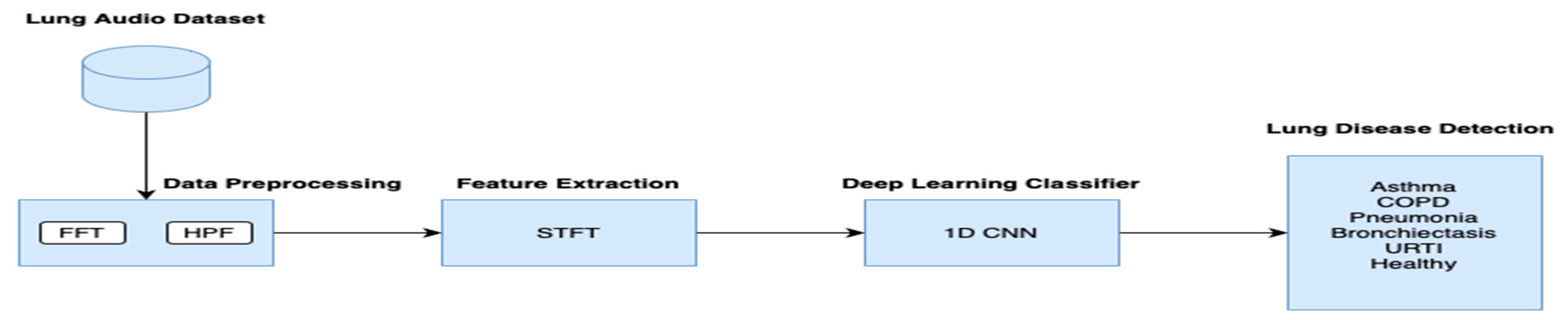
3. Materials and Methods

3.1. Data Description
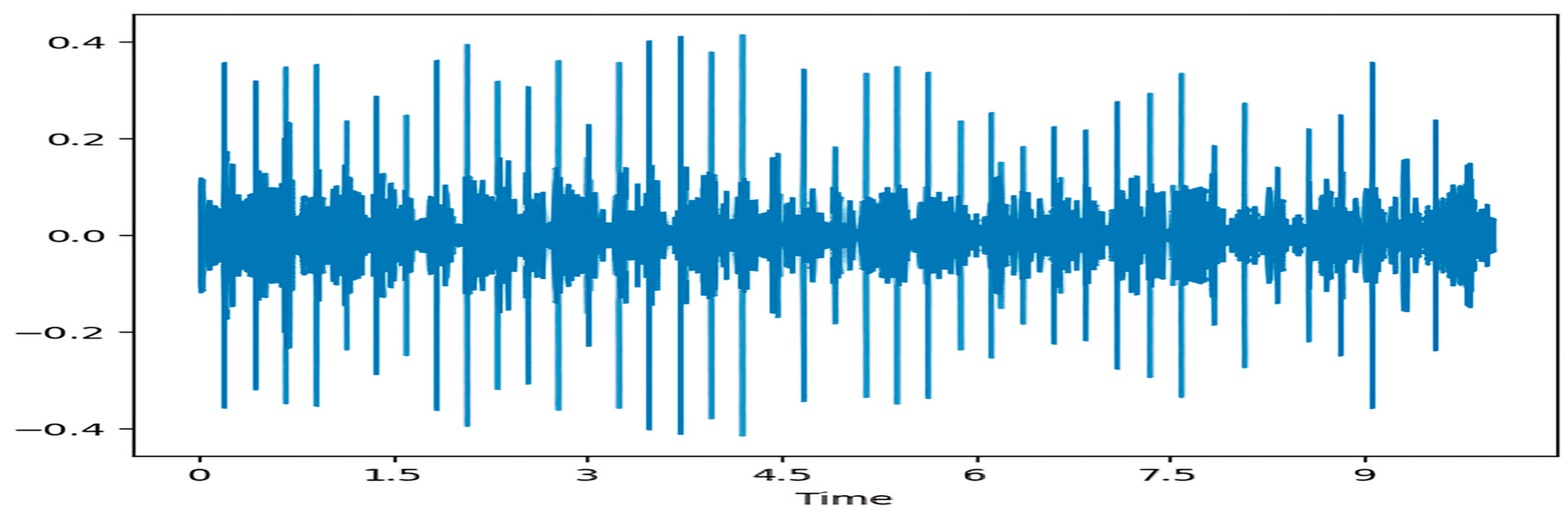
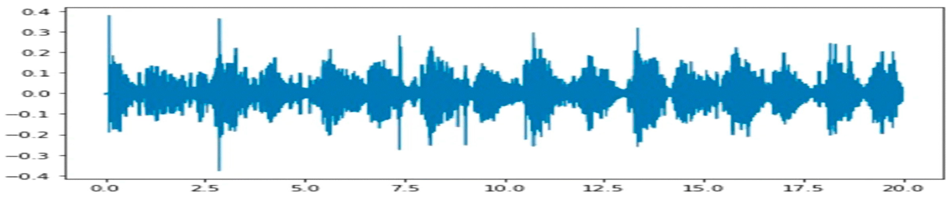
The research uses the ICBHI 2017 [12] breathing sound database, comprising 5.5 h of recorded breathing sounds from 126 individuals with diverse ages and medical problems. The database comprises 6898 manually annotated respiratory cycles, of which 3642 are categorised as normal, while the rest records are deemed problematic, encompassing crackles and wheezes. These anomalies are significant indications of respiratory disorders such as asthma, COPD, pneumonia, and pulmonary fibrosis. The dataset was produced by two autonomous research teams in Portugal and Greece, guaranteeing diversity and robustness. The recordings were acquired via diverse stethoscope models, enhancing the dataset’s practical relevance. The database has established itself as a standard for respiratory sound analysis, utilised in machine learning, artificial intelligence, and signal processing research to create automated diagnostic instruments for respiratory ailments. The comprehensive annotations and organised data render it a significant resource for academics and doctors in respiratory health studies.

3.2. Preprocessing

The double denoising method is a two-phase procedure that enhances signal integrity by mitigating noise through Fast Fourier Transform (FFT) and High-Pass Filtering (HPF). This approach, shown in Figure 1, is particularly useful in biological signal processing, such as respiratory and cardiac sound analysis, where removing low-frequency noise and unwanted artefacts is essential for accurate diagnosis. FFT converts the signal from the time domain to the frequency domain, allowing for the identification and removal of undesirable noise components. Afterward, Inverse Fast Fourier Transform (IFFT) is applied to reconstruct the purified signal. Following this, a high-pass filter is used to eliminate low-frequency components, filtering out interference while preserving the high-frequency components critical for signal processing. This dual denoising method improves signal clarity, making it highly beneficial for machine learning applications, diagnostic systems, and the real-time monitoring of physiological signals.
Figure 2 displays a randomly chosen lung sound sample from our dataset, illustrating the existence of noise prior to the use of any denoising methods. Figure 3 illustrates the outcomes achieved following the preliminary denoising phase utilising FFT-based [13] noise reduction, successfully attenuating undesirable frequency components. Figure 4 illustrates the results of the High-Pass Filtering (HPF) technique [14], which functions as the second denoising stage, enhancing the signal by removing residual low-frequency noise. Collectively, these data demonstrate the incremental improvement of signal clarity by the suggested double denoising method.

3.3. Feature Extraction

Short-Time Fourier Transform (STFT) is utilised on the previously pre-processed respiratory cycles to examine their frequency components in both the temporal and frequency domains. It disaggregates non-stationary data into localised frequency representations across brief temporal intervals. STFT segments respiratory sound waves into overlapping frames and utilises Fourier Transform to generate a comprehensive spectrogram that reflects temporal fluctuations in frequency content. This method is essential for detecting respiratory irregularities such as crackles and wheezes, which possess unique spectral attributes. The selection of window function, overlap percentage, and frequency resolution substantially affects the efficacy of STFT [15].

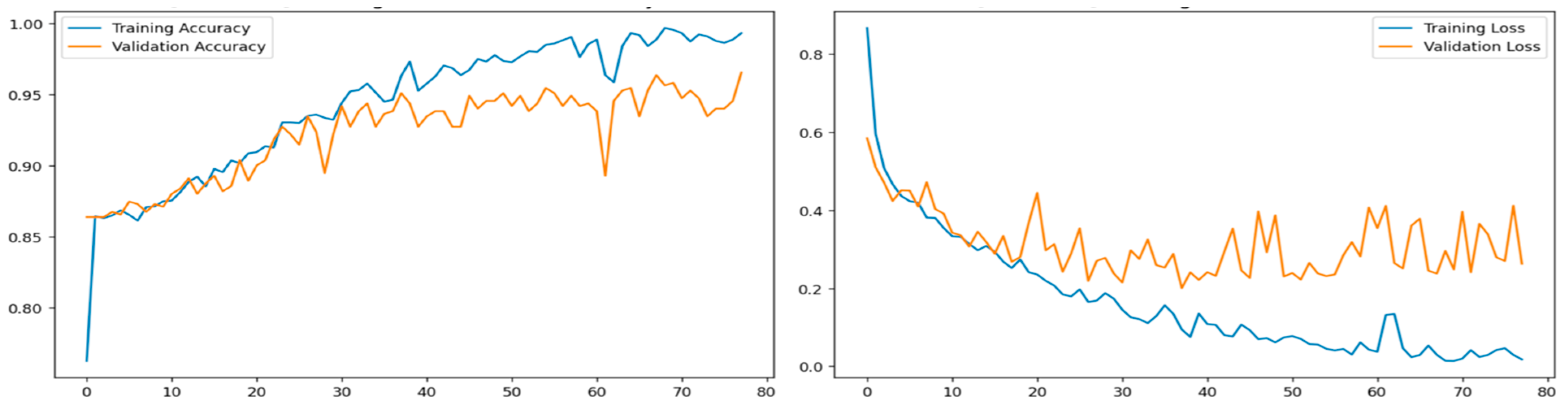
3.4. Training and Testing

Following Short-Time Fourier Transform (STFT) analysis, a one-dimensional Convolutional Neural Network (1D CNN) [16] was used to train and categorise audio characteristics for detecting pulmonary illnesses. The CNN identifies spatial and temporal relationships in the feature set, making it suitable for analysing sequential data like respiration sound waves [17]. The network comprises several convolutional layers, each using learnable filters to identify frequency patterns and differentiate between normal and pathological respiratory sounds. Batch normalisation and activation functions were employed to improve feature learning and mitigate overfitting. Pooling layers were used to diminish dimensionality while preserving lung sound characteristics [18]. The 1D CNN-based classification framework demonstrated significant efficacy in identifying disease-associated auditory patterns, making it a reliable instrument for detecting respiratory diseases from audio recordings. The classifier identified various respiratory conditions such as bronchiectasis, pneumonia, bronchiolitis, asthma, COPD, healthy and URTI as well as a healthy category of sounds using CNN features extracted from audio samples. The model uses a sequential architecture consisting of four 1D convolutional layers and a dense layer for the final output. CNN utilises a method of smoothly moving a filter window across the input to accurately identify and detect features.

4. Result Analysis

The implementation of the double denoising technique, which includes Fast Fourier Transform (FFT) and High-Pass Filtering (HPF), significantly improved the signal quality of respiratory sounds, hence increasing the accuracy of the classification model. The initial lung sound samples were affected by multiple types of noise, as illustrated in Figure 2, where the raw data displayed considerable unwanted noise elements that could disrupt the ensuing analysis and categorisation [18]. Following the initial phase of denoising, employing FFT-based noise reduction, the frequency components responsible for the noise were effectively attenuated, as illustrated in Figure 3. The FFT approach proficiently transformed the signal into the frequency domain, facilitating the detection and removal of undesirable low-frequency noise. Nevertheless, some residual low-frequency noise persisted. The second phase of the denoising procedure, High-Pass Filtering (HPF), enhanced the signal by removing low-frequency interferences while preserving the vital high-frequency components necessary for precise lung disease classification [19]. Figure 4 illustrates the results of the HPF approach, demonstrating a markedly clearer signal that is more concentrated on the pertinent frequency ranges. Through the implementation of these two denoising phases, the model demonstrated improved clarity in the respiratory signals, aiding the 1D Convolutional Neural Network (CNN) in discerning the essential aspects linked to diverse lung illnesses [20]. The enhanced signal quality resulted in increased classification accuracy, enabling the model to more effectively distinguish between normal and abnormal sounds, including wheezes, crackles, and other respiratory irregularities. The model got a validation accuracy of 96%, as shown in Figure 5. This shows that the double denoising strategy had a big impact on its performance. The accuracy loss was significantly reduced, suggesting that the refined and concentrated signals enabled the model to prevent overfitting and sustain strong generalisation to novel data [21]. In conclusion, the double denoising methodology enhanced the quality of respiratory signals and resulted in a quantifiable improvement in classification accuracy. The CNN demonstrated superior capability in identifying a broader spectrum of pulmonary disorders with clearer signals, including bronchiectasis, pneumonia, asthma, COPD, URTI, and others. This indicates that this preprocessing method is effective for enhancing machine learning-based diagnostic systems.

5. Conclusions

This study investigated an advanced methodology for detecting respiratory disorders by analysing lung sound recordings, employing a double denoising technique in conjunction with a 1D Convolutional Neural Network (CNN) for classification purposes. The double denoising method, comprising Fast Fourier Transform (FFT) and High-Pass Filtering (HPF), demonstrated significant efficacy in improving the quality of respiratory signals by eliminating noise and low-frequency disturbances. The preprocessing steps made the lung sounds much clearer, which made it easier for the model to tell the difference between normal and abnormal breathing states. The incorporation of STFT for feature extraction enhanced the process by delivering localised frequency representations of respiratory sounds, enabling the CNN to learn and identify critical signal properties. Utilising both strategies in conjunction resulted in a remarkable validation accuracy of 96%. This indicates that the suggested approach is dependable in identifying lung disorders such as bronchiectasis, pneumonia, asthma, COPD, and URTI, in addition to normal lung sounds. This method illustrates the capability of utilising machine learning techniques for the study of respiratory sounds to facilitate non-invasive and real-time disease identification. The findings underscore the significance of efficient signal preprocessing and facilitate further progress in diagnostic instruments reliant on audio analysis. Future endeavours will concentrate on augmenting the dataset, optimising the model, and investigating the system’s application in practical clinical environments to achieve more precise and efficient diagnoses of respiratory diseases.

Author Contributions

Funding acquisition, R.K.R.; methodology, R.K.R. and W.-N.M.-I.; supervision, R.K.R., W.-N.M.-I., and J.A.; writing—original draft, R.S. All authors have read and agreed to the published version of the manuscript.

Funding

Telekom Research & Development Sdn Bhd: RDTC/241124.

Institutional Review Board Statement

This study does not include human participants or any experimental procedures involving personal data. It is a review that uses publicly available data from peer-reviewed research that has already been published. No sensitive data, private information, or personally identifiable information was collected or analysed. Since this study complies with ethical guidelines for non-intervention studies, it does not require formal ethics approval. The analysed research cited in this article was carried out in accordance with the ethical guidelines established by the corresponding ethics committees or institutional review boards (IRBs). This ensures that the original research adhered to the principles of informed consent, data privacy, and participant protection.

Informed Consent Statement

Not applicable.

Data Availability Statement

This study does not create ethical issues about data security, privacy, or confidentiality because of the nature of the review and the lack of human participants in the research process.

Conflicts of Interest

The authors declare no conflicts of interest.

References

  1. Kim, Y.; Hyon, Y.; Jung, S.S.; Lee, S.; Yoo, G.; Chung, C.; Ha, T. Respiratory sound classification for crackles, wheezes, and rhonchi in the clinical field using deep learning. Sci. Rep. 2021, 11, 17186. [Google Scholar] [CrossRef] [PubMed]
  2. Horimasu, Y.; Ohshimo, S.; Yamaguchi, K.; Sakamoto, S.; Masuda, T.; Nakashima, T.; Miyamoto, S.; Iwamoto, H.; Fujitaka, K.; Hamada, H.; et al. A machine-learning based approach to quantify fine crackles in the diagnosis of interstitial pneumonia. Medicine 2021, 100, e24738. [Google Scholar] [CrossRef] [PubMed]
  3. Rocha, B.; Pessoa, D.; Marques, A.; Carvalho, P.; Paiva, R.P. Automatic Classification of Adventitious Respiratory Sounds: A (Un)Solved Problem? Sensors 2020, 21, 57. [Google Scholar] [CrossRef] [PubMed]
  4. Zulfiqar, R.; Majeed, F.; Irfan, R.; Rauf, H.T.; Benkhelifa, E.; Belkacem, A.N. Abnormal Respiratory Sounds Classification Using Deep CNN Through Artificial Noise Addition. Front. Med. 2021, 8, 714811. [Google Scholar] [CrossRef] [PubMed]
  5. Aykanat, M.; Kılıç, Ö.; Kurt, B.; Saryal, S. Classification of lung sounds using convolutional neural networks. EURASIP J. Image Video Process. 2017, 2017, 65. [Google Scholar] [CrossRef]
  6. Demir, F.; Ismael, A.M.; Sengur, A. Classification of lung sounds with CNN model using parallel pooling structure. IEEE Access 2020, 8, 105376–105383. [Google Scholar] [CrossRef]
  7. Hazra, R.; Majhi, S. Detecting respiratory diseases from recorded lung sounds by 2D CNN. In Proceedings of the 2020 5th International Conference on Computing, Communication and Security (ICCCS), Patna, India, 14–16 October 2020; pp. 1–6. [Google Scholar] [CrossRef]
  8. Fraiwan, M.; Fraiwan, L.; Alkhodari, M.; Hassanin, O. Recognition of pulmonary diseases from lung sounds using convolutional neural networks and long short-term memory. J. Ambient. Intell. Humaniz. Comput. 2021, 13, 4759–4771. [Google Scholar] [CrossRef] [PubMed]
  9. Choi, Y.; Lee, H. Interpretation of lung disease classification with light attention connected module. Biomed. Signal Process. Control. 2023, 84, 104695. [Google Scholar] [CrossRef] [PubMed]
  10. Bacanin, N.; Jovanovic, L.; Stoean, R.; Stoean, C.; Zivkovic, M.; Antonijevic, M.; Dobrojevic, M. Respiratory condition detection using audio analysis and convolutional neural networks optimized by modified metaheuristics. Axioms 2024, 13, 335. [Google Scholar] [CrossRef]
  11. Wang, Z.; Sun, Z. Performance evaluation of lung sounds classification using deep learning under variable parameters. EURASIP J. Adv. Signal Process. 2024, 2024, 1–21. [Google Scholar] [CrossRef]
  12. Sun, Z. ICBHI 2017 Challenge; Harvard Dataverse; Harvard University: Cambridge, MA, USA, 1 January 2023. [Google Scholar] [CrossRef]
  13. Kong, Z.; Deng, F.; Zhuang, H.; Yang, X.; Yu, J.; He, L. A Comparison of Image Denoising Methods. arXiv 2023, arXiv:2304.08990. [Google Scholar] [CrossRef]
  14. Liang, C.; He, N. Research on ultrasound image of interventional catheterization processing method based on wavelet transform and fuzzy theory. Filomat 2020, 34, 5187. [Google Scholar] [CrossRef]
  15. Srivastava, A.; Jain, S.; Miranda, R.; Patil, S.; Pandya, S.; Kotecha, K. Deep learning based respiratory sound analysis for detection of chronic obstructive pulmonary disease. PeerJ Comput. Sci. 2021, 7, e369. [Google Scholar] [CrossRef] [PubMed]
  16. Casado, C.Á.; Cañellas, M.L.; Pedone, M.; Wu, X.; López, M.B. Audio-Based Classification of Respiratory Diseases using Advanced Signal Processing and Machine Learning for Assistive Diagnosis Support. arXiv 2023, arXiv:2309.07183. [Google Scholar] [CrossRef]
  17. Borwankar, S.; Verma, J.P.; Jain, R.; Nayyar, A. Improvise approach for respiratory pathologies classification with multilayer convolutional neural networks. Multimed. Tools Appl. 2022, 81, 39185. [Google Scholar] [CrossRef] [PubMed]
  18. Balamurali, B.T.; Hee, H.I.; Kapoor, S.; Teoh, O.H.; Teng, S.S.; Lee, K.P.; Herremans, D.; Chen, J.M. Deep Neural Network-Based Respiratory Pathology Classification Using Cough Sounds. Sensors 2021, 21, 5555. [Google Scholar] [CrossRef] [PubMed]
  19. Hoang, T.; Sinh, N.T.A.; Nguyen, C.; Phong, N.X. Sound-Dr: Reliable Sound Dataset and Baseline Artificial Intelligence System for Respiratory Illnesses. arXiv 2022, arXiv:2201.04581. [Google Scholar] [CrossRef]
  20. Sabry, A.H.; Bashi, O.I.D.; Ali, N.N.; Al Kubaisi, Y.M. Lung disease recognition methods using audio-based analysis with machine learning. Heliyon 2024, 10, e26218. [Google Scholar] [CrossRef] [PubMed]
  21. Albiges, T.; Sabeur, Z.; Arbab-Zavar, B. Compressed Sensing Data with Performing Audio Signal Reconstruction for the Intelligent Classification of Chronic Respiratory Diseases. Sensors 2023, 23, 1439. [Google Scholar] [CrossRef] [PubMed]
Figure 1. Deep learning network-based classifier for lung diseases based on audio.
Csmf 10 00007 g001
Figure 2. Lung sound audio before denoising.
Csmf 10 00007 g002
Figure 3. Lung audio signal following Fast Fourier Transform denoising process.
Csmf 10 00007 g003
Figure 4. Lung sound audio after FFT denoising and High-Pass Filtering.
Csmf 10 00007 g004
Figure 5. Evaluation results: accuracy and validation loss of lung audio classification after testing.
Csmf 10 00007 g005
Disclaimer/Publisher’s Note: The statements, opinions and data contained in all publications are solely those of the individual author(s) and contributor(s) and not of MDPI and/or the editor(s). MDPI and/or the editor(s) disclaim responsibility for any injury to people or property resulting from any ideas, methods, instructions or products referred to in the content.

