INTRODUCTION
Mastication is one of the main functions of the stomatognathic system, and is considered the first step of the digestive process and characterized by the set of phenomena which aim at the mechanical degradation of food into gradually smaller potions (English, Buschang, Throckmorton, 2002; Pereira, Gavião, Englen, Bilt, 2007; Martinez-Gomis, Lujan-Climent, Palau, Bizar, Salsench, Peraire, 2009; Magalhães, Pereira, Marques, Gameiro, 2010). The act of biting and grinding food is a physiological and complex action (Lepley, Throckmorton, Parker, Buschang, 2010) involving neuromuscular and digestive activities (Lepley et al, 2010; Magalhães et al, 2010).
The masticatory act is responsible for providing stimulus to the masticatory muscles and jaw bones (Turcio, Zuim, Guiotti, Santos, Goiato, Brandini, 2016). Physiologically, in mastication, there is predominance of mandibular rotatory, alternate and drop-shaped movements. The jaw moves to the side where the food is located. This movement is characterized by bilateral muscle activity and uniform pressure on the supporting tissues (Hitos, Solé, Periotto, Fernandes, Weckx, Guedes, 2011).
Generally, in normal mastication, the individual switches the food bolus between the right and left sides until it is ready to be swallowed (Paphangkorakit, Thothongkam, Supanont, 2006). The alternate bilateral masticatory pattern keeps the occlusal balance, with extensive excursions and physiological occlusal contacts, bilaterally synchronous muscle activity and uniform force on the teeth supporting tissues, providing appropriate stimulus for the sagittal and transverse normal development of the mandible and maxilla and participate directly and indirectly in the prevention of periodontal problems and temporomandibular disorders (Ramfjord, Ash, 1983).
However, it is common that some people present a preferred chewing side (Christensen, Radue, 1985; Martinez-Gomis et al, 2009; Turcio et al, 2016). These changes in masticatory pattern may be related to several factors: absence of maxillomandibular harmony, joint disorders, muscle dysfunction, malocclusion, lack of occlusal contacts and missing teeth (Hitos et al, 2011; Barcellos, Silva, Batista, Pleffken, Pucci, Borges, Torres, Gonçalves, 2012). The performance of the masticatory function may be significantly impaired in the presence of malocclusions, especially when there is a reduction of occlusal contacts between the upper and lower teeth (Magalhães et al, 2010).
The masticatory function engages several structures of the stomatognathic system. Therefore, an efficient evaluation of this function can be a powerful asset in diagnosing disorders in different parts of the system (Kuwahara, 1989). Conditions like the temporomandibular disorder (TMD) can be associated to orofacial muscles dysfunction, affecting chewing, swallowing and speech processes (Greene, Klasser, Epstein, 2010), and representing the orofacial myofunctional disorder (OMD) (Ferreira, Da Silva, de Felício, 2009).
The most common methods used for the diagnosis of masticatory dysfunctions are: visual assessment with video recording, electromyography and electrognathography (Christensen et al., 1985; Hennequin, Allison, Veyrune, Peyron, 2005; Paphangkorakit et al., 2006; Lepley et al., 2010; Rovira-Lastra, Flores-Orozco, Salsench, Peraire, Martinez-Gomis, 2014). The visual assessment is a subjective method with few evaluation criteria norms. However, it is a low-cost accessible method that enables registration and repetition of the analysis without the presence of the patient (Hitos et al., 2011). Objective methods such as electromyography and electrognathography also present some disadvantages, since they require expensive equipment and depend on specialized and trained professionals in the field of expertise, which hinder its application in clinical practice. Furthermore, they are unsuitable for individuals who have cognitive impairments, such as Down’s syndrome, and cerebral palsy, due to their difficulty in cooperating (Hennequin et al., 2005). This study aims to develop a computer-based method for assessing masticatory pattern.
Material and Methods:
This study was submitted to and approved by the Ethics Committee of Juiz de Fora Federal University (under number 231.104). All subjects signed an informed consent form. The sample consisted of 44 videos of the masticatory process of individuals aged 24 to 37 years, with complete permanent dentition (the third molars were not considered), symmetrical occlusion and absence of functional deviations, crossbite and temporomandibular disorders.
Individuals were filmed performing 90-seconds masticatory sequences, eating a portion of bread roll. Four masticatory pattern groups were defined, each of them with 11 videos. The subjects were instructed to perform a specific masticatory sequence according to the group (
Table 1).
The subjects were instructed to remain with the head still and looking at the camera in the first 5 seconds of footage, so that this initial position could be adopted as a reference for evaluation of lateral deviations of the mandible by the computerized method proposed. In addition, individuals should look directly at the camera during filming, avoiding excessive head movements.
In order to standardize the masticatory process filming, the positioning recommended by Felício, Folha, Ferreira, Medeiros (2010) was adopted, and individuals were asked to remain seated on a chair with the back upright, feet flat on the floor, the upper and lower limbs relaxed and uncrossed, hands on the thighs and head without support, encouraging a more spontaneous posture. The chair was positioned before a white background, 50 cm away from it. The room used had good lighting and, at the time of the filming, only the subject and the researcher were there.
Table 1.
Mean values of the number and amplitude of deviation in groups 1 and 2 masticatory cycles.
Table 1.
Mean values of the number and amplitude of deviation in groups 1 and 2 masticatory cycles.
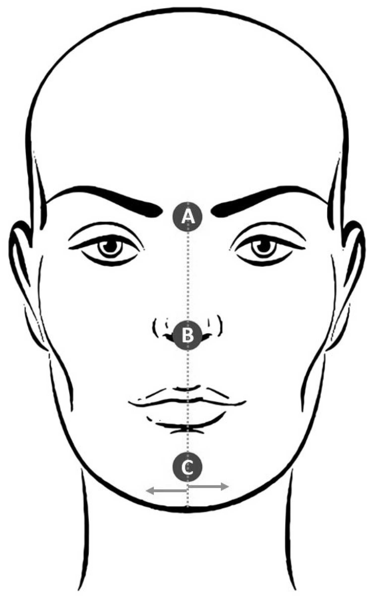
The face of each individual was marked with three red colored adhesive labels with a diameter of 12mm (Pimaco, TP12-017611) on the glabella, nose and chin centers (
Figure 1).
During the tests, all individuals used a white cotton coat. The use of the background and white coat in white was intended to avoid interference in the electronic identification of facial markings (labels) by the computerized method proposed.
Masticatory sequences were filmed in high resolution (1920x1080 pixels) with a camcorder (Sony MHS-PM5 model) positioned in fixed support 1 meter away from the individual and at the height of the mandible (Felicio et al., 2010).
Digital Video Processing
Each recorded video file was uploaded into a data analysis computing environment, processed and analyzed frame by frame through a digital processing algorithm. In each video, the algorithm detected the centroids of the labels placed in the patient’s face, and it estimated a linear function (straight line) that passed through the centroids of the patient’s glabella (
Figure 1A) and nose (
Figure 1B) labels. These two labels remained static during the chewing process (glabella-nose). Subsequently, the algorithm computed, in pixels, the perpendicular distance from the centroid of the chin (
Figure 1C) and the straight line previously estimated. This parameter corresponds to the lateral chin deviation due to chewing, and it is used for the data analysis carried out later on. The computation of the chin deviation, per frame, was repeated along the video, describing the behavior of the chin as a function of the time.
In order to reduce the noise introduced by the estimation of the labels’ centroid, a pre-processing step based on a moving-average digital filter (Mitra, 1998) was applied on the acquired data. A forth-order moving-average digital filter was used where each data point corresponds to the arithmetic mean of the present value and the three past ones. The computation is performed according to the equation in
Figure 2, where
di corresponds to the value in time sample
i. The order of the moving-average digital filter was chosen based on the data autocorrelation function (Box, Jenkins, Reinsel, 1994).
Regardless the chewing pattern, the most frequent value corresponds to the chin central position, as it repeats the most during the chewing process. Therefore, this value is subtracted from the chin deviation of each frame, and the process becomes zero-mean.
At this point, several low amplitude peaks were observed in the data. These peaks did not configure a complete chewing cycle and, therefore, they were removed from the chin deviation analysis. The strategy chosen to eliminate the region that comprises such undesirable information was based on the number of chewing cycles per minute. 80% of the data points around the central position were discarded from the analysis, resulting in 59.2 chewing cycles per minute.
The remaining data points were used by the algorithm to detect and quantify the individual chewing cycles. The positive peaks correspond to deviations of the chin to the right, whereas the negative peaks indicate chin deviations to the left (
Figure 3). In the end, the algorithm provides the percentage of the chewing cycles performed to each side (right and left). These two values are used to infer the chewing pattern. Finally, the block diagram shown in Figure 4 describes the algorithm developed for both feature extraction and data analysis.
Statistics
The distribution pattern of the number of deviations and total amplitude of the cycles was assessed using the Kolmogorov-Smirnov test, in which the variables showed a normal distribution. The t test for paired data was used to compare the preferential side (or chewing side) and the opposite side in groups 1 and 2 videos, and the right and left sides in group 3 videos. The t test for paired data was also used to compare the proportion of chewing cycles on the side identified in the video with the proportion of cycles required to groups 1 and 2.
Figure 2.
Moving-average digital filter used for data smoothing.
Figure 2.
Moving-average digital filter used for data smoothing.
Figure 3.
Chin deviation as a function of the time. The discarded region around the central position and the valid peaks are highlighted.
Figure 3.
Chin deviation as a function of the time. The discarded region around the central position and the valid peaks are highlighted.
RESULTS
Table 2 shows the number and the total amplitude of the deviations in the masticatory cycles as well as the comparison between the chewing side and the opposite side in the groups 1 and 2. The comparison between the sides of both groups presented a statistically significant difference (p <0.001), both for the number of cycles and for the total amplitude, with the chewing side showing higher average values than the opposite side.
Table 3 expresses the mean and the Student’s t-test results for paired data of the mean of number of cycles and total amplitude deviation of the cycles in group 3. There was no significant difference between the right and left sides regarding the number of masticatory cycler and total amplitude.
In groups 1 and 2, it was assessed whether the proportion of cycles identified in the video was equal to the proportion of required cycles
(
Table 4). It was found in both groups that the average percentage of deviation to the chewing side identified in the videos (Group 1, 75.41%; Group 2, 64.82%) was significantly lower (p <0.001) than the percentage of deviations requested at the time of shooting (Group 1, 100%; Group 2, 71.42%).
Table 2.
Mean values of the number and amplitude of deviation in group 3 masticatory cycles.
Table 2.
Mean values of the number and amplitude of deviation in group 3 masticatory cycles.
Table 3.
Comparison between the percentage of deviation to the chewing side with the percentage required to groups 1 and 2.
Table 3.
Comparison between the percentage of deviation to the chewing side with the percentage required to groups 1 and 2.
Table 4.
Comparison between the percentage of deviation to the chewing side with the percentage required to groups 1 and 2.
Table 4.
Comparison between the percentage of deviation to the chewing side with the percentage required to groups 1 and 2.
DISCUSSION
Among the methods described for the evaluation of the masticatory pattern, visual assessment is one of the most used (Hennequin et al 2005; Paphangkorakit et al, 2006; Felício et al, 2010; Hitos et al, 2011; Felício et al, 2012; Rovira-Lastra et al, 2014). However, the result of its application is associated with the experience of the professional, being subjective and susceptible to failures in the assessment. Regarding the objective methods such as electromyography and electrognathography, these are less commonly used and the presence of equipment can interfere with the execution of the masticatory process (Christensen et al., 1985; Hennequin et al., 2005; Nicolas, Veyrune, Lassauzay, Peyron, Hennequin, 2007; Gomes, Custódio, Jufer, Cury, Garcia, 2010; Gomes, Custódio, Faot, Cury, Garcia, 2011; Lepley et al., 2010; Turcio et al., 2016). The evaluation method of the masticatory pattern described in this study requires non-specialized equipment and presents little or no interference on the patient, differing from electromyography and electrognathography and making it simple and accessible.
The facial marking with adhesive labels on the glabella, nose and chin enabled digital identification of these structures by the proposed method. This way, we could not only identify but also quantify the lateral deviation of the mobile reference (the center of the mentum) in relation to the fixed structures of the face (glabella and nose) during the masticatory act.
Since the identification of the centroids of facial markings was based on the recognition of the intensity of the pixels of the labels, this may have been affected by the ambient light, incorporating small noise in the measurement of mandibular deviations, which can be related to potential instrumental biases. The determination of a rest band excluding 80% of the deviation peaks with lowest amplitude allowed the elimination of noises associated with the identification of the centroids of the labels and of non-representative lateral movements of the cycles. Thus, the videos of the masticatory processes used in this study showed an average of 59.26 masticatory cycles per minute, frequency of near 55 cycles/ min described by Berretin-Felix, Genaro, Trindade, Trindade Júnior (2005) for mastication of bread roll.
There is a wide range of chewing pattern classification, with no consensus or standardization among the authors. Unilateral mastication has been described as preferred when the occurrence of the cycles is observed in 61% to 94% on one side (Gomes et al., 2010; Felício et al., 2010; Gomes et al., 2011; Felício, Medeiros, Melchior, 2012) and as chronic when this occurrence is higher than 95% (Felício et al., 2010; Felício et al., 2012). In this study, the preference for one side during the chewing process has been identified by the proposed method, affecting 75.41% in group 1 and 64.82% in group 2, both significantly higher than in the opposite side to the preference. Thus, considering that there is a masticatory dysfunction potentially harmful to the components of the stomatognathic system (Hitos et al., 2011; Barcellos et al., 2012) when the prevalence of cycles on one side exceeds 60% (Ramfjord et al., 1983; Christensen et al., 1985; Martinez-Gomis et al., 2009; Turcio et al., 2016), the computational method proposed was efficient in identifying the masticatory dysfunction.
The pattern of bilateral distribution of masticatory process was identified, and found no significant difference between the right and left sides regarding prevalence and extent of lateral displacement peaks, p = 0.128 and p = 0.069, respectively, in the group 3 videos.
It must be taken into account that there is the possibility of errors by the subjects in the performance of the masticatory cycles, as well as difficulty in keeping the predetermined chewing pattern and the head stable throughout filming, allowing for some extent of participant-related biases. Masticatory patterns misidentifications are also expected when visual methods are used, as they are subjective and results depend on a number of factors related to the professionals who use them. The computerized method proposed allowed an objective analysis of the masticatory pattern without the influence of interpretations related to the examiner, allowing, thus, the standardization of evaluations with reduction of errors inherent from observational methods. Therefore, the evaluation results of mandibular movements during the masticatory process become reproducible and free of human errors, allowing clinical comparisons intra and inter-patients.
Future research must compare the diagnostic capacity of the computerized method shown in this study with other clinical methods currently in use with the intent of identifying the factors that can influence the results of analysis, which will benefit patients with myofunctional disorders.
CONCLUSION
The use of filming of the masticatory process associated with the proposed computerized method was effective in identifying the bilateral masticatory pattern and able to recognize the preference to use one side during the masticatory cycles.


