Next Article in Journal
Orofacial Myofunctional Disorders: Guidelines for Assessment and Treatment
Previous Article in Journal
Thumbsucking as Related to Placement and Acoustic Aspects of /s,z/ and Lingual Rest Postures
 
 
International Journal of Orofacial Myology and Myofunctional Therapy is published by MDPI from Volume 51 Issue 1 (2025). Previous articles were published by another publisher in Open Access under a CC-BY (or CC-BY-NC-ND) licence, and they are hosted by MDPI on mdpi.com as a courtesy and upon agreement with the previous journal publisher.
Article

Speech-Language Pathology Findings in Patients with Mouth Breathing: Multidisciplinary Diagnosis According to Etiology

by
Patricia Junqueira
1,*,
Irene Queiroz Marchesan
1,
Luciana Regina de Oliveira
1,
Emilio Ciccone
2,
Leonardo Haddad
2 and
Maria Cândida Rizzo
2
1
CEFAC Institute, São Paulo, Brazil
2
Menino Jesus Hospital, São Paulo, Brazil
*
Author to whom correspondence should be addressed.
Int. J. Orofac. Myol. Myofunct. Ther. 2010, 36(1), 27-32; https://doi.org/10.52010/ijom.2010.36.1.3
Submission received: 1 November 2010 / Revised: 1 November 2010 / Accepted: 1 November 2010 / Published: 1 November 2010

Abstract

:
The purpose of this study was to identify and compare the results of the findings from speech-language pathology evaluations for orofacial function including tongue and lip rest postures, tonus, articulation and speech, voice and language, chewing, and deglutition in children who had a history of mouth breathing. The diagnoses for mouth breathing included: allergic rhinitis, adenoidal hypertrophy, allergic rhinitis with adenoidal hypertrophy; and/or functional mouth breathing. This study was conducted with on 414 subjects of both genders, from 2 to 16-years old. A team consisting of 3 speech-language pathologists, 1 pediatrician, 1allergist, and 1 otolaryngologist, evaluated the patients. Multidisciplinary clinical examinations were carried out (complete blood counting, X-rays, nasofibroscopy, audiometry). The two most commonly found etiologies were allergic rhinitis, followed by functional mouth breathing. Of the 414 patients in the study, 346 received a speech-language pathology evaluation. The most prevalent finding in this group of 346 subjects was the presence of orofacial myofunctional disorders. The most frequently orofacial myofunctional disorder identified in these subjects who also presented mouth breathing included: habitual open lips rest posture, low and forward tongue rest posture and lack of adequate muscle tone. There were also no statistically significant relationships identified between etiology and speech-language diagnosis. Therefore, the specific type of etiology of mouth breathing does not appear to contribute to the presence, type, or number of speech-language findings which may result from mouth breathing behavior.

INTRODUCTION

Mouth breathing (MB) is one of the most common symptoms in childhood and a great deal of the literature relates it directly to different etiologies (Cintra, Castro, Cintra, 2000; Motonaga, Berti, Anselmo-Lima, 2000; Rizzo, Hauache, Naspitz, 2002; Takahashi, Ono, Ishiwata, Kuroda, 2002; Paulo, Conceição, 2003; Di Francesco, Passeroti, Paulucci, Miniti, 2004; Andrade, Andrade, Araújo, Ribeiro, Deccax, Nemr, 2005; Lessa, Enoki, Feres, Valera, Lima, Matsumoto, 2005; Bicalho, Motta, Vicente, 2006; Vera, Conde, Wajnsztejn, Nemr, 2006). While the most common cause for mouth-breathing behavior is allergic rhinitis, there are other etiologies, which include: pharyngeal tonsil (adenoid) and/or palatine tonsils (amygdales) hypertrophy, non-allergic rhinitis, turbinal bone hypertrophy, and septum deviations (Lund, 1988; Motonaga et al, 2000; Paulo et al, 2003; Di Francesco et al, 2004). In addition to etiological factors, which contribute to this behavior, MB may occur as an established habit pattern that persists without an obstructive anatomical factor that prevents nasal breathing (Cintra et al, 2000; Motonaga et al, 2000). Many authors refer to the ‘mouth-breathing syndrome’ to represent the characteristics found in individuals who use their mouths as their predominant manner of breathing. However, it is know that MB has various causes and characteristics, with distinct pathophysiologies. This makes it difficult to include all MB patients within a homogeneous group (Brodsky, 1993).
It is not uncommon for some authors to claim that the following concerns are characteristic of children with MB: articulation disorders, impaired vocal quality, alterations in the orofacial structures and functions, as well as language difficulties (Rizzo et al., 2002; Di Francesco et al., 2004). Because MB is a symptom that frequently presents in association with multifactorial causes associated with various pathologies, it is necessary to clarify details of the clinical aspects of MB. This study aims to identify and compare the results of the findings from speech-language pathology evaluations in children from 2 to 16-years of age with a history of mouth breathing who were diagnosed by a multidisciplinary team with one or more of the following: allergic rhinitis; adenoidal hypertrophy; allergic rhinitis with adenoidal hypertrophy and/or functional MB.

METHODS

This study was conducted with 414 subjects of genders, 269 (65.0%) males and 145 (35.0%) females, from 2 to 16-years old, attending the Care Center for the Mouth-Breather at the Cefac Institute. A team of three speech-language pathologists, one pediatrician, one allergist, and one otolaryngologist, who are staff members at the Care Center for the Mouth-Breather, evaluated the patients over several years from March 2004 to April 2009. Multidisciplinary clinical examinations were carried out which included complete blood counting, cavum X-rays, nasofibroscopy, audiometry, with all subjects submitted to all the examinations. Additional examinations were required after the medical evaluation to determine the cause of MB. Then a speech-language evaluation was administered.
An etiology of MB was determined by the physicians based on the following objective measurements: specific IgE (immunoglobulin E) serum, cavum X-ray and/or nasofibroscopy. Based on the identified etiology, the subjects were divided into four groups:
  • Group 1. Allergic Rhinitis: subjects were assigned to this group when signs and symptoms characteristic of allergic rhinitis was documented in their clinical history and was accompanied by specific IgE results greater than or equal to Class 3, according ImmunoCAP - Phadia technique - level minimum 0.35 KU/ml - Class 0 (Lund, 1988).
  • Group 2. Adenoidal Hypertrophy: subjects were assigned to this group when their X-rays indicated that the aerial column of the nasal cavity was decreased by ¾ (three quarters) or more, (i.e., obstruction of more than 80%;) or when in the nasofibroscopy it was determined that the adenoids occupied over three quarters of the nasopharynx, (i.e., 80% or more, for adenoidal hypertrophy) (Lund, 1998).
  • Group 3. Allergic Rhinitis and Adenoidal Hypertrophy: subjects were assigned to this group when signs of both allergic rhinitis and adenoidal hypertrophy were diagnosed.
  • Group 4. Functional Mouth Breathing (FMB): subjects were assigned to this group when mouth-breathing behavior was diagnosed with no signs of allergic rhinitis or obstruction (Di Francesco et al., 2004).
Of the 414 subjects in the total study, 346 subjects met the criteria to undergo the speech-language pathology evaluation. A speech-language pathologist trained in orofacial myofunctional disorders performed the speech-language evaluation. The assessment included a speech/articulation, voice, language, and orofacial myofunctional examination. A hearing evaluation was also administered. Subjects with hearing loss, neurological and/or motor disabilities and/or related problems were excluded from the speech evaluation. For the speech-language pathology evaluation, protocol established by Marchesan (2003) was used, which included specific observations concerning the subjects’ usual position of the lips, tongue, orofacial tonicity, breathing, swallowing, speech, voice and spoken language. Based on the results of this evaluation, subjects diagnosed as mouth-breathers were classified into three groups:
  • Group 1 - Orofacial Myology: subjects were assigned to this group when orofacial myofunctional disorders were identified in one or more of the evaluated areas (for example: habitual position of lips and/or tongue, tonus, chewing and/or swallowing).
  • Group 2 – Alterations in Orofacial Myology with Speech and/or Voice and/or Language: subjects were assigned to this group when orofacial myofunctional deficits were identified in conjunction with any other speech, voice and/or language impairments.
  • Group 3 - Normal: subjects were assigned to this group when no deficiencies were identified in either orofacial myofunctional areas or speech-language areas.
Ethical Committee approved the study (number 078/09). Data were statistically analyzed using the chi-square test (p = 0.05).

RESULTS

The study included 414 subjects, from 2 to 16-years of age, 269 (65.0%) males and 145 (35.0%) females. The subjects were divided according to their etiological mouth-breathing diagnosis. The most prevalent identified etiology was allergic rhinitis which was found in 148 (35.7%) subjects. This was followed by 102 (24.6%) subjects diagnosed with functional mouth breathing. The group of subjects with the adenoidal hypertrophy, and the group of subjects with both allergic rhinitis and adenoidal hypertrophy were equally significant as they each had 82 (19.8%) subjects.
Of the 346 subjects receiving the speech-language evaluation, which included an orofacial myofunctional evaluation, 82.2% were found to have at least one disorder. Subjects were assigned to one of three groups: Group 1 only OMD; Group 2 - OMD in conjunction with another speech-language disorders such as speech articulation, voice and/or language; and Group 3 – Normal. The most prevalent finding was the presence of Orofacial Myofunctional Disorders in 216 (62.4%) subjects. There were no statistically significant associations found between the patients’ etiology and their respective speech-language pathology diagnosis group (p = 0.218) (Table 1).
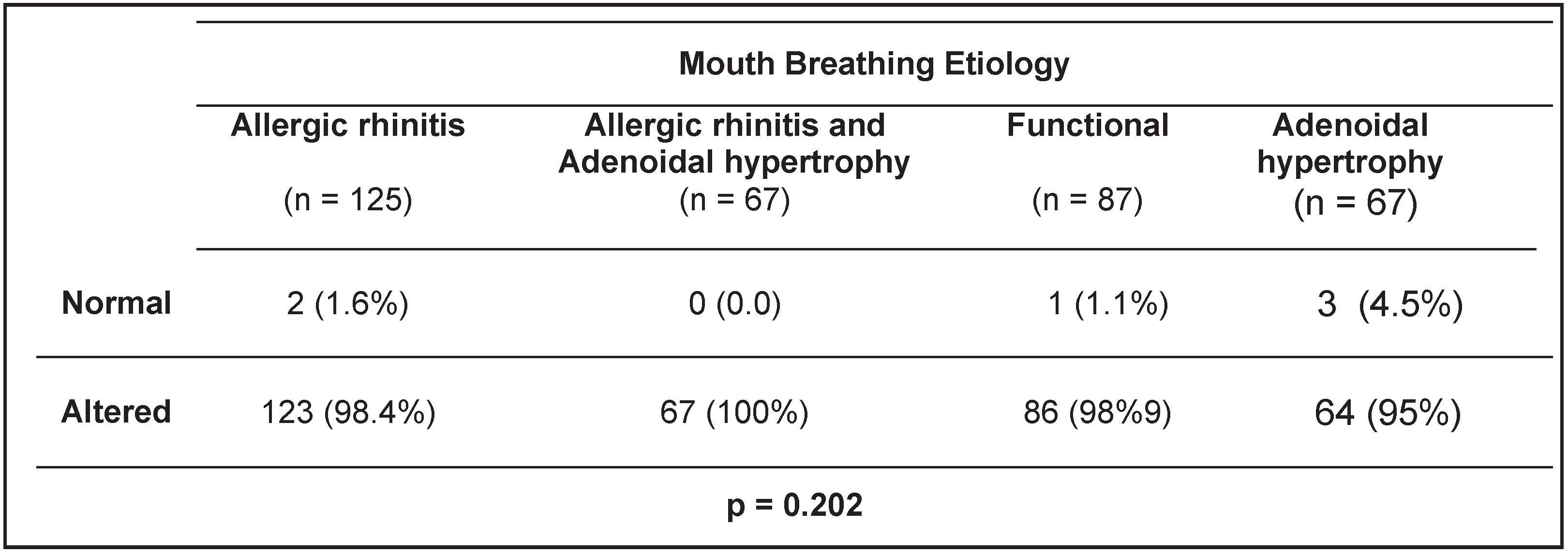
An analysis of results for all the patients who received a speech language evaluation was completed based on etiology. There were no statistically significant associations between the patients’ etiology and the presence of any alteration, either OMD or others (p = 0.202) (Table 2).

DISCUSSION

The speech-language difficulties found in MB subjects have been widely reported in the last decade (Cintra et al., 2000; Rizzo et al., 2002; Paulo et al, 2003; Valera, Trawitzki, Mattar, Matsumoto, Elias, Anselmo-Lima, 2003; Valera Trawitzki, Anselmo-Lima, 2006). However, it should be emphasized that much of the published research is carried out with no data proof. Some authors repeat in their publications the reports of earlier authors who did not support their findings based on quantitative data analysis. In addition, other studies show an often-subjective criterion to define MB. The lack of objectivity in order to define nasal obstruction can lead to misdiagnosis and, consequently, to inappropriate treatment. Instrumental assessment of MB is of critical importance for accurate diagnosis.
This study sought to identify the causes of the MB behaviors presented by subjects through a multidisciplinary approach using instrumental assessments, which allowed the documentation of the presence or absence of an anatomical obstruction. Through the clinical evaluation and standardized complementary examinations, findings indicated that among the most prevalent causes of MB were: allergic rhinitis (35.7%), functional MB (24.6%), adenoidal obstruction (19.8%), and allergic rhinitis with adenoidal obstruction (19.8 %). This last finding indicates an association of respiratory pathologies resulting in nasal obstruction in the same subject. This is consistent with findings in previous research studies that sought to identify the primary causes of MB (Motanaga et al., 2000; Valera et al., 2006).
Other authors have reported in their studies that the presence of MB may be habitual in nature, with subjects persisting in MB behavior even when the permeability of their upper airways were clear (Cintra et al., 2000; Motanaga et al., 2000; Di Francesco et al., 2004). The current results support this finding. Of the subjects who presented as mouth breathers, 24.6% were classified as functional mouth breathers who did not have obstructive causes as confirmed by instrumental multidisciplinary assessment. Attention should be called to this diagnosis of functional mouth breathing, because many children may present with MB behaviors even after a previous obstruction has been medically treated and is no longer present. Functional mouth breathing is a disorder that should be addressed using therapeutic measures.
Whatever the MB etiology, orofacial myofunctional disorders were almost always identified. These disorders are associated with the subject persisting in mouth breathing behaviors either in an attempt to compensate for the deficiency of inspired air, or as a habit pattern. One of the consequences may be that the function of the tongue to shape the oral vestibule is greatly restricted when it is in a breathers may impact the interactions of the orofacial musculature. The end result of this incorrect and inefficient breathing pattern is the potential interactive effect on surrounding orofacial musculature, which may result in generating a functional deficiency. (Cintra et al., 2000; Motanaga et al., 2000; Rizzo et al., 2002; Paulo et al, 2003; Valera et al., 2003).
Disorders in voice quality, speech, and language, which may be related to the presence of MB, have also been reported by several authors (Cintra et al., 2000; Motanaga et al., 2000; Rizzo et al., 2002). In concurrence with the literature, a high incidence of orofacial myofunctional disorders was found in subjects who were mouth breathers. This study also identified a higher incidence of both orofacial myofunctional disorders and speech-language disorders among the participants identified as mouth breathers than previous studies. A significant number of subjects with isolated voice, speech and language disorders were not identified in this study.
Given these results, an analysis was completed to determine if there was any correlation between the MB etiology and the degree of severity in the subjects’ speech and low and forward rest posture which frequently occurs in mouth breathers, as it is difficult to breathe through one’s mouth with the tongue in position against the palate. This low and forward tongue rest posture in mouth language deficits. No statistically significant relationship was identified between the subject’s etiology and the presence or the number of their respective speech-language pathology disorder/s. However, MB regardless of its cause was a factor associated with speech-language/orofacial myofunctional impairments.
Since impairments in the orofacial myofunctional system were the most prevalent findings in this study population, regardless of the etiology, future studies could focus on identifying if there is any relationship between the severity of MB and the most common causes of nasal obstruction.

CONCLUSIONS

Impairments in oral function, tonus and habitual lips and tongue rest postures are frequently found in mouth breathers. Etiology does not contribute to the presence, severity and/or the number of disorders found by SLPs in mouth breathers. Mouth breathing and the associated OMD are factors that may lie at the very foundation of a variety of speech and language disorders

References

  1. Andrade, F.V., D.V. Andrade, A.S. Araújo, A.C.C. Ribeiro, L.D.G. Deccax, and K. Nemr. 2005. Alterações estruturais de órgãos fonoarticulatórios e más oclusões dentárias em respiradores orais de 6 a 10 anos. Revista Cefac 7: 318–25. [Google Scholar]
  2. Bicalho, G.P., A.R. Motta, and L.C. Vicente. 2006. Avaliação da deglutição em crianças respiradoras orais. Revista Cefac 8: 50–5. [Google Scholar]
  3. Brodsky, L. 1993. Tonsillitis, tonsillectomy and adenoidectomy. In Head and Neck Surgery-Otolaryngology. Edited by B.J. Bailey. Philadelphia: JB Lippincott, pp. 838–47. [Google Scholar]
  4. Cintra, C.F., F.F. Castro, and P.P. Cintra. 2000. As alterações orofaciais apresentadas em pacientes respiradores bucais. Revista Brasileira de Alergia e Imunopatologia 23: 78–83. [Google Scholar]
  5. Di Francesco, R.C., G. Passeroti, B. Paulucci, and A. Miniti. 2004. Respiração oral na criança: repercussões diferentes de acordo com o diagnóstico. Revista Brasileira de Otorrinolaringologia 70, 5: 667–70. [Google Scholar] [CrossRef]
  6. Lessa, F.C., C. Enoki, M.F. Feres, F.C. Valera, W.T. Lima, and M.A. Matsumoto. 2005. Influência do padrão respiratório na morfologia craniofacial. Revista Brasileira de Otorrinolaringologia 71: 156–60. [Google Scholar] [CrossRef] [PubMed]
  7. Lund, V. 1998. Allergic rhinitis: making the correct diagnosis. Journal of the British Society for Allergy & Clinical Immunology 28, 6: 25–8. [Google Scholar]
  8. Marchesan, I.Q. 2003. Protocolo de Avaliação Miofuncional Orofacial. In Respiração Oral. Edited by L.H. Krakauer, R.C. Di Francesco and I.Q. Marchesan. Ed. Pulso: pp. 55–80. [Google Scholar]
  9. Motonaga, S.M., L.C. Berti, and W.T. Anselmo-Lima. 2000. Respiração bucal: causas e conseqüências. Revista Brasileira de Otorrinolaringologia 66: 115–119. [Google Scholar]
  10. Paulo, C.B., and C.A. Conceição. 2003. Sintomatologia do respirador oral. Revista Cefac 5: 219–22. [Google Scholar]
  11. Rizzo, C., S. Hauache, C. Naspitz, S. Pignatari, P. Junqueira, and M. Hallinan. 2002. Characteristics of children with allergic rhinitis and chronic mouth breathing. Journal of the British Society for Allergy & Clinical Immunology 109: S263. [Google Scholar]
  12. Takahashi, S., T. Ono, Y. Ishiwata, and T. Kuroda. 2002. Breathing modes, body positions, and suprahyoid muscle activity. Journal Orthodontic 29: 307–13. [Google Scholar] [CrossRef] [PubMed]
  13. Valera, F.C.P., L.V.V. Trawitzk, and W.T. Anselmo-Lima. 2006. Myofunctional evaluation after surgery for tonsils hypertrophy and its correlation to breathing pattern: A 2-year-follow up. International Journal Pediatric Otorhinolayngology 70: 221–25. [Google Scholar] [CrossRef] [PubMed]
  14. Valera, F.C.P., L.V.V. Trawitzki, S.E.M. Mattar, A.M. Matsumoto, W.T. Elias, and W.T. Anselmo-Lima. 2003. Muscular, functional and orthodontic changes in preschool children with enlarged adenoids and tonsils. International Journal Pediatric Otorhinolayngology 67: 761–70. [Google Scholar] [CrossRef] [PubMed]
  15. Vera, C.F.D., G.E. Conde, R. Wajnsztejn, and K. Nemr. 2006. Transtornos de aprendizagem e presença de respiração oral em indivíduos com diagnóstico de transtornos de déficit de atenção/hiperatividade (TDAH). Revista Cefac 8: 441–55. [Google Scholar] [CrossRef]
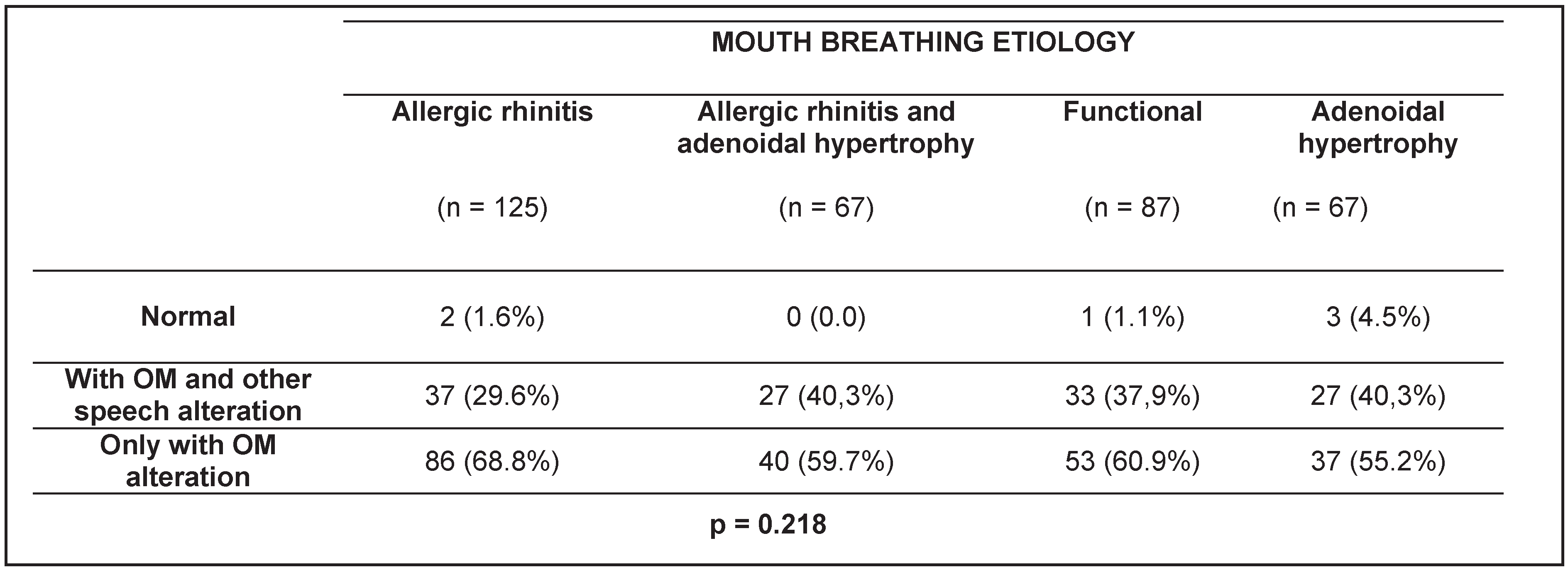
Table 1. Association between Mouth-Breathing etiologies and Speech-Language Pathology diagnosis in 346 patients.
Ijom 36 00027 i001
Table 2. Overall association between Mouth Breathing etiologies and Speech-Language Pathology diagnosis (including OMD and other alterations) in 346 patients.
Ijom 36 00027 i002