Review

Non-Animal Technologies to Study and Target the Tumour Vasculature and Angiogenesis

1
IRCCS S Raffaele, 20132 Milan, Italy
2
National Dental Center, Singapore 168938, Singapore
3
Faculty of Dentistry Oral & Craniofacial Sciences, King’s College London, London WC2R 2LS, UK
*
Author to whom correspondence should be addressed.
Organoids 2025, 4(2), 12; https://doi.org/10.3390/organoids4020012
Submission received: 3 April 2025 / Revised: 28 May 2025 / Accepted: 29 May 2025 / Published: 4 June 2025

Abstract

Tumour-associated angiogenesis plays a key role at all stages of cancer development and progression by providing a nutrient supply, promoting the creation of protective niches for therapy-resistant cancer stem cells, and supporting the metastatic cascade. Therapeutic strategies aimed at vascular targeting, including vessel disruption and/or normalisation, have yielded promising but inconsistent results, pointing to the need to set up reliable models dissecting the steps of the angiogenic process, as well as the ways to interfere with them, to improve patients’ outcomes while limiting side effects. Murine models have successfully contributed to both translational and pre-clinical cancer research, but they are time-consuming, expensive, and cannot recapitulate the genetic heterogeneity of cancer inside its native microenvironment. Non-animal technologies (NATs) are rapidly emerging as invaluable human-centric tools to reproduce the complex and dynamic tumour ecosystem, particularly the tumour-associated vasculature. In the present review, we summarise the currently available NATs able to mimic the vascular structure and functions with progressively increasing complexity, starting from two-dimensional static cultures to the more sophisticated tri-dimensional dynamic ones, patient-derived cultures, the perfused engineered microvasculature, and in silico models. We emphasise the added value of a “one health” approach to cancer research, including studies on spontaneously occurring tumours in companion animals devoid of the ethical concerns associated with traditional animal studies. The limitations of the present tools regarding broader use in pre-clinical oncology, and their translational potential in terms of new target identification, drug development, and personalised therapy, are also discussed.

1. Introduction

Cancer emerges when mutated cells grow uncontrollably within a permissive tissue environment [1]. Initially, natural safeguards inhibit neoplastic formation; however, prolonged exposure to carcinogenic factors favours the accumulation of further mutations in cancer cells until they become able to evade these defences. Concurrently, growing tumours alter the local and systemic environments, enabling cancer progression and spread [2].
Cancers are complex dynamic ecosystems composed of tumour cells and different non-cancerous cells, all embedded in an extracellular matrix (ECM), exhibiting distinctive and unique physical, biochemical, and mechanical properties [3] (Figure 1).
The tumour microenvironment (TME) includes immune cells, cancer-associated fibroblasts, endothelial cells, pericytes, and tissue-resident cell types, all playing critical roles in each stage of cancer progression [3,4] (Figure 1). Tumour–TME crosstalk, mediated by cellular interactions, soluble factors, and metabolite availability [5], affects cancer cell survival, proliferation, responses to anti-cancer drugs, and immune evasion, and is emerging as an attractive therapeutic target.
Typically, all cancers elicit a certain degree of local tissue inflammation. Inflammation initially combats cancer development, but, at the same time, it exercises selective pressure on cancer cells [6,7,8] (Figure 1B,C). Those cells that survive become tolerant to inflammation and ultimately shape the immune landscape both in the TME [2] and systemically. In solid tumours, these initial malignant lesions are called carcinoma in situ (Figure 1B), whose prognosis is favourable if they are promptly detected [9,10].
Tumour-associated angiogenesis (TAA) and vascular alterations are hallmarks of the TME, and, following the work of J. Folkman [11,12], microvascular involvement is included among the criteria defining invasive versus non-invasive lesions (Figure 1B vs. Figure 1C). A hypovascular, hypoxic TME can limit the efficacy of chemo-/radiotherapy by hampering the delivery of oxygen and drugs [13,14]. On the other hand, uneven intratumour angiogenesis and angiocrine signalling further promote cancer cells’ phenotypic heterogeneity [2] (Figure 1D).
Overall, the TME behaves like a chronic, never-healing wound [15], because the sources of tissue damage and inflammation are not removed. The cellular and molecular dynamics in such systems are complex to predict as they depend on many contextual signals that vary widely over time, across different genetic backgrounds, across different organs, and even within the same tissue. Moreover, by means of systemic signalling, mediated by soluble factors like cyto-/chemokines and extracellular vesicles [16], cancer may induce changes in distant tissue types, including vascular leakiness, angiogenesis, and immunosuppression. All of this forms a pre-metastatic niche [17]. Pre-metastatic niches promote the seeding of metastases and are tumour-specific (both within the same cancer type and across different types), dictating preferences for specific target organs (seed and soil theory [18]).
Notably, the tumour-associated microvasculature (TAMV; Figure 1C,D) plays a key role at all stages of cancer development and progression, including the “angiogenic switch”, the creation of protective niches for therapy-resistant cancer stem cells (CSCs), the creation of pre-metastatic niches, and metastasis.

1.1. Tumour-Associated Microvasculature

Malignant cancer cells have a high metabolic demand to sustain their abnormal growth, and rapid cell growth promotes intratumoural hypoxia and chronic inflammation [19]. All these features contribute to the “angiogenic switch”, i.e., the sustained and uncontrolled induction of pathological angiogenesis, leading to the formation of an aberrant TAMV (Figure 1D). Hypoxia activates the cellular oxygen-sensing machinery (hypoxia-inducible factor (HIF) pathway) [20] in endothelial, immune, stromal, and cancer cells, directly regulating the transcription of pro-angiogenic genes like vascular endothelial growth factor (VEGF-A), stromal-derived factor (SDF-1), and angiopoietin-2 [19]. However, due to the unbalanced signalling in the TME, the TAMV does not fully mature, is morphologically and functionally abnormal, and exhibits the suboptimal diffusion of oxygen and nutrients, contributing to a self-sustaining hypoxic–inflammatory loop. In such environments, cancer cells can acquire distinct phenotypes, including fast-growing clones, slow growing CSCs with self-renewal potential, and migratory clones with high metastatic potential (Figure 1D).
Endothelial cells (ECs) are the main cellular components of the microvasculature. ECs are physiologically heterogeneous across different organs and within the same organ, reflecting a variety of different functions [21]. ECs composing the TAMV are phenotypically distinct from their healthy counterparts [22] and have distinct metabolism [23] and angiocrine signalling [24].
Dysregulated angiocrine signalling to immune cells contributes to an immunosuppressive TME, which hampers natural anti-tumour responses [25,26,27]. Concurrently, a leaky TAMV favours the intravasation and dissemination of metastasis-initiating cells, which travel as circulating tumour cells to distant organs along the blood or lymphatic streams [28].
In breast cancer, ECs, tumour-associated perivascular macrophages (TAMs), and specific cancer cells form a functional triad, named the tumour microenvironment of metastasis (TMEM) doorway. The number of TMEM doorways in the primary tumour has been found to correlate with the propensity for metastasis [29] (Figure 1D). It is tempting to assume that similar doorways might exist within other cancer types, possibly with distinct molecular signatures; however, this has not been demonstrated so far.
Upon seeding in multiple organs, metastasis-initiating cells may generate overt, clinically evident metastases via the co-option of permissive microenvironments, mimicking the native niches, including a primed microvasculature [30,31,32,33].

1.2. Therapeutic Implications

Given the central role of the TAMV in tumour development, vascular targeting has been explored as a therapeutic option in the past 20 years.
Anti-angiogenic therapy (AAT), i.e., attempting to prune the abnormal TAMV via interference with pro-angiogenic signalling, especially the VEGF pathway, has been widely studied and tested experimentally and clinically [32]. Despite the initial promise and some clinical success, AAT has yielded inconsistent clinical results. Not all cancer types are sensitive to AAT, and, in those sensitive types, AATs like the monoclonal antibody against VEGF-A, Bevacizumab, cause a host of escape mechanisms, like the “angiogenic rebound”, i.e., the compensatory upregulation of VEGF or other angiogenic molecules. Additionally, interference with the VEGF axis causes vessel disruption locally and systemically, producing severe side effects and increasing the propensity for metastasis [22,32].
More recently, vascular normalisation therapy (VNT), i.e., attempting to modulate rather than prune the TMV via the fine-tuning of AATs, has been proposed as a promising new route. Animal studies have shown that VNT could alleviate intratumour hypoxia, facilitate the delivery of drugs, increase the oxygen-dependent effects of radiotherapy, and favourably modulate the immune TME [13,34,35,36].
The clinical testing of VNT is currently ongoing [36]; however, previous studies have already highlighted that the clinical success of VNT will depend on precisely identifying and measuring the timeframe of its therapeutic activity (TAMV normalisation window).
In summary, it is now clear that VNTs are potent tools to modulate the TAMV, with huge therapeutic potential. However, their clinical efficacy is currently limited by our capacity to measure and understand the dynamic crosstalk between cancer, the TME, and therapeutic agents.
For example, we do not know how to timely and precisely deliver therapeutic agents to maximise their beneficial effects while minimising their side effects.
Thanks to intensive research and powerful technologies like omics, we are rapidly learning which cell types and molecules are involved in these processes. However, we do not fully understand how these elements crosstalk before, during, and after therapy in different tissue types and organs.
Characterising the cellular and molecular dynamics driving TAMV functions is thus a central and unmet clinical need, as TAMV-associated features, like metastases, still represent the leading cause of cancer-associated death [30,33].
Deciphering the dynamic interplay between cancer cells and the TAMV may offer the opportunity to identify and target pathways common to most cancers. For this purpose, reliable models of the TAMV are urgently needed.
Murine models are a cornerstone of biological research and have been successfully used in both translational and pre-clinical research [37]. However, pre-clinical animal models are typically complex, time-consuming, expensive, and difficult to standardise [38]. More importantly, they do not fully capture the complexity and genetic heterogeneity of human cancers, and they cannot recapitulate the molecular signalling and cancer–TME cellular crosstalk, limiting their translation potential. Indeed, US Food and Drug Administration (FDA) data show that over 90% of drugs that are successful in pre-clinical animal models fail to demonstrate safety or effectiveness in humans [39].
We need to develop affordable research tools that are able to increase our mechanistic understanding of the TME, to facilitate the development of new therapeutic strategies and to customise treatment to individual patients. Non-animal technologies (NATs) able to reproduce selected aspects of human biology are rapidly emerging to fill these gaps.
In the following sections of this review, we discuss the advances in modelling the TME, the TAMV, and their dynamic crosstalk in vitro, ex vivo, and in silico, focusing on those that are promising to advance cancer research, therapy, and care.

2. Non-Animal Technologies

2.1. Static Cultures

Cancer research and drug development have long relied upon experiments performed using in vitro cultures of cell lines and primary tumour cells grown in two-dimensional (2D) static cultures [40,41,42]. While these culture systems have allowed us to elucidate the basic molecular signatures of cancer and still are the gold standard in primary drug screenings [43], they do not adequately reproduce the spatial and functional complexity of the TME; hence, they do not enable us to predict the impacts of drugs in individual patients. The pioneering work of Bissell and colleagues [44,45] has clearly demonstrated that normal and tumour cells grown in 2D culture significantly differ from those kept in 3D in terms of morphology, biological behaviour, gene expression profiles, and responses to drugs [46,47].
Experimental cancer models should incorporate elements of the surrounding milieu to recapitulate the tissue-specific multicellularity and architecture, biochemical and mechanical signals, and cell–cell and cell–ECM interactions [40,41,42]. Moreover, from the perspective of patient-specific cancer therapy, the need for personalised tumour models is rapidly emerging [48].
Personalised 3D cell cultures include tumour spheroids, organoids, and explants. Spheroids are clusters of cells, aggregated under static conditions via hanging drop techniques or cultures on non-adhesive substrates [49,50,51]. Spheroids derived from tumour cell lines or primary tumour cells recapitulate the main features of human solid tumours, like their multi-layered structures, where peripheral proliferative cells surround a necrotic core, and hypoxia and nutrient gradients, making them suitable for drug screening [52,53]. More complex approaches that incorporate the ECM take advantage of natural or synthetic polymeric substrates with tuneable compositions and stiffness. Such substrates can be formed as hydrogels or solid scaffolds, which provide mechanical support and a biomimetic physical and chemical environment [54]. Scaffolds allow tumour and stromal cells to attach and proliferate, mimicking cell-to-cell and cell-to-ECM signals [54].
A better approximation of tumours’ multicellular complexity is represented by patient-derived organoids, which are 3D cell aggregates generated by culturing pluripotent stem cells (PSC) or cells derived from patients’ tumour tissue (tumouroids) under appropriate conditions [48,55]. Compared to tumour spheroids, which are typically monocultures [50,51], organoids more closely reflect the TME’s heterogeneity and composition and the structures of the tumours that they originate from. However, they still do not incorporate key elements of the TME, like the microvasculature. Moreover, the current protocols to generate patient-derived tumour organoids have inconsistent yields, are complex and time-consuming to implement, and are scarcely standardised across laboratories [52,55].
Finally, 3D bioprinting technology is emerging as a promising strategy to manufacture tissue-engineered constructs with well-defined 3D geometries [56]. For example, 3D bioprinting can be used to build tumour constructs via the sequential layering of living cells (both tumour and stroma) within functionalised biomaterials (bioinks) [56,57].

2.2. Ex Vivo Culture of Tumour Explants

Patient-derived pre-clinical models that can preserve tumour features and predict outcomes in a personalised manner are greatly needed in the field of cancer drug discovery and development. Compared to tumour-derived organoids, tumour explants obtained from resected tumour/metastasis or biopsy specimens fully incorporate the native genomic, histological, and functional profiles of the cancer cells within their TMEs [58,59]. For example, tissue slices of human gliomas have been successfully cultured and used to detect metabolically active EC-associated “hot-spots” in otherwise low-grade lesions [59].
Tumour explants cultured under static conditions have a limited lifespan in culture (less than 3 days), limiting their applicability. This limitation has been addressed through the application of dynamic culture technologies, including the Rotary Cell Culture System (RCCS) bioreactor.

2.3. Dynamic Cultures

The 3D models described above have a common limitation in that they do not allow the appropriate transport of nutrients, solutes, and metabolic waste in and out of the cultured tissue—all prerequisites to sustain long-term cell viability and prevent cell necrosis. Thus, complex cancer cultures typically have a short lifespan in culture.
Seminal discoveries and inventions in recent years have fostered the transition from static to dynamic cultures. Dynamic cultures can better reproduce the changes occurring in a growing tumour by providing efficient mass transfer, i.e., nutrients’ delivery and metabolic waste removal, thus preserving the cell viability within 3D cell/tissue masses [60]. These culture conditions can be obtained using dynamic bioreactors [50,61], including roller bottles, spinner flasks, gyratory shakes, perfusion [62,63], and microgravity bioreactors [48,59,60,61].
Among these systems, the microgravity-based RCCS bioreactor stands out as suitable for culturing functional 3D tissue-like bioconstructs and explants of various origins. RCCS bioreactors were primarily used to facilitate the self-assembly and culture of scaffold-free spheroids [64] and then further tuned to obtain tumour–TME co-cultures on scaffolds [65] and tissue explants to study the molecular mechanisms involved in cancer development and biology, as well as for drug testing in a patient-specific context [66,67,68]. Culture in an RCCS bioreactor allows us to perform downstream analyses, including “omics” approaches, to dissect the metabolic and functional pathways operating within TMEs [69].
In recent years, advances in microfluidics technology, i.e., the study of fluid flow within small (<100 micrometres in diameter) conduits and the development of affordable devices operating in such conditions, have paved the way for the development of more biomimetic systems.
Moreover, 3D cell culture based on microfluidic devices (often called lab-on-chip or microphysiological systems (MPSs)) allows the study of key processes like cancer migration, invasion, and angiogenesis and drug responses in a miniaturised yet precisely defined culture environment [70].

3. Modelling Tumour Vasculature and Angiogenesis

Microvessels, i.e., blood vessels, or capillaries with a calibre smaller than 100 microns, sit at the interface between the blood and all tissue types in the body. They mediate oxygen and nutrient diffusion, they regulate tissue development and homeostasis via angiocrine signalling, and they are the gatekeepers of immunity and inflammation [22,32].
Comprehensively modelling such functions in vitro is challenging, and many of the currently available models [71,72] typically focus on aspects of only one specific function.

3.1. EC Cultures and PSCs

The first step towards modelling the microvasculature in vitro is the ability to culture ECs and vascular cells like pericytes under physiological conditions to retain their original phenotypes and functions. After over 20 years of research, it is now possible to robustly isolate and subculture vascular and microvascular ECs and pericytes from human samples (primary cells). Primary EC 2D cultures have been extensively characterised, standardised, and validated to model several functions, like the endothelial barrier and angiogenesis (see sections below).
A new perspective in culturing human ECs comes from pluripotent stem cell (PSC) technology, whereby PSCs can be differentiated into ECs (PSC-ECs) via directed differentiation [73,74] or by inducing transcriptional regulators of ECs’ fate via transgenic strategies [75]. PSC-ECs can be generated in large quantities, they can be genetically edited via CRISPR-Cas9 (which is impossible with primary cells), and thus they are amenable to genetic/pharmacological screening for drug development.
PSC-ECs are also ideal tools to study developmental vasculogenesis/angiogenesis in vitro, to study tissue-specific vascularisation, and to create vascularised organoids. However, the current protocols to generate PSC-ECs are still imperfect, and PSC-ECs are more expensive and complicated to culture and are less robust in culture than primary ECs.

3.2. In Vitro Tools to Study Vascular Permeability and Trans-Endothelial Cell Migration

As discussed, a leaky TAMV mediates the aberrant extravasation of solutes and immune cells in the TME while offering an entry/exit point for metastatic cancer cells (Figure 1D).
The transit of solutes and the trans-endothelial migration of cells have been studied in vitro by leveraging EC monolayer cultures (Figure 2A, upper panels) upon semi-permeable membranes [76]. Using such assays, it is possible to observe and measure the effects of experimental perturbations (including patient-derived samples [77]) on endothelial integrity, its permeability to macromolecules, and its propensity to allow cells’ transmigration. Trans-endothelial migration assays are amenable to high-throughput screening, which, in turn, can be efficiently analysed with dedicated image analysis tools [78,79] to achieve detailed molecular insights at the cellular and subcellular levels.
Despite their utility, initial trans-endothelial migration assays cannot fully reproduce haemodynamic forces, which are key determinants of the above functions. To overcome this challenge, more advanced experimental platforms like MPSs are required. In recent years, several seminal works have established the utility of MPSs to model endothelial barriers in different organs, like the lungs [80], liver [81], and brain [82], under physiological perfusion. Recent work has also demonstrated how multiple MPSs could be joined together to study the systemic crosstalk between different tissue types via vascular transport [83,84].

3.3. In Vitro Tools to Study Angiogenesis

The microvasculature, especially the TAMV, constantly remodels in response to homeostatic or pathological stimuli. As discussed above, the angiogenic switch is a hallmark of cancer with therapeutic potential, and assays to study angiogenesis in vitro have been developed since the early 2000s.
Historically, beyond 2D EC monolayers, ECs’ self-organisation in tubule-like structures has been appraised by culturing them on extracellular matrices containing appropriate growth factors (Matrigel; Figure 2A middle panel). However, the Matrigel assay can only recapitulate the early phases of vascular assembly, and it is inadequate to study sprouting angiogenesis.
Hydrogel-based angiogenesis models like the fibrin gel assay [85] (Figure 2A, middle panel) offer a more developed alternative to study the initial phases of sprouting angiogenesis. The fibrin gel assay leverages the robust self-assembly of ECs into patent microvascular networks within fibrin hydrogels, and it has been used to study endothelial signalling during sprouting angiogenesis in different contexts [86,87]. However, microvascular networks grown in fibrin gels cannot mature into a well-formed microvasculature, mainly because such hydrogels do not possess appropriate mechanical properties.
Developmental and reparative angiogenesis mostly occurs within cell assemblies including matrix-producing stromal cells, which offer appropriate biomechanical support and signalling to nascent microvessels. This fact has inspired the development of a more biomimetic assay, the organotypic vasculo-/angiogenesis assay (OVAA), able to create patent capillaries within layers of matrix-producing fibroblasts or stromal cells [88]. The OVAA generates an interconnected network of patent and functional microvessels resembling the in vivo microvasculature. The OVAA is amenable to high-throughput and timelapse imaging and can be used to observe and measure fine cellular and subcellular details during sprouting angiogenesis, like tip cells, endothelial filopodia, and the associated signalling (Figure 2A, bottom panels).

3.4. Vascularised Organoids and 3D Dynamic Cultures

Organoids are multicellular assemblies created by culturing adult or pluripotent stem cells (ASCs or PSCs, respectively) under appropriate in vitro conditions, which promotes their self-organisation into organ-like microtissue that is able to recapitulate selected tissue functions. Organoids are promising tools for applications like disease modelling and drug screening. Organoids’ lifespans and viability in culture are limited due to an inappropriate vascular supply, and novel strategies are being developed to overcome this limitation [89,90].
Vascularised organoids can be created by forming organoids in the presence of primary or PSC-derived ECs and perivascular cells like pericytes or smooth muscle cells [91,92]. Using these strategies, several authors have reported the successful creation of organoids embedding patent capillary-like structures [89,90]. Such systems can be used to study intratumour angiogenesis and angiocrine signalling; however, they cannot reproduce perfusion-dependent functions like mass transfer unless bioreactors like the RCCS described above (Figure 2B,C) are used to facilitate it. Notably, the RCCS technology also allows the long-term evaluation of native tumour-associated angiogenic vessels inside cultured tumour explants, including their histochemical identification, quantification, and functions [93] (Figure 2B), and it may be exploited to identify and validate new molecular targets, thus improving the efficacy of anti-angiogenic therapies.

3.5. Microphysiological Systems to Perfuse Engineered Microvasculature

Research in the past few years has yielded robust and flexible assays to create biomimetic capillaries that are amenable to high-throughput screening and thus can be leveraged in drug discovery. However, until recently, the perfusion of remodelling capillaries, such as in fibrin gels or the OVAA, aiming to enable physiological mass transfer, has been very challenging.
The work of R. Kamm and other groups has pioneered the development of a perfusable self-assembling microvasculature in vitro by leveraging microfluidic chips and the fibrin gel system described in previous sections. These works have provided many proof-of-principle applications to demonstrate their potential and utility in microvascular and cancer research [94,95,96,97,98]. For example, vascularised MPSs can be used to reproduce aspects of the TAMV, like its development and its responses to anti-cancer drugs [98,99].
As discussed above, fibrin gel systems cannot support microvascular remodelling and maturation, yielding EC-lined sinusoidal structures rather than well-formed capillaries (Figure 2A, middle panel). Large tubule-like structures have low hydraulic resistance, and they can be easily perfused using gravity-driven flow, without the need to create secure microfluidic connections. All of this greatly simplifies chip manufacture and the creation of a perfusable vasculature, which allowed all of the seminal advances outlined above.
Building upon this research, we address two critical components that have remained unresolved until recently: the need for continuous perfusion and the appropriate mechanical stimulation of engineered capillaries, both chief modulators of the microvascular architecture and functions. Via the OVAA co-culture model (Figure 2A, bottom panels), it is possible to provide appropriate extravascular signalling and mechanical stimulation to nascent vessels. To perfuse OVAA capillaries and thus provide intravascular flow-dependent mechanical forces, we have developed the Vasculature-on-Chip (VoC) platform [100]. The VoC is based on optimised cell culture substrates, a system to create secure microfluidic connections, a fluidic design allowing medium recirculation, and a compact flow driver fitting standard cell culture incubators (Figure 2C, right panel).
Through the VoC-OVAA, we have successfully demonstrated the creation of perfusable microvascular networks reminiscent of those observed in vivo and capable of remodelling over weeks under continuous flow, with a balance of intravascular and extravascular biomechanical forces (Figure 2D) [100]. The VoC-OVAA allows the long-term culture of perfused microvessels and can be combined with other tissue culture techniques including organoids, representing a promising platform to study angiogenic/angiocrine signalling in tissue microenvironments under physiological conditions, including mass transfer.
These technical advances have been made possible by the recent development of affordable microfluidic devices, which enable the creation of appropriate interfaces between microfluidic channels and biological systems of interest. Despite these advances, MPS technology is still in its infancy, and future research will need to address key issues like improving the biocompatibility, robustness, standardisation, and parallelisation of MPS platforms.

3.6. Spontaneous Tumour Models in Companion Animals

Spontaneous companion animal cancers (SCACs) [101,102,103] are emerging as an alternative to traditional laboratory animal models (LAMs) for cancer research [104]. Although SCACs involve animals, they do not pose ethical concerns (as LAMs do) because they are spontaneously occurring due to similar risk factors as seen in humans, they are treated with curative intent, and they do not involve any additional procedure on the “patient”. Inspired by the concepts of “one health” and “one medicine”, SCACs can be included in “clinical trials” and help us to shed new light on the fundamental mechanisms of oncogenesis that are common to all higher animals, including companion animals and humans.
LAMs typically use an immunocompromised animal, altering the immune response to the tumour, while chemically induced tumours do not fully recapitulate the complex multifactorial nature of carcinogenesis [105]. On the other hand, spontaneously occurring cancers in domestic animals mimic carcinogenesis and the body’s immune response more naturally, and the close relationship between humans and companion animals results in shared cancer risk factors. For example, second-hand tobacco exposure has been identified as a risk factor for both human and feline oral squamous cell carcinomas [106,107]. The parallel advancements in human and veterinary medical research have dramatically increased companion animals’ life expectancies, without the possibility to evolve tumour-suppressive mechanisms, leading to similarities in carcinogenesis in humans and companion animals [108].
As human and veterinary oncology research continues to progress, we are uncovering several prognostic cancer biomarkers that are common to humans and animals. There is a mutual benefit to a comparative oncological approach, as the findings from human cancer research can guide investigations in veterinary research and vice versa.
Diagnostic biopsies and tumour resections of spontaneously occurring cancers as part of the surgical management of cancer provide a wealth of information, including information about the tumour vasculature. For example, the microvessel density (MVD) has been investigated in both human and canine breast cancers and carries similar prognostic significance [109,110]. In the case of prostate cancer, studies have identified numerous cancer-related genes that are altered in both humans and dogs [104,111]. In addition, VEGFR-2 and VEGF-A are evolutionarily conserved, and both exhibit similar prognostic trends in the two species [104,112].
Furthermore, certain cancers are rare in humans but relatively common in companion animals. For example, oral melanomas are extremely rare in humans (1–2% of all oral cancers [113]). However, in canines, oral melanomas are the most common oral malignancies [114]. Dogs have been identified as a promising model to study human oral melanomas, and their high incidence provides ample data to study an otherwise very rare cancer in humans.
Combined with advances in computational pathology, as highlighted in the following sections, cancer tissue can be further interrogated to identify biologically relevant prognostic biomarkers from the tumour microenvironment and associated vasculature.
There is potential utility in performing veterinary clinical trials in spontaneously occurring tumours in companion animal models to form an evidence base for subsequent human clinical trials. This allows drugs to be tested with curative and therapeutic intent in a system that more closely mimics the disease in humans. Such clinical trials would also provide researchers with the opportunity to obtain longer-term follow-up and quality of life metrics from owner-reported questionnaires.

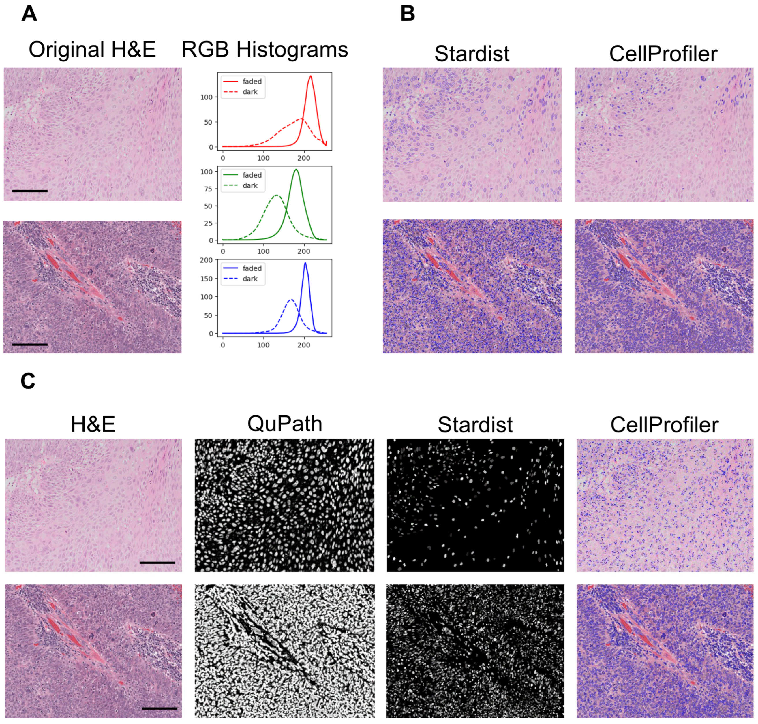
3.7. Computational Pathology and Artificial Intelligence

Human cancers are highly heterogeneous—morphologically, genotypically, and phenotypically—and this represents the main obstacle in understanding their biology and delivering appropriate treatment.
Bioptic and excisional samples are an invaluable resource to better understand tumour heterogeneity, because they encapsulate much of the key information regarding each patient’s specific cancer. In parallel with the ex vivo culture systems described above, the histopathologic analysis of diagnostic or cultured samples allows the detection and measurement of architectural and cytomorphological features of the TME at the single-cell level. An analysis of the resulting data via univariate or multivariate statistics offers valuable diagnostic/prognostic information [67,115,116].
The systematic quantification of features of interest in bioptic samples is a daunting task if performed manually, relying on dedicated professionals outside their diagnostic routines and strongly limited by human capabilities and biases. Recent computational pathology and image analysis techniques, including AI, are very promising and could automate and standardise tissue histomorphometry. Automation can remove observer biases, thus aiding the compilation of high-quality, large, and potentially multicentric datasets [117].
For example, we have previously used image analysis to automatically measure the MVD in cancerous, healthy, or irradiated tissue [115,118]. Translationally, this analysis allowed us to correlate the MVD and irradiation dose in jaw bones, with direct clinical implications for the oral rehabilitation of cancer patients receiving radiation therapy in the head and neck region [115].
However, standard image analysis requires homogeneous samples in terms of tissue preparation, the staining quality, and the intensity, which is a limitation in reliably comparing different experiments or samples from different sources.
Previous work has reported a performance decrease in AI models in segmenting heterogeneous haematoxylin and eosin (H&E) images without stain normalisation prior to analysis [119]. We have encountered similar challenges in our own work [120] using archival H&E slides.
For example, the images shown in Figure 3A have different staining intensities, as evident from the corresponding intensity distribution RGB histograms. Standard segmentation techniques and even a convolutional neural network (CNN)-based segmentator (Stardist [121]) might fail to appropriately segment heterogeneous data (Figure 3B). To overcome this issue, a variety of image restoration techniques can be applied. Figure 3C shows an example workflow where the images are enhanced with a random forest-based probability mapper prior to segmentation, and these predictions are further refined with Stardist to accurately identify all nuclei, including correctly segmenting overlapping objects.
Using this strategy, we have demonstrated how single-cell analysis in HPV-positive oropharyngeal squamous cell carcinoma patients can help to predict therapy outcomes solely via the analysis of diagnostic histological slides [120]. Thus, proper image pre-processing and the accurate segmentation of biologically relevant structures in H&E images are the first step in extracting the rich data encoded within the tumour islands, the TME, and the TAMV.
AI, especially CNNs [122], is rapidly reshaping cancer research and personalised clinical care through a wide range of applications, encompassing the detection, staging, and classification of cancer and the molecular characterisation of tumours and their microenvironments [123]. Specifically, machine learning and AI have been successfully used to detect and measure microvessels in cancer patients’ histopathological samples [124]. Advances in this field, together with the integration of additional data types, including genomics, epigenomics, and proteomics, are likely to yield even more accurate models to automatically detect and measure many features of interest, enabling us to diagnose and monitor patients’ responses to therapy or clinical outcomes.
The rapid growth of AI technology has also led to criticisms and concerns. For example, most CNNs developed for image segmentation and classification do not allow experimenters to backtrack the features or weights that led to specific decisions. This poses ethical and legal concerns to the use of such models in clinical practice, where decisions must be always justified. The current research is focused on optimising AI algorithms and on developing robust validation strategies for the current NNs and explainable AI technologies to overcome these limitations [125].
One method to introduce a degree of interpretability in “black box” deep learning models is to use class activation mapping, which highlights areas of the images that contribute strongly to the model’s predictions. While this provides some insight regarding the most important image features, it does not provide clear explanations regarding how the overall decisions are made [126]. Furthermore, in some instances, the category mapping areas do not correspond to biologically relevant features that the pathologist may expect. Another method to improve the explainability of AI models is by utilising biologically significant features established in the literature as model inputs. Indeed, in our previous work [120], we quantified several pathologically known prognostic biomarkers, such as the number of tumour-infiltrating lymphocytes and the tumour morphology, as inputs to train an AI model to predict patient outcomes. This can help clinicians to build confidence in AI models trained on evidence-based and explainable features.

3.8. Mechanistic Modelling

As discussed above, we need to understand the dynamic evolution of cancer within its TME to treat it with maximal efficacy and minimal side effects. Biological experimentation, including the in vitro systems discussed in previous paragraphs, is often designed and interpreted via statistical methods, providing a qualitative understanding of the underlying phenomena. Achieving a comprehensive mechanistic understanding is more challenging.
Mathematical biology, i.e., the study of biological processes defined as mathematical rules, offers an exciting perspective from which to address this problem. By defining, calibrating, and using suitable sets of rules (mathematic equations), we can model complex biological systems and predict their evolution via computer simulations. For example, physiologically based pharmacokinetic modelling, which allows the prediction of the adsorption, distribution, metabolism, and excretion of drugs, is now widely used to investigate new cancer-targeting drugs [127].
Physiologically based pharmacokinetic models are ideal to study molecules’ biodistribution and activity at the organ and system scales; however, they are not suitable to model cell–cell interactions at the tissue, cellular, and subcellular scales. Powerful tools developed in the past 20 years now allow the modelling of complex cell assemblies and their signalling.
For example, biochemical network modelling (e.g., Boolean networks, ODE systems), which captures the dynamics of molecular reactions (e.g., enzyme kinetics), can help in investigating intracellular signalling qualitatively and quantitatively [128,129]. On the other hand, spatial modelling (e.g., agent-based, cellular automata) can help in studying cell–cell and cell–environment interactions dependent on relative cell adhesiveness or intrinsic motility [130]. Finally, multiscale simulations combine the advantages of spatial and non-spatial models to recreate virtual tissue composed of different cell types, where the behaviour of each cell can be regulated by cell-specific signalling, juxtacrine, and paracrine signalling among cells in the simulation or interactions with the environment. Typically, multiscale tools offer facilities to simulate molecular transport within tissue, including cell secretion and uptake, allowing us to model paracrine and long-range signalling or the delivery of therapeutics.
Currently, there are several software tools able to build and execute multiscale simulations with different foci. Notable examples like Morpheus [131] and Compucell 3D [132] offer a multiscale approach to virtual tissue development, including the ability to create complex models of angiogenesis and cancer microenvironments [133,134,135].
For example, Figure 4A,B show results obtained by reimplementing the simulation developed by R. Merks and colleagues [135] in Compucell 3D. The simulation allows the exploration of the role of contact-inhibited EC chemotaxis and cell adhesion in self-organising networks like that observed in the in vitro Matrigel assay. The simulation reproduces experimental scenarios, paralleling the results of perturbation experiments. However, due to its simplicity, and similarly to its in vitro counterpart, this simulation cannot reproduce more complex scenarios like sprouting angiogenesis.
More complex TME simulations can be created; for example, Figure 4C shows the qualitative results of a CC3D model reproducing cancer-induced sprouting angiogenesis promoted by tumour-secreted VEGF, which induces migratory tip cells and proliferating stalk cells selected by a lateral inhibition mechanism (adaptation from Shirinifard et al. [133]).
This kind of simulation can be invaluable to better interpret experimental results or clinical data, to optimally design new experiments, and, in the future, even to predict disease evolution, anticipate the response to therapy, and design personalised therapeutic programs for each patient [136].
The potential is considerable; however, selecting and calibrating appropriate rules is a formidable challenge, as shown by substantial literature in the field. For example, it is established that simple models of cancer growth limited by immune responses can generate complex and difficult-to-predict dynamic behaviours [137,138]. Much has been done to address these and other problems, like appropriately leveraging omics “big data” [139]. However, there is still an urgent need to standardise in silico experiments across different platforms and to establish suitable metrics to enable meaningful comparisons with experimental data and cross-validation.
The purposefully designed dynamic cell culture systems described throughout this review can help in experimentally measuring the dynamics of multicellular environments like the TME. Co-developing in vitro and in silico models, including suitable metrics that are common to both, will enable the cross-validation of simulations against experimental evidence, increasing their predictive potential. Validated simulations will then allow the inexpensively formulation and testing of new hypotheses and the optimal planning of informative experiments.

4. Conclusions

The recent shift in the US FDA’s stance towards non-animal pre-clinical testing underscores a critical need for innovative biomedical research tools [39]. While fully replicating human tissue’s complexity in vitro remains ambitious, emerging NATs such as patient-derived organoids, tumouroids, ex vivo cultures, and microphysiological systems (MPSs) offer promising advancements. Human-based models focused on the tumour vasculature and angiogenesis are particularly valuable, as they enable the detailed exploration of the molecular mechanisms within the tumour microenvironment. Ultimately, these tools (summarised in Table 1) could significantly enhance our understanding of cancer progression and foster the development of personalised therapeutic strategies.
Intratumour hypoxia and biochemical imbalances, along with the resulting phenotypic heterogeneity and co-option of physiological functions towards metastasis, are still key unresolved issues in cancer therapy. The TAMV is central to all of these processes, and, although the results have been inconsistent regarding the first generation of VTTs observed in clinical trials, it is still one of the most promising targets for novel cancer therapies.
Despite the significant recent progress, numerous challenges still need to be addressed to bring vascularised tumour models closer to clinical use, including the implementation and standardisation of suitable in vitro techniques, as well as the development of analysis tools and metrics for quantification [140].
We envisage that novel microfluidics technologies, bioreactor systems, and biomaterials will help in the development of increasingly biomimetic MPSs, where engineered or patient-derived tissue, such as tissue slices [59], could be robustly cultured for extended time periods.
Mechanistically, new, complex assays and MPSs are very promising in terms of replacing animal experimentation in both basic and pre-clinical research. Such systems leverage the simplicity of in vitro experimental manipulation, including transgenic approaches, the possibility to appreciate variability in human cell phenotypes, and the ability to model complex functions. Furthermore, just as 2D culture systems are the gold standard in primary drug/toxicology screening [43], we envisage that more complex but robust in vitro screening will take the lead in modelling functions like angiogenesis and microvascular homeostasis, reducing the need for animal testing in such contexts.
Tissue-, system-, and organism-level interactions have been very challenging to model in vitro until recently, leaving animal experimentation as the only viable option. New technologies like ex vivo cultures [68] offer an exciting translational perspective from which to model and study tissue-level functions, and new multiorgan MPSs are being developed to achieve a system-level perspective [83]. We envisage that, by fostering close collaboration between clinicians, biologists, and engineers, such technologies will mature until eventually surpassing in vivo experiments in delivering research or prognostic value.
Capturing, studying, and understanding the variability in cancer phenotypes and manifestations among different patients, with different genetic backgrounds, lifestyles, and exposure to risk factors, is a key unmet need that eludes the capabilities of typical animal experiments. Organoid technology can aid in unravelling these aspects in vitro [141], and AI-powered digital pathology and omics are powerful tools to tackle this challenge, providing tissue-level insights. These technologies are revealing the complexity of the human cancer tissue composition [142], and, via appropriate statistic modelling, they are highlighting useful biomarkers for patient stratification and therapy optimisation [120,143]. In addition, taking inspiration from the “one health” concept (https://www.who.int/health-topics/one-health, accessed on 30 May 2025), there is a clear opportunity to investigate cancers and their development in domesticated animals [104]. In this way, we can bypass the ethical concerns associated with traditional animal models, mine an existing and potentially huge source of new data, and promote integrated healthcare for humans and companion animals.
The need to mine large volumes of data generated via high-throughput screenings, omics, and computational pathology has sparked intense research in bioinformatics, yielding numerous tools to infer mechanistic knowledge from large volumes of data. Beyond this, mechanistic mathematical modelling is offering exciting perspectives from which to model and simulate multiscale functions, from genes, through cells and tissue, to organs and systems in silico. Despite the potential, creating, calibrating, validating, and standardising these models is still a huge and only partially addressed challenge.
As discussed above, in vitro and in silico technologies can complement and empower each other to create NATs for research, which can be further developed into pre-clinical or point-of-care precision medicine applications [144]. We believe that all of this will ultimately yield a better understanding of cancer, the TME, and their dynamic responses to therapies for the benefit of patients.
In conclusion, NATs can help us to tackle unmet research and clinical needs, emphasising sustainability and translation, enabling us to understand the dynamic evolution of the TAMV and TME, devise new therapeutic strategies, aid drug discovery, expedite the diagnostic process, and personalise therapy.

Author Contributions

Conceptualization, E.F., J.H., M.F., and L.V.; writing—original draft preparation, E.F., J.H., M.F., and L.V.; writing—review and editing, E.F., J.H., M.F., and L.V.; visualization, J.H. and L.V. All authors have read and agreed to the published version of the manuscript.

Funding

This research received no external funding.

Data Availability Statement

No new data were generated for this review.

Acknowledgments

The authors wish to acknowledge the work of F. Pedica, and F. Chesnais, who contributed with fruitful discussions and experimental work. During the preparation of this manuscript, the authors used ChatGPT 4o for syntax and grammar checks. The authors have reviewed and edited each AI output and take full responsibility for the content of this publication.

Conflicts of Interest

The authors declare no conflicts of interest.

Abbreviations

The following abbreviations are used in this manuscript:
Cell Types Others
MacrophagesMVDMicrovessel density
ASCAdult stem cellsOMICSOmics technologies (genomics, proteomics, etc.)
CAFCancer-associated fibroblastsOVAAOrganotypic vasculogenesis/angiogenesis assay
PCSPericytesRCCSRotary Cell Culture System
ECEndothelial cellRGBRed, Green, Blue
CSCCancer stem cellsSCACSpontaneous companion animal cancers
PSC-ECPluripotent stem cell-derived endothelial cellTAATumour-associated angiogenesis
PSCPluripotent stem cellsTAMVTumour-associated microvasculature
TAMTumour-associated Mφ
Drugs TMETumour microenvironment
SFSorafenibMPSMicrophysiological system
MCSMonte Carlo Steps
Proteins HPVHuman papillomavirus
PGFPlacental growth factorAATAnti-angiogenic therapy
VEGF-AVascular endothelial growth factor AAIArtificial intelligence
EGFREpidermal growth factor receptorCC3DCompuCell3D (simulation software)
HIFHypoxia-inducible factorCNNConvolutional Neural Network
VEGFVascular endothelial growth factorVNTVascular normalization therapy
COX-2Cyclooxygenase-2ECMExtracellular matrix
RFPRed fluorescent proteinFDAFood and Drug Administration
H&EHematoxylin and eosin
Cell markers
CD34+Cluster of Differentiation 34 positive
CD31Platelet endothelial cell adhesion molecule
CD14+Cluster of Differentiation 14 positive

References

  1. Hanahan, D.; Weinberg, R.A. The hallmarks of cancer. Cell 2000, 100, 57–70. [Google Scholar] [CrossRef] [PubMed]
  2. de Visser, K.E.; Joyce, J.A. The evolving tumor microenvironment: From cancer initiation to metastatic outgrowth. Cancer Cell 2023, 41, 374–403. [Google Scholar] [CrossRef] [PubMed]
  3. Mantovani, A.; Allavena, P.; Marchesi, F.; Garlanda, C. Macrophages as tools and targets in cancer therapy. Nat. Rev. Drug Discov. 2022, 21, 799–820. [Google Scholar] [CrossRef] [PubMed]
  4. Chen, Y.; McAndrews, K.M.; Kalluri, R. Clinical and therapeutic relevance of cancer-associated fibroblasts. Nat. Rev. Clin. Oncol. 2021, 18, 792–804. [Google Scholar] [CrossRef]
  5. Arner, E.N.; Rathmell, J.C. Metabolic programming and immune suppression in the tumor microenvironment. Cancer Cell 2023, 41, 421–433. [Google Scholar] [CrossRef]
  6. Mantovani, A.; Allavena, P.; Sica, A.; Balkwill, F. Cancer-related inflammation. Nature 2008, 454, 436–444. [Google Scholar] [CrossRef]
  7. Colotta, F.; Allavena, P.; Sica, A.; Garlanda, C.; Mantovani, A. Cancer-related inflammation, the seventh hallmark of cancer: Links to genetic instability. Carcinogenesis 2009, 30, 1073–1081. [Google Scholar] [CrossRef]
  8. Hanahan, D.; Weinberg, R.A. Hallmarks of cancer: The next generation. Cell 2011, 144, 646–674. [Google Scholar] [CrossRef]
  9. van Seijen, M.; Lips, E.H.; Thompson, A.M.; Nik-Zainal, S.; Futreal, A.; Hwang, E.S.; Verschuur, E.; Lane, J.; Jonkers, J.; Rea, D.W.; et al. Ductal carcinoma in situ: To treat or not to treat, that is the question. Br. J. Cancer 2019, 121, 285–292. [Google Scholar] [CrossRef]
  10. Knoblauch, M.; Kühn, F.; von Ehrlich-Treuenstätt, V.; Werner, J.; Renz, B.W. Diagnostic and Therapeutic Management of Early Colorectal Cancer. Visc. Med. 2023, 39, 10–16. [Google Scholar] [CrossRef]
  11. Folkman, J.; Merler, E.; Abernathy, C.; Williams, G. Isolation of a tumor factor responsible for angiogenesis. J. Exp. Med. 1971, 133, 275–288. [Google Scholar] [CrossRef] [PubMed]
  12. Folkman, J. Tumor angiogenesis: Therapeutic implications. N. Engl. J. Med. 1971, 285, 1182–1186. [Google Scholar] [CrossRef] [PubMed]
  13. Leroi, N.; Lallemand, F.; Coucke, P.; Noel, A.; Martinive, P. Impacts of Ionizing Radiation on the Different Compartments of the Tumor Microenvironment. Front. Pharmacol. 2016, 7, 78. [Google Scholar] [CrossRef] [PubMed]
  14. Minchinton, A.I.; Tannock, I.F. Drug penetration in solid tumours. Nat. Rev. Cancer 2006, 6, 583–592. [Google Scholar] [CrossRef]
  15. Deyell, M.; Garris, C.S.; Laughney, A.M. Cancer metastasis as a non-healing wound. Br. J. Cancer 2021, 124, 1491–1502. [Google Scholar] [CrossRef]
  16. Whiteside, T.L. Tumor-Derived Exosomes and Their Role in Cancer Progression. Adv. Clin. Chem. 2016, 74, 103–141. [Google Scholar] [CrossRef]
  17. Patras, L.; Shaashua, L.; Matei, I.; Lyden, D. Immune determinants of the pre-metastatic niche. Cancer Cell 2023, 41, 546–572. [Google Scholar] [CrossRef]
  18. Fidler, I.J.; Nicolson, G.L. Organ selectivity for implantation survival and growth of B16 melanoma variant tumor lines. J. Natl. Cancer Inst. 1976, 57, 1199–1202. [Google Scholar] [CrossRef]
  19. Wicks, E.E.; Semenza, G.L. Hypoxia-inducible factors: Cancer progression and clinical translation. J. Clin. Investig. 2022, 132, e159839. [Google Scholar] [CrossRef]
  20. Mole, D.R.; Ratcliffe, P.J. Cellular oxygen sensing in health and disease. Pediatr. Nephrol. 2008, 23, 681–694. [Google Scholar] [CrossRef]
  21. Kalucka, J.; de Rooij, L.P.M.H.; Goveia, J.; Rohlenova, K.; Dumas, S.J.; Meta, E.; Conchinha, N.V.; Taverna, F.; Teuwen, L.-A.; Veys, K.; et al. Single-Cell Transcriptome Atlas of Murine Endothelial Cells. Cell 2020, 180, 764–779.e20. [Google Scholar] [CrossRef] [PubMed]
  22. Eelen, G.; Treps, L.; Li, X.; Carmeliet, P. Basic and Therapeutic Aspects of Angiogenesis Updated. Circ. Res. 2020, 127, 310–329. [Google Scholar] [CrossRef] [PubMed]
  23. Li, X.; Sun, X.; Carmeliet, P. Hallmarks of Endothelial Cell Metabolism in Health and Disease. Cell Metab. 2019, 30, 414–433. [Google Scholar] [CrossRef] [PubMed]
  24. Butler, J.M.; Kobayashi, H.; Rafii, S. Instructive role of the vascular niche in promoting tumour growth and tissue repair by angiocrine factors. Nat. Rev. Cancer 2010, 10, 138–146. [Google Scholar] [CrossRef]
  25. Gajewski, T.F.; Schreiber, H.; Fu, Y.-X. Innate and adaptive immune cells in the tumor microenvironment. Nat. Immunol. 2013, 14, 1014–1022. [Google Scholar] [CrossRef]
  26. Whiteside, T.L.; Demaria, S.; Rodriguez-Ruiz, M.E.; Zarour, H.M.; Melero, I. Emerging Opportunities and Challenges in Cancer Immunotherapy. Clin. Cancer Res. 2016, 22, 1845–1855. [Google Scholar] [CrossRef]
  27. Mantovani, A.; Marchesi, F.; Jaillon, S.; Garlanda, C.; Allavena, P. Tumor-associated myeloid cells: Diversity and therapeutic targeting. Cell. Mol. Immunol. 2021, 18, 566–578. [Google Scholar] [CrossRef]
  28. Kang, Y.; Pantel, K. Tumor cell dissemination: Emerging biological insights from animal models and cancer patients. Cancer Cell 2013, 23, 573–581. [Google Scholar] [CrossRef]
  29. Coste, A.; Karagiannis, G.S.; Wang, Y.; Xue, E.A.; Lin, Y.; Skobe, M.; Jones, J.G.; Oktay, M.H.; Condeelis, J.S.; Entenberg, D. Hematogenous Dissemination of Breast Cancer Cells From Lymph Nodes Is Mediated by Tumor MicroEnvironment of Metastasis Doorways. Front. Oncol. 2020, 10, 571100. [Google Scholar] [CrossRef]
  30. Lambert, A.W.; Pattabiraman, D.R.; Weinberg, R.A. Emerging Biological Principles of Metastasis. Cell 2017, 168, 670–691. [Google Scholar] [CrossRef]
  31. Massagué, J.; Ganesh, K. Metastasis-Initiating Cells and Ecosystems. Cancer Discov. 2021, 11, 971–994. [Google Scholar] [CrossRef] [PubMed]
  32. Potente, M.; Gerhardt, H.; Carmeliet, P. Basic and therapeutic aspects of angiogenesis. Cell 2011, 146, 873–887. [Google Scholar] [CrossRef] [PubMed]
  33. Ganesh, K.; Massagué, J. Targeting metastatic cancer. Nat. Med. 2021, 27, 34–44. [Google Scholar] [CrossRef]
  34. Fan, P.; Zhang, N.; Candi, E.; Agostini, M.; Piacentini, M.; TOR Centre; Shi, Y.; Huang, Y.; Melino, G. Alleviating hypoxia to improve cancer immunotherapy. Oncogene 2023, 42, 3591–3604. [Google Scholar] [CrossRef] [PubMed]
  35. Jain, R.K. Antiangiogenesis strategies revisited: From starving tumors to alleviating hypoxia. Cancer Cell 2014, 26, 605–622. [Google Scholar] [CrossRef]
  36. Huang, Y.; Kim, B.Y.S.; Chan, C.K.; Hahn, S.M.; Weissman, I.L.; Jiang, W. Improving immune-vascular crosstalk for cancer immunotherapy. Nat. Rev. Immunol. 2018, 18, 195–203. [Google Scholar] [CrossRef]
  37. Zitvogel, L.; Pitt, J.M.; Daillère, R.; Smyth, M.J.; Kroemer, G. Mouse models in oncoimmunology. Nat. Rev. Cancer 2016, 16, 759–773. [Google Scholar] [CrossRef]
  38. Bédard, P.; Gauvin, S.; Ferland, K.; Caneparo, C.; Pellerin, È.; Chabaud, S.; Bolduc, S. Innovative Human Three-Dimensional Tissue-Engineered Models as an Alternative to Animal Testing. Bioengineering 2020, 7, 115. [Google Scholar] [CrossRef]
  39. Moutinho, S. Researchers and regulators plan for a future without lab animals. Nat. Med. 2023, 29, 2151–2154. [Google Scholar] [CrossRef]
  40. Pampaloni, F.; Reynaud, E.G.; Stelzer, E.H.K. The third dimension bridges the gap between cell culture and live tissue. Nat. Rev. Mol. Cell Biol. 2007, 8, 839–845. [Google Scholar] [CrossRef]
  41. Kapałczyńska, M.; Kolenda, T.; Przybyła, W.; Zajączkowska, M.; Teresiak, A.; Filas, V.; Ibbs, M.; Bliźniak, R.; Łuczewski, Ł.; Lamperska, K. 2D and 3D cell cultures—A comparison of different types of cancer cell cultures. Arch. Med. Sci. 2018, 14, 910–919. [Google Scholar] [CrossRef] [PubMed]
  42. Zimmer, J.; Castriconi, R.; Scaglione, S. Editorial: Recent 3D Tumor Models for Testing Immune-Mediated Therapies. Front. Immunol. 2021, 12, 798493. [Google Scholar] [CrossRef] [PubMed]
  43. Allemang, A.; Lester, C.; Roth, T.; Pfuhler, S.; Peuschel, H.; Kosemund, K.; Mahony, C.; Bergeland, T.; O’Keeffe, L. Assessing the genotoxicity and carcinogenicity of 2-chloroethanol through structure activity relationships and in vitro testing approaches. Food Chem. Toxicol. 2022, 168, 113290. [Google Scholar] [CrossRef] [PubMed]
  44. Lee, G.Y.; Kenny, P.A.; Lee, E.H.; Bissell, M.J. Three-dimensional culture models of normal and malignant breast epithelial cells. Nat. Methods 2007, 4, 359–365. [Google Scholar] [CrossRef]
  45. Roskelley, C.D.; Desprez, P.Y.; Bissell, M.J. Extracellular matrix-dependent tissue-specific gene expression in mammary epithelial cells requires both physical and biochemical signal transduction. Proc. Natl. Acad. Sci. USA 1994, 91, 12378–12382. [Google Scholar] [CrossRef]
  46. Chang, T.T.; Hughes-Fulford, M. Monolayer and spheroid culture of human liver hepatocellular carcinoma cell line cells demonstrate distinct global gene expression patterns and functional phenotypes. Tissue Eng. Part A 2009, 15, 559–567. [Google Scholar] [CrossRef]
  47. Riedl, A.; Schlederer, M.; Pudelko, K.; Stadler, M.; Walter, S.; Unterleuthner, D.; Unger, C.; Kramer, N.; Hengstschläger, M.; Kenner, L.; et al. Comparison of cancer cells in 2D vs 3D culture reveals differences in AKT-mTOR-S6K signaling and drug responses. J. Cell Sci. 2017, 130, 203–218. [Google Scholar] [CrossRef]
  48. van Renterghem, A.W.J.; van de Haar, J.; Voest, E.E. Functional precision oncology using patient-derived assays: Bridging genotype and phenotype. Nat. Rev. Clin. Oncol. 2023, 20, 305–317. [Google Scholar] [CrossRef]
  49. Weiswald, L.-B.; Bellet, D.; Dangles-Marie, V. Spherical cancer models in tumor biology. Neoplasia 2015, 17, 1–15. [Google Scholar] [CrossRef]
  50. Bray, L.J.; Hutmacher, D.W.; Bock, N. Addressing Patient Specificity in the Engineering of Tumor Models. Front. Bioeng. Biotechnol. 2019, 7, 217. [Google Scholar] [CrossRef]
  51. Gunti, S.; Hoke, A.T.K.; Vu, K.P.; London, N.R. Organoid and Spheroid Tumor Models: Techniques and Applications. Cancers 2021, 13, 874. [Google Scholar] [CrossRef] [PubMed]
  52. Lee, K.-H.; Kim, T.-H. Recent Advances in Multicellular Tumor Spheroid Generation for Drug Screening. Biosensors 2021, 11, 445. [Google Scholar] [CrossRef] [PubMed]
  53. Nunes, A.S.; Barros, A.S.; Costa, E.C.; Moreira, A.F.; Correia, I.J. 3D tumor spheroids as in vitro models to mimic in vivo human solid tumors resistance to therapeutic drugs. Biotechnol. Bioeng. 2019, 116, 206–226. [Google Scholar] [CrossRef] [PubMed]
  54. Carletti, E.; Motta, A.; Migliaresi, C. Scaffolds for tissue engineering and 3D cell culture. Methods Mol. Biol. 2011, 695, 17–39. [Google Scholar] [CrossRef]
  55. Drost, J.; Clevers, H. Organoids in cancer research. Nat. Rev. Cancer 2018, 18, 407–418. [Google Scholar] [CrossRef]
  56. Datta, P.; Dey, M.; Ataie, Z.; Unutmaz, D.; Ozbolat, I.T. 3D bioprinting for reconstituting the cancer microenvironment. npj Precis. Oncol. 2020, 4, 18. [Google Scholar] [CrossRef]
  57. Augustine, R.; Kalva, S.N.; Ahmad, R.; Zahid, A.A.; Hasan, S.; Nayeem, A.; McClements, L.; Hasan, A. 3D Bioprinted cancer models: Revolutionizing personalized cancer therapy. Transl. Oncol. 2021, 14, 101015. [Google Scholar] [CrossRef]
  58. Powley, I.R.; Patel, M.; Miles, G.; Pringle, H.; Howells, L.; Thomas, A.; Kettleborough, C.; Bryans, J.; Hammonds, T.; MacFarlane, M.; et al. Patient-derived explants (PDEs) as a powerful preclinical platform for anti-cancer drug and biomarker discovery. Br. J. Cancer 2020, 122, 735–744. [Google Scholar] [CrossRef]
  59. Kirby, A.J.; Lavrador, J.P.; Bodi, I.; Vergani, F.; Bhangoo, R.; Ashkan, K.; Finnerty, G.T. Multicellular “hotspots” harbor high-grade potential in lower-grade gliomas. Neuro-Oncol. Adv. 2021, 3, vdab026. [Google Scholar] [CrossRef]
  60. Navran, S. The application of low shear modeled microgravity to 3-D cell biology and tissue engineering. Biotechnol. Annu. Rev. 2008, 14, 275–296. [Google Scholar] [CrossRef]
  61. Selden, C.; Fuller, B. Role of Bioreactor Technology in Tissue Engineering for Clinical Use and Therapeutic Target Design. Bioengineering 2018, 5, 32. [Google Scholar] [CrossRef] [PubMed]
  62. Muraro, M.G.; Muenst, S.; Mele, V.; Quagliata, L.; Iezzi, G.; Tzankov, A.; Weber, W.P.; Spagnoli, G.C.; Soysal, S.D. Ex-vivo assessment of drug response on breast cancer primary tissue with preserved microenvironments. Oncoimmunology 2017, 6, e1331798. [Google Scholar] [CrossRef] [PubMed]
  63. Manfredonia, C.; Muraro, M.G.; Hirt, C.; Mele, V.; Governa, V.; Papadimitropoulos, A.; Däster, S.; Soysal, S.D.; Droeser, R.A.; Mechera, R.; et al. Maintenance of Primary Human Colorectal Cancer Microenvironment Using a Perfusion Bioreactor-Based 3D Culture System. Adv. Biosyst. 2019, 3, e1800300. [Google Scholar] [CrossRef] [PubMed]
  64. Ferreira, L.P.; Gaspar, V.M.; Mano, J.F. Design of spherically structured 3D in vitro tumor models -Advances and prospects. Acta Biomater. 2018, 75, 11–34. [Google Scholar] [CrossRef]
  65. Belloni, D.; Heltai, S.; Ponzoni, M.; Villa, A.; Vergani, B.; Pecciarini, L.; Marcatti, M.; Girlanda, S.; Tonon, G.; Ciceri, F.; et al. Modeling multiple myeloma-bone marrow interactions and response to drugs in a 3D surrogate microenvironment. Haematologica 2018, 103, 707–716. [Google Scholar] [CrossRef]
  66. Grimm, D.; Wehland, M.; Pietsch, J.; Aleshcheva, G.; Wise, P.; van Loon, J.; Ulbrich, C.; Magnusson, N.E.; Infanger, M.; Bauer, J. Growing tissues in real and simulated microgravity: New methods for tissue engineering. Tissue Eng. Part B Rev. 2014, 20, 555–566. [Google Scholar] [CrossRef]
  67. Guzzeloni, V.; Veschini, L.; Pedica, F.; Ferrero, E.; Ferrarini, M. 3D Models as a Tool to Assess the Anti-Tumor Efficacy of Therapeutic Antibodies: Advantages and Limitations. Antibodies 2022, 11, 46. [Google Scholar] [CrossRef]
  68. Ferrarini, M.; Steimberg, N.; Boniotti, J.; Berenzi, A.; Belloni, D.; Mazzoleni, G.; Ferrero, E. 3D-Dynamic Culture Models of Multiple Myeloma. Methods Mol. Biol. 2017, 1612, 177–190. [Google Scholar] [CrossRef]
  69. Ferrero, E.; Villa, A.; Stefanoni, D.; Nemkov, T.; D’Alessandro, A.; Tengesdal, I.; Belloni, D.; Molteni, R.; Vergani, B.; De Luca, G.; et al. Immunometabolic activation of macrophages leads to cytokine production in the pathogenesis of KRAS-mutated histiocytosis. Rheumatology 2022, 61, e93–e96. [Google Scholar] [CrossRef]
  70. Holton, A.B.; Sinatra, F.L.; Kreahling, J.; Conway, A.J.; Landis, D.A.; Altiok, S. Microfluidic Biopsy Trapping Device for the Real-Time Monitoring of Tumor Microenvironment. PLoS ONE 2017, 12, e0169797. [Google Scholar] [CrossRef]
  71. Marei, I.; Abu Samaan, T.; Al-Quradaghi, M.A.; Farah, A.A.; Mahmud, S.H.; Ding, H.; Triggle, C.R. 3D Tissue-Engineered Vascular Drug Screening Platforms: Promise and Considerations. Front. Cardiovasc. Med. 2022, 9, 847554. [Google Scholar] [CrossRef] [PubMed]
  72. Simons, M.; Alitalo, K.; Annex, B.H.; Augustin, H.G.; Beam, C.; Berk, B.C.; Byzova, T.; Carmeliet, P.; Chilian, W.; Cooke, J.P.; et al. State-of-the-Art Methods for Evaluation of Angiogenesis and Tissue Vascularization: A Scientific Statement from the American Heart Association. Circ. Res. 2015, 116, 99–132. [Google Scholar] [CrossRef] [PubMed]
  73. Oh, J.E.; Jung, C.; Yoon, Y.-S. Human Induced Pluripotent Stem Cell-Derived Vascular Cells: Recent Progress and Future Directions. J. Cardiovasc. Dev. Dis. 2021, 8, 148. [Google Scholar] [CrossRef] [PubMed]
  74. Palpant, N.J.; Pabon, L.; Friedman, C.E.; Roberts, M.; Hadland, B.; Zaunbrecher, R.J.; Bernstein, I.; Zheng, Y.; Murry, C.E. Generating high-purity cardiac and endothelial derivatives from patterned mesoderm using human pluripotent stem cells. Nat. Protoc. 2016, 12, 15–31. [Google Scholar] [CrossRef]
  75. Kumar, M.; Toprakhisar, B.; Van Haele, M.; Antoranz, A.; Boon, R.; Chesnais, F.; De Smedt, J.; Tricot, T.; Idoype, T.I.; Canella, M.; et al. A fully defined matrix to support a pluripotent stem cell derived multi-cell-liver steatohepatitis and fibrosis model. Biomaterials 2021, 276, 121006. [Google Scholar] [CrossRef]
  76. Ferrero, E.; Bondanza, A.; Leone, B.E.; Manici, S.; Poggi, A.; Zocchi, M.R. CD14+ CD34+ peripheral blood mononuclear cells migrate across endothelium and give rise to immunostimulatory dendritic cells. J. Immunol. 1998, 160, 2675–2683. [Google Scholar] [CrossRef]
  77. Langheim, S.; Dreas, L.; Veschini, L.; Maisano, F.; Foglieni, C.; Ferrarello, S.; Sinagra, G.; Zingone, B.; Alfieri, O.; Ferrero, E.; et al. Increased expression and secretion of resistin in epicardial adipose tissue of patients with acute coronary syndrome. Am. J. Physiol. Heart Circ. Physiol. 2010, 298, H746–H753. [Google Scholar] [CrossRef]
  78. Chesnais, F.; Hue, J.; Roy, E.; Branco, M.; Stokes, R.; Pellon, A.; Le Caillec, J.; Elbahtety, E.; Battilocchi, M.; Danovi, D.; et al. High-content image analysis to study phenotypic heterogeneity in endothelial cell monolayers. J. Cell Sci. 2022, 135, jcs259104. [Google Scholar] [CrossRef]
  79. Veschini, L.; Sailem, H.; Malani, D.; Pietiäinen, V.; Stojiljkovic, A.; Wiseman, E.; Danovi, D. High-Content Imaging to Phenotype Human Primary and iPSC-Derived Cells. Methods Mol. Biol. 2021, 2185, 423–445. [Google Scholar] [CrossRef]
  80. Kerns, S.J.; Belgur, C.; Petropolis, D.; Kanellias, M.; Barrile, R.; Sam, J.; Weinzierl, T.; Fauti, T.; Freimoser-Grundschober, A.; Eckmann, J.; et al. Human immunocompetent Organ-on-Chip platforms allow safety profiling of tumor-targeted T-cell bispecific antibodies. eLife 2021, 10, e67106. [Google Scholar] [CrossRef]
  81. Ewart, L.; Apostolou, A.; Briggs, S.A.; Carman, C.V.; Chaff, J.T.; Heng, A.R.; Jadalannagari, S.; Janardhanan, J.; Jang, K.-J.; Joshipura, S.R.; et al. Performance assessment and economic analysis of a human Liver-Chip for predictive toxicology. Commun. Med. 2022, 2, 154. [Google Scholar] [CrossRef] [PubMed]
  82. Pediaditakis, I.; Kodella, K.R.; Manatakis, D.V.; Le, C.Y.; Barthakur, S.; Sorets, A.; Gravanis, A.; Ewart, L.; Rubin, L.L.; Manolakos, E.S.; et al. A microengineered Brain-Chip to model neuroinflammation in humans. iScience 2022, 25, 104813. [Google Scholar] [CrossRef] [PubMed]
  83. Ronaldson-Bouchard, K.; Baldassarri, I.; Tavakol, D.N.; Graney, P.L.; Samaritano, M.; Cimetta, E.; Vunjak-Novakovic, G. Engineering complexity in human tissue models of cancer. Adv. Drug Deliv. Rev. 2022, 184, 114181. [Google Scholar] [CrossRef] [PubMed]
  84. Ronaldson-Bouchard, K.; Teles, D.; Yeager, K.; Tavakol, D.N.; Zhao, Y.; Chramiec, A.; Tagore, S.; Summers, M.; Stylianos, S.; Tamargo, M.; et al. A multi-organ chip with matured tissue niches linked by vascular flow. Nat. Biomed. Eng. 2022, 6, 351–371. [Google Scholar] [CrossRef]
  85. Nakatsu, M.N.; Davis, J.; Hughes, C.C.W. Optimized fibrin gel bead assay for the study of angiogenesis. J. Vis. Exp. 2007, 186. [Google Scholar] [CrossRef]
  86. Jauhiainen, S.; Ilmonen, H.; Vuola, P.; Rasinkangas, H.; Pulkkinen, H.H.; Keränen, S.; Kiema, M.; Liikkanen, J.J.; Laham-Karam, N.; Laidinen, S.; et al. ErbB signaling is a potential therapeutic target for vascular lesions with fibrous component. eLife 2023, 12, e82543. [Google Scholar] [CrossRef]
  87. Francis, C.R.; Kincross, H.; Kushner, E.J. Rab35 governs apicobasal polarity through regulation of actin dynamics during sprouting angiogenesis. Nat. Commun. 2022, 13, 5276. [Google Scholar] [CrossRef]
  88. Hetheridge, C.; Mavria, G.; Mellor, H. Uses of the in vitro endothelial-fibroblast organotypic co-culture assay in angiogenesis research. Biochem. Soc. Trans. 2011, 39, 1597–1600. [Google Scholar] [CrossRef]
  89. Strobel, H.A.; Moss, S.M.; Hoying, J.B. Vascularized Tissue Organoids. Bioengineering 2023, 10, 124. [Google Scholar] [CrossRef]
  90. Yu, J. Vascularized Organoids: A More Complete Model. Int. J. Stem Cells 2021, 14, 127–137. [Google Scholar] [CrossRef]
  91. Orlova, V.V.; van den Hil, F.E.; Petrus-Reurer, S.; Drabsch, Y.; Ten Dijke, P.; Mummery, C.L. Generation, expansion and functional analysis of endothelial cells and pericytes derived from human pluripotent stem cells. Nat. Protoc. 2014, 9, 1514–1531. [Google Scholar] [CrossRef] [PubMed]
  92. Kumar, A.; D’Souza, S.S.; Moskvin, O.V.; Toh, H.; Wang, B.; Zhang, J.; Swanson, S.; Guo, L.-W.; Thomson, J.A.; Slukvin, I.I. Specification and Diversification of Pericytes and Smooth Muscle Cells from Mesenchymoangioblasts. Cell Rep. 2017, 19, 1902–1916. [Google Scholar] [CrossRef] [PubMed]
  93. Ferrarini, M.; Steimberg, N.; Ponzoni, M.; Belloni, D.; Berenzi, A.; Girlanda, S.; Caligaris-Cappio, F.; Mazzoleni, G.; Ferrero, E. Ex-vivo dynamic 3-D culture of human tissues in the RCCSTM bioreactor allows the study of Multiple Myeloma biology and response to therapy. PLoS ONE 2013, 8, e71613. [Google Scholar] [CrossRef]
  94. Osaki, T.; Sivathanu, V.; Kamm, R.D. Vascularized microfluidic organ-chips for drug screening, disease models and tissue engineering. Curr. Opin. Biotechnol. 2018, 52, 116–123. [Google Scholar] [CrossRef]
  95. Chen, M.B.; Whisler, J.A.; Fröse, J.; Yu, C.; Shin, Y.; Kamm, R.D. On-chip human microvasculature assay for visualization and quantification of tumor cell extravasation dynamics. Nat. Protoc. 2017, 12, 865–880. [Google Scholar] [CrossRef]
  96. Yamamoto, K.; Tanimura, K.; Watanabe, M.; Sano, H.; Uwamori, H.; Mabuchi, Y.; Matsuzaki, Y.; Chung, S.; Kamm, R.D.; Tanishita, K.; et al. Construction of Continuous Capillary Networks Stabilized by Pericyte-like Perivascular Cells. Tissue Eng. Part A 2019, 25, 499–510. [Google Scholar] [CrossRef]
  97. Boussommier-Calleja, A.; Atiyas, Y.; Haase, K.; Headley, M.; Lewis, C.; Kamm, R.D. The effects of monocytes on tumor cell extravasation in a 3D vascularized microfluidic model. Biomaterials 2019, 198, 180–193. [Google Scholar] [CrossRef]
  98. Sobrino, A.; Phan, D.T.T.; Datta, R.; Wang, X.; Hachey, S.J.; Romero-López, M.; Gratton, E.; Lee, A.P.; George, S.C.; Hughes, C.C.W. 3D microtumors in vitro supported by perfused vascular networks. Sci. Rep. 2016, 6, 31589. [Google Scholar] [CrossRef]
  99. Hachey, S.J.; Sobrino, A.; Lee, J.G.; Jafari, M.D.; Klempner, S.J.; Puttock, E.J.; Edwards, R.A.; Lowengrub, J.S.; Waterman, M.L.; Zell, J.A.; et al. A human vascularized microtumor model of patient-derived colorectal cancer recapitulates clinical disease. Transl. Res. J. Lab. Clin. Med. 2023, 255, 97–108. [Google Scholar] [CrossRef]
  100. Chesnais, F.; Joel, J.; Hue, J.; Shakib, S.; Di Silvio, L.; Grigoriadis, A.E.; Coward, T.; Veschini, L. Continuously perfusable, customisable, and matrix-free vasculature on a chip platform. Lab. Chip 2023, 23, 761–772. [Google Scholar] [CrossRef]
  101. Razavirad, A.; Rismanchi, S.; Mortazavi, P.; Muhammadnejad, A. Canine Mammary Tumors as a Potential Model for Human Breast Cancer in Comparative Oncology. Vet. Med. Int. 2024, 2024, 9319651. [Google Scholar] [CrossRef] [PubMed]
  102. Palma, S.D.; McConnell, A.; Verganti, S.; Starkey, M. Review on Canine Oral Melanoma: An Undervalued Authentic Genetic Model of Human Oral Melanoma? Vet. Pathol. 2021, 58, 881–889. [Google Scholar] [CrossRef] [PubMed]
  103. Wong, K.; van der Weyden, L.; Schott, C.R.; Foote, A.; Constantino-Casas, F.; Smith, S.; Dobson, J.M.; Murchison, E.P.; Wu, H.; Yeh, I.; et al. Cross-species genomic landscape comparison of human mucosal melanoma with canine oral and equine melanoma. Nat. Commun. 2019, 10, 353. [Google Scholar] [CrossRef] [PubMed]
  104. Oh, J.H.; Cho, J.-Y. Comparative oncology: Overcoming human cancer through companion animal studies. Exp. Mol. Med. 2023, 55, 725–734. [Google Scholar] [CrossRef]
  105. Onaciu, A.; Munteanu, R.; Munteanu, V.C.; Gulei, D.; Raduly, L.; Feder, R.-I.; Pirlog, R.; Atanasov, A.G.; Korban, S.S.; Irimie, A.; et al. Spontaneous and Induced Animal Models for Cancer Research. Diagnostics 2020, 10, 660. [Google Scholar] [CrossRef]
  106. Mariano, L.C.; Warnakulasuriya, S.; Straif, K.; Monteiro, L. Secondhand smoke exposure and oral cancer risk: A systematic review and meta-analysis. Tob. Control 2022, 31, 597–607. [Google Scholar] [CrossRef]
  107. Zaccone, R.; Renzi, A.; Chalfon, C.; Lenzi, J.; Bellei, E.; Marconato, L.; Ros, E.; Rigillo, A.; Bettini, G.; Faroni, E.; et al. Environmental risk factors for the development of oral squamous cell carcinoma in cats. J. Vet. Intern. Med. 2022, 36, 1398–1408. [Google Scholar] [CrossRef]
  108. Sarver, A.L.; Makielski, K.M.; DePauw, T.A.; Schulte, A.J.; Modiano, J.F. Increased risk of cancer in dogs and humans: A consequence of recent extension of lifespan beyond evolutionarily-determined limitations? Aging Cancer 2022, 3, 3–19. [Google Scholar] [CrossRef]
  109. Carvalho, M.I.; Guimarães, M.J.; Pires, I.; Prada, J.; Silva-Carvalho, R.; Lopes, C.; Queiroga, F.L. EGFR and microvessel density in canine malignant mammary tumours. Res. Vet. Sci. 2013, 95, 1094–1099. [Google Scholar] [CrossRef]
  110. Queiroga, F.L.; Pires, I.; Parente, M.; Gregório, H.; Lopes, C.S. COX-2 over-expression correlates with VEGF and tumour angiogenesis in canine mammary cancer. Vet. J. 2011, 189, 77–82. [Google Scholar] [CrossRef]
  111. Laufer-Amorim, R.; Fonseca-Alves, C.E.; Villacis, R.A.R.; Linde, S.A.D.; Carvalho, M.; Larsen, S.J.; Marchi, F.A.; Rogatto, S.R. Comprehensive Genomic Profiling of Androgen-Receptor-Negative Canine Prostate Cancer. Int. J. Mol. Sci. 2019, 20, 1555. [Google Scholar] [CrossRef] [PubMed]
  112. Nordby, Y.; Andersen, S.; Richardsen, E.; Ness, N.; Al-Saad, S.; Melbø-Jørgensen, C.; Patel, H.R.H.; Dønnem, T.; Busund, L.-T.; Bremnes, R.M. Stromal expression of VEGF-A and VEGFR-2 in prostate tissue is associated with biochemical and clinical recurrence after radical prostatectomy. Prostate 2015, 75, 1682–1693. [Google Scholar] [CrossRef] [PubMed]
  113. Warszawik-Hendzel, O.; Słowińska, M.; Olszewska, M.; Rudnicka, L. Melanoma of the oral cavity: Pathogenesis, dermoscopy, clinical features, staging and management. J. Dermatol. Case Rep. 2014, 8, 60–66. [Google Scholar] [CrossRef] [PubMed]
  114. Bergman, P.J. Canine oral melanoma. Clin. Tech. Small Anim. Pract. 2007, 22, 55–60. [Google Scholar] [CrossRef]
  115. Patel, V.; Di Silvio, L.; Kwok, J.; Burns, M.; Henley Smith, R.; Thavaraj, S.; Veschini, L. The impact of intensity-modulated radiation treatment on dento-alveolar microvasculature in pharyngeal cancer implant patients. J. Oral Rehabil. 2020, 47, 1411–1421. [Google Scholar] [CrossRef]
  116. Nafe, R.; Schlote, W. Histomorphometry of brain tumours. Neuropathol. Appl. Neurobiol. 2004, 30, 315–328. [Google Scholar] [CrossRef]
  117. Mahmood, H.; Shaban, M.; Indave, B.I.; Santos-Silva, A.R.; Rajpoot, N.; Khurram, S.A. Use of artificial intelligence in diagnosis of head and neck precancerous and cancerous lesions: A systematic review. Oral Oncol. 2020, 110, 104885. [Google Scholar] [CrossRef]
  118. Veschini, L.; Crippa, L.; Dondossola, E.; Doglioni, C.; Corti, A.; Ferrero, E. The vasostatin-1 fragment of chromogranin A preserves a quiescent phenotype in hypoxia-driven endothelial cells and regulates tumor neovascularization. FASEB J. 2011, 25, 3906–3914. [Google Scholar] [CrossRef]
  119. Madusanka, N.; Jayalath, P.; Fernando, D.; Yasakethu, L.; Lee, B.-I. Impact of H&E Stain Normalization on Deep Learning Models in Cancer Image Classification: Performance, Complexity, and Trade-Offs. Cancers 2023, 15, 4144. [Google Scholar] [CrossRef]
  120. Hue, J.; Valinciute, Z.; Thavaraj, S.; Veschini, L. Multifactorial estimation of clinical outcome in HPV-associated oropharyngeal squamous cell carcinoma via automated image analysis of routine diagnostic H&E slides and neural network modelling. Oral Oncol. 2023, 141, 106399. [Google Scholar] [CrossRef]
  121. Graham, S.; Vu, Q.D.; Jahanifar, M.; Weigert, M.; Schmidt, U.; Zhang, W.; Zhang, J.; Yang, S.; Xiang, J.; Wang, X.; et al. CoNIC Challenge: Pushing the frontiers of nuclear detection, segmentation, classification and counting. Med. Image Anal. 2024, 92, 103047. [Google Scholar] [CrossRef] [PubMed]
  122. Graham, S.; Vu, Q.D.; Raza, S.E.A.; Azam, A.; Tsang, Y.W.; Kwak, J.T.; Rajpoot, N. Hover-Net: Simultaneous segmentation and classification of nuclei in multi-tissue histology images. Med. Image Anal. 2019, 58, 101563. [Google Scholar] [CrossRef] [PubMed]
  123. Cabral, B.P.; Braga, L.A.M.; Syed-Abdul, S.; Mota, F.B. Future of Artificial Intelligence Applications in Cancer Care: A Global Cross-Sectional Survey of Researchers. Curr. Oncol. 2023, 30, 3432–3446. [Google Scholar] [CrossRef] [PubMed]
  124. Timakova, A.; Ananev, V.; Fayzullin, A.; Makarov, V.; Ivanova, E.; Shekhter, A.; Timashev, P. Artificial Intelligence Assists in the Detection of Blood Vessels in Whole Slide Images: Practical Benefits for Oncological Pathology. Biomolecules 2023, 13, 1327. [Google Scholar] [CrossRef]
  125. Gniadek, T.; Kang, J.; Theparee, T.; Krive, J. Framework for Classifying Explainable Artificial Intelligence (XAI) Algorithms in Clinical Medicine. Online J. Public Health Inform. 2023, 15, e50934. [Google Scholar] [CrossRef]
  126. Ali, S.; Akhlaq, F.; Imran, A.S.; Kastrati, Z.; Daudpota, S.M.; Moosa, M. The enlightening role of explainable artificial intelligence in medical & healthcare domains: A systematic literature review. Comput. Biol. Med. 2023, 166, 107555. [Google Scholar] [CrossRef]
  127. Wang, X.; Chen, F.; Guo, N.; Gu, Z.; Lin, H.; Xiang, X.; Shi, Y.; Han, B. Application of physiologically based pharmacokinetics modeling in the research of small-molecule targeted anti-cancer drugs. Cancer Chemother. Pharmacol. 2023, 92, 253–270. [Google Scholar] [CrossRef]
  128. Calzone, L.; Noël, V.; Barillot, E.; Kroemer, G.; Stoll, G. Modeling signaling pathways in biology with MaBoSS: From one single cell to a dynamic population of heterogeneous interacting cells. Comput. Struct. Biotechnol. J. 2022, 20, 5661–5671. [Google Scholar] [CrossRef]
  129. Choi, K.; Medley, J.K.; König, M.; Stocking, K.; Smith, L.; Gu, S.; Sauro, H.M. Tellurium: An extensible python-based modeling environment for systems and synthetic biology. Biosystems 2018, 171, 74–79. [Google Scholar] [CrossRef]
  130. Graner, F.; Glazier, J.A. Simulation of biological cell sorting using a two-dimensional extended Potts model. Phys. Rev. Lett. 1992, 69, 2013–2016. [Google Scholar] [CrossRef]
  131. Starruß, J.; de Back, W.; Brusch, L.; Deutsch, A. Morpheus: A user-friendly modeling environment for multiscale and multicellular systems biology. Bioinformatics 2014, 30, 1331–1332. [Google Scholar] [CrossRef]
  132. Swat, M.H.; Thomas, G.L.; Belmonte, J.M.; Shirinifard, A.; Hmeljak, D.; Glazier, J.A. Multi-scale modeling of tissues using CompuCell3D. Methods Cell Biol. 2012, 110, 325–366. [Google Scholar] [CrossRef]
  133. Shirinifard, A.; Gens, J.S.; Zaitlen, B.L.; Popławski, N.J.; Swat, M.; Glazier, J.A. 3D multi-cell simulation of tumor growth and angiogenesis. PLoS ONE 2009, 4, e7190. [Google Scholar] [CrossRef]
  134. Merks, R.M.H.; Glazier, J.A. Dynamic mechanisms of blood vessel growth. Nonlinearity 2006, 19, C1–C10. [Google Scholar] [CrossRef]
  135. Merks, R.M.H.; Perryn, E.D.; Shirinifard, A.; Glazier, J.A. Contact-Inhibited Chemotaxis in De Novo and Sprouting Blood-Vessel Growth. PLOS Comput. Biol. 2008, 4, e1000163. [Google Scholar] [CrossRef]
  136. Niarakis, A.; Laubenbacher, R.; An, G.; Ilan, Y.; Fisher, J.; Flobak, Å.; Reiche, K.; Rodríguez Martínez, M.; Geris, L.; Ladeira, L.; et al. Immune digital twins for complex human pathologies: Applications, limitations, and challenges. npj Syst. Biol. Appl. 2024, 10, 141. [Google Scholar] [CrossRef]
  137. Yang, H.M. Mathematical modeling of solid cancer growth with angiogenesis. Theor. Biol. Med. Model. 2012, 9, 2. [Google Scholar] [CrossRef]
  138. Moffett, A.S.; Deng, Y.; Levine, H. Modeling the Role of Immune Cell Conversion in the Tumor-Immune Microenvironment. Bull. Math. Biol. 2023, 85, 93. [Google Scholar] [CrossRef]
  139. Uatay, A.; Gall, L.; Irons, L.; Tewari, S.G.; Zhu, X.S.; Gibbs, M.; Kimko, H. Physiological Indirect Response Model to Omics-Powered Quantitative Systems Pharmacology Model. J. Pharm. Sci. 2023, 113, 11–21. [Google Scholar] [CrossRef]
  140. Pereira, M.; Pinto, J.; Arteaga, B.; Guerra, A.; Jorge, R.N.; Monteiro, F.J.; Salgado, C.L. A Comprehensive Look at In Vitro Angiogenesis Image Analysis Software. Int. J. Mol. Sci. 2023, 24, 17625. [Google Scholar] [CrossRef]
  141. Yan, H.H.N.; Siu, H.C.; Law, S.; Ho, S.L.; Yue, S.S.K.; Tsui, W.Y.; Chan, D.; Chan, A.S.; Ma, S.; Lam, K.O.; et al. A Comprehensive Human Gastric Cancer Organoid Biobank Captures Tumor Subtype Heterogeneity and Enables Therapeutic Screening. Cell Stem Cell 2018, 23, 882–897.e11. [Google Scholar] [CrossRef] [PubMed]
  142. Oyoshi, H.; Du, J.; Sakai, S.A.; Yamashita, R.; Okumura, M.; Motegi, A.; Hojo, H.; Nakamura, M.; Hirata, H.; Sunakawa, H.; et al. Comprehensive single-cell analysis demonstrates radiotherapy-induced infiltration of macrophages expressing immunosuppressive genes into tumor in esophageal squamous cell carcinoma. Sci. Adv. 2023, 9, eadh9069. [Google Scholar] [CrossRef] [PubMed]
  143. Shen, Y.; Ni, S.; Li, S.; Lv, B. Role of stemness-related genes TIMP1, PGF, and SNAI1 in the prognosis of colorectal cancer through single-cell RNA-seq. Cancer Med. 2023, 12, 11611–11623. [Google Scholar] [CrossRef] [PubMed]
  144. Laubenbacher, R.; Niarakis, A.; Helikar, T.; An, G.; Shapiro, B.; Malik-Sheriff, R.S.; Sego, T.J.; Knapp, A.; Macklin, P.; Glazier, J.A. Building digital twins of the human immune system: Toward a roadmap. npj Digit. Med. 2022, 5, 64. [Google Scholar] [CrossRef]
Figure 1. Schematic representation of cancer development. (A) Healthy stratified epithelial tissue encompassing sub-epithelial connective tissue and epithelium separated by a continuous basal membrane. (B) Carcinoma in situ eliciting tissue inflammation. (C) Invasive carcinoma composed of distinct cancer clones (CEpCs) with differential properties. Invasive CEpCs subvert the microenvironment, generating cancer-associated fibroblasts (CAFs) and an altered ECM. (D) Close-up view of an invasive TME and its relations with the TAMV. Inflammation can control cancer growth in some but not all CEpCs. Hypoxia and CEpC mutations favour the “angiogenic switch”, and CEpC-EC interactions can generate further phenotypic heterogeneity. Cancer cells can co-opt TME components like ECs and macrophages (MΦ) to intravasate and then disseminate to distant organs. In breast cancer, a pro-metastatic Tie2-expressing-MΦ /EC/cancer cell triad, i.e., the tumour microenvironment of metastasis doorway (TMEM doorway), has been identified.
Organoids 04 00012 g001
Figure 2. Overview of in vitro NATs to study the TME and TAMV. (A) EC cultures in monolayer, Matrigel, fibrin gel, or the OVAA system. The OVAA can be imaged at high magnification, allowing us to appreciate details like individual tip cells (red arrow) and their filopodia. (B) Immunostained slides of RCCS-cultured human hepatocarcinoma at diagnosis or upon 3 days of RCCS culture in the presence or absence of Sorafenib (SF). CD31 marks microvessels. (C) Photographs of the RCCS and VoC systems within standard tissue incubators. (D) RFP-labelled EC in the VoC-OVAA system upon 10 days of perfusion.
Organoids 04 00012 g002
Figure 3. Strategies to improve pixel-level segmentation of H&E images. (A) Two examples of images with different staining intensities (faded and dark) and relative RGB intensity histograms. Solid line for faded images and dashed lines for dark ones. (B) Nuclei segmentations using either Stardist (CNN-based) or CellProfiler (intensity-based). Stardist (default weights) under-segments faded images and over-segments dark ones. A cell profiler pipeline tailored to darker images fails to segment faded images. (C) A sequential workflow to segment both dark stained and faded haematoxylin and eosin (H&E) images employing a random forest pixel classifier (as implemented in QuPAth), followed by refinement with Stardist, where the results are plugged into CellProfiler for object analysis and classification. Scale bars = 100 μm.
Organoids 04 00012 g003
Figure 4. Agent-based cell models of vascular assembly and tumour angiogenesis. (A,B) Simulations calibrated to reproduce results of the in vitro tubulogenesis assay on Matrigel (reimplementation of ref. [135]) based on cell adhesion energies and contact-inhibited chemotaxis of ECs. (A) Time course evolution (600 MCS~12 h, 1 MCS~90 s) of “tubulogenesis” simulations with varying initial cell densities. Paralleling in vitro assays, the simulation demonstrates the importance of the cell density to achieve structured networks. (B) Replicated simulations (n = 5) with varying λ-chemotaxis parameters, i.e., the responsiveness of ECs to VEGF chemoattraction. High values represent standard experimental conditions where networks of tubular structures are formed efficiently. Decreasing values reflect experimental conditions where VEGF signalling is inhibited—for example, with anti-KDR (human VEGF receptor 2) monoclonal antibodies. The simulation’s results parallel in vitro observations, where interference with VEGF signalling prevents tubule organisation, forming blob-like structures. (C) The 2D reimplementation of a tumour angiogenesis simulation [133]. The simulation recapitulates the crosstalk between tumour cells (TCs, brown) and ECs (green) mediated by VEGF signalling and oxygen shown as dimensionless values (0–1 scale). Hypoxic TCs produce long-range diffusing VEGF, promoting angiogenic sprouting, which in turn is regulated by VEGF induction and lateral inhibition from neighbouring ECs. As new vessels reach the TCs, these proliferate and decrease VEGF production. The depicted time course (1 MCS~2 h) displays the efficient formation of a vascular network, which, via steps of intermittent hypoxia, reaches a stable state where most TCs are oxygenated and cease to produce VEGF.
Organoids 04 00012 g004
Table 1. Comparisons between NATs to study angiogenesis and the TME.
NAT CategoryAdvantagesLimitationsTranslational ChallengesRegulatory/Commercial AspectsValue in Studying EC Biology/AngiogenesisValue in Studying Tumour-Associated Angiogenesis
2D Static CulturesSimple, cost-effective, high-throughputLack of 3D architecture and TME complexityModerate clinical predictivityEstablished for first-line screensUsed in toxicity testing, barrier function assays, leukocyte transmigration, and endothelial signalling studies. Applied in screening VEGF signalling, endothelial permeability, and immune cell transmigration; foundational in early angiogenesis modelling.
3D Spheroids and OrganoidsBetter mimicry of tumour architecture; patient-derivedVariable yield and standardisation; lack vasculatureScale-up and patient-specific validationGaining traction in precision oncologyEC spheroids used to investigate angiogenesis in matrices or microtissues.Support indirect exploration of angiogenic signalling under hypoxia or drug conditions
Ex Vivo Tumour ExplantsPreserve native TME; clinically relevantShort culture lifespan; access to specimensIntegration with drug screening pipelinesValuable for personalised medicine; yet under-utilisedEnable investigation of native endothelial structures, vessel morphology, and angiogenic responses.Preserve native tumour vasculature enabling direct evaluation of angiogenic features, drug effects, and EC-TME interaction in patient tissues.
Dynamic Bioreactors (e.g., RCCS)Sustain viability in complex 3D tissuesComplex handling; limited throughputStandardising protocols for clinical translationIncreasingly explored under FDA Modernization ActAllow monitoring of endothelial and vascular behaviour over time in viable 3D cultures; Angiogenic response studies and drug testing.Used to culture tumour explants with intact vasculature; facilitate real-time observation of angiogenic modulation under therapeutic conditions.
Microphysiological Systems (MPS)Allow perfusion, mass transfer; scalableCostly, complex fabrication and operationInter-device reproducibility, regulatory acceptanceKey to non-animal preclinical validation; high priorityControlled study of EC function under flow, including vessel formation, barrier properties, and signalling.Allow reproduction of tumour vascular environments with flow; applied in mechanistic studies, drug screening, and metastasis research.
Vascularised OrganoidsIntegrate vascular features into tissue modelsPerfusion and maturation still limitedDemonstrating consistent vascularisationPotential to fulfil unmet modelling needsSupport formation of capillary-like structures within organoids; Study EC-stroma interaction and vascular self-organisation.Enable angiocrine and vessel remodelling studies within patient-derived or stem-cell-based tumour constructs
In Silico/Computational ModelsHypothesis generation and testing; multi-scale integrationRequire high-quality experimental validationValidation, regulatory uncertaintySeen as decision-support tools; still unregulatedEnable in silico experimentation on EC dynamics, angiogenic pathways, and network behaviour.Support modelling of tumour-induced angiogenesis, VEGF diffusion, and EC-tumour cell crosstalk at multiple scales.
Spontaneous Tumours in Companion AnimalsHuman-relevant, ethically viable, naturally occurring cancersLogistics, sample standardisation, limited availabilityData harmonisation across speciesSupports One Health approach; gaining interestPhysiologic endothelial diversity and vascular changes in spontaneous diseases; informative for natural history and treatment response studies.Offer clinically relevant insights into tumour angiogenesis and vascular responses in natural disease; valuable for translational and comparative research.
Disclaimer/Publisher’s Note: The statements, opinions and data contained in all publications are solely those of the individual author(s) and contributor(s) and not of MDPI and/or the editor(s). MDPI and/or the editor(s) disclaim responsibility for any injury to people or property resulting from any ideas, methods, instructions or products referred to in the content.

