Proceeding Paper

Chenopodium quinoa to Modulate Innate Myeloid Cells in the Induction of Obesity †

by
José Moisés Laparra
1,*,
Elena Aguilar-Aguilar
2 and
Claudia Monika Haros
3
1
Molecular Immunonutrition Group, Madrid Institute for Advanced Studies in Food (IMDEA-Food), Ctra. de Canto Blanco No 8, 28049 Madrid, Spain
2
Nutrition and Clinical Trials Unit, GENYAL Platform IMDEA-Food Institute, CEI UAM + CSIC, 28049 Madrid, Spain
3
Instituto de Agroquímica y Tecnología de Alimentos (IATA), Consejo Superior de Investigaciones Científicas (CSIC), Av. Agustín Escardino 7, Parque Científico, Paterna, 46980 Valencia, Spain
*
Author to whom correspondence should be addressed.
Presented at the III Conference la ValSe-Food and VI Symposium Chia-Link Network, online, 15–17 November 2021.
Biol. Life Sci. Forum 2021, 8(1), 13; https://doi.org/10.3390/blsf2021008013
Published: 14 January 2022
(This article belongs to the Proceedings of III Conference la ValSe-Food and VI Symposium Chia-Link Network)

Abstract

:
Complex interactions between innate and adaptive immune effectors are an important component in the induction of obesity. Particularly, different subsets of myeloid cells play key roles in metabolic liver diseases and, therefore, are promising targets for intervention strategies. Chenopodium quinoa seeds constitute a good source of immunonutritional compounds, which help prevent high-fat, diet-enhanced innate immune signaling via TLR4/MyD88 that boosts inflammation. Herein, two metabolic mouse models—wild type (WT) and tributyltin treated (TBT)—were used to examine the effects associated with non-alcoholic fatty liver disease (NAFLD); mice were fed with a high-fat diet (HFD) and administered with wheat or C. quinoa bread. Variations in myeloid cells were obtained from a hemogram analysis, and rt-qPCR (mRNA) served to evaluate macrophage markers (i.e., CD68/CD206 ratio) as well as liver inflammation (i.e., Lyve-1) to gain insights into their selective functional differentiation into metabolically injured livers. Only administration of C. quinoa bread prevented alterations in the liver/body weight ratio either in WT animals or those treated with TBT. These effects were associated with significantly increased variations in the peripheral myeloid cell population. Hepatic mRNA markers revealed that C. quinoa enables a selective functional differentiation and function of intrahepatic monocyte-derived macrophages preserving tissue integrity and function.

1. Introduction

Obesity is recognized as overweight caused by the dysfunctional accumulation of energy reserves as fat depots, and its prevalence appears associated with an increased incidence of metabolic disorders. Complex interactions between innate and adaptive immune effectors are an important component in the induction of obesity. Accordingly, recent research demonstrated that myeloid cells accumulate in the liver as monocytes and macrophages during the progression of obesity-related non-alcoholic fatty liver disease to steatohepatitis [1]. These cells contribute to either worsening or improving tissue homeostasis following impairment of liver function. Specific environmental signals within the gut–liver axis further determine the selective functional differentiation and function of hepatic macrophages. Different subsets of these myeloid cells have pivotal roles in metabolic liver diseases; thus, they provide promising targets for intervention strategies with a preventive and/or therapeutic application.
Under a high-fat diet, innate immune Toll-like receptor (TLR)-4/MyD88 signaling leads to an inhibited macrophage proliferation to infiltrate into adipose tissue boosting inflammation [2]. C. quinoa seeds constitute a good source of immunonutritional serine-type protease inhibitors (SETIs), which enable innate immune events mediated by TLR4 downstream signaling that can be associated with a delayed wave, implying adaptor molecules such as TRAM/TRIF [3,4].
In view of the pivotal role of the hepatic immune–metabolic crosstalk, thereby influencing the natural history of obesity, this study evaluates the impact of the inclusion of C. quinoa flour into bread formulations in the variations and polarization of the myeloid population in metabolic mouse models.

2. Materials and Methods

2.1. Metabolic Mouse Models

C57BL/6 mice (6 weeks of age) born from untreated females or receiving obesogen tributyltin (50 nM) via drinking water to develop a state of NAFLD were used [5]. Afterward, both F1_A and F1_B generations were kept under an HFD until reaching 6 weeks of age. Bread formulations were administered (14 mg/day/animal) to the different animal models 3 times per week for 3 weeks.
Animal experiments were carried out in strict accordance with the recommendations in the Guide for the Care and Use of Laboratory Animals of CSIC (Consejo Superior de Investigaciones Científicas), and the protocol was approved by its ethics committee (Proex No. 080/19).

2.2. Hemogram

The whole blood count was performed on an automated hemocytometer (Abacus Junior Vet, ELECTROMEDINTER SL).

2.3. Markers of Selective Functional Differentiation Intrahepatic Macrophages

Validated gene for murine CD68 (M1-like phenotype) (forward 5′- AGA AGT GCA ATG GTG GGT CT-3′, reverse 5′- TGG GGC TTA AAG AGG GCA AG -3′), CD206 (M2-like phenotype) (forward 5′- TGC AAG CTT GTA GGA AGG AGG -3′, reverse 5′- GAT TAG AGT GGT GAG CAG GC -3′); Lyve-1 (forward 5′- CCC TCC ATT ACC AGT TGT CCC -3′, reverse 5′- ACG GCT CAT CAT CAC CAT TCT C -3′), and β-actin (forward 5′- GGC TCC TAG CAC CAT GAA GAT CAA -3′, reverse 5′- AGC TCA GTA ACA GTC CGC CTA GAA -3′) was purchased from Applied Biosystems (Foster City, CA, USA). RT-qPCR was performed with 500 ng of cDNA from liver sections, using the Universal PCR Master Mix (Applied Biosystems, ThermoFisher®). Quantitative values were calculated by using the 2−ΔCt method [3].

2.4. Statistical Analyses

The statistical analysis between the different groups of treatment within the same experimental model was conducted using one-way analysis of variance (ANOVA) and the Kruskal–Wallis post hoc test by ranks. Analyses were performed with the software Statgraphics Centurion XVI, and significance was established at p < 0.05 for all comparisons.

3. Results

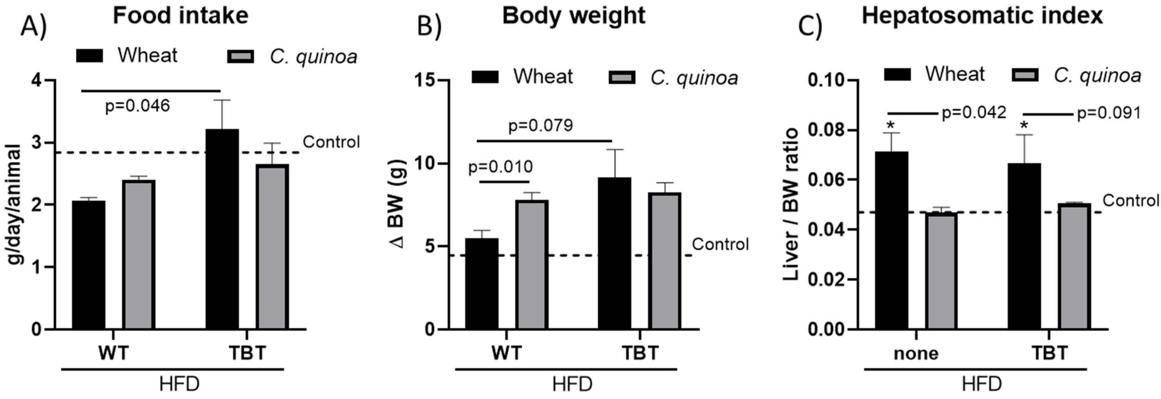
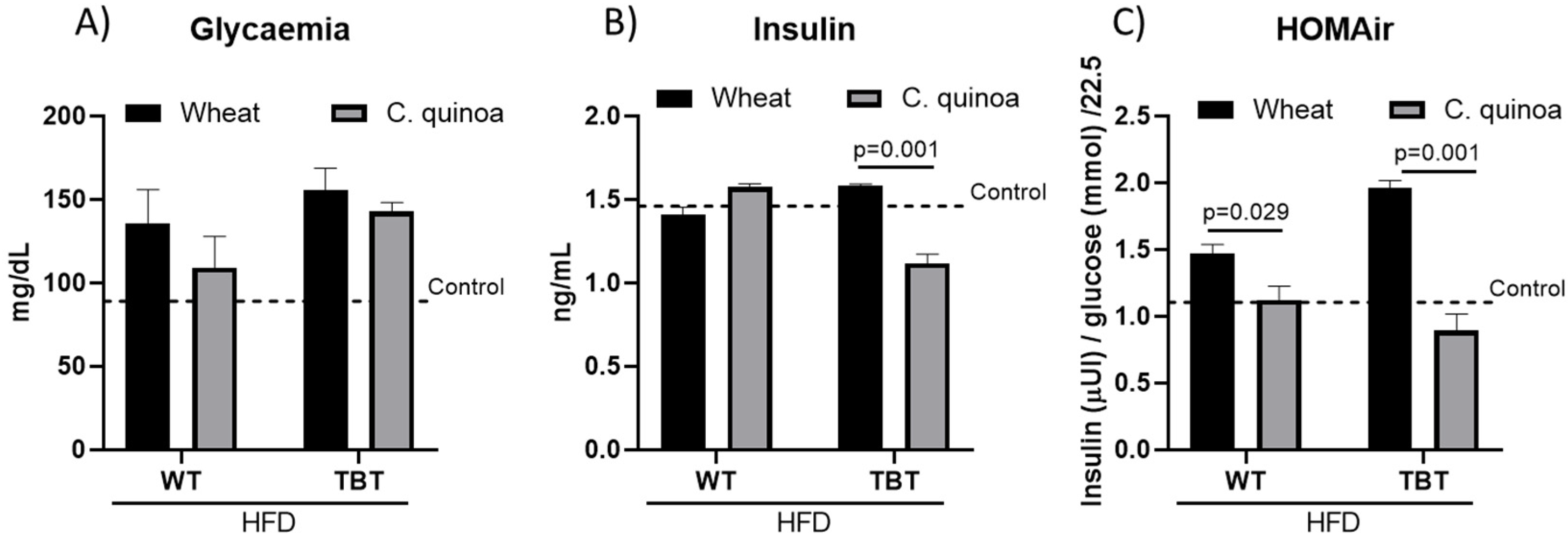
3.1. Food Intake and Morphometric Measurements

Animals administered with either wheat or C. quinoa bread formulations displayed reduced consumption rates in relation to controls (Figure 1A. With C. quinoa bread, the food intake rate did not reach statistical significance between animals under different treatments (i.e., WT vs. TBT). Upward trends for food intake rates in TBT-affected animals could reflect decreased nutrient utilization derived from NAFLD-associated liver dysfunction.
Administration of C. quinoa bread to wild-type (WT) animals favored higher body weight gain than those fed with wheat bread (Figure 1B). However, these differences were abolished in TBT-treated animals. Notably, animals receiving C. quinoa bread showed similar effects in both metabolic mouse models. Administration of C. quinoa bread prevented alterations in the hepatosomatic index either in wild-type animals or those displaying a transgenerational inheritance to develop NAFLD-associated obesity (Figure 1C). Altogether, data may interpret the results as a differential engagement of metabolic processes mainly derived from the different compositions of immunonutritional bioactive proteins.

3.2. Variations on Innate Immune Myeloid Cell Population

Animals administered with C. quinoa bread displayed significantly increased variations in the myeloid cell population (Figure 2A). Only a downward trend was calculated in the hepatic infiltrating monocyte-derived macrophages in relation to that in wheat-bread-fed mice (Figure 2B). In both cases, a favorable CD68/CD206 ratio was found, reflecting the more prevalent M1-like phenotype of the infiltrated cells.
Notably, hepatic transcripts for Lyve-1 suggest the existence of two distinct functional macrophage populations when feeding C. quinoa or wheat bread (Figure 2C).

4. Discussion

This study investigated the associations between administration of C. quinoa (20%, w/w) bread, in comparison with wheat bread, and the variability of selective functional phenotypes of hepatic infiltrating monocyte-derived macrophages in metabolic mouse models with diet-induced obesity. The associations were independent of body weight gain and the CD68/CD206 proportion. The risk of cardiovascular disease and liver fibrosis in animals displaying a transgenerational inheritance to develop NAFLD was lower in those fed with C. quinoa bread.
Based on these findings, administration of C. quinoa bread enabled a better preserved hepatosomatic index, decreasing the risk of liver dysfunction and NAFLD development, which are important factors favoring the metabolic syndrome, in both metabolic mouse models [6]. The mechanism behind the associations of administration of C. quinoa bread and alleviation of liver inflammation and NAFLD are diverse. First, hyaluronan accumulation with HFD feeding [7], to contribute to insulin resistance and exacerbate liver inflammation by interacting with Lyve-1, is reduced by the administration of C. quinoa bread in WT animals. Increased insulin resistance plays a crucial role in the progression of NAFLD, which is related to adverse health outcomes. Second, preservation of Lyve-1 in TBT-treated mice, which exhibit transgenerational inheritance of disturbances in glucose homeostasis [5] and inhibition of the insulin receptor expression [8] as well as a permanently metabolic (re)programming toward hepatic fat accumulation [5], allow suggesting the amelioration of chronic inflammation and long-term deterioration of liver function. Third, distinct interstitial macrophage populations coexist across tissues in specific subtissular niches where the absence of Lyve-1 macrophages exacerbates fibrosis [9].

5. Conclusions

In metabolic mouse models, the immunonutritional potential of C. quinoa enables a selective functional differentiation and function of myeloid cell population toward resolutive macrophage. This influence favors tissue integrity in conditions of caloric excess. These data warrant further human-based research.

Author Contributions

All authors contribute equally to this paper. All authors have read and agreed to the published version of the manuscript.

Funding

This research received no external funding.

Institutional Review Board Statement

Animal experiments were carried out in strict accordance with the recommendations in the Guide for the Care and Use of Laboratory Animals of CSIC (Consejo Superior de Investigaciones Científicas) and the protocol was approved by its Ethic Committee and the regional government (Ethic code, Proex 220/17).

Data Availability Statement

The data that support the findings of this study are available from the corresponding author upon reasonable request.

Acknowledgments

This work was financially supported by grants La ValSe-Food CYTED, (119RT0S67) and Food4ImNut (PRP_PID-2019-107650RBC22, Ministry of Science, Innovation and Universities—MICIU).

Conflicts of Interest

The authors declare no conflict of interest.

References

  1. Krenkel, O.; Hundertmark, J.; Abdallah, A.T.; Kohlhepp, M.; Puengel, T.; Roth, T.; Branco, D.P.P.; Mossanen, J.C.; Luedde, T.; Trautwein, C.; et al. Myeloid cells in liver and bone marrow acquire a functionally distinct inflammatory phenotype during obesity-related steatohepatitis. Gut 2020, 69, 551–563. [Google Scholar] [CrossRef] [PubMed]
  2. Griffin, C.; Eter, L.; Lanzetta, N.; Abrishami, S.; Varghese, M.; McKernan, K.; Muir, L.; Lane, J.; Lumeng, C.N.; Singer, K. TLR4, TRIF, and MyD88 are essential for myelopoiesis and CD11c+ adipose tissue macrophage production in obese mice. J. Biol. Chem. 2018, 293, 8775–8786. [Google Scholar] [CrossRef] [PubMed] [Green Version]
  3. Selma-Gracia, R.; Haros, C.M.; Laparra Llopis, J.M. Inclusion of Salvia hispanica L. and Chenopodium quinoa into bread formulations improves metabolic imbalances derived from a high-fat intake in hyperglycaemic mice. Food Funct. 2020, 11, 7994–8002. [Google Scholar] [CrossRef]
  4. Srdić, M.; Ovčina, I.; Fotschki, B.; Haros, C.M.; Laparra, J.M. C. quinoa and S. hispanica L. seeds provide immunonutritional agonists to selectively polarize macrophages. Cells 2020, 9, 593. [Google Scholar] [CrossRef] [Green Version]
  5. Chamorro-García, R.; Sahu, M.; Abbey, R.J.; Laude, J.; Pham, N.; Blumberg, B. Transgenerational inheritance of increased fat depot size, stem cell reprogramming, and hepatic steatosis elicited by prenatal exposure to the obesogen tributyltin in mice. Environ. Health Perspect. 2013, 121, 359–366. [Google Scholar] [CrossRef] [PubMed] [Green Version]
  6. Yki-Järvinen, H. Non-alcoholic fatty liver disease as a cause and a consequence of metabolic syndrome. Lancet Diabetes Endocrinol. 2014, 2, 901–910. [Google Scholar] [CrossRef]
  7. Kang, L.; Lantier, L.; Kennedy, A.; Bonner, J.S.; Mayes, W.H.; Bracy, D.P.; Bookbinder, L.H.; Hasty, A.H.; Thompson, C.B.; Wasserman, D.H. Hyaluronan accumulates with high-fat feeding and contributes to insulin resistance. Diabetes 2013, 62, 1888–1896. [Google Scholar] [CrossRef] [PubMed] [Green Version]
  8. Li, B.; Guo, J.; Xi, Z.; Xu, J.; Zuo, Z.; Wang, C. Tributyltin in male mice disrupts glucose homeostasis as well as recovery after exposure: Mechanism analysis. Arch. Toxicol. 2017, 91, 3261–3269. [Google Scholar] [CrossRef] [PubMed]
  9. Chakarov, S.; Lim, H.Y.; Tan, L.; Lim, S.Y.; See, P.; Lum, J.; Zhang, X.-M.; Foo, S.; Nakamizo, S.; Duan, D.; et al. Two distinct interstitial macrophage populations coexist across tissues in specific subtissular niches. Science 2019, 363, eaau0964. [Google Scholar] [CrossRef] [PubMed]
Figure 1. Food intake (A) and morphometric measurements, body weight gain (BW) (B), and hepatosomatic index (C) of in wild-type (WT) and tributyltin (TBT)-exposed mice fed a high-fat diet. Results are expressed as mean ± standard error (SEM) (n = 6). Untreated controls are represented by the dotted line.
Blsf 08 00013 g001
Figure 2. Innate immune adaptation: (A) variations in myeloid cells in wild-type (WT) and tributyltin (TBT)-exposed mice; (B) hepatic rt-qPCR analysis (mRNA) of macrophage marker genes CD68/CD206 and (C) the homing cell adhesion molecule (Lyve-1/CD44) in wild-type (WT) and tributyltin (TBT)-exposed mice fed a high-fat diet. Results are expressed as mean ± mean standard error (n = 6).
Blsf 08 00013 g002
Publisher’s Note: MDPI stays neutral with regard to jurisdictional claims in published maps and institutional affiliations.

