Proceeding Paper

Acute and Chronic Effects of Medium-Chain Triglyceride Supplementation on Metabolic Parameters and Working Memory in Rats †

1
Institute of Experimental Medicine, 12 Akad. Pavlova St., 197022 St. Petersburg, Russia
2
I. M. Sechenov Institute of Evolutionary Physiology and Biochemistry, Russian Academy of Sciences, 44 Prospekt Toreza, 194223 St. Petersburg, Russia
*
Authors to whom correspondence should be addressed.
Presented at the 1st International Electronic Conference on Biomedicine, 1–26 June 2021; Available online: https://ecb2021.sciforum.net.
Biol. Life Sci. Forum 2021, 7(1), 22; https://doi.org/10.3390/ECB2021-10282
Published: 1 June 2021
(This article belongs to the Proceedings of The 1st International Electronic Conference on Biomedicine)

Abstract

:
Medium-chain triglycerides (MCT) have demonstrated a wide range of neuroprotective effects, although the mechanisms still remain poorly understood. Animal models are indispensable for such research. Metabolic effects of regular diet supplementation with fats must be considered. Male Wistar rats aged 2.5 months received (o/g) 3 g/kg/day of MCT oil, lard, or water (control) as a supplement to standard chow for 28 days. On the 17th day, the animals were tested in Y-maze. On the 28th day, blood was collected for biochemical testing (glucose, triglycerides (TG), total cholesterol (TC), HDL cholesterol). In a separate experiment, animals received 3 g/kg MCT, lard, or water, and were then sacrificed 30 or 120 min after. Blood was collected for biochemical testing (glucose, lactate, pyruvate, acetoacetate, β-hydroxybutyrate (BHB), TC, TG, aspartate transaminase (AST), alanine transaminase (ALT)). In the Y-maze test, the MCT-fed rats demonstrated an increased frequency of spontaneous alterations compared to both the control and lard groups, indicating improved working memory. Chronic administration of neither fat affected the blood glucose, TG, TC, and HDL cholesterol. Acutely, MCT supplementation elevated blood BHB, while lard did not. Lard increased blood TG, TC, and ALT, while MCT did not. Daily supplementation of standard feed with MCT led to mild intermittent ketosis and improved working memory in rats. Neither chronic nor acute MCT administration had any adverse effect on metabolic health markers. This animal model may be used to study the mechanisms of the cognitive-enhancing effects of MCT.

1. Introduction

Medium-chain triglycerides (MCT) are triglycerides (TG) that contain saturated fatty acids with a chain length from 6 to 10 carbon atoms (medium-chain fatty acids, MCFAs). MCFAs and long-chain fatty acids (LCFAs) are metabolized differently. When absorbed by enterocytes, LCFAs trigger chylomicron formation and are transported in the lymph, while MCFAs are primarily directed to the liver through the portal vein. In hepatocytes, MCFAs mostly avoid activation in the cytosol and enter mitochondria bypassing the carnitine transport system, which is limiting the LCFA transport to mitochondria when enough glucose is available [1]. Therefore, the main fate of MCFAs in hepatocytes is to be oxidized in the mitochondria, generating an excess of acetyl-CoA, while LCFAs are primarily esterified for TG storage, phospholipid synthesis, and excretion in very-low-density lipoprotein (VLDL) particles [2]. When MCFAs are rapidly oxidized in the mitochondria, the amount of acetyl-CoA, which exceeds the tricarboxylic acid (TCA) cycle capacity, can be redirected to various metabolic pathways, including ketogenesis in the mitochondria and de novo lipogenesis and cholesterol synthesis in the cytosol. The ketone bodies (KB) produced in the liver (including acetoacetate (AcAc) and β-hydroxybutyrate (BHB)) can be excreted into the blood and transported to other organs, including the brain, where the KB can be converted back to acetyl-CoA and enter the TCA cycle to produce ATP [1]. For LCFAs, the ketogenesis pathway only becomes significant under conditions such as starvation, high-fat–low-carbohydrate ketogenic diet (KD), or diabetes. MCFAs, on the other hand, are ketogenic even in a well-fed state in the presence of carbohydrates [3,4].
It is well established that KD and its mechanistic mediators, KB, possess neuroprotective properties [5,6,7]. Various approaches have been developed to raise the blood KB levels without the challenge of adhering to KD, including the use of MCT as well as BHB salts and esters [6]. Aging and Alzheimer’s disease (AD) are often accompanied by reduced glucose but intact KB metabolism in the brain [8,9]. Both single and chronic ingestion of MCT have been shown to increase brain energy metabolism [10,11]. In several human studies, both chronic supplementation and acute oral administration of MCT offered beneficial effects on cognition in elderly individuals with normal cognition [12], elderly individuals suffering from mild cognitive impairment (MCI) [13,14], and APOE4-negative AD patients [15]. Although MCT supplementation studies are usually conducted with the rationale that the KB produced by the liver are the mediators of the beneficial effects, MCFAs may also exert certain protective effects on nervous cells via mechanisms independent of KB metabolism [16,17]. In fact, some studies reported that MCFA but not BHB concentrations were elevated in the brain of animals fed diets enriched with MCT [17,18]. One study reported that the biochemical effects of MCT supplementation were brain-region-specific and phenotype-specific [19]. While MCT supplementation is undoubtedly a promising approach to support cognitive function, its mechanisms remain poorly understood.
A potential concern with the MCT supplementation approach (as opposed to a KD) is that although MCT doses of less than 1 g/kg are generally considered safe [20], the excess of acetyl-CoA, produced during rapid MCFA oxidation in the liver, has the potential to feed the de novo lipogenesis and cholesterol synthesis pathways in the cytosol and may also limit the LCFA oxidation, pushing the LCFAs towards TG storage and VLDL excretion, depending on many factors such as the underlying physiological condition and the proportions of carbohydrates and various fatty acids in the diet. In various studies, MCFA-rich diet compared to LCFA-rich diet increased [21], decreased [22], or had no effect [23] on TG accumulation in the liver; increased [24], decreased [21], or had no effect on [25] fasting plasma TG concentration; and increased [24], decreased [26], or had no effect on [27] fasting plasma total cholesterol concentrations. While human clinical trials of MCT’s effects on cognitive functions generally do not include participants with metabolic disorders [13,14,28], obesity, insulin resistance, and diabetes are common risk factors for developing AD, and they share dyslipidemia as a pathological mechanism [29]. The long-term effects of MCT supplementation of a regular diet on metabolic and cardiovascular health are still unknown.
The present study aimed to assess metabolic effects of acute and chronic administration of MCT in concentrations high enough to elicit neuroprotective effects in young adult male rats.

2. Materials and Methods

The study was performed on male Wistar rats aged 2.5–3.5 months with an average weight (M ± SD) of 316.3 ± 28.03 g at the beginning of the experiments and 371.5 ± 38.64 g at the end of the experiments. The animals with an average weight of about 160–180 g and 4–5 weeks of age were purchased from the State Breeding Farm “Rappolovo” (Leningrad region, Russia), and housed under standard conditions with ad libitum access to standard chow and tap water for 1 month prior the study and 1.5 months during the study. The room temperature was controlled at 24–26 °C in a 12 h light/dark cycle. All experiments were conducted in compliance with the regulations of the European Communities Council Directive 2010/63/EU and were approved by the ethics authorities of the Institute of Experimental Medicine, St. Petersburg, Russia.
To study the acute effects of medium-chain triglyceride supplementation on biochemical markers in the blood serum, 2.5 m.o., animals received 3 g/kg MCT (C8 & C10 mixture, “Jarrow Formulas® MCT Oil”, n = 5), lard (n = 5), or water (n = 11) through oral gavage and were then sacrificed by decapitation 30 or 120 min after. Blood was collected for biochemical testing, stored at +4 °C overnight, centrifuged at 2000× g for 20 min. Serum was distributed into 0.6 ml tubes and stored at −70 °C until biochemical analyses.
The levels of β-hydroxybutyrate (BHB), acetoacetate (AcAc), glucose, lactate, pyruvate, total cholesterol, HDL cholesterol, triglycerides (TG), malondialdehyde (MDA), and aspartate transaminase (AST) and alanine transaminase (ALT) activities were measured in the serum samples without deproteinization using commercial kits according to the manufacturers’ instructions (“Sigma-Aldrich”, St. Louis, MO, USA (BHB, AcAc); “Olvex Diagnosticum” (AST, ALT), St. Petersburg, Russia; “Vital Development Corp.,” St. Petersburg, Russia). All analyses were performed by enzymatic colorimetric assays. The final colored products’ absorbance was measured on ImmunoChem-2100 Microplate Reader (High Technology, Inc., North Attleborough, MA, USA) at 340 nm (BHB, AcAc, AST, ALT), 532 nm (MDA), or 500–510 nm.
To study the effects of chronic MCT supplementation on the biochemical markers in the blood serum and the performance in the Y-maze, the animals were first tested in Y-maze (8 min, 3 equal arms) to evaluate the baseline total number of arm entries and spontaneous alternations performance (consecutive visits to three different arms). Nine days after the testing, animals started to receive orogastrically 3 g/kg/day of either MCT oil (n = 8), lard (n = 8), or water (control, n = 9) as a supplement to standard chow for 28 days. On Day 17 of the supplementation regime, the Y-maze task was repeated (prior to the supplement administration on that day). On Day 28, animals were sacrificed (20 h after the final orogastric administration) and blood was collected for biochemical testing (glucose, triglycerides, total cholesterol, HDL cholesterol, MDA, AST and ALT activity), performed as described above.
Statistical analysis and graph plotting were performed using GraphPad Prism v.8 (GraphPad Software, Inc., San Diego, CA, USA). D’Agostino–Pearson and Shapiro–Wilk tests were used for assessing the normality of the data distribution. Inter-group differences were analyzed by ANOVA with Tukey’s post hoc test and 2-way repeated measures (rm)-ANOVA with Holm-Sidak post hoc test. Linear regression differences with slope comparison were used to assess the effects of MCT and Lard treatment on the dynamics of the blood biochemical parameters after single administration. The graphs are plotted as M ± SEM. Significant differences were accepted at p < 0.05.

3. Results

In the Y-maze test, the number of total arm entries was lower during the second trial in all experimental groups (Figure 1a; 2-way rm-ANOVA: F(1, 22) = 36.9, p < 0.0001; Holm-Sidak post hoc Before vs. After: Ctrl t = 4.7, p = 0.0003, Lard t = 3.4, p = 0.0045, MCT t = 2.3, p = 0.02) with no statistical difference among groups (Figure 1b; ANOVA: F(2, 22) = 1.18, p = 0.32), indicating that the fat supplements had no effect on locomotive activity. The frequency of spontaneous alterations tended to decrease significantly during the second trial in the control and the lard-fed animals but not in the MCT-fed animals (Figure 1c; 2-way rm-ANOVA: F(1, 22) = 36.9, p < 0.0012 (effect of trial), F(2, 22) = 3.24, p = 0.05 (effect of trial × treatment interaction); Holm-Sidak post hoc Before vs. After: Ctrl t = 3.1, p = 0.011, Lard t = 3.2, p = 0.011, MCT t = 0.04, p = 0.96). The value of the treatment-induced change in the frequency of spontaneous alternations relative to baseline was greater in the MCT-fed rats (compared to both the control and the lard groups), indicating improved working memory (Figure 1d; ANOVA: F(2, 22) = 4.809, p = 0.0185; Tukey’s post hoc: Ctrl vs. Lard q = 0.161, p = 0.99, Ctrl vs. MCT q = 3.79, p = 0.035, Lard vs. MCT q = 3.84, p = 0.0325).
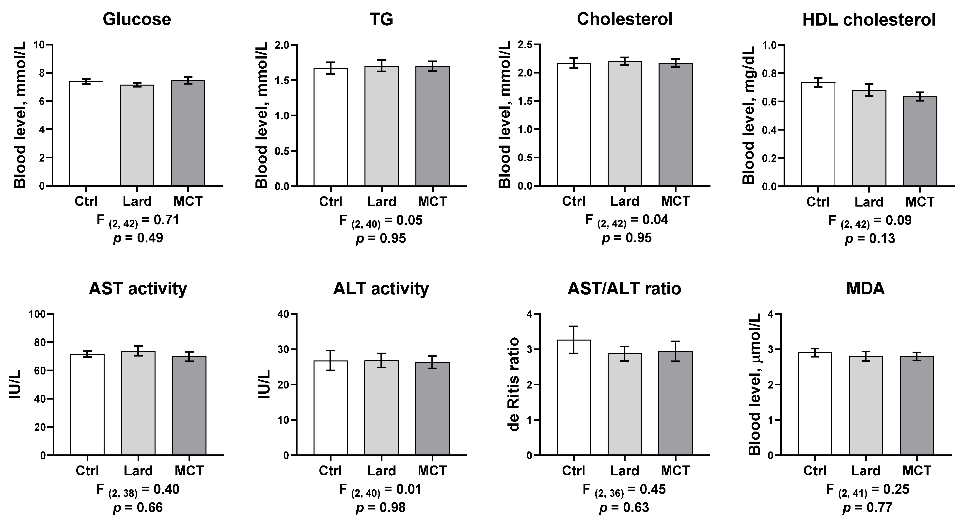
Chronic administration of neither fat affected the blood glucose, triglycerides, total cholesterol, HDL cholesterol levels, the AST and ALT activities and their ratio (de Ritis ratio), and the MDA level (Figure 2; p > 0.05).
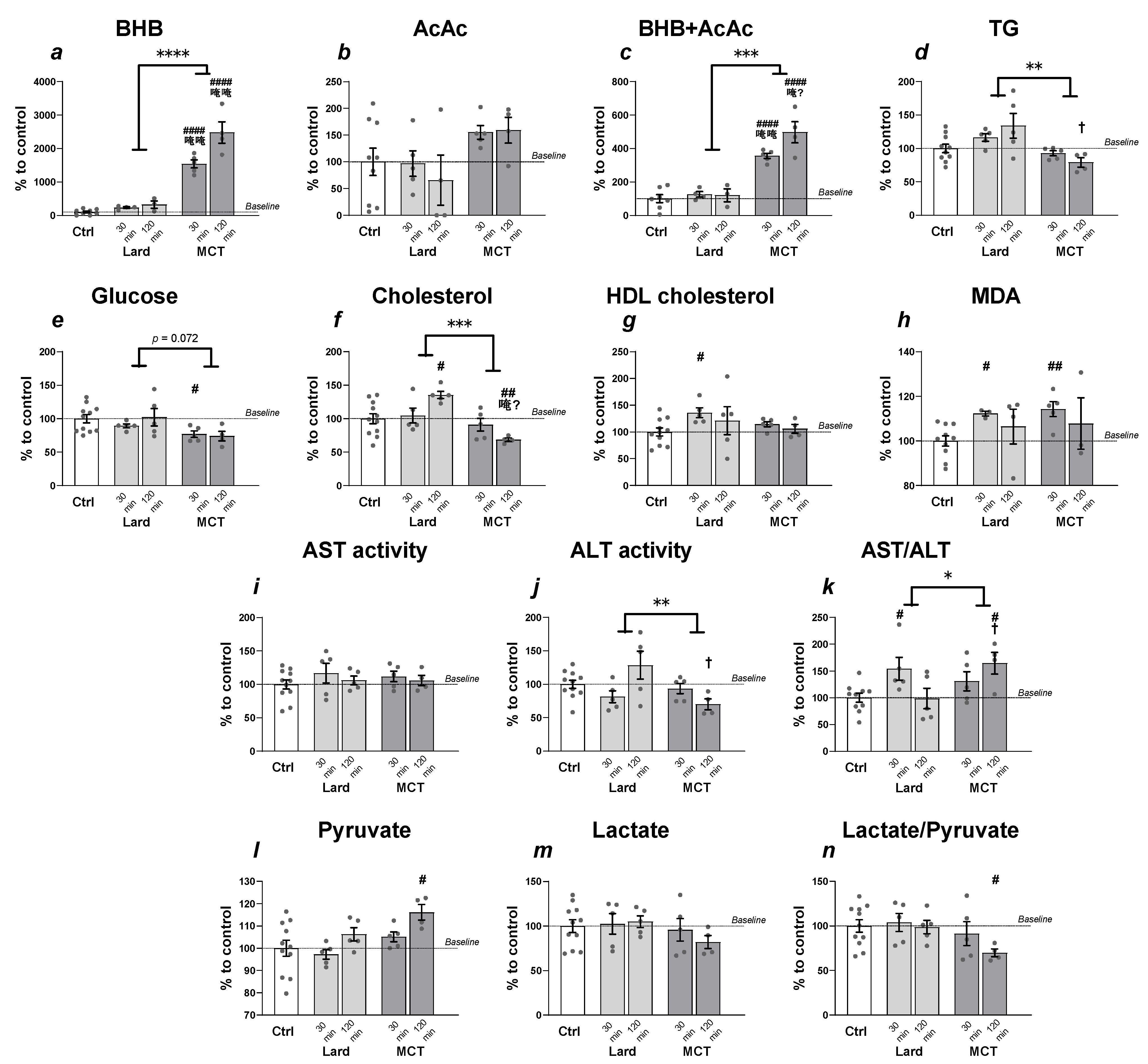
Acutely, MCT supplementation elevated blood BHB level, while lard did not (Figure 3a; linear regression comparison: F(1, 28) = 34.32, p < 0.0001; ANOVA 30 min: F(2, 14) = 140.4, p < 0.0001; ANOVA 120 min: F(2, 11) = 63.56, p < 0.0001). The AcAc level did not significantly change after the administration of either fat (Figure 3b; p > 0.05). The cumulative level of both ketone bodies was significantly elevated after the MCT but not the lard treatment (Figure 3c; linear regression comparison: F(1, 26) = 19.5, p = 0.0002; ANOVA 30 min: F(2, 13) = 43.1, p < 0.0001; ANOVA 120 min: F(2, 11) = 30.01, p < 0.0001).
Lard administration increased blood TG (Figure 3d; linear regression comparison: F(1, 35) = 10.18, p = 0.003), cholesterol levels (Figure 3f; linear regression comparison: F(1, 37) = 15.00, p = 0.0004), and ALT activity (Figure 3j; linear regression comparison: F(1, 37) = 9.34, p = 0.004), while MCT did not.
The glucose level was found to be decreased 30 min after MCT ingestion compared to control (Figure 3e; ANOVA: F(2, 18) = 3.67, p = 0.046, Tukey’s post hoc Control vs. MCT: q = 3.78, p = 0.039), while this parameter was unchanged in the lard-fed animals. The HDL cholesterol level increased 30 min after lard but not MCT treatment compared to control animals (Figure 3g; ANOVA: F(2, 18) = 4.95, p = 0.019, Tukey’s post hoc Control vs. Lard: q = 4.43, p = 0.015).
The level of pyruvate increased 120 min after the MCT but not the lard treatment compared to control (Figure 3l; ANOVA: F(2, 17) = 3.81, p = 0.043, Tukey’s post hoc Control vs. MCT: q = 3.87, p = 0.036). As the lactate level did not change after either fat administration (Figure 3m; p > 0.05), the ratio of lactate/pyruvate in MCT-fed animals was lower compared to control 120 min after the treatment (Figure 3n; ANOVA: F(2, 17) = 3.63, p = 0.048, Tukey’s post hoc Control vs. MCT: q = 3.67, p = 0.045).

4. Discussion

The MCT dose used in this study (3 g/kg) corresponds to the amount typically given to human subjects in clinical trials [12,15,30], adjusted for metabolic rate [31]. This dose was sufficient to achieve mild ketosis, elevating the blood KB levels (AcAc + BHB) 4–5-fold with the peak concentration at around 2 h. Our results demonstrate that this amount of MCT, when given as a supplement to standard feed for 28 days, is sufficient to achieve a measurable beneficial effect on cognition in young adult rats. A higher frequency of spontaneous alternations (SA) in the Y-maze test indicates better working memory [32]. In our study, chronic MCT supplementation increased the SA frequency by 37% compared to baseline. The test was conducted before MCT was administered on that day. Therefore, the improved performance cannot be attributed to KBs acting as an alternative energy source immediately during the test. Instead, it should be regarded as a cumulative effect of 16 days of MCT supplementation. In line with our findings, LCFA-based KD improved performance in the Y-maze [33], and the supplementation with MCFA-rich oil improved spatial memory [34] in healthy adult rats. Conversely, MCT-based KD failed to improve cognitive performance in two murine transgenic models of AD [35], while MCT supplementation reduced anxiety in rats with high but not normal baseline anxiety levels [19]. In our study, MCT had no effect on locomotive activity. The effects of MCT treatment on cognitive functions appear to depend on the investigated trait and the phenotype. More research is needed to define the scope of impairments, for which the neuroprotective effects can be achieved with MCT supplementation.
In our study, 28 days of supplementation of standard feed with MCT (3 g/kg) had no significant effect on any of the measured markers of metabolic health. In contrast, 4 g/kg MCT added to a fructose-rich diet for 12 weeks has been reported to exacerbate the liver damage associated with fructose feeding in mice [36]. In another study, when 10 g/kg/day MCT were given to juvenile rats for 28 days with standard feed, the livers of the rats were enlarged, and the HDH cholesterol level was reduced [37]. It appears that the dose used in our study is high enough to improve cognition in young adult rats fed with standard rodent chow, but low enough to avoid hepatotoxic effects.
We assessed the acute effects of our supplementation protocol. The effects of a single oral dose of MCT on serum glucose, lactate, and pyruvate mostly agree with the classical studies in non-fasted rats [38]. However, in our study, we observed an increase (instead of a decrease) in pyruvate level at 120 min post-administration and a parallel decrease in the lactate/pyruvate ratio. These discrepancies may be due to differences in experimental protocols. The elevation of pyruvate level may be related to the inhibitory effect of KB on pyruvate dehydrogenase [39]. Enzymatic activities of ALT and AST are commonly used as biochemical markers of liver injury. MCT administration reduced blood ALT activity, while lard administration increased it, consistent with some previous reports [40,41]. In our study, MCT had a slight lowering effect on TG and cholesterol levels 120 min after MCT administration. This is consistent with some previous reports where MCT was given to healthy human subjects together with a carbohydrate-rich product and can be explained by reduced chylomicron formation when ingesting MCT [42,43]. Both MCT and lard ingestion resulted in a small increase in malondialdehyde (MDA), which is the main end product of lipid peroxidation used as a marker of oxidative stress. Fats typically affect postprandial MDA levels stronger than proteins and carbohydrates [44]. Although 28 days of our protocol had no lasting effect on the blood MDA level, it is unknown what the effect might be over a longer period or in the case of pre-existing pathological conditions. Elevated MDA levels are associated with heart disease [45]. More studies are necessary to determine which doses offer the most benefits under varying conditions, as well as whether long-term MCT supplementation is safe for cardiovascular health.

5. Conclusions

Daily supplementation of regular feed with 3 g/kg/day MCT improved working memory in young adult rats without adverse effects on markers of metabolic health. The working memory assessment test was conducted when the serum KB concentration had already returned to baseline; therefore, MCT supplementation resulted in a long-lasting effect on the brain. Similar administration procedures and concentrations may be used in future research to investigate the mechanisms of the MCT effects on cognition. More studies are needed to determine long-term consequences of MCT supplementation of regular diet on cardio-vascular health and metabolic health.

Supplementary Materials

The following supporting information can be downloaded at: https://www.mdpi.com/article/10.3390/ECB2021-10282/s1.

Author Contributions

Conceptualization, K.S. and A.T.; methodology, A.T., V.N., and D.K.; validation, A.T.; formal analysis, A.T.; investigation, A.T., I.I., V.N., and D.K.; writing—original draft preparation, K.S. and A.T.; writing—review and editing, K.S., A.T., A.S., M.K., and S.A.; visualization, A.T.; project administration, A.T. All authors have read and agreed to the published version of the manuscript.

Funding

This research was funded by the Russian Science Foundation, grant number 19-75-10076.

Institutional Review Board Statement

The study was conducted in accordance with the regulations of the European Communities Council Directive 2010/63/EU, and approved by the Institutional Ethics Committee of the Institute of Experimental Medicine (protocol no. 2/22).

Informed Consent Statement

Not applicable.

Data Availability Statement

Data is available upon request from the authors.

Conflicts of Interest

The authors declare no conflict of interest.

References

  1. Bach, A.C.; Babayan, V.K. Medium-chain triglycerides: An update. Am. J. Clin. Nutr. 1982, 36, 950–962. [Google Scholar] [CrossRef] [PubMed]
  2. Nguyen, P.; Leray, V.; Diez, M.; Serisier, S.; Le Bloc’h, J.; Siliart, B.; Dumon, H. Liver lipid metabolism. J. Anim. Physiol. Anim. Nutr. 2008, 92, 272–283. [Google Scholar] [CrossRef] [PubMed]
  3. Seaton, T.B.; Welle, S.L.; Warenko, M.K.; Campbell, R.G. Thermic effect of medium-chain and long-chain triglycerides in man. Am. J. Clin. Nutr. 1986, 44, 630–634. [Google Scholar] [CrossRef] [PubMed]
  4. Courchesne-Loyer, A.; Fortier, M.; Tremblay-Mercier, J.; Chouinard-Watkins, R.; Roy, M.; Nugent, S.; Castellano, C.-A.; Cunnane, S.C. Stimulation of mild, sustained ketonemia by medium-chain triacylglycerols in healthy humans: Estimated potential contribution to brain energy metabolism. Nutrition 2013, 29, 635–640. [Google Scholar] [CrossRef] [PubMed]
  5. Maalouf, M.; Rho, J.M.; Mattson, M.P. The neuroprotective properties of calorie restriction, the ketogenic diet, and ketone bodies. Brain Res. Rev. 2009, 59, 293–315. [Google Scholar] [CrossRef] [PubMed] [Green Version]
  6. Kovács, Z.; D’Agostino, D.P.; Diamond, D.; Kindy, M.S.; Rogers, C.; Ari, C. Therapeutic Potential of Exogenous Ketone Supplement Induced Ketosis in the Treatment of Psychiatric Disorders: Review of Current Literature. Front. Psychiatry 2019, 10, 363. [Google Scholar] [CrossRef] [PubMed]
  7. Puchalska, P.; Crawford, P.A. Multi-dimensional Roles of Ketone odies in Fuel Metabolism, Signaling, and Therapeutics. Cell Metab. 2017, 25, 262–284. [Google Scholar] [CrossRef] [Green Version]
  8. Croteau, E.; Castellano, C.A.; Fortier, M.; Bocti, C.; Fulop, T.; Paquet, N.; Cunnane, S.C. A cross-sectional comparison of brain glucose and ketone metabolism in cognitively healthy older adults, mild cognitive impairment and early Alzheimer’s disease. Exp. Gerontol. 2018, 107, 18–26. [Google Scholar] [CrossRef]
  9. Nugent, S.; Tremblay, S.; Chen, K.W.; Ayutyanont, N.; Roontiva, A.; Castellano, C.-A.; Fortier, M.; Roy, M.; Courchesne-Loyer, A.; Bocti, C.; et al. Brain glucose and acetoacetate metabolism: A comparison of young and older adults. Neurobiol. Aging 2014, 35, 1386–1395. [Google Scholar] [CrossRef]
  10. Xin, L.; Ipek, Ö.; Beaumont, M.; Shevlyakova, M.; Christinat, N.; Masoodi, M.; Greenberg, N.; Gruetter, R.; Cuenoud, B. Nutritional Ketosis Increases NAD+/NADH Ratio in Healthy Human Brain: An in Vivo Study by 31P-MRS. Front. Nutr. 2018, 5, 62. [Google Scholar] [CrossRef] [Green Version]
  11. Croteau, E.; Castellano, C.-A.; Richard, M.A.; Fortier, M.; Nugent, S.; Lepage, M.; Duchesne, S.; Whittingstall, K.; Turcotte, É.E.; Bocti, C.; et al. Ketogenic Medium Chain Triglycerides Increase Brain Energy Metabolism in Alzheimer’s Disease. J. Alzheimers Dis. 2018, 64, 551–561. [Google Scholar] [CrossRef] [PubMed]
  12. Ota, M.; Matsuo, J.; Ishida, I.; Hattori, K.; Teraishi, T.; Tonouchi, H.; Ashida, K.; Takahashi, T.; Kunugi, H. Effect of a ketogenic meal on cognitive function in elderly adults: Potential for cognitive enhancement. Psychopharmacology 2016, 233, 3797–3802. [Google Scholar] [CrossRef] [PubMed]
  13. Rebello, C.J.; Keller, J.N.; Liu, A.G.; Johnson, W.D.; Greenway, F.L. Pilot feasibility and safety study examining the effect of medium chain triglyceride supplementation in subjects with mild cognitive impairment: A randomized controlled trial. BBA Clin. 2015, 3, 123–125. [Google Scholar] [CrossRef] [Green Version]
  14. Fortier, M.; Castellano, C.-A.; Croteau, E.; Langlois, F.; Bocti, C.; St-Pierre, V.; Vandenberghe, C.; Bernier, M.; Roy, M.; Descoteaux, M.; et al. A ketogenic drink improves brain energy and some measures of cognition in mild cognitive impairment. Alzheimers. Dement. 2019, 15, 625–634. [Google Scholar] [CrossRef]
  15. Ota, M.; Matsuo, J.; Ishida, I.; Takano, H.; Yokoi, Y.; Hori, H.; Yoshida, S.; Ashida, K.; Nakamura, K.; Takahashi, T.; et al. Effects of a medium-chain triglyceride-based ketogenic formula on cognitive function in patients with mild-to-moderate Alzheimer’s disease. Neurosci. Lett. 2019, 690, 232–236. [Google Scholar] [CrossRef] [PubMed]
  16. Page, K.A.; Williamson, A.; Yu, N.; McNay, E.C.; Dzuira, J.; McCrimmon, R.J.; Sherwin, R.S. Medium-chain fatty acids improve cognitive function in intensively treated type 1 diabetic patients and support in vitro synaptic transmission during acute hypoglycemia. Diabetes 2009, 58, 1237–1244. [Google Scholar] [CrossRef] [Green Version]
  17. Tan, K.N.; Carrasco-Pozo, C.; McDonald, T.S.; Puchowicz, M.; Borges, K. Tridecanoin is anticonvulsant, antioxidant, and improves mitochondrial function. J. Cereb. Blood Flow Metab. 2017, 37, 2035–2048. [Google Scholar] [CrossRef]
  18. Wang, D.; Mitchell, E.S. Cognition and Synaptic-Plasticity Related Changes in Aged Rats Supplemented with 8- and 10-Carbon Medium Chain Triglycerides. PLoS ONE 2016, 11, e0160159. [Google Scholar] [CrossRef] [Green Version]
  19. Hollis, F.; Mitchell, E.S.; Canto, C.; Wang, D.; Sandi, C. Medium chain triglyceride diet reduces anxiety-like behaviors and enhances social competitiveness in rats. Neuropharmacology 2018, 138, 245–256. [Google Scholar] [CrossRef]
  20. Traul, K.A.; Driedger, A.; Ingle, D.L.; Nakhasi, D. Review of the toxicologic properties of medium-chain triglycerides. Food Chem. Toxicol. 2000, 38, 79–98. [Google Scholar] [CrossRef]
  21. Wein, S.; Wolffram, S.; Schrezenmeir, J.; Gasperiková, D.; Klimes, I.; Seböková, E. Medium-chain fatty acids ameliorate insulin resistance caused by high-fat diets in rats. Diabetes. Metab. Res. Rev. 2009, 25, 185–194. [Google Scholar] [CrossRef] [PubMed]
  22. Lieber, C.S.; DeCarli, L.M.; Leo, M.A.; Mak, K.M.; Ponomarenko, A.; Ren, C.; Wang, X. Beneficial effects versus toxicity of medium-chain triacylglycerols in rats with NASH. J. Hepatol. 2008, 48, 318–326. [Google Scholar] [CrossRef] [PubMed]
  23. Baba, N.; Bracco, E.F.; Hashim, S.A. Enhanced thermogenesis and diminished deposition of fat in response to overfeeding with diet containing medium chain triglyceride. Am. J. Clin. Nutr. 1982, 35, 678–682. [Google Scholar] [CrossRef] [Green Version]
  24. Tholstrup, T.; Ehnholm, C.; Jauhiainen, M.; Petersen, M.; Høy, C.-E.; Lund, P.; Sandström, B. Effects of medium-chain fatty acids and oleic acid on blood lipids, lipoproteins, glucose, insulin, and lipid transfer protein activities. Am. J. Clin. Nutr. 2004, 79, 564–569. [Google Scholar] [CrossRef] [PubMed] [Green Version]
  25. Asakura, L.; Lottenberg, A.M.; Neves, M.Q.; Nunes, V.S.; Rocha, J.C.; Passarelli, M.; Nakandakare, E.R.; Quintão, E.C. Dietary medium-chain triacylglycerol prevents the postprandial rise of plasma triacylglycerols but induces hypercholesterolemia in primary hypertriglyceridemic subjects. Am. J. Clin. Nutr. 2000, 71, 701–705. [Google Scholar] [CrossRef] [PubMed]
  26. Han, J.R.; Deng, B.; Sun, J.; Chen, C.G.; Corkey, B.E.; Kirkland, J.L.; Ma, J.; Guo, W. Effects of dietary medium-chain triglyceride on weight loss and insulin sensitivity in a group of moderately overweight free-living type 2 diabetic Chinese subjects. Metabolism 2007, 56, 985–991. [Google Scholar] [CrossRef]
  27. Schwartz, R.M.; Boyes, S.; Aynsley-Green, A. Metabolic effects of three ketogenic diets in the treatment of severe epilepsy. Dev. Med. Child Neurol. 1989, 31, 152–160. [Google Scholar] [CrossRef]
  28. Reger, M.A.; Henderson, S.T.; Hale, C.; Cholerton, B.; Baker, L.D.; Watson, G.S.; Hyde, K.; Chapman, D.; Craft, S. Effects of beta-hydroxybutyrate on cognition in memory-impaired adults. Neurobiol. Aging 2004, 25, 311–314. [Google Scholar] [CrossRef]
  29. Salameh, T.S.; Rhea, E.M.; Banks, W.A.; Hanson, A.J. Insulin resistance, dyslipidemia, and apolipoprotein E interactions as mechanisms in cognitive impairment and Alzheimer’s disease. Exp. Biol. Med. (Maywood) 2016, 241, 1676–1683. [Google Scholar] [CrossRef] [Green Version]
  30. Henderson, S.T.; Vogel, J.L.; Barr, L.J.; Garvin, F.; Jones, J.J.; Costantini, L.C. Study of the ketogenic agent AC-1202 in mild to moderate Alzheimer’s disease: A randomized, double-blind, placebo-controlled, multicenter trial. Nutr. Metab. 2009, 6, 31. [Google Scholar] [CrossRef] [Green Version]
  31. Nair, A.B.; Jacob, S. A simple practice guide for dose conversion between animals and human. J. Basic Clin. Pharm. 2016, 7, 27–31. [Google Scholar] [CrossRef] [PubMed] [Green Version]
  32. Kraeuter, A.-K.; Guest, P.C.; Sarnyai, Z. The Y-Maze for Assessment of Spatial Working and Reference Memory in Mice. Methods Mol. Biol. 2019, 1916, 105–111. [Google Scholar] [CrossRef] [PubMed]
  33. Fukushima, A.; Ogura, Y.; Furuta, M.; Kakehashi, C.; Funabashi, T.; Akema, T. Ketogenic diet does not impair spatial ability controlled by the hippocampus in male rats. Brain Res. 2015, 1622, 36–42. [Google Scholar] [CrossRef]
  34. Rahim, N.S.; Lim, S.M.; Mani, V.; Abdul Majeed, A.B.; Ramasamy, K. Enhanced memory in Wistar rats by virgin coconut oil is associated with increased antioxidative, cholinergic activities and reduced oxidative stress. Pharm. Biol. 2017, 55, 825–832. [Google Scholar] [CrossRef] [PubMed]
  35. Brownlow, M.L.; Benner, L.; D’Agostino, D.; Gordon, M.N.; Morgan, D. Ketogenic diet improves motor performance but not cognition in two mouse models of Alzheimer’s pathology. PLoS ONE 2013, 8, e75713. [Google Scholar] [CrossRef] [PubMed] [Green Version]
  36. Guimarães, J.; Bargut, T.C.L.; Mandarim-de-Lacerda, C.A.; Aguila, M.B. Medium-chain triglyceride reinforce the hepatic damage caused by fructose intake in mice. Prostaglandins. Leukot. Essent. Fatty Acids 2019, 140, 64–71. [Google Scholar] [CrossRef] [PubMed]
  37. Kesl, S.L.; Poff, A.M.; Ward, N.P.; Fiorelli, T.N.; Ari, C.; Van Putten, A.J.; Sherwood, J.W.; Arnold, P.; D’Agostino, D.P. Effects of exogenous ketone supplementation on blood ketone, glucose, triglyceride, and lipoprotein levels in Sprague-Dawley rats. Nutr. Metab. 2016, 13, 9. [Google Scholar] [CrossRef] [Green Version]
  38. Bach, A.; Weryha, A.; Schirardin, H. Influence of oral MCT or LCT load on glycemia in Wistar and Zucker rats and in guinea pigs. Ann. Biol. Anim. Biochim. Biophys. 1979, 19, 625–635. [Google Scholar] [CrossRef] [Green Version]
  39. Robinson, A.M.; Williamson, D.H. Physiological roles of ketone bodies as substrates and signals in mammalian tissues. Physiol. Rev. 1980, 60, 143–187. [Google Scholar] [CrossRef]
  40. Zhang, L.; Wang, X.; Chen, S.; Wang, S.; Tu, Z.; Zhang, G.; Zhu, H.; Li, X.; Xiong, J.; Liu, Y. Medium-Chain Triglycerides Attenuate Liver Injury in Lipopolysaccharide-Challenged Pigs by Inhibiting Necroptotic and Inflammatory Signaling Pathways. Int. J. Mol. Sci. 2018, 19, 3697. [Google Scholar] [CrossRef] [Green Version]
  41. Gao, Y.; Li, X.; Gao, Q.; Fan, L.; Jin, H.; Guo, Y. Differential effects of olive oil, soybean oil, corn oil and lard oil on carbon tetrachloride-induced liver fibrosis in mice. Biosci. Rep. 2019, 39, BSR20191913. [Google Scholar] [CrossRef] [PubMed] [Green Version]
  42. Calabrese, C.; Myer, S.; Munson, S.; Turet, P.; Birdsall, T.C. A cross-over study of the effect of a single oral feeding of medium chain triglyceride oil vs. canola oil on post-ingestion plasma triglyceride levels in healthy men. Altern. Med. Rev. 1999, 4, 23–28. [Google Scholar]
  43. Lee, A.; Yoo, H.J.; Kim, M.; Kim, M.; Choi, J.-H.; Lee, C.; Lee, J.H. Effects of equivalent medium-chain diacylglycerol or long-chain triacylglycerol oil intake via muffins on postprandial triglycerides and plasma fatty acids levels. J. Funct. Foods 2019, 53, 299–305. [Google Scholar] [CrossRef]
  44. Fisher-Wellman, K.H.; Bloomer, R.J. Exacerbated postprandial oxidative stress induced by the acute intake of a lipid meal compared to isoenergetically administered carbohydrate, protein, and mixed meals in young, healthy men. J. Am. Coll. Nutr. 2010, 29, 373–381. [Google Scholar] [CrossRef] [PubMed]
  45. Khan, M.A.; Baseer, A. Increased malondialdehyde levels in coronary heart disease. J. Pak. Med. Assoc. 2000, 50, 261–264. [Google Scholar] [PubMed]
Figure 1. Effects of chronic MCT and lard administration (3 g/kg) on locomotive activity (a,b) and working memory (c,d). (a) The number of arm entries in the Y-maze test before and after treatment, plotted for each animal separately; (b) the average change in the number of arm entries compared to the baseline value in each group; (c) the frequency of spontaneous alternations, plotted for each animal separately; (d) the average change in the frequency of spontaneous alternations compared to the baseline value in each respective group. †—significant difference vs. Lard; #—significant difference vs. Control; *, †, #—p < 0.05, **—p < 0.01, ***—p < 0.001; ns—non significant; n = 8–9 rats per group.
Blsf 07 00022 g001
Figure 2. MCT or Lard oral gavage during 28 consecutive days (3 g/kg/day) did not change the blood glucose, triglycerides, total cholesterol, HDL cholesterol level, or AST and ALT activity 20 h after the last orogastric injection. n = 8–9 rats per group.
Blsf 07 00022 g002
Figure 3. Acute effects of MCT and lard administration (3 g/kg) on the biochemical and metabolic parameters in the blood. (a) MCT but not lard increased beta-hydroxybutyrate (BHB) level; (b) neither fat significantly increased acetoacetate (AcAc) level; (c) MCT but not lard increased total blood ketone body level; (d) triglyceride level was lower after MCT administration compared to lard; (e) MCT but not lard decreased glucose level; (f) 120 min post-administration, MCT decreased while lard increased total cholesterol; (g) lard but not MCT increased HDL cholesterol; (h) both fats acutely increased malondialdehyde level; (i) neither fat affected aspartate transaminase (AST) activity; (j) MCT lowered alanine transaminase (ALT) activity compared both to lard and control; (k) both fats elevated the AST/ALT ratio compared to control but at different time points; (l) MCT but not lard elevated pyruvate level; (m) neither fat affected lactate level; (n) MCT but not lard reduced the lactate/pyruvate ratio. *—MCT vs. Lard difference: linear regression; #—fat vs. control difference: ANOVA and Tukey’s post hoc; †—MCT vs. the respective-time-point Lard difference: ANOVA and Tukey post hoc. *, #, †—p < 0.05; **, ##—p < 0.01; ***, †††—p < 0.001; ****, ####, ††††—p < 0.0001. n = 5–11 rats per group.
Blsf 07 00022 g003
Publisher’s Note: MDPI stays neutral with regard to jurisdictional claims in published maps and institutional affiliations.