Proceeding Paper

Structural Analysis of Saxitoxin and Neosaxitoxin Toxins with Potential Therapeutic Targets †

by
Vanessa dos Santos Silva
1,*,
Beatriz de Paiva Mendes
1,2,
Daniel Vinicius Neves de Lima
3,4,
Tatiana Lúcia Santos Nogueira
1 and
Virginia Sara Grancieri do Amaral
1
1
Center for Biodefense Studies, Army Biology Institute, Rio de Janeiro 20911-270, Brazil
2
Nova Iguaçu Campus, Estacio de Sa University, Rio de Janeiro 26220-099, Brazil
3
Laboratory of Ecophysiology and Toxicology of Cyanobacteria Carlos Chagas Filho Institute of Biophysics, Health Sciences Center—CCS, Federal University of Rio de Janeiro, Rio de Janeiro 21941-902, Brazil
4
Biocorp Environmental Solutions, Rio de Janeiro 27913-270, Brazil
*
Author to whom correspondence should be addressed.
Presented at the 3rd International Online Conference on Toxins, 10–12 September 2025; Available online: https://sciforum.net/event/IOCT2025.
Biol. Life Sci. Forum 2025, 52(1), 1; https://doi.org/10.3390/blsf2025052001
Published: 18 November 2025

Abstract

Historically, saxitoxin (STX) and its analogs, like neosaxitoxin (NeoSTX), have been restricted to scientific research due to their potent toxicity, despite their potential as therapeutic agents. This study aimed to evaluate the pharmacological versatility of STX and NeoSTX through molecular docking. Using the SwissTargetPrediction server, 18 potential human therapeutic targets were identified, with the 5-HT6 receptor being the primary target. Molecular docking assays were performed with AutoDock Vina version 1.1.2, and the lowest-energy pose for NeoSTX (−5.7 kcal/mol) suggested a stronger binding affinity compared to STX (−5.5 kcal/mol). The analysis of the binding mode revealed key interactions, including hydrogen bonds with Thr199 and hydrophobic contacts with Phe285. These findings provide robust hypotheses regarding the therapeutic potential of NeoSTX as a 5-HT6 receptor antagonist, supporting the development of novel bioactive agents derived from these marine toxins.

1. Introduction

Historically, several toxins have been explored as potential bioterrorism agents [1]. These include marine toxins produced by dinoflagellates and cyanobacteria, which are known for their ecological impact and association with outbreaks of paralytic shellfish poisoning (PSP), present in fresh water from Southern America [2]. Due to their potent toxicity and difficulty in detection, some of these molecules, including saxitoxin (STX), have been listed in the annexes of the Chemical Weapons Convention (CWC)—an international treaty implemented by the United Nations (UN)—prohibiting their use for warfare purposes and restricting their application exclusively to scientific research [3].
Nevertheless, STX and some of its analogs, such as neosaxitoxin (NeoSTX), possess significant chemical diversity and biological functionality, making them promising reservoirs for the discovery of new therapeutic agents [4] (Figure 1). STX is recognized as one of the most potent blockers of neuronal voltage-dependent sodium channels (NaVs), causing rapid muscle paralysis. Structurally, it is a hydrophilic toxin formed by a tricyclic guanidine structure, a feature that confers high polarity and affinity for NaVs [5].
Conversely, NeoSTX differs from STX by the substitution of a hydroxyl group with a carbamoyl group in the tricyclic ring of the molecule, slightly altering its pharmacokinetic profile. Consequently, NeoSTX is being investigated as an analgesic, anti-inflammatory, and even anesthetic agent [6,7]. In this regard, molecular docking becomes an essential tool for understanding the therapeutic potential of these substances. This in silico approach allows the prediction of ligand–receptor binding modes, the identification of intermolecular interactions, and the estimation of binding affinity [8]. This capability provides robust hypotheses, even before conducting in vitro or in vivo assays [8].
Therefore, this study aims to evaluate, using docking assays, the binding profile of STX and NeoSTX to various therapeutic targets and to discuss the potential in vitro consequences of these interactions. Furthermore, we intend to provide a strategy for exploring the pharmacological versatility of these marine toxins, thereby proposing new approaches for the development of bioactive agents derived from these structures.

2. Methods

The structures of Saxitoxin (STX) and Neosaxitoxin (NeoSTX) were used as ligands, retrieved from the Protein Data Bank (PDB) database [9]. To identify novel human therapeutic targets, we utilized the SwissTargetPrediction server (http://www.swisstargetprediction.ch/ (accessed on 19 June 2024)) [10,11,12], which employs the principle of ligand-based similarity searching. The 3D structure of 5-HT6 was selected from PDB ID 7XTB [13] at 3.30 Å resolution as a receptor. Although the structure exhibited a relatively high resolution for a membrane protein, its validation metrics were satisfactory, including acceptable side chain and Ramachandran outliers. The 3D structure of STX (used as a ligand) was obtained from PDB ID 600F [14], and the 3D structure of NeoSTX (used as a ligand) was obtained from PubChem ID 21117946 [15]. Subsequently, molecular docking simulations were prepared using the AutoDock Vina 1.1.2 program [16,17]. The receptor and ligand were prepared using AutoDock Tools version 1.5.6. Polar hydrogens and partial charges were added using the Gasteiger method.
The docking grid box was constructed using AutoGrid version 4 integrated into AutoDock Tools with a spacing of 0.25 Å and was centered on the structure’s co-crystallized ligand (SRO). A docking simulation was performed in AutoDock Vina, setting the exhaustiveness parameters to 150 and an energy range of 5. The analysis of the protein–ligand interaction profiler was performed from the Protein-Ligand Interaction Profiler (PLIP) server online (https://plip-tool.biotec.tu-dresden.de/plip-web/plip/index/ (accessed on 9 July 2024)) [18].

3. Results

The potent paralytic toxins, STX and NeoSTX, were used as ligands. The 2D structures of the toxins were uploaded to the SwissTargetPrediction server, which identified 18 potential therapeutic targets in humans, belonging to different classes (Table 1). Considering these identified targets, the PDB was subsequently searched for structures featuring STX and targets reported by SwissTargetPrediction server online. According to Table 1, the primary potential therapeutic target identified was the 5-HT6 receptor. The 5-HT6 receptor is one of several subtypes of serotonin (5-hydroxytryptamine, or 5-HT) receptors, and a crucial biogenic neurotransmitter in the Central Nervous System (CNS). It stands out for its function and location, making it an important target for the development of new drugs [19].
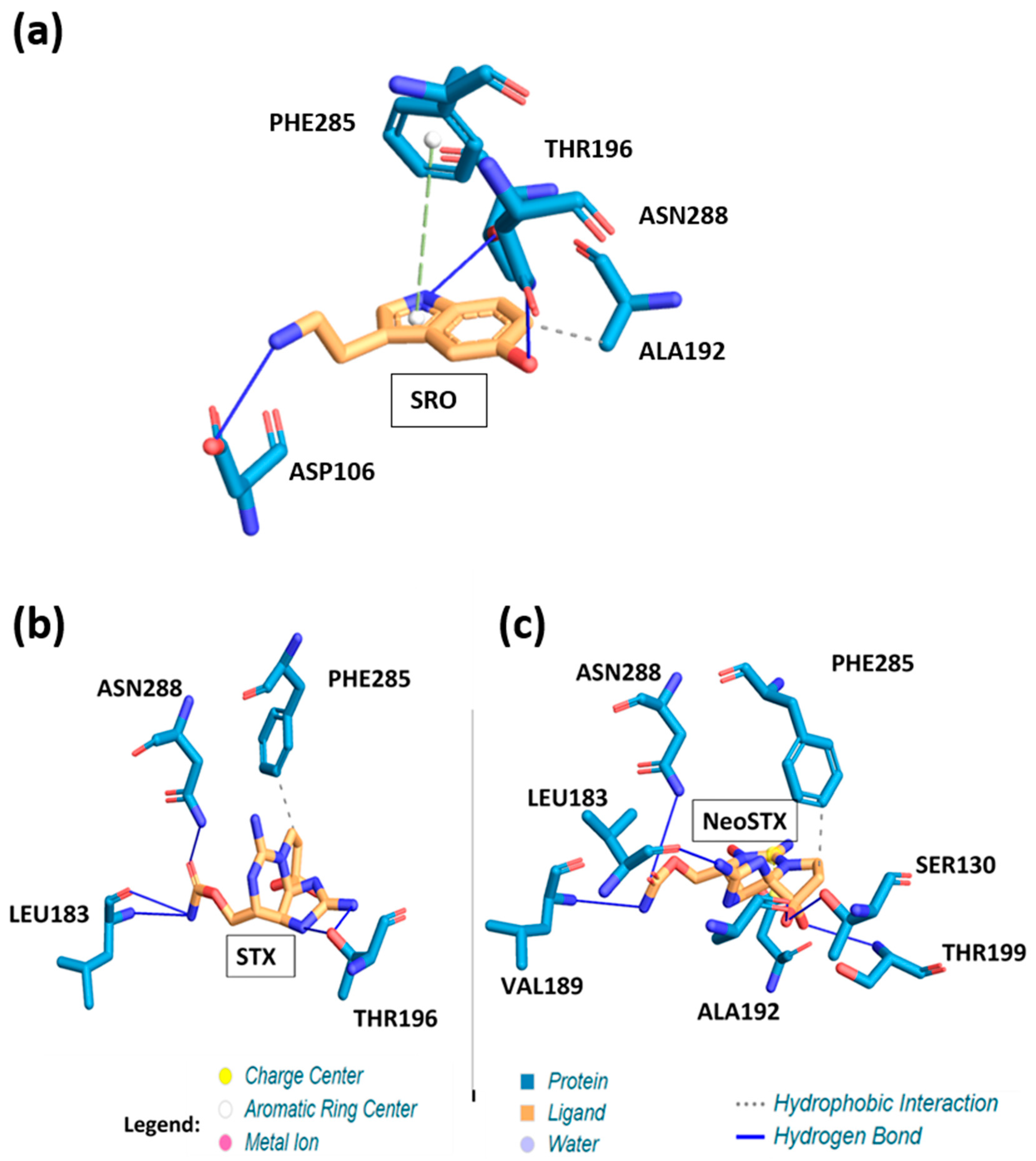
The redocking reported estimated free-energy values of the 10 poses from −6.3 kcal/mol to −4.3 kcal/mol (Table 2). The lowest-energy solution pose was −6.3 kcal/mol. This pose showed three specific hydrogen bonds between the indole group of SRO and Thr196’s side chain; between the amine group’s nitrogen atom on SRO and the side chain of Asp106; and the oxygen atom on SRO and Asn288. A hydrophobic interaction was identified between the purine ring of SRO and the side chain of Ala192. And finally, a π-stacking interaction between the indole group of SRO and Phe285 was identified (Figure 2).
The estimated free-energy values of the 10 poses reported by AutoDock Vina ranged from −5.5 kcal/mol to −4.1 kcal/mol (Table 2). The lowest-energy solution was calculated to be −5.5 kcal/mol (Figure 2). This pose exhibited a specific hydrogen bond between the amide group’s nitrogen atom on STX and the side chain of Leu183.
Additionally, hydrogen bonds were identified between the guanidine group of STX and Thr196’s side chain and between the amide group’s oxygen atom on STX and Asn288’s side chain. Furthermore, a hydrophobic interaction was identified between the purine ring of STX and the side chain of Phe285.
For the NeoSTX ligand, docking on the same target resulted in estimated free-energy values for the 10 poses ranging from −5.7 kcal/mol to −3.7 kcal/mol (Table 2). The lowest-energy pose exhibited a binding affinity of −5.7 kcal/mol. This pose showed a specific hydrogen bond between the purine ring of NeoSTX and the side chain of Leu183. Two additional hydrogen bonds were formed between the guanidine group of NeoSTX and the side chain of Thr199, and between the amide group’s oxygen atom and Asn288. A fourth hydrogen bond was observed between the amide group’s nitrogen atom on NeoSTX and Val189’s side chain. Furthermore, residues Ser130, Ala192, and Thr199 established specific hydrogen bonds with the oxygen atoms attached to the pyrrolidine ring. Finally, a hydrophobic interaction between the purine ring of NeoSTX and the side chain of Phe285 was identified.

4. Conclusions

As a conclusion, the toxins exhibited affinity for different potential receptor targets, which were identified by in silico target prediction. Molecular docking showed that STX and NeoSTX interact with Leu183, Asn288, and Phe285 residues; although NeoSTX interacted with more residues than the HTR6 receptor, maybe its structure allows a large number of interactions. However, the interactions were similar between SRO, STX, and NeoSTX with Asn288 and Phe285 residues.
We show that the wealth of existing complexes and of molecular docking results provides deep insights into the design principles for drug targeting.
These findings provide robust hypotheses about the therapeutic potential of STX and NeoSTX as 5-HT6 receptor antagonists, supporting the development of new bioactive agents derived from these marine toxins. Molecular dynamics simulations will be performed for the most promising complexes, and atomic force microscopy tests will be performed to identify the interaction between STX and neosaxitoxin with the targets.

Author Contributions

Writing—original draft preparation, V.d.S.S. and B.d.P.M.; writing—review and editing, V.d.S.S., D.V.N.d.L. and V.S.G.d.A.; and supervision, V.S.G.d.A. and T.L.S.N.; investigation, V.d.S.S. All authors have read and agreed to the published version of the manuscript.

Funding

This work was funded by the Center for Biodefense Studies, Army Biology Institute.

Institutional Review Board Statement

Not applicable.

Informed Consent Statement

Not applicable.

Data Availability Statement

No new data were created or analyzed in this study. Data sharing is not applicable to this article.

Conflicts of Interest

Author Daniel Vinicius Neves de Lima was employed by the company Biocorp Environmental Solutions. The remaining authors declare that the research was conducted in the absence of any commercial or financial relationships that could be construed as a potential conflict of interest.

Abbreviations

STXSaxitoxin
NeoSTXNeosaxitoxin
PDBProtein Data Bank
5-HT6Serotonin 6 Receptor

References

  1. Janik, E.; Ceremuga, M.; Saluk-Bijak, J.; Bijak, M. Biological Toxins as the Potential Tools for Bioterrorism. Int. J. Mol. Sci. 2019, 20, 1181. [Google Scholar] [CrossRef] [PubMed]
  2. Guillotin, S.; Delcourt, N. Marine Neurotoxins’ Effects on Environmental and Human Health: An OMICS Overview. Mar. Drugs 2021, 20, 18. [Google Scholar] [CrossRef] [PubMed]
  3. Chemical Weapons Convention—Annex on Chemicals: Schedule 1. Available online: https://www.opcw.org/chemical-weapons-convention/annexes/annex-chemicals/schedule-1 (accessed on 10 September 2025).
  4. Deng, H.; Shang, X.; Zhu, H.; Huang, N.; Wang, L.; Sun, M. Saxitoxin: A Comprehensive Review of Its History, Structure, Toxicology, Biosynthesis, Detection, and Preventive Implications. Mar. Drugs 2025, 23, 277. [Google Scholar] [CrossRef] [PubMed]
  5. Flores-Holguín, N.; Salas-Leiva, J.S.; Nuñes-Vázquez, E.J.; Tovar-Ramírez, D.; Glossman-Mitnik, D. Marine Toxins as Pharmaceutical Treasure Troves: A Focus on Saxitoxin Derivatives from a Computational Point of View. Molecules 2024, 29, 275. [Google Scholar] [CrossRef] [PubMed]
  6. Montero, C.; Riquelme, G.; Del Campo, M.; Lagos, N. Neosaxitoxin, a Paralytic Shellfish Poison Phycotoxin, Blocks Pain and Inflammation in Equine Osteoarthritis. Toxicon 2021, 204, 5–8. [Google Scholar] [CrossRef] [PubMed]
  7. Oyaneder, L.; Lagos, N.; Dörner, C. Systemic, Hemodynamic and Neurological Effects of Caudal Epidural Administration of Neosaxitoxin in Horses. Toxicon 2025, 257, 108303. [Google Scholar] [CrossRef] [PubMed]
  8. Paggi, J.M.; Pandit, A.; Dror, R.O. The Art and Science of Molecular Docking. Annu. Rev. Biochem. 2024, 93, 389–410. [Google Scholar] [CrossRef] [PubMed]
  9. Berman, H.; Henrick, K.; Nakamura, H. Announcing the worldwide Protein Data Bank. Nat. Struct. Mol. Biol. 2003, 10, 980. [Google Scholar] [CrossRef] [PubMed]
  10. Gfeller, D.; Michielin, O.; Zoete, V. Shaping the interaction landscape of bioactive molecules. Bioinformatics 2013, 29, 3073–3079. [Google Scholar] [CrossRef] [PubMed]
  11. Gfeller, D.; Grosdidier, A.; Wirth, M.; Daina, A.; Michielin, O.; Zoete, V. SwissTargetPrediction: A web server for target prediction of bioactive small molecules. Nucleic Acids Res. 2014, 42, W32–W38. [Google Scholar] [CrossRef] [PubMed] [PubMed Central]
  12. Daina, A.; Michielin, O.; Zoete, V. SwissTargetPrediction: Updated data and new features for efficient prediction of protein targets of small molecules. Nucleic Acids Res. 2019, 47, W357–W364. [Google Scholar] [CrossRef] [PubMed] [PubMed Central]
  13. Huang, S.; Xu, P.; Shen, D.D.; Simon, I.A.; Mao, C.; Tan, Y.; Zhang, H.; Harpsøe, K.; Li, H.; Zhang, Y.; et al. GPCRs steer Gi and Gs selectivity via TM5-TM6 switches as revealed by structures of serotonin receptors. Mol. Cell 2022, 82, 2681–2695.e6. [Google Scholar] [CrossRef] [PubMed]
  14. Yen, T.J.; Lolicato, M.; Thomas-Tran, R.; Du Bois, J.; Minor, D.L., Jr. Structure of the saxiphilin:saxitoxin (STX) complex reveals a convergent molecular recognition strategy for paralytic toxins. Sci. Adv. 2019, 5, eaax2650. [Google Scholar] [CrossRef] [PubMed] [PubMed Central]
  15. National Center for Biotechnology Information. PubChem Compound Summary for CID 21117946, Neosaxitoxin. 2025. Available online: https://pubchem.ncbi.nlm.nih.gov/compound/NeoSTX (accessed on 13 July 2024).
  16. Eberhardt, J.; Santos-Martins, D.; Tillack, A.F.; Forli, S. AutoDock Vina 1.2.0: New Docking Methods, Expanded Force Field, and Python Bindings. J. Chem. Inf. Model. 2021, 61, 3891–3898. [Google Scholar] [CrossRef] [PubMed] [PubMed Central]
  17. Trott, O.; Olson, A.J. AutoDock Vina: Improving the speed and accuracy of docking with a new scoring function, efficient optimization, and multithreading. J. Comput. Chem. 2010, 31, 455–461. [Google Scholar] [CrossRef] [PubMed] [PubMed Central]
  18. Schake, P.; Bolz, S.N.; Linnemann, K.; Schroeder, M. PLIP 2025: Introducing protein-protein interactions to the protein-ligand interaction profiler. Nucleic Acids Res. 2025, 53, W463–W465. [Google Scholar] [CrossRef] [PubMed]
  19. Sharp, T.; Barnes, N.M. Central 5-HT receptors and their function; present and future. Neuropharmacology 2020, 177, 108155. [Google Scholar] [CrossRef] [PubMed]
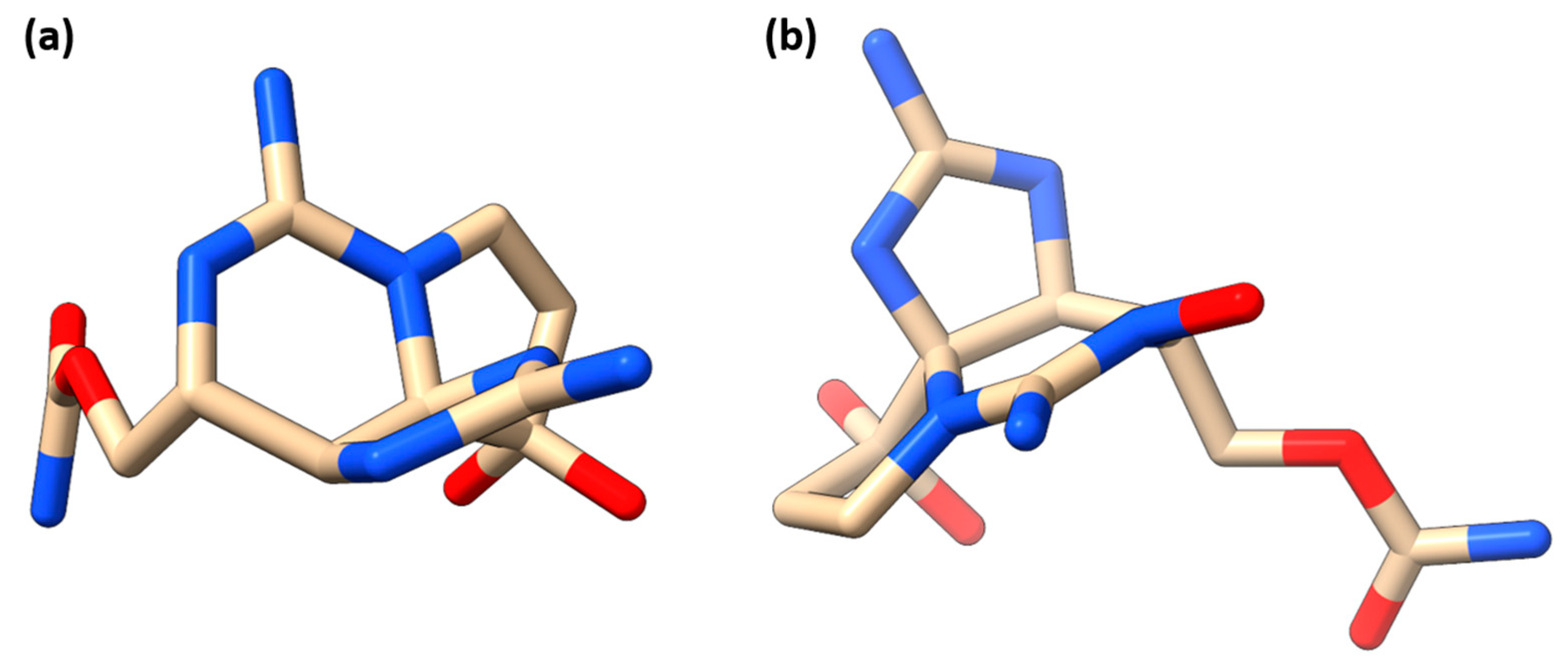
Figure 1. The three-dimensional structures of the STX and NeoSTX ligands were prepared for molecular docking assays. (a) The STX structure was obtained from the PDB database (ID 600F), while (b) the NeoSTX structure was retrieved from PubChem (ID 21117946). These structures were used as ligands in the docking simulations.
Blsf 52 00001 g001
Figure 2. Binding modes of SRO, STX, and NeoSTX within the 5-HT6 receptor. Binding mode of (a) SRO, native ligand; (b) STX; and (c) NeoSTX within the 5-HT6 receptor, highlighting surrounding residues (shown in blue) within a cutoff radius of 5 Å.
Blsf 52 00001 g002
Table 1. Potential human therapeutic targets for STX and NeoSTX identified by the SwissTargetPrediction server analysis.
TargetsUNIPROT ID
Serotonin 6 (5-HT6) receptorP50406
Carbonic anhydrase IIP00918
Carbonic anhydrase IP00915
Carbonic anhydrase IXQ16790
Carbonic anhydrase VIIP43166
Carbonic anhydrase XIIO43570
Carbonic anhydrase XIVQ9ULX7
Steryl-sulfataseP08842
C-X-C chemokine receptor type 3P49682
Carbonic anhydrase IIIP07451
Carbonic anhydrase VIP23280
Carbonic anhydrase IVP22748
Carbonic anhydrase VBQ9Y2D0
Carbonic anhydrase VAP35218
Carbonic anhydrase XIII (by homology)Q8N1Q1
TyrosinaseP14679
Nuclear receptor subfamily 4 group A member 1P22736
Protein tyrosine kinase 2 betaQ14289
Table 2. Docking scores from AutoDock Vina for the top 10 poses of the native ligand, SRO, and STX and NeoSTX ligands on the 5-HT6 receptor.
PosesScore (kcal/mol)
SRO—Native LigandSTXNeoSTX
1−6.3−5.5−5.7
2−6.1−5.2−5.4
3−6.0−5.0−5.1
4−5.9−4.9−4.8
5−5.9−4.7−4.6
6−5.7−4.6−4.5
7−5.7−4.6−4.4
8−4.3−4.5−4.2
9−5.4−4.2−4.0
10−4.3−4.1−3.7
Disclaimer/Publisher’s Note: The statements, opinions and data contained in all publications are solely those of the individual author(s) and contributor(s) and not of MDPI and/or the editor(s). MDPI and/or the editor(s) disclaim responsibility for any injury to people or property resulting from any ideas, methods, instructions or products referred to in the content.